Performance Characteristics and Validation of
Next-Generation Sequencing for Human
Leucocyte Antigen Typing
Eric T. Weimer,*yMaureen Montgomery,*Rosanne Petraroia,*John Crawford,*and John L. Schmitz*y
From the Human Leukocyte Antigen Laboratory,* McLendon Clinical Laboratories, University of North Carolina Hospitals, Chapel Hill; and the Department of Pathology and Laboratory Medicine,yUniversity of North Carolina at Chapel Hill School of Medicine, Chapel Hill, North Carolina
Accepted for publication March 23, 2016.
Address correspondence to Eric T. Weimer, Ph.D., Department of Pathology and Laboratory Medicine, University of North Carolina at Chapel Hill School of Medicine, 101 Manning Dr, Room 1035 E. Wing, Chapel Hill, NC 27514. E-mail:eric_ [email protected].
High-resolution human leukocyte antigen (HLA) matching reduces graft-versus-host disease and im-proves overall patient survival after hematopoietic stem cell transplant. Sanger sequencing has been the gold standard for HLA typing since 1996. However, given the increasing number of new HLA alleles identified and the complexity of the HLA genes, clinical HLA typing by Sanger sequencing requires several rounds of additional testing to provide allele-level resolution. Although next-generation sequencing (NGS) is routinely used in molecular genetics, few clinical HLA laboratories use the tech-nology. The performance characteristics of NGS HLA typing using TruSight HLA were determined using Sanger sequencing as the reference method. In total, 211 samples were analyzed with an overall accuracy of 99.8% (2954/2961) and 46 samples were analyzed for precision with 100% (368/368) reproducibility. Most discordant alleles were because of technical error rather than assay performance. More important, the ambiguity rate was 3.5% (103/2961). Seventy-four percentage of the ambiguities were within the DRB1andDRB4loci. HLA typing by NGS saves approximately $6000 per run when compared to Sanger sequencing. Thus, TruSight HLA assay enables high-throughput HLA typing with an accuracy, precision, ambiguity rate, and cost savings that should facilitate adoption of NGS technology in clinical HLA laboratories.(J Mol Diagn 2016, 18: 668e675;http://dx.doi.org/10.1016/j.jmoldx.2016.03.009)
It is widely accepted that human leukocyte antigen (HLA) matching reduces patient morbidity and mortality after he-matopoietic stem cell transplantation (HSCT).1The current standard of care is high-resolution HLA typing by Sanger sequencing or sequence-based typing (SBT). High-resolution HLA typing is defined as a set of alleles that specifies and encodes the same protein sequence for the peptide-binding region of an HLA molecule. However, SBT cannot accu-rately phase heterozygous alleles and provides limited sequencing information. Traditionally, HLA typing by SBT typically involves sequencing only exons 2, 3, and 4 of HLA class I genes and exons 2 and 3 of HLA class II genes. Since the regulatory requirement was established for high-resolution HLA typing for HLA-A/B/C/DRB1 in 2005, many clinical laboratories are putting significant resources toward ambiguity resolution.2 Even as several restrictions were removed, such as the requirement to exclude rare al-leles, there is still a growing list of ambiguities that require
additional testing and delay patient results. The issue of ambiguities is a testament to the complexity of the HLA re-gion in the human genome with>13,000 alleles identified to date.3The combination of the inability to phase heterozygous alleles and the growing number of HLA alleles has led to a significant number of ambiguities in HLA typing that require time-consuming and costly additional tests to be performed. In 2004, Adams et al4reported the ambiguity rate forHLA-A, HLA-B, and HLA-C of 24% to 41%. Three years later, Voorter et al5reported that ambiguities forHLA-A,HLA-B, andHLA-Chad increased to approximately 50%.
Supported by institutional support from University of North Carolina Hospitals (M.M., R.P., J.C.) and University of North Carolina School of Medicine Department of Pathology and Laboratory Medicine (E.T.W., J.L.S.). Disclosures: E.T.W. reports nonfinancial support from Illumina, Inc., dur-ing the conduct of the study. Illumina, Inc. generously provided the reagents for conducting the study.
Currently, our experience is that ambiguity resolution is required in 53% of patient specimens to determine a specific allele (E.T. Weimer and J.L. Schmitz, unpublished obser-vation). This high level of additional testing delays patient HLA typing results and increases the cost of HLA typing. With no sign that the number of HLA alleles identified will decline and the increasing application of typing of addi-tional loci (DPB1andDRB3/4/5), there is a great need for a technology that allows for accurate high-resolution HLA typing without the requirement of additional testing.3,6e8
Next-generation sequencing (NGS) technology is the massive parallel sequencing of clonal DNA molecules. By using unique molecular signatures or barcodes, many sam-ples can be pooled together and sequenced simultaneously. A key feature of the NGS method is the ability to generate massive amounts of genetic data from many DNA mole-cules simultaneously.9,10 The combination of clonal DNA sequencing and application of long-range PCR techniques to increase HLA genomic information aids in the reduction in HLA allele ambiguities. For example, Danzer et al11 used long-range PCR of HLA genes and NGS to demonstrate an average ambiguity reduction of 93.5% forHLA-A,HLA-B, HLA-C,DRB1,DQB1, andDPB1. Although the ambiguity reduction varied by locus [ie, the reduction of DRB1 was less pronounced (46.1%)], this was still considered signifi -cant given the amount of additional testing required by SBT. HLA typing by NGS using long-range PCR techniques to increase genomic coverage of HLA genes has proven effec-tive at improving ambiguity resolution.11e14 The increased genomic coverage is partly what enables the accurate phasing of heterozygous bp positions often observed in SBT.13,15 Several recent reports have demonstrated the feasibility of using NGS technology to provide>97% concordance with SBT.11,13,15e17Although the high-throughput nature of NGS is thought to be cost-effective for HLA typing, this has yet to be shown. More important, previous reports focused on laboratory-developed assays rather than commercially available reagents.11e14,17,18With the increasing availability of commercial NGS HLA reagents, there is a need to better understand each assay’s characteristics and their utility to solve the limitations of SBT.
In this study, we evaluated the performance of the TruSight HLA assay, a commercially available NGS assay for HLA typing, to not only accurately type HLA alleles but also identify a set of quality control criteria required to ensure accurate HLA allele determination. More important, we also performed a cost analysis between NGS and SBT for HLA typing that provides thefirst evidence that NGS is a cost-effective alternative to SBT.
Materials and Methods
Patient Samples, DNA Extraction, and HLA Gene
Ampli
fi
cation
Two-hundred and eleven samples that were already high-resolution HLA typed were used for comparison and clinical
validation. An additional 79 samples of genomic DNA extracted from buccal swabs from patients and donors under evaluation for HSCT were also used. Genomic DNA was extracted from each sample using Qiagen DNA Tissue Extraction kits (Qiagen, Valencia, CA). The study was approved by the Institutional Review Board of the Univer-sity of North Carolina at Chapel Hill.
For HLA gene amplification, primers specific for each HLA gene were used in a long-range PCR. DNA was quantified using a QuBit fluorometer (Life Technologies, Carlsbad, CA). After quantification, sample DNA was diluted to 10 ng/mL. For buccal swabs, 40 of the 79 samples received additional purification according to the manufac-turer’s instructions. Fifty nanogram of genomic DNA was used for each HLA locus and amplified according to the manufacturer’s instructions. PCR amplicons were visualized using 2% agarose gel electrophoresis before preparing NGS libraries. Twenty-four samples (192 HLA loci) were run in a single NGS experiment. A sample with a known HLA typing was run on each NGS run to ensure library prepa-ration, data quality, and analysis were of sufficient quality to ensure accurate HLA typing.
TruSight HLA Library Preparations and NGS Sequencing
Amplified DNA for NGS sequencing was prepared according to the supplied instructions (Illumina, Inc., San Diego, CA). Briefly, amplified DNA was purified using magnetic AMPure XP beads, fragmented, and Illumina-specific adap-tors were applied using a tagmentation enzyme supplied by Illumina. After tagmentation, fragments were purified using AMPure XP beads and patient-specific indices were added to individual HLA loci by a short PCR, followed by magnetic bead purification. Post-barcoding samples were pooled and the libraries quantified using QuBitfluorimeter. The size of HLA libraries was determined using TapeStation Bioanalyzer 2200.
To assess sequencer-based errors, a 1% to 5% concen-tration of 12.5 pmol/L PhiX control (Illumina, San Diego, CA) was spiked into pooled HLA libraries. Pooled HLA libraries and PhiX control were loaded onto the cartridge and 2250 bp sequencing was performed using a regular
flow cell on an Illumina MiSeq. Demultiplexing and gen-eration of FASTQ files was performed on the MiSeq system. A total of 34 MiSeq runs were performed and used for quantification of sequencing metrics.
Sample Analysis
genes. Mismatches that represented potential novel HLA alleles were noted. Ambiguity resolution was performed by Sanger sequencing with analysis on uTYPE 6.0 (SeCore HLA kit; Life Technologies).
Cost Analysis
The cost analysis between SBT and NGS was performed using the available list price for each reagent necessary for the respective technologies. For NGS, reagents included HLA locusespecific primers, library preparation, and sequencing reagents. For Sanger sequencing, reagents included HLA locusespecific primers, ExoSAP, sequencing reagents (cathode, anode, polymer), sequence-specific oli-gonucleotides, and group-specific sequencing primers for ambiguity resolution. Ambiguity resolution was added to each technology using laboratory-specific 53% for Sanger sequencing and 3% for NGS. Instrumentation cost was excluded for the analysis. An estimation of time required for each method was determined by averaging the hands-on time from amplification to completed analysis (including ambiguity resolution) for each technologist (n Z 5) per-forming the assay. The average labor time was multiplied by the average hourly rate for HLA technologists at UNC Hospitals. Assay time was defined as the time from PCR to completion of sequencing analysis. Turnaround-time (TAT) was determined for all HSCT-related HLA typings from receiving in laboratory to verification of results in calendar days. Date range for NGS (n Z 323) was August to December 2015 and the same time period 1 year prior for Sanger sequencing (n Z324). NGS runs were between 10 and 23 patients per run, withHLA-A,HLA-B,HLA-C, HLA-DRB1, HLA-DQB1, HLA-DQA1, HLA-DPB1, HLA-DPA1 typed. Only HLA-A, HLA-B, HLA-C, HLA-DRB1, HLA-DQB1, HLA-DPB1 were reported clinically. HLA-DQA1 and HLA-DPA1 typing were used for academic purposes. For comparison, a maximum offive patients were run using Sanger sequencing.
Statistical Analysis
Significant differences between two proportions were determined by two-tailed probability test from the calculated z-ratio. Significant differences between NGS and Sanger for TAT were calculated using unpaired t-test with Welch’s correction.P0.05 was considered significant.
Results
HLA Gene Ampli
fi
cation Rates
To enrich HLA genes, genomic DNA was amplified by long-range PCR. A known HLA-typed sample and negative control were used with every PCR to ensure proper HLA amplification and to assess for potential contamination. There were no cases of PCR-based contamination
throughout the validation. The PCR fragment length for each HLA gene is shown inTable 1, and a representative gel electrophoresis of HLA amplicons is shown in Figure 1A. Overall, the amplification success rate of HLA genes was 95.0% (2399/2526) (Figure 1B). Only 37 HLA class I genes failed to amplify of 945 amplifications (3.9%) and 31 of the 37 were from buccal swabs. There were a total of 127 in-cidences of no detectable PCR band on electrophoresis that were subsequently sequenced and HLA typed (Figure 1, C and D).DPA1andDPB1were the most difficult HLA genes to amplify, most likely because of their length (Figure 1, BeD). In addition, DPA1 and DPB1 had the lowest HLA typing success overall. Amplification success was signifi -cantly lower forDPA1(P<0.001),DPB1(PZ0.001), and DQB1(PZ0.006) when using DNA isolated from buccal swabs (BSs) compared to DNA from peripheral blood19 (Figure 1B). To determine the reason for decreased HLA amplification rate from BSs, the effect of concentration and quality of DNA on HLA typing was evaluated. Additional purification of BSs significantly increased HLA typing rates forDQB1(PZ0.048) and DPB1 (PZ0.002) compared to nonpurified samples (Figure 1E). Similarly, BSs with higher concentrations were significantly more often successfully HLA typed forDPA1(P<0.001) andDPB1(PZ0.001) compared to lower concentrations (Figure 1F).
Characteristics of HLA Library Fragments and NGS Data
NGS library fragments vary in size and DNA fragments
>1000 bp are less efficient at cluster generation on the MiSeq than smaller fragments.19 To determine the DNA fragment size generated using TruSight HLA, three (576 loci) pooled HLA libraries were analyzed using a TapeS-tation Bioanalyzer. The average library fragment size was 1268 bp (95% CI, 856e1680 bp) (Figure 2A). Larger fragment sizes aid in correct phasing of HLA alleles.15Next, overall read quality and depth of coverage were determined for each HLA locus. The average percentage of reads Q30 was 96.1% (95% CI, 95.4%e96.7%) for all HLA loci (Figure 2B). The lowest quality reads were consistently observed with HLA-B and HLA-DQB1. The average depth of coverage was 280 (95% CI, 270e289) for
Table 1 Amplification Size for HLA Genes Using TruSight HLA
Loci
Sequencer region (kb)
A 4.1
B 2.6
C 4.2
DPA1 10.3
DPB1 9.7
DQA1 7.3
DQB1 7.1
DRB1/3/4/5 4.1
all HLA loci (Figure 2C). These results indicate that NGS data generated by TruSight HLA are high quality and core exons within each HLA are covered beyond 250.
NGS Data Quality Metrics
To determine the quality of each library preparation and sequencing run, cluster density, percentage of clusters passing filter, percentage of reads Q30, and error rate were monitored using Illumina’s sequencing analysis
viewer. The acceptable range for each parameter was determined to be the average plus or minus 2 SDs or using the MiSeq performance specifications provided by the manufacturer. There were two runs in which PhiX was not added to the pooled libraries before sequencing and thus no error rate could be determined. The shaded areas inFigure 3
show the acceptable range for each parameter. For a run to be considered high quality, it must fall within specific ranges for each category. There were two runs that required repeat sequencing because of reagent issues (Figure 3, B
A
B
C
D
E
F
Figure 1 HLA typing rates for peripheral blood and buccal swabs.A:Representative gel electrophoresis of TruSight HLA amplicons.B:Comparison between peripheral blood (PB) and buccal swab (BS) HLA successful typing rates.C:Peripheral blood.D:Buccal swabs.CandD:Absence of a visible electrophoresis band does not prevent accurate HLA typing. Gel electrophoresis bands were counted and compared to the overall ability of each sample to accurately HLA type. E:Additional purification of buccal swabs provides better HLA typing. HLA typing was compared between buccal swab samples that were either used as extract (nonpurified) or received additional magnetic bead-based purification (purified).F:Higher concentration buccal swabs outperform low concentration samples. *P<0.05, **P<0.005.
A
B
C
and D). The average cluster density and proportion of clusters passing filter were 1071 35 K/mm2 and 88%1%, respectively (Figure 3, A and B). The average percentage of readQ30 was 80%1% (Figure 3C). The average error rate was 1.65%0.1% (Figure 3D). After run 7, there was a 54.5% reduction in error rate variability compared to the first six runs (Figure 3D). Establishing these criteria is crucial for ensuring library preparation consistency and sequencing performance over time.
HLA Typing Comparison between Sanger and NGS
Clinical validation of HLA typing by NGS involves deter-mination of the assay’s accuracy and precision compared to the gold standard, Sanger sequencing. To determine TruSight HLA assay accuracy, 211 samples for which existing genomic DNA and high-resolution HLA typing was known for each locus were used. Fifty of the 211 samples were blinded (all authors were blinded). Forty-six samples were used to assess assay reproducibility. HLA alleles were considered equivalent on the basis of the National Marrow Donor Program HLA reporting criteria. The National Marrow Donor Program requires identification of eight null (nonexpressed) HLA alleles within specific HLA G groups.20 All eight null HLA alleles could be identified or ruled out as potential HLA alleles using TruSight HLA. Overall, accu-racy for the assay was (2954/2961) 99.8% with a precision of 100% (Table 2). Two-hundred and sixty-five unique HLA alleles were accurately identified by TruSight HLA
(Supplemental Table S1). Only ambiguities outside of HLA G groups are considered significant because those alter the peptide-binding region of the HLA protein. There were 103 (3.5%) ambiguities of 2961 alleles identified. Of 103 ambi-guities, 91 (88.4%) were from DRB1 (26.3% of all DRB1 typings) and 11 (10.7%) were from DRB4 (47.8% of all DRB4 typings) loci (Table 2). Because DRB4 is not commonly part of HLA matching for HSCT, the impact on routine clinical use is minimal. There was one ambiguity in
A
B
C
D
Clusters Passing Filter
Individual NGS Runs
Individual NGS Runs
Reads
≥
Q30
Figure 3 Characterization of next-generation sequencing (NGS) data metrics. For 34 NGS runs, cluster density, frequency of clusters passingfilter, percentage of reads Q30, and error rate were determined. All of the parameters were determined using Sequence Analysis Viewer (SAV) after each run. Error was determined by spiking in 1% to 5% PhiX into pooled HLA libraries before sequencing. Gray regionsindicate acceptable range for each parameter. AeC: Line indicates mean value. A: Cluster density.B: Clusters passingfilter.C: Fre-quency of reads at or above Q30.D:PhiX error rate. Two separate sequencing runs fell outside acceptable range because of reagent issues.
Table 2 Accuracy and Ambiguities for TruSight HLA Assay
Gene N
Allele level
mismatch % correct Ambiguities
Ambiguity rate (%)
HLA-A 353 1 99.7 1* 0.3
HLA-B 353 1 99.7 0 0.0
HLA-C 353 0 100.0 0 0.0
HLA-DPA1 345 1 99.7 0 0.0
HLA-DPB1 354 0 100.0 0 0.0
HLA-DQA1 346 1 99.7 0 0.0
HLA-DQB1 345 0 100.0 0 0.0
HLA-DRB1 346 1 99.7 91y 26.3
HLA-DRB3 100 0 100.0 0 0.0
HLA-DRB4 23 0 100.0 11z 47.8
HLA-DRB5 43 1 97.7 0 0.0
Total 2961 6 99.8 103 3.5
*Ambiguity exists between 03:01:01, 11:01:01 pair, and 03:63/11:12. yAmbiguities exist for the following HLA alleles: 03:01; 03:50, 08:04;
08:59, 15:01; 15:110, 13:01; 13:112, 16:02; 16:22, 14:54; 14:113; 14:125; 14:157, 15:02; 15:19, 13:02; 13:128.
the Alocus (0.3%). All remaining HLA loci had no ambi-guities. HLA typing by TruSight HLA demonstrated a 93.4% reduction in ambiguities compared to Sanger sequencing.
The seven cases of incorrect HLA typing emphasized the need for HLA-specific data criteria. Three of the seven in-stances were because of allele dropout. Given those results, HLA-specific data quality criteria were established. A depth of coverage (DOC) of at least 100 and at least 81% of the reads Q30 were determined to be necessary forHLA-A, HLA-B, HLA-C, HLA-DRB1, HLA-DQB1, HLA-DQA1, HLA-DPB1, HLA-DPA1 typing. Higher quality data (88% of the reads areQ30) was necessary forHLA-DRB3/4/5to prevent false identification of those alleles (Figure 2, B and C). The sequencing and HLA-specific data quality metrics were validated using the 50 blinded samples. Although the above criteria are crucial for increased accuracy in HLA typing results, samples could be accurately typed below those thresholds. Samples were accurately typed with as few as 67 reads and a 74% frequency ofQ30 reads (data not shown). Higher DOC was a better HLA typing quality predictor compared to quality scores (Table 3). Of note, if a sample meets the sequencing run parameters but fails HLA-specific criteria, HLA typing for that sample is not reported and must be repeated. More important, repeat amplification and NGS library preparation accurately typed all incorrect samples, suggesting the incorrect HLA typing was because of a technical error issue rather than an assay issue (Table 3).
HLA Typing Cost Comparison between Sanger
Sequencing and NGS
As mentioned above, 53% of the HLA typings by Sanger sequencing require additional tests to resolve ambiguities in
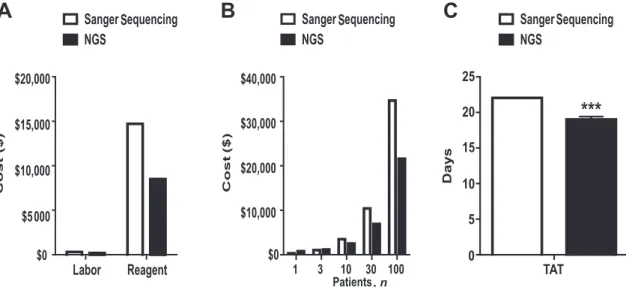
our laboratory. Ambiguity resolution delays patient results and increases patient cost for HSCT. Given that there was significant reduction in the number of ambiguities encoun-tered by TruSight HLA, we hypothesized that NGS sequencing would be more cost effective than typing by Sanger sequencing. The relative cost of HLA typing by Sanger sequencing and TruSight HLA was determined and compared. For each technology, all reagent costs and rela-tive hands-on time were considered. The average time for an assay and hourly rate of the technologists were used to calculate labor costs. TruSight HLA reagent costs were 41.8% ($270/patient) lower compared to Sanger sequencing for HLA typing at all loci (Figure 4A). There was a 25% ($5/patient) reduction in labor (library preparation and analysis) required for TruSight HLA compared to Sanger sequencing. High-resolution HLA typing data for HLA-A/ HLA-B/HLA-C/HLA-DRB1/HLA-DQB1 costs between NGS and Sanger were determined. The total cost to type those HLA loci by NGS is approximately $250 ($50/HLA locus) per patient and $380 ($76/HLA locus) per patient for Sanger sequencing (Figure 4B). Thus, the more patients run together for NGS the more cost savings that are realized. To examine the impact of batching samples TAT for HLA typing by NGS was compared to Sanger sequencing during the same time period. The average TAT using NGS was significantly faster (19 days,P<0.001) compared to Sanger sequencing (22 days) (Figure 4C). Thus, in addition to being highly accurate, TruSight HLA is cost-effective while not delaying patient HLA typing.
Discussion
The analysis presented herein is not only the first charac-terization and validation of the TruSight HLA assay for HLA typing by NGS, but is also thefirst report of specific sequencing and HLA criteria necessary for clinical reporting of HLA results. TruSight HLA demonstrated efficient gen-eration of long-range HLA amplicons (Figure 1) and NGS libraries that consistently generated high-quality data (Figures 2and3). The degree of concordance is consistent with previous reports comparing different sequencing plat-forms and NGS HLA assays17,21,22 (Table 2). The cost-effectiveness of HLA typing by NGS is consistent with a report by Stoddard et al23on the use of NGS compared to Sanger for diseases with multiple candidate genes (Figure 4). Overall, TruSight HLA is a robust assay that consistently provides accurate high-resolution HLA typing with a dramatically reduced ambiguity rate, cost, and TAT. Clinical implementation of new HLA typing technology requires strict validation to provide HLA typing for pa-tients. There are few clinical HLA laboratories performing HLA typing by NGS compared to Sanger sequencing. Establishing criteria for what should be considered suffi -cient validation is a vital part of the process for clinical laboratories. Several of the NGS runs experienced reagent
Table 3 Quality Metrics on Incorrectly Typed Samples
ID*
Average %
of readQ30 Average DOC HLA typing
84 93 30 DRB3 present
Repeaty No DRB3 present
141 91 77 DRB1*15:01
Repeaty 97 293 DRB1*12:01G
UNC9 DQB1*06 dropout
Repeaty 96 277 DQB1*06:02
122 94 49 DRB3 present
Repeaty No DRB3 present
2417 DQB1*06 dropout
Repeaty 95 277 DQB1*06:02
4609 DQB1*06 dropout
Repeaty 96 284 DQB1*06:03
UNC7 DRB1*04 dropout
Repeaty 89 164 DRB1*04:04
*All incorrect samples wereflagged by the Conexio Assign for TruSight HLA software.
yAll repeat testing was consistent with Sanger sequencing results.
issues that highlighted the need to establish guidelines for an acceptable sequencing run to proceed to HLA analysis (Figure 3, C and D). This report demonstrates one such manner of approaching the issue. Without the criteria presented herein, the accuracy of HLA typing would have been significantly decreased. In addition, clinical labora-tories must have a quality assurance system in place to monitor assay performance. More important, DOC was a better indicator for data quality than quality score (Table 3). Higher quality scores were associated with less phasing issues for the HLA region and thus incorporated for quality assurance measures (data not shown). Tracking the sequencing metrics meets this clinical laboratory standard (Figure 3). As an additional level of quality control, HLA-specific data quality criteria were deter-mined (Figure 2). Application of these criteria allowed for increased accuracy of HLA typing and reduced the po-tential for false-positive HLA typings. In our experience, failed HLA typing is most often because of poor sample quality (Figure 1) or improper handling of the magnetic beads (M. Montgomery and E.T. Weimer, unpublished observation). Laboratories considering NGS for HLA typing should examine their workload and the TAT wanted. Thus far, NGS has improved the TAT for high-resolution HLA typing, this will be dependent on the frequency of samples the laboratory receives and the TAT wanted. The cost benefit of NGS is greatly diminished as the batching of samples is reduced (Figure 4C).
A thorough validation should not only determine whether an assay performs robustly, but also identify areas of concern. Consistent with previous reports, HLA typing ofDRB1/3/4/5 was the most difficult22,24,25 (Table 2). These issues arise
because of primer locations inhibiting the ability to resolve certain HLA alleles. All instances where the software inap-propriately identified DRB3/4/5 alleles had DOC of <80. After application, the proposed HLA-specific criteria (Figure 2, B and C), those samples would have been repeated before reporting results. A known issue with PCR amplifi -cation methods is the potential for allelic drop out.DQB1*06 (5% of DQB1*06 typings), DRB1*04 (2.9% of total DRB1*04 typings), andB*27:05:02 (low amplification rate) were found to be most susceptible to allelic drop out. This occurred most often from low DNA concentration buccal swabs and additional purification of buccal swabs before PCR significantly reduced allele drop out (M. Montgomery and E.T. Weimer, unpublished observations). Although several reports have demonstrated that cluster generation and MiSeq sequencing are less efficient with long DNA fragment sizes,19,26we and others have clearly demonstrated that DNA fragments >1000 bp can effectively and reproducibly generate high-quality NGS data using MiSeq (Figures 2
and 3).15 More work is needed to improve HLA typing of DQB1,DRB1, andDRB4to further reduce ambiguities and cost. In addition, more time is needed to examine the impact transitioning to HLA typing by NGS will have on patient care and the utility of whole gene sequencing.
Acknowledgments
We thank the University of North Carolina Human Leukocyte Antigen laboratory for their support and careful review of the manuscript, the 17thInternational HLA and Immunogenetics
A
B
C
, n
workshop for providing the 50 blinded samples, and Illumina for providing the reagents used in this study.
Supplemental Data
Supplemental material for this article can be found at http://dx.doi.org/10.1016/j.jmoldx.2016.03.009.
References
1. Lee SJ, Klein J, Haagenson M, Baxter-Lowe LA, Confer DL, Eapen M, Fernandez-Vina M, Flomenberg N, Horowitz M, Hurley CK, Noreen H, Oudshoorn M, Petersdorf E, Setterholm M, Spellman S, Weisdorf D, Williams TM, Anasetti C: High-resolution donor-recipient HLA matching contributes to the success of unrelated donor marrow transplantation. Blood 2007, 110:4576e4583
2. National Marrow Donor Program: NMDP Policy for Confirmatory Typing. Minneapolis, MN: National Marrow Donor Program, 2005. 3. Robinson J, Halliwell JA, Hayhurst JD, Flicek P, Parham P, Marsh SG:
The IPD and IMGT/HLA database: allele variant databases. Nucleic Acids Res 2015, 43:D423eD431
4. Adams SD, Barracchini KC, Chen D, Robbins F, Wang L, Larsen P, Luhm R, Stroncek DF: Ambiguous allele combinations in HLA Class I and Class II sequence-based typing: when precise nucleotide sequencing leads to imprecise allele identification. J Transl Med 2004, 2:30
5. Voorter CE, Mulkers E, Liebelt P, Sleyster E, van den Berg-Loonen EM: Reanalysis of sequence-based HLA-A, -B and -Cw typings: how ambiguous is today’s SBT typing tomorrow. Tissue Antigens 2007, 70:383e389
6. Crivello P, Zito L, Sizzano F, Zino E, Maiers M, Mulder A, Toffalori C, Naldini L, Ciceri F, Vago L, Fleischhauer K: The impact of amino acid variability on alloreactivity defines a functional distance predictive of permissive HLA-DPB1 mismatches in hematopoietic stem cell transplantation. Biol Blood Marrow Transplant 2015, 21: 233e241
7. Fleischhauer K, Shaw BE, Gooley T, Malkki M, Bardy P, Bignon J-D, Dubois V, Horowitz MM, Madrigal JA, Morishima Y, Oudshoorn M, Ringden O, Spellman S, Velardi A, Zino E, Petersdorf EW: Effect of T-cell-epitope matching at HLA-DPB1 in recipients of unrelated-donor haemopoietic-cell transplantation: a retrospective study. Lancet Oncol 2012, 13:366e374
8. Fernandez-Vina MA, Klein JP, Haagenson M, Spellman SR, Anasetti C, Noreen H, Baxter-Lowe LA, Cano P, Flomenberg N, Confer DL, Horowitz MM, Oudshoorn M, Petersdorf EW, Setterholm M, Champlin R, Lee SJ, de Lima M: Multiple mismatches at the low expression HLA loci DP, DQ, and DRB3/4/5 associate with adverse outcomes in hematopoietic stem cell transplantation. Blood 2013, 121:4603e4610
9. Metzker ML: Sequencing technologies: the next generation. Nat Rev Genet 2010, 11:31e46
10. Gabriel C, Furst D, Fae I, Wenda S, Zollikofer C, Mytilineos J, Fischer GF: HLA typing by next-generation sequencing: getting closer to reality. Tissue Antigens 2014, 83:65e75
11. Danzer M, Niklas N, Stabentheiner S, Hofer K, Proll J, Stuckler C, Raml E, Polin H, Gabriel C: Rapid, scalable and highly automated HLA genotyping using next-generation sequencing: a transition from research to diagnostics. BMC Genomics 2013, 14:221
12. Wang C, Krishnakumar S, Wilhelmy J, Babrzadeh F, Stepanyan L, Su LF, Levinson D, Fernandez-Vina MA, Davis RW, Davis MM, Mindrinos M: High-throughput, high-fidelity HLA genotyping with deep sequencing. Proc Natl Acad Sci U S A 2012, 109:8676e8681
13. Shiina T, Suzuki S, Ozaki Y, Taira H, Kikkawa E, Shigenari A, Oka A, Umemura T, Joshita S, Takahashi O, Hayashi Y, Paumen M, Katsuyama Y, Mitsunaga S, Ota M, Kulski JK, Inoko H: Super high resolution for single molecule-sequence-based typing of classical HLA loci at the 8-digit level using next generation sequencers. Tissue An-tigens 2012, 80:305e316
14. Lind C, Ferriola D, Mackiewicz K, Heron S, Rogers M, Slavich L, Walker R, Hsiao T, McLaughlin L, D’Arcy M, Gai X, Goodridge D, Sayer D, Monos D: Next-generation sequencing: the solution for high-resolution, unambiguous human leukocyte antigen typing. Hum Immunol 2010, 71:1033e1042
15. Hosomichi K, Jinam TA, Mitsunaga S, Nakaoka H, Inoue I: Phase-defined complete sequencing of the HLA genes by next-generation sequencing. BMC Genomics 2013, 14:355
16. Smith AG, Pyo CW, Nelson W, Gow E, Wang R, Shen S, Sprague M, Pereira SE, Geraghty DE, Hansen JA: Next generation sequencing to determine HLA class II genotypes in a cohort of he-matopoietic cell transplant patients and donors. Hum Immunol 2014, 75:1040e1046
17. Holcomb CL, Hoglund B, Anderson MW, Blake LA, Bohme I, Egholm M, Ferriola D, Gabriel C, Gelber SE, Goodridge D, Hawbecker S, Klein R, Ladner M, Lind C, Monos D, Pando MJ, Proll J, Sayer DC, Schmitz-Agheguian G, Simen BB, Thiele B, Trachtenberg EA, Tyan DB, Wassmuth R, White S, Erlich HA: A multi-site study using high-resolution HLA genotyping by next gen-eration sequencing. Tissue Antigens 2011, 77:206e217
18. Lange V, Bohme I, Hofmann J, Lang K, Sauter J, Schone B, Paul P, Albrecht V, Andreas JM, Baier DM, Nething J, Ehninger U, Schwarzelt C, Pingel J, Ehninger G, Schmidt AH: Cost-efficient high-throughput HLA typing by MiSeq amplicon sequencing. BMC Ge-nomics 2014, 15:63
19. Duke JL, Lind C, Mackiewicz K, Ferriola D, Papazoglou A, Derbeneva O, Wallace D, Monos DS: Towards allele-level human leucocyte antigens genotyping - assessing two next-generation sequencing platforms: Ion Torrent Personal Genome Machine and Illumina MiSeq. Int J Immunogenet 2015, 42:346e358
20. National Marrow Donor Program. NMDP policy for HLA confirmatory typing requirements for adult donor and patient HLA confirmatory typing. Number P00079. 2015. Available athttps://bioinformatics.bethematch clinical.org/workarea/downloadasset.aspx?id=10528 (accessed March 10, 2016)
21. Barone JC, Saito K, Beutner K, Campo M, Dong W, Goswami CP, Johnson ES, Wang ZX, Hsu S: HLA-genotyping of clinical specimens using Ion Torrent-based NGS. Hum Immunol 2015, 76:903e909
22. Lane JA, Johnson JR, Noble JA: Concordance of next generation sequence-based and sequence specific oligonucleotide probe-based HLA-DRB1 genotyping. Hum Immunol 2015, 76:939e944
23. Stoddard JL, Niemela JE, Fleisher TA, Rosenzweig SD: Targeted NGS: a cost-effective approach to molecular diagnosis of PIDs. Front Immunol 2014, 5:531
24. Ozaki Y, Suzuki S, Kashiwase K, Shigenari A, Okudaira Y, Ito S, Masuya A, Azuma F, Yabe T, Morishima S, Mitsunaga S, Satake M, Ota M, Morishima Y, Kulski JK, Saito K, Inoko H, Shiina T: Cost-efficient multiplex PCR for routine genotyping of up to nine classical HLA loci in a single analytical run of multiple samples by next gen-eration sequencing. BMC Genomics 2015, 16:318
25. Wittig M, Anmarkrud JA, Kassens JC, Koch S, Forster M, Ellinghaus E, Hov JR, Sauer S, Schimmler M, Ziemann M, Gorg S, Jacob F, Karlsen TH, Franke A: Development of a high-resolution NGS-based HLA-typing and analysis pipeline. Nucleic Acids Res 2015, 43:e70