Phase II Trial of Lapatinib for Brain Metastases in Patients
With Human Epidermal Growth Factor Receptor 2–Positive
Breast Cancer
Nancy U. Lin, Lisa A. Carey, Minetta C. Liu, Jerry Younger, Steven E. Come, Matthew Ewend, Gordon J. Harris, Elizabeth Bullitt, Annick D. Van den Abbeele, John W. Henson, Xiaochun Li, Rebecca Gelman, Harold J. Burstein, Elizabeth Kasparian, David G. Kirsch, Ann Crawford, Fred Hochberg, and Eric P. Winer
FromtheDana-FarberCancerInstitute; MassachusettsGeneralHospital;Beth IsraelDeaconessMedicalCenter, Boston,MA;Lineberger Comprehen-siveCancerCenter,UniversityofNorth CarolinaatChapelHill,ChapelHill,NC; andtheLombardiComprehensive CancerCenter,GeorgetownUniversity, Washington,DC.
SubmittedMay1,2007;accepted December19,2007.
SupportedbyanAVONPartnersfor ProgressAwardCA89393-AV-55P, NationalCancerInstituteSpecialized PrograminResearchExcellencein BreastCanceratDana-FarberCancer Institute/HarvardCancerCenter (DF/HCC;GrantNo.CA89393)andthe UniversityofNorthCarolina(GrantNo. CA58223);AmericanSocietyofClinical OncologyYoungInvestigatorAward (N.U.L.);BreastCancerResearch Foun-dation(E.P.W.andN.U.L.);LeafFund (D.G.K.);NationalInstitutesofHealth (NIH)GrantNo.R01EB000219-NIBIB (E.B.);TumorImagingMetricsCore (G.J.HandA.V.D.A.)underDF/HCC NationalCancerInstitute Comprehen-siveCancerCengerGrantNo.P30 CA006516;andNIHGrantNo. M01RR00046(L.A.C).
Presentedinpartatthe42ndAnnual MeetingoftheAmericanSocietyof ClinicalOncology,Atlanta,GA,June 2-6,2006.
Authors’disclosuresofpotential con-flictsofinterestandauthor contribu-tionsarefoundattheendofthis article.
Correspondingauthor:EricP.Winer, MD,Dana-FarberCancerInstitute,44 BinneySt,Boston,MA02115;e-mail: [email protected].
0732-183X/08/2612-1993/$20.00
DOI:10.1200/JCO.2007.12.3588
A B S T R A C T
Purpose
Onethirdofwomenwithadvancedhumanepidermalgrowthfactorreceptor2(HER-2)–positive
breast cancer develop brain metastases; a subset progress in the CNS despite standard
approaches. Medical therapies for refractory brain metastases are neither well-studied nor
established.Weevaluatedthesafetyandefficacyoflapatinib,anoralinhibitorofepidermalgrowth
factorreceptor(EGFR)andHER-2,inpatientswithHER-2–positivebrainmetastases.
Patients and Methods
Patients had HER-2–positive breast cancer, progressive brain metastases, prior trastuzumab
treatment,andatleastonemeasurablemetastaticbrainlesion.Patientsreceivedlapatinib750mg
orallytwiceaday.Tumorresponsewasassessedbymagneticresonanceimagingevery8weeks.
Theprimaryendpointwasobjectiveresponse(completeresponse[CR]pluspartialresponse[PR])
in the CNS by Response Evaluation Criteria in Solid Tumors (RECIST). Secondary end points
includedobjectiveresponseinnon-CNSsites,timetoprogression,overallsurvival,andtoxicity.
Results
Thirty-ninepatientswereenrolled.Allpatientshaddeveloped brainmetastaseswhilereceiving
trastuzumab;37hadprogressedafterpriorradiation.Onepatientachieved aPRinthebrainby
RECIST(objectiveresponserate2.6%,95%conditionalCI,0.21%to26%).Sevenpatients(18%)
wereprogressionfreeinbothCNSandnon-CNSsitesat16weeks.Exploratoryanalysesidentified
additional patientswithsomedegree ofvolumetricreduction inbraintumor burden.Themost
commonadverseevents(AEs)werediarrhea(grade3,21%)andfatigue(grade3,15%).
Conclusion
Thestudydidnotmeetthepredefinedcriteriaforantitumoractivityinhighlyrefractorypatients
with HER-2–positive brain metastases. Because of the volumetric changes observed in our
exploratoryanalysis,furtherstudiesareunderwayutilizingvolumetricchangesasaprimaryend
point.
JClinOncol26:1993-1999.
INTRODUCTION
Amplification of human epidermal growth factor receptor 2 (HER-2) occurs in approximately 25% of breast carcinomas and has historically been
associ-ated with poorer disease-free and overall survival.1
Over time, approximately one third of women treated with trastuzumab for advanced cancer will
develop brain metastases.2-5Although trastuzumab
reduces the risk of distant relapse in patients with HER-2–positive, early-stage breast cancer, the CNS
re-mains a site of initial and subsequent relapse.6These
and other data suggest that trastuzumab has limited
penetration through the blood-brain barrier.7
Despite the use of whole-brain radiotherapy (WBRT) or stereotactic radiosurgery (SRS), a sub-stantial percentage of patients with HER-2–positive, metastatic breast cancer succumb from
pro-gressive cancer within the CNS.2 At present,
chemotherapeutic agents have been prospectively evaluated in
the breast cancer population.4,8-11
Lapatinib is a small-molecule inhibitor of epidermal growth
fac-tor recepfac-tor (EGFR) and HER-2.12 In heavily pretreated patients,
lapatinib achieved an investigator-reported objective response rate of
5% to 8% for systemic metastatic disease.13Objective responses in the
CNS have been observed with gefitinib, a structurally similar com-pound, in patients with brain metastases from non–small-cell lung
cancer (NSCLC).14,15 Although neither gefitinib nor lapatinib
cross the intact blood-brain barrier to a significant degree in pre-clinical models, the blood-tumor barrier may be more permissive, leading to the hypothesis that lapatinib may have activity in
estab-lished CNS disease.16,17
We conducted a phase II study to evaluate the clinical efficacy and adverse-effect profile of lapatinib in the treatment of women with brain metastases from HER-2–positive breast cancer. On the basis of the activity of lapatinib in refractory breast cancer, and its structural similarity to gefitinib, we hypothesized that lapatinib would be active in women with HER-2–positive breast cancer metastatic to the brain. This report summarizes the clinical outcomes of the study.
PATIENTS AND METHODS
taken off protocol. Patients with confirmed grade 3 or 4 interstitial pneumo-nitis were also taken off protocol.
Study Analysis
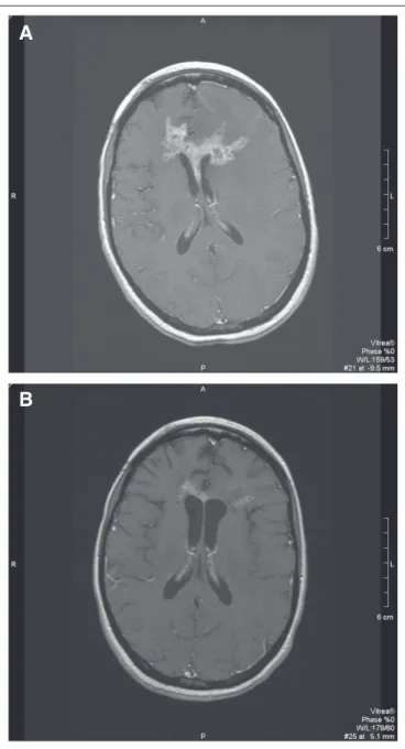
Patients were assessed for toxicity according to National Cancer Institute Common Terminology Criteria for Adverse Events, version 3.0 (NCI-CTCAE v. 3.0). Staging procedures were completed every 8 weeks and included brain magnetic resonance imaging (MRI) and computed tomography (CT) scans of the chest, abdomen, and pelvis. MRI scans were performed using 3-mm slices for axial T1-weighted, contrast-enhanced images, and 5-mm slices for the other sequences. Patients continued study treatment until they withdrew consent, experienced unacceptable toxicity, or had progressive dis-ease (PD).
The primary end point was objective response (complete response [CR] plus partial response [PR]) in the CNS. All measurable lesions, up to a maximum of five target lesions, were assessed. CNS responses were classified accord-ing to modified Response Evaluation Criteria in Solid Tumors (RECIST). CR was defined as the disappearance of all target and nontarget lesions. PR was defined as at least a 30% decrease in the sum longest dimension (LD) of target lesionsandan absolute decrease of at least 5 mm in at least one target lesion. PD was defined as at least a 20% increase in the sum LD of target lesionsandan absolute increase in size of at least 5 mm in at least one target lesion,orthe appearance of one or more new lesions of at least 6 mm in size. Non-CNS response was assessed by RECIST (CR, disappearance of all measurable and nonmeasurable disease; PR,ⱖ30% decrease in sum LD of target lesions; PD,ⱖ20% increase in sum LD of target lesions). Date of progression was recorded as the first documented progression at any site (either CNS or non-CNS), as assessed by the local investigator. Patients were considered to have progressed if they were taken off study for clinical deterioration or died as a result of any cause, regardless of whether there was documented radiographic evidence of progression.
Evaluation of CNS lesions for response categorization was performed centrally at the Tumor Imaging Metrics Core of Dana-Farber/Harvard Cancer Center. For RECIST, images were transferred to a Voxar imaging workstation (Barco, Kortrijk, Belgium), where target lesions were measured using a linear digital caliper tool.
For volumetric analyses, MRI scans were transmitted to a Vitrea2 work-station (Vital Images, Minnetonka, MN). The contrast-enhancing portions of the target lesions were outlined across all MRI image slices in which the lesion appeared, and edited manually to fit the exact perimeter. The software calcu-lated the tumor volumes by multiplying the outlined area by the slice thickness, and then by adding values across slices.
Patients were accrued in a two-stage design. The accrual goal was 37 patients (n⫽12, first stage; n⫽25, second stage); at least one CNS response was required in the first 12 patients to proceed to full accrual. The protocol-stipulated criteria indicated that four responders among 37 patients would be considered adequate to justify further study. With this study design, the trial had a 90% chance of positive findings if the true response rate was 20%, and a 10% chance of positive findings if the true response rate was 5%. Calculation of CIs was performed according to the Atkinson and Brown procedure.18
Time-to-event variables were summarized using the Kaplan-Meier method. The point-wise confidence curves for time-to-event variables were generated using the Greenwood formula. The estimation of the median and its CI was as described by Therneau and Grambsch.19Comparison of time-to-event
vari-ables between subgroups was performed using the log-rank test.
RESULTS
Patients and Treatment Characteristics
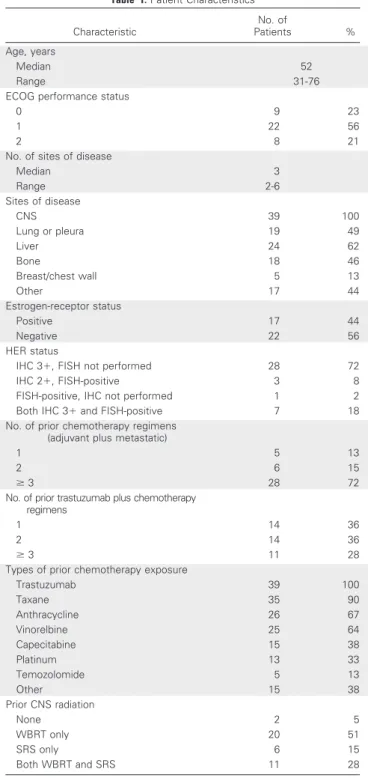
A CNS response was observed in the third patient enrolled onto the study; hence the study proceeded to full accrual. Thirty-nine women were enrolled (two more than the accrual goal because they had already consented to enter the study when the 37th patient was registered). Table 1 lists the clinical characteristics of these women. All
Eligibility
Patientswererequiredtobeatleast18yearsofage,providewritten informedconsent,andhaveHER-2–positivebreastcancer,definedas3⫹ immunohistochemistryorevidenceofgeneamplificationbyfluorescencein situhybridization.Priortrastuzumabwasrequired.Patientswereeligibleif theyhaddocumentedCNSprogressionafterWBRT,SRS,orboth.Patients werealsoeligibleiftheyhadnotpreviouslyreceivedradiationtherapy, pro-videdthattheywereasymptomatic.
Eligiblepatientshadatleastonemeasurablelesioninthebrain(defined asanylesionⱖ1.0cminlongestdimension),anEasternCooperative Oncol-ogyGroupperformancestatus0to2,lifeexpectancyⱖ12weeks,theabilityto swalloworalmedications,andtheabsenceofapriormalignancybesidesbreast cancerunlesstreatedwithcurativeintent.Patientswererequiredtohavealeft ventricularejectionfraction(LVEF)withininstitutionalnormallimits, abso-luteneutrophilcountofatleast1,000/L,plateletcountofatleast75,000/L, bilirubinnomorethan1.5⫻upperlimitofnormal(ULN),ASTandALTno morethan5⫻ULN,andcreatinineclearanceofatleast25mL/min.Patients withleptomeningealcarcinomatosisastheonlysiteofCNSinvolvement wereexcluded.
Allradiotherapy,chemotherapy,and/orhormonaltherapyhadtobe completedatleast2weeksbeforeprotocoltreatment.Concurrent administra-tionofotherantineoplasticagentswasnotpermitted.Patientswereexcluded fromtakinginducersorinhibitorsofCYP3A4,includingphenytoin. Cortico-steroidswerepermitted.
Thisstudywasconductedinaccordancewithguidelinesestablishedby theUnitedStatesDepartmentofHealthandHumanServices.TheNational CancerInstituteCancerTherapyEvaluationProgramandinstitutionalreview boardsofDana-FarberCancerInstitute/HarvardCancerCenter(Boston, MA),UniversityofNorthCarolinaatChapelHill(ChapelHill,NC),and GeorgetownUniversity(Washington,DC)approvedthestudy.Patientswere enrolledbetweenSeptember2004andSeptember2005.
Treatment Plan
Thestartingdoseoflapatinibwas750mgtwicedailyadministeredorally incontinuous4-weekcycles.Lapatinibdosewasheld,thenreducedto500mg twiceadayandsubsequentlyto1,250mgoncedaily,forinitialorrecurrent grade3to4toxicity,orclinicallysignificantgrade2toxicity.
but two patients had experienced progression after CNS-directed ra-diation therapy. Among patients who had previously been irradiated, median time from last radiotherapy was 5.97 months. Patients re-ceived a median of two prior trastuzumab-containing chemother-apy regimens.
At the time of study analysis, 136 4-week cycles of treatment had been administered. Fifteen patients (38%) required at least one dose reduction, most commonly for diarrhea. Seventy-four percent of cy-cles were administered at full dose. In 23% of cycy-cles, lapatinib dose was reduced to 500 mg twice a day. One patient required a further dose reduction to 1,250 mg once daily.
Toxicity
All patients were assessable for toxicity. The worst grades of treatment-related toxicity are listed in Table 2. The most common adverse event was diarrhea, which improved with supportive mea-sures and/or dose reductions in most cases.
Three patients were removed from study because of toxicity. One patient experienced grade 3 elevation of transaminases; one patient experienced grade 3 diarrhea and anorexia; and one patient developed endocarditis, which was ultimately judged unlikely to be related to lapatinib.
One patient died suddenly while receiving her third cycle of lapatinib. Restaging studies performed after cycle 2 had demon-strated stable disease. The research staff spoke with the patient the day before her death and she reported feeling well, with the excep-tion of a mild headache. An autopsy was declined, and the cause of death is unknown.
Cardiac Surveillance and Cardiotoxicity
No patient developed symptomatic congestive heart failure on study. Four patients developed asymptomatic declines in LVEF to less than 50% (range, 44% to 49%). Of the four patients, only one experi-enced a 10% or greater decline in LVEF from baseline. In two patients, lapatinib was continued per protocol, and repeat evaluation demon-strated normalization of LVEF. One patient was taken off study after two cycles because of PD, and was lost to follow-up before reevalua-tion of LVEF. One patient died of an acute intracerebral hemorrhage 2 weeks after the last dose of study drug, before repeat cardiac evalua-tion. The event was judged related to anticoagulation with low molecular-weight heparin and warfarin for a pulmonary embolus in the setting of progressive CNS disease.
Efficacy
The principal end point of the study was the rate of CNS response by RECIST. Of 39 patients, one achieved a PR, for an overall CNS response rate of 2.6% (95% conditional CI, 0.21% to
Table 1.Patient Characteristics
Characteristic
No. of
Patients %
Age, years
Median 52
Range 31-76
ECOG performance status
0 9 23
1 22 56
2 8 21
No. of sites of disease
Median 3
Range 2-6
Sites of disease
CNS 39 100
Lung or pleura 19 49
Liver 24 62
Bone 18 46
Breast/chest wall 5 13
Other 17 44
Estrogen-receptor status
Positive 17 44
Negative 22 56
HER status
IHC 3⫹, FISH not performed 28 72
IHC 2⫹, FISH-positive 3 8
FISH-positive, IHC not performed 1 2
Both IHC 3⫹and FISH-positive 7 18
No. of prior chemotherapy regimens (adjuvant plus metastatic)
1 5 13
2 6 15
ⱖ3 28 72
No. of prior trastuzumab plus chemotherapy regimens
1 14 36
2 14 36
ⱖ3 11 28
Types of prior chemotherapy exposure
Trastuzumab 39 100
Taxane 35 90
Anthracycline 26 67
Vinorelbine 25 64
Capecitabine 15 38
Platinum 13 33
Temozolomide 5 13
Other 15 38
Prior CNS radiation
None 2 5
WBRT only 20 51
SRS only 6 15
Both WBRT and SRS 11 28
Abbreviations: ECOG, Eastern Cooperative Oncology Group; HER-2, human epidermal growth factor receptor 2; IHC, immunohistochemistry; FISH, fluo-rescence in situ hybridization; WBRT, whole-brain radiotherapy; SRS, stereo-tactic radiosurgery.
Table 2.Worst Grade of Toxicity on Study (N⫽39)
Toxicity
%
Grade 2 Grade 3 Grade 4
Diarrhea 23 21 0
Fatigue 15 15 0
Headache 8 10 0
Rash 10 5 0
Anorexia 8 3 0
AST/ALT 5 8 0
Nausea 5 3 0
NOTE. Frequency of treatment-related toxicity according to National Cancer Institute Common Terminology Criteria for Adverse Events, version 3.0, worst grade per patient.
26%; Fig 1; Table 3). This patient had received her last radiotherapy 12.6 months before study entry and did not require corticosteroids during the study period.
Sixteen patients (41%) had measurable non-CNS disease at base-line. Four (25%) achieved a PR in non-CNS sites (Table 4). Of the patients that responded in non-CNS sites, all were eventually taken off study for CNS progression. The relatively small proportion of pa-tients with measurable non-CNS disease likely reflects that the study attracted a group of patients whose dominant problem was CNS progression.
Time to progression (TTP) and overall survival are shown in Figure 2. Median TTP was 3.0 months (95% CI, 2.3 to 3.7 months).
For the patient with CNS objective response, TTP was 11.3 months. Seven patients (18%) were free of any progression at 16 weeks. One
additional patient had stable CNS disease at 16 weeks, but progressed in her adrenal lesions. Of these patients, three received corticosteroids at some point during the study. Two patients sustained temporary increases in corticosteroid dose beginning during cycle 1 of therapy, but which subsequently decreased, and one patient had a sustained decrease in steroid dose from baseline.
At the time of this final study analysis, all patients had completed protocol-directed therapy. Patients were removed from the study for
PD in the CNS only (n⫽24), PD in non-CNS sites only (n⫽4), PD
in both CNS and non-CNS sites (n⫽5), toxicity (n⫽3), death (n⫽
1), or other (n⫽2; physician-patient decision and generalized clinical
deterioration, respectively).
Volumetric Analysis of CNS Lesions
An exploratory analysis was conducted of volumetric changes in CNS target lesions. For this analysis, 34 of 39 patients were included. Of the remaining five patients, one patient was excluded because of technical problems that did not allow calculation of lesion volumes, and four were excluded because they were taken off study before the week 8 evaluation.
Figure 3 illustrates the best volumetric change among the 34 patients who were included in the analysis. Three patients achieved at least 30% volumetric reductions in CNS target lesions, and an addi-tional seven patients achieved reductions of 10% to 30%. It is not known what cutoff of volumetric change is clinically significant. We therefore conducted an exploratory analysis to correlate volumetric change and TTP. To avoid bias, we employed a “landmark method,” restricting the analysis to patients who had a follow-up MRI at 8 weeks
and no progression before or at that time point (n⫽27). Patients with
at least a certain percentage of volumetric reduction at the 8-week point were compared with patients with lesser or no reduction at 8 weeks in terms of TTP from the end of 8 weeks (rather than from protocol entry). We found a trend toward a longer TTP for patients
Table 3.Overall CNS Activity Rate for Lapatinib
Clinical Category
Response
No. %
Overall response 1 2.6
Complete response 0 0
Partial response 1 2.6
Stable diseaseⱖ16 weeks (in both CNS and non-CNS sites)
6 15.4
Table 4.Overall Non-CNS Activity Rate for Lapatinib
Clinical Category No. of Patients
Measurable disease 16
Overall response 4
Complete response 0
Partial response 4
Nonmeasurable disease 23
NOTE. Patients were not required to have measurable non-CNS disease to enter onto the study.
A
B
with at least 30% volumetric reduction versus others (median TTP
from 8-week MRI, 1.8v5.4 months;P⫽.16). Similar results were seen
when patients were dichotomized according to at least 10%
volumet-ric reduction versus others (median TTP from 8-week MRI, 1.8v3.5
months;P⫽.04).
DISCUSSION
In this prospective, multicenter, phase II study, we evaluated the safety and efficacy of lapatinib in women with HER-2–positive breast cancer and brain metastases. One patient achieved a PR in the CNS by
RECIST, for a response rate of 2.6%. The study did not meet the primary efficacy goal, which would have required at least four re-sponders. However, we did observe volumetric reductions in CNS target lesions in some patients. On the basis, in part, of results from this study, a large, international study was initiated to further evaluate the role of lapatinib monotherapy in women with HER-2–positive breast
cancer and progressive CNS disease after cranial radiotherapy.20This
study will utilize volumetric changes as a primary end point. The CNS response rate in this study was similar to that ob-served for lapatinib for systemic disease in phase II studies of
trastuzumab-refractory patients.21,22In a phase III study evaluating
capecitabine versus capecitabine plus lapatinib in women with meta-static disease, the addition of lapatinib led to a statistically significant improvement in TTP, and numerically fewer patients experienced
CNS progression.23Given these data, and past data demonstrating
improvements in response rate when cytotoxic agents are added to trastuzumab (compared with trastuzumab monotherapy), future studies of lapatinib for CNS disease should include an evaluation of lapatinib combined with cytotoxic agents with the potential to cross
the blood-tumor barrier.24-26Lapatinib could also be evaluated with
cranial radiotherapy, on the basis of preclinical data indicating that
lapatinib may act as a radiosensitizer.27Studies of lapatinib in less
refractory patients or studies to determine whether the agent can prevent the appearance of CNS disease may also be of interest.
Our study had several limitations. First, we cannot exclude the possibility that CNS penetration of lapatinib was suboptimal. Indeed, since the study was initiated, a high incidence of CNS-only recurrence in non–small-cell lung cancer patients with an initial response to
therapy to gefitinib has been reported.28-30In designing the trial, we
chose the dosing schedule on the basis of pharmacokinetic data indi-cating that the same total daily dose, when divided twice a day, leads to approximately twice the area under the curve compared with a once
daily schedule.31As in any phase II trial, our results apply only to the
dose and schedule that were used. Further optimization of lapatinib dose was outside the scope of this trial. However, the rate of grade 3 diarrhea was higher in this study compared with other trials of
lapa-tinib, which supports the pharmacokinetic data.32In phase I studies,
A
1.0
0.8
0.6
0.4
0.2
0 2 4 6 8 10
Time to Progression (months)
Probability of Time to Progression
B
1.0
0.8
0.6
0.4
0.2
0 5 10 15
Survival (months)
Probability of Survival
Fig 2.(A) Proportion of patients without progression. Progression in either CNS or non-CNS sites is counted as progressive disease. Dashed lines indicate the upper and lower bounds of the 95% CI. (B) Overall survival for all 39 patients, from time of study entry. Dashed lines indicate the upper and lower bounds of the 95% CI.
Change From Baseline (%)
-100 -50 0 50 100 150 200
*
* **
Fig 3.Best volumetric change in sum of CNS target lesions with lapatinib. Each bar represents an individual patient with at least a baseline and week 8 magnetic resonance imaging scan (n⫽34). Pale yellow bars indicate patients with 10% to 30% volumetric reduction. Yellow bars indicate patients with at least 30% volumetric reduction. The arrow denotes the patient who achieved a partial response in the CNS by Response Evaluation Criteria in Solid Tumors. (*) Concomitant increase in corticosteroid dose at the time of the restaging scan.
dose-limiting toxicity was reached at 900 mg twice a day; therefore, from a practical standpoint, we do not believe the dose could be
further escalated without a corresponding increase in toxicity.33
Another limitation was the choice of CNS response criteria. We prospectively developed modified RECIST to evaluate CNS response, but acknowledge that these are not the standard neuro-oncology cri-teria. In a patient population with limited options, we did not want to take patients off study for small changes in size that could be in the range of interobserver variation, and therefore required a 5-mm abso-lute change in the size of at least one CNS target lesion, in addition to standard RECIST. Conversely, we aimed to be more conservative in ascertaining response (for example, a patient with a single target lesion measuring 10 mm would be required to have shrinkage to 5 mm or less
to qualify as a PR [ie,⬎30% decrease in LD and at least 5 mm absolute
change]). Volumetric measurements may ultimately provide a more accurate estimate of tumor burden; however, it is unclear what degree
of volumetric change is clinically significant.21,34,35In this study, there
was a suggestion of clinical benefit, as ascertained by longer TTP, associated with volumetric changes of at least 10%. This hypothesis could be tested prospectively in future trials, as in trials of biologic agents, TTP may be a more relevant end point than objective response. Although we believe that the absence of progression at 16 weeks in 18% of patients is suggestive of clinical benefit, we are not aware of any studies in which patients with progressive CNS disease are fol-lowed with imaging studies without treatment. Without an untreated reference population, we cannot make any definite conclusions. Of note, the median time from last radiation to study entry was 5.9 months, and patients were required to have progressive CNS disease for study entry. Therefore, we do not believe that the stabilizations that were observed were a result of prior radiation.
In conclusion, to our knowledge, this is the first prospective study evaluating a targeted agent for the treatment of brain metastases in patients with HER-2–positive breast cancer. Results of this study have led to a multicenter phase II trial of lapatinib in patients with HER-2– positive brain metastases, and studies of lapatinib in combination with cytotoxic agents are being initiated. This trial also underscores the feasibility of medical oncology treatment trials for brain metastases, and the urgent need to identify new treatment approaches for patients with CNS involvement, particularly for patients with HER-2–posi-tive disease.
AUTHORS’ DISCLOSURES OF POTENTIAL CONFLICTS OF INTEREST
Although all authors completed the disclosure declaration, the following author(s) indicated a financial or other interest that is relevant to the subject
matter under consideration in this article. Certain relationships marked with a “U” are those for which no compensation was received; those relationships marked with a “C” were compensated. For a detailed description of the disclosure categories, or for more information about ASCO’s conflict of interest policy, please refer to the Author Disclosure Declaration and the Disclosures of Potential Conflicts of Interest section in Information for Contributors.
Employment or Leadership Position:NoneConsultant or Advisory Role:Nancy U. Lin, GlaxoSmithKline (C); Lisa A. Carey, Genentech (U), GlaxoSmithKline (U), Pfizer (U), Bristol Meyers Squib (U); John W. Henson, GlaxoSmithKline (C); Elizabeth Kasparian, GlaxoSmithKline (C); Eric P. Winer, Genentech (C), GlaxoSmithKline (C)Stock Ownership:NoneHonoraria:Minetta C. Liu, GlaxoSmithKline; Elizabeth Kasparian, GlaxoSmithKlineResearch Funding:Nancy U. Lin, GlaxoSmithKline; Lisa A. Carey, Genentech, GlaxoSmithKline, Bristol Meyers Squib; Minetta C. Liu, GlaxoSmithKline; Eric P. Winer, Genentech, GlaxoSmithKlineExpert Testimony:NoneOther Remuneration:None
AUTHOR CONTRIBUTIONS
Conception and design:Nancy U. Lin, Lisa A. Carey, Minetta C. Liu, Jerry Younger, Matthew Ewend, Elizabeth Bullitt, Annick D. Van den Abbeele, Rebecca Gelman, David G. Kirsch, Fred Hochberg, Eric P. Winer
Financial support:Nancy U. Lin, Lisa A. Carey, Annick D. Van den Abbeele, Eric P. Winer
Administrative support:Annick D. Van den Abbeele, Elizabeth Kasparian, Ann Crawford
Provision of study materials or patients:Nancy U. Lin, Lisa A. Carey, Minetta C. Liu, Jerry Younger, Steven E. Come, Harold J. Burstein, Elizabeth Kasparian, Fred Hochberg, Eric P. Winer
Collection and assembly of data:Nancy U. Lin, Lisa A. Carey, Minetta C. Liu, Jerry Younger, Annick D. Van den Abbeele, Elizabeth Kasparian, Ann Crawford
Data analysis and interpretation:Nancy U. Lin, Lisa A. Carey, Minetta C. Liu, Jerry Younger, Steven E. Come, Matthew Ewend, Gordon J. Harris, Elizabeth Bullitt, Annick D. Van den Abbeele, John W. Henson, Xiaochun Li, Rebecca Gelman, Eric P. Winer
Manuscript writing:Nancy U. Lin, Lisa A. Carey, Minetta C. Liu, Jerry Younger, Steven E. Come, Gordon J. Harris, Annick D. Van den Abbeele, John W. Henson, Xiaochun Li, Rebecca Gelman, Harold J. Burstein, Fred Hochberg, Eric P. Winer
Final approval of manuscript:Nancy U. Lin, Lisa A. Carey, Minetta C. Liu, Jerry Younger, Steven E. Come, Matthew G. Ewend, Gordon J. Harris, Elizabeth Bullitt, Annick D. Van den Abbeele, John W. Henson, Xiaochun Li, Rebecca Gelman, Harold J. Burstein, Elizabeth Kasparian, David G. Kirsch, Ann Crawford, Fred Hochberg, Eric P. Winer
REFERENCES trastuzumab for metastatic breast cancer. Br J Can-cer 91:639-643, 2004
4.Lin NU, Bellon JR, Winer EP: CNS metasta-ses in breast cancer. J Clin Oncol 22:3608-3617, 2004
5.Lin NU, Winer EP: Brain metastases: The HER2 paradigm. Clin Cancer Res 13:1648-1655, 2007
6.Romond EH, Perez EA, Bryant J, et al: Tras-tuzumab plus adjuvant chemotherapy for operable HER2-positive breast cancer. N Engl J Med 353: 1673-1684, 2005
7.Pestalozzi BC, Brignoli S: Trastuzumab in CSF. J Clin Oncol 18:2349-2351, 2000
8.Boogerd W, Dalesio O, Bais EM, et al: Re-sponse of brain metastases from breast cancer to systemic chemotherapy. Cancer 69:972-980, 1992
9.Trudeau ME, Crump M, Charpentier D, et al: Temozolomide in metastatic breast cancer (MBC): A phase II trial of the National Cancer Institute of Canada - Clinical Trials Group (NCIC-CTG). Ann On-col 17:952-956, 2006
10.Rivera E, Meyers C, Groves M, et al: Phase I study of capecitabine in combination with temozolomide in the treatment of patients with brain metastases from breast carcinoma. Cancer 107:1348-1354, 2006
1. Slamon DJ, Clark GM, Wong SG, et al: Human breast cancer: Correlation of relapse and survival withamplificationoftheHER-2/neuoncogene. Sci-ence 235:177-182, 1987
2. Bendell JC, Domchek SM, Burstein HJ, et al: Central nervous system metastases in women who receive trastuzumab-basedtherapyformetastatic breast carcinoma. Cancer 97:2972-2977, 2003
11.Oberhoff C, Kieback DG, Wurstlein R, et al: Effectiveness and tolerance of primary topotecan chemotherapy in patients with breast cancer and brain metastases: Results of a pilot study. Proc Am Soc Clin Oncol 20:72b, 2001 (abstr 2036)
12.Xia W, Mullin RJ, Keith BR, et al: Anti-tumor activity of GW572016: a dual tyrosine kinase inhibi-tor blocks EGF activation of EGFR/erbB2 and down-stream Erk1/2 and AKT pathways. Oncogene 21: 6255-6263, 2002
13.Burstein HJ, Storniolo AM, Salazar VM, et al: A phase II, open-label, multicenter study of lapatinib (GW572016) in two cohorts of patients with ad-vanced or metastatic breast cancer who have pro-gressed while receiving trastuzumab-containing regimens: Interim analysis. Presented at the 29th Annual Congress of the European Society for Med-ical Oncology, October 29 –November 2, 2004, Vi-enna, Austria
14.Katz A, Zalewski P: Quality-of-life benefits and evidence of antitumour activity for patients with brain metastases treated with gefitinib. Br J Cancer 89:S15-S18, 2003 (suppl)
15.Ceresoli GL, Cappuzzo F, Gregorc V, et al: Gefitinib in patients with brain metastases from non-small-cell lung cancer: A prospective trial. Ann Oncol 15:1042-1047, 2004
16.Stewart DJ, Mikhael NZ, Nair RC, et al: Plati-num concentrations in human autopsy tumor sam-ples. Am J Clin Oncol 11:152-158, 1988
17.Stewart DJ, Molepo JM, Green RM, et al: Factors affecting platinum concentrations in human surgical tumour specimens after cisplatin. Br J Can-cer 71:598-604, 1995
18.Atkinson EN, Brown BW: Confidence limits for probability of response in multistage phase II clinical trials. Biometrics 41:741-744, 1985
19.Therneau TM, Grambsch PM: Modeling Sur-vival Data. New York, NY, Springer, 2000
20.Lin NU, Dieras V, Paul D, et al: EGF105084, a phase II study of lapatinib for brain metastases in patients (pts) with HER2⫹breast cancer following trastuzumab (H) based systemic therapy and cranial radiotherapy (RT). J Clin Oncol 25:35s, 2007 (suppl; abstr 1012)
21.Blackwell KL, Kaplan EH, Franco SX, et al: A phase II, open-label, multicenter study of GW572016 in patients with trastuzumab-refractory metastatic breast cancer. J Clin Oncol 22:196s, 2004 (suppl; abstr 3006)
22.Blackwell KL, Burstein HJ, Pegram M, et al: De-termining relevant biomarkers from tissue and serum that may predict response to single agent lapatinib in trastuzumab refractory metastatic breast cancer. J Clin Oncol 23:193s, 2005 (suppl; abstr 3004)
23.Geyer CE, Forster J, Lindquist D, et al: Lapa-tinib plus capecitabine for HER2-positive advanced breast cancer. N Engl J Med 355:2733-2743, 2006 24.Vogel CL, Cobleigh MA, Tripathy D, et al: Efficacy and safety of trastuzumab as a single agent in first-line treatment of HER2-overexpressing met-astatic breast cancer. J Clin Oncol 20:719-726, 2002 25.Slamon DJ, Leyland-Jones B, Shak S, et al: Use of chemotherapy plus a monoclonal antibody against HER2 for metastatic breast cancer that overexpresses HER2. N Engl J Med 344:783-792, 2001
26.Burstein HJ, Harris LN, Marcom PK, et al: Trastuzumab and vinorelbine as first-line therapy for HER2-overexpressing metastatic breast cancer: Multicenter phase II trial with clinical outcomes, analysis of serum tumor markers as predictive fac-tors, and cardiac surveillance algorithm. J Clin Oncol 21:2889-2895, 2003
27.Zhou H, Kim YS, Peletier A, et al: Effects of the EGFR/HER2 kinase inhibitor GW572016 on EGFR- and HER2-overexpressing breast cancer cell
line proliferation, radiosensitization, and resistance. Int J Radiat Oncol Biol Phys 58:344-352, 2004
28.Omuro AM, Kris MG, Miller VA, et al: High incidence of disease recurrence in the brain and leptomeninges in patients with nonsmall cell lung carcinoma after response to gefitinib. Cancer 103: 2344-2348, 2005
29.Namba Y, Kijima T, Yokota S, et al: Gefitinib in patients with brain metastases from non-small-cell lung cancer: Review of 15 clinical cases. Clin Lung Cancer 6:123-128, 2004
30.Jackman DM, Holmes AJ, Lindeman N, et al: Response and resistance in a non-small-cell lung cancer patient with an epidermal growth factor receptor mutation and leptomeningeal metastases treated with high-dose gefitinib. J Clin Oncol 24: 4517-4520, 2006
31.Nativg B, Paul E, Koch K, et al: GW572016 Investigator’s Brochure, version 5. Upper Provi-dence, PA, GlaxoSmithKline, 2005, pp 75
32.Gomez HL, Chavez MA, Doval DC, et al: A Phase II, randomized trial using the small mole-cule tyrosine kinase inhibitor lapatinib as a first-line treatment in patients with FISH-positive advanced or metastatic breast cancer. J Clin Oncol 23:203s, 2005 (suppl; abstr 3046)
33.Burris HA, Taylor C, Jones SE, et al: A phase I study of GW572016 in patients with solid tumors. Proc Am Soc Clin Oncol 22:248, 2003 (abstr 994)
34.Miranpuri SS, Schulz CA, Chappell RJ, et al: Comparison of methods for response analysis of central nervous system neoplasms. J Radiosurg 2:153-161, 2004
35.Kilpatrick MR, Lin NU, Smith JK, et al: Supe-riority of bi-dimensional measurements compared to RECIST in measuring response of metastatic brain tumors. J Clin Oncol 24:68s, 2006 (suppl; abstr 1542)
■ ■ ■
Acknowledgment
We thank Janet Dancey, MD, and Steve Rubin, MD, for their expertise and guidance; William Hanlon, Trinity Urban, and Jeffrey Yap, PhD, for assistance with imaging analysis; and Kristen Kindsvogel, Judy Fogleman, Karla Hurtley, and Julie Castle for assistance with study conduct.