35
Protists
Concept Outline
35.1 Eukaryotes probably arose by endosymbiosis.
Endosymbiosis. Mitochondria and chloroplasts are
thought to have arisen by endosymbiosis from aerobic bacteria.
35.2 The kingdom Protista is by far the most diverse of any kingdom.
The Challenge of Classifying the Protists. There is no
general agreement among taxonomists about how to classify the protists.
General Biology of the Protists. Protista contains
members exhibiting a wide range of methods of locomotion, nutrition, and reproduction.
35.3 Protists can be categorized into five groups.
Five Groups of Protists. The 15 major phyla of protists
can be conveniently discussed in seven general groups that share certain characteristics.
Heterotrophs with No Permanent Locomotor
Apparatus. Amoebas and other sarcodines have no
permanent locomotor apparatus.
Photosynthetic Protists. The flagellates are
photosynthesizers that propel themselves through the water with flagella. Diatoms are photosynthesizers with hard shells of silica. Algae are photosynthetic protists, some are multicellular.
Heterotrophs with Flagella. Flagellates propel
themselves through the water. Single cells with many cilia, the ciliates possess highly complex and specialized
organelles.
Nonmotile Spore-Formers. The sporozoans are
nonmotile parasites that spread by forming spores.
Heterotrophs with Restricted Mobility. Heterotrophs
with restricted mobility, molds have cell walls made of carbohydrate.
F
or more than half of the long history of life on earth, all life was microscopic in size. The biggest organisms that existed for over 2 billion years were single-celled bac-teria fewer than 6 micrometers thick. The first evidence of a different kind of organism is found in tiny fossils in rock 1.5 billion years old. These fossil cells are much larger than bacteria (some as big as 60 micrometers in diameter) and have internal membranes and what appear to be small, membrane-bounded structures. Many have elaborate shapes, and some exhibit spines or filaments. These new, larger fossil organisms mark one of the most important events in the evolution of life, the appearance of a new kind of organism, the eukaryote (figure 35.1). Flexible and adaptable, the eukaryotes rapidly evolved to produce all of the diverse large organisms that populate the earth today, including ourselves—indeed, all organisms other than bac-teria are eukaryotes.FIGURE 35.1
Volvox,a colonial protist. The protists are a large, diverse group of primarily single-celled organisms, a group from which the other three eukaryotic kingdoms each evolved.
contained a complex system of inter-nal membranes. The inner membrane of mitochondria is folded into numer-ous layers, resembling the folded membranes of nonsulfur purple bacte-ria; embedded within this membrane are the proteins that carry out oxida-tive metabolism. The engulfed bacte-ria became the interior portion of the mitochondria we see today. Host cells were unable to carry out the Krebs cycle or other metabolic reactions necessary for living in an atmosphere that contained increasing amounts of oxygen before they had acquired these bacteria.
During the billion and a half years in which mitochondria have existed as en-dosymbionts within eukaryotic cells, most of their genes have been trans-ferred to the chromosomes of the host cells—but not all. Each mitochondrion still has its own genome, a circular, closed molecule of DNA similar to that found in eubacteria, on which is located genes encoding the essential proteins of oxidative metabolism. These genes are transcribed within the mitochondrion, using mitochondrial ribosomes that are smaller than those of eukaryotic cells, very much like bacte-rial ribosomes in size and structure. Mitochondria divide by simple fission, just as bacteria do, replicating and sorting their DNA much as bacteria do. However, nuclear genes direct the process, and mitochondria cannot be grown out-side of the eukaryotic cell, in cell-free culture.
The theory of endosymbiosis has had a controversial history but has now been accepted by all but a few biolo-gists. The evidence supporting the theory is so extensive that in this text we will treat it as established.
What of mitosis, the other typical eukaryotic process that Pelomyxa lacks? The mechanism of mitosis, now so common among eukaryotes, did not evolve all at once. Traces of very different, and possibly intermediate, mechanisms survive today in some of the eukaryotes. In fungi and some groups of protists, for example, the nu-clear membrane does not dissolve and mitosis is confined to the nucleus. When mitosis is complete in these organ-isms, the nucleus divides into two daughter nuclei, and only then does the rest of the cell divide. This separate nuclear division phase of mitosis does not occur in most protists, or in plants or animals. We do not know if it represents an intermediate step on the evolutionary jour-ney to the form of mitosis that is characteristic of most
694
Endosymbiosis
What was the first eukaryote like? We cannot be sure, but a good model is
Pelomyxa palustris, a single-celled, nonphotosynthetic organism that ap-pears to represent an early stage in the evolution of eukaryotic cells (fig-ure 35.2). The cells of Pelomyxa are much larger than bacterial cells and contain a complex system of internal membranes. Although they resemble some of the largest early fossil eukary-otes, these cells are unlike those of any other eukaryote: Pelomyxa lacks mitochondria and does not undergo mitosis. Its nuclei divide somewhat as do those of bacteria, by pinching apart into two daughter nuclei, around which new membranes form. Al-though Pelomyxa cells lack mitochon-dria, two kinds of bacteria living within them may play the same role that mitochondria do in all other eu-karyotes. This primitive eukaryote is so distinctive that it is assigned a phy-lum all its own, Caryoblastea.
Biologists know very little of the origin of Pelomyxa,except that in many
of its fundamental characteristics it resembles the archae-bacteria far more than the euarchae-bacteria. Because of this gen-eral resemblance, it is widely assumed that the first eukary-otic cells were nonphotosynthetic descendants of archaebacteria.
What about the wide gap between Pelomyxa and all other eukaryotes? Where did mitochondria come from? Most biologists agree with the theory of endosymbiosis, which proposes that mitochondria originated as symbiotic, aerobic (oxygen-requiring) eubacteria (figure 35.3). Sym-biosis (Greek, syn,“together with” + bios,“life”) means liv-ing together in close association. Recall from chapter 5 that mitochondria are sausage-shaped organelles 1 to 3 microm-eters long, about the same size as most eubacteria. Mito-chondria are bounded by two membranes. Aerobic eubac-teria are thought to have become mitochondria when they were engulfed by ancestral eukaryotic cells, much like
Pelomyxa, early in the history of eukaryotes.
The most similar eubacteria to mitochondria today are the nonsulfur purple bacteria, which are able to carry out oxidative metabolism (described in chapter 9). In mi-tochondria, the outer membrane is smooth and is thought to be derived from the endoplasmic reticulum of the host cell, which, like Pelomyxa, may have already
35.1
Eukaryotes probably arose by endosymbiosis.
FIGURE 35.2
Pelomyxa palustris.This unique, amoeba-like protist lacks mitochondria and does not undergo mitosis. Pelomyxamay represent a very early stage in the evolution of eukaryotic cells.
eukaryotes today or if it is simply a different way of solv-ing the same problem. There are no fossils in which we can see the interiors of dividing cells well enough to be able to trace the history of mitosis.
Endosymbiosis Is Not Rare
Many eukaryotic cells contain other endosymbiotic bacteria in addition to mitochondria. Plants and algae contain chloroplasts, bacteria-like organelles that were apparently derived from symbiotic photosynthetic bacteria. Chloro-plasts have a complex system of inner membranes and a cir-cle of DNA. Centrioles, organelles associated with the as-sembly of microtubules, resemble in many respects spirochete bacteria, and they contain bacteria-like DNA in-volved in the production of their structural proteins.
While all mitochondria are thought to have arisen from a single symbiotic event, it is difficult to be sure with
chloro-plasts. Three biochemically distinct classes of chloroplasts exist, each resembling a different bacterial ancestor. Red algae possess pigments similar to those of cyanobacteria; plants and green algae more closely resemble the photosyn-thetic bacteria Prochloron;while brown algae and other pho-tosynthetic protists resemble a third group of bacteria. This diversity of chloroplasts has led to the widely held belief that eukaryotic cells acquired chloroplasts by symbiosis at least three different times. Recent comparisons of chloroplast DNA sequences, however, suggest a single origin of chloro-plasts, followed by very different postendosymbiotic histo-ries. For example, in each of the three main lines, different genes became relocated to the nucleus, lost, or modified.
The theory of endosymbiosis proposes that mitochondria originated as symbiotic aerobic eubacteria.
Photosynthetic bacterium
Ancestral eukaryotic cell Eukaryotic cell with mitochondrion Internal membrane system Aerobic bacterium Mitochondrion Chloroplast
Eukaryotic cell with chloroplasts
Endosymbiosis
Endosymbiosis
FIGURE 35.3
The theory of endosymbiosis.Scientists propose that ancestral eukaryotic cells, which already had an internal system of membranes,
engulfed aerobic eubacteria, which then became mitochondria in the eukaryotic cell. Chloroplasts may also have originated this way, with eukaryotic cells engulfing photosynthetic eubacteria.
The Challenge of Classifying
the Protists
Protists are the most diverse of the four kingdoms in the domain Eukaryota. The kingdom Protista contains many unicellular, colonial, and multicellular groups. Probably the most important statement we can make about the kingdom Protista is that it is an artificial group; as a matter of conve-nience, single-celled eukaryotic organisms have typically been grouped together into this kingdom. This lumps many very different and only distantly related forms to-gether. The “single-kingdom” classification of the Protista is not representative of any evolutionary relationships. The phyla of protists are, with very few exceptions, only dis-tantly related to one another.
New applications of a wide variety of molecular methods are providing important insights into the relationships among the protists. Of all the groups of organisms biolo-gists study, protists are probably in the greatest state of flux when it comes to classification. There is little consensus, even among experts, as to how the different kinds of protists should be classified. Are they a single, very diverse kingdom, or are they better considered as several different kingdoms, each of equal rank with animals, plants, and fungi?
Because the Protista are still predominantly considered part of one diverse, nonunified group, that is how we will treat them in this chapter, bearing in mind that biologists are rapidly gaining a better understanding of the evolution-ary relationships among members of the kingdom Protista (figure 35.4). It seems likely that within a few years, the tra-ditional kingdom Protista will be replaced by another more illuminating arrangement.
The taxonomy of the protists is in a state of flux as new information shapes our understanding of this kingdom.
696
35.2
The kingdom Protista is by far the most diverse of any kingdom.
Animals
Ciliates and dinoflagellates Euglenoids, cellular slime molds, water molds, trypanosomes Rhizopods, plasmodial slime molds, fungi (basidiomycetes) Plants
Fungi (ascomycetes) Red algae
Analysis Based on Ribosomal Subunits Red algae
KINGDOM CHROMISTA 10 protist phyla, including the more familiar 7 “heterokont” phyla: brown algae, slime nets, and diatoms
Photosynthetic protists (Volvox, Spirogyra ) and plants
Most rhizopods, water molds, diatoms, brown algae, heliozoans, slime nets Heterotrophic symbiotic flagellates (Trichomonas, Trichonympha, Giardia) Choanoflagellates and animals Amoeboflagellates and cellular slime molds
Euglenoids
Dinoflagellates and ciliates
Cladistic Analysis
(Prokaryotic Ancestor) Six Eukaryotic Kingdoms KINGDOM ARCHEZOA
Primitive amitochondrial forms, including Pelomyxa, Giardia
KINGDOM FUNGI Fungi and 1 phylum of saprophytic protists (chytridiomycota) KINGDOM ANIMALIA (no protist phyla) KINGDOM PLANTAE
Plants and 5 protist phyla,including the green algae (Volvox, Ulva, Spirogyra ) and the red algae
KINGDOM PROTOZOA 14 protist phyla,including hypermastigotes (Trichonympha), euglenoids, slime molds, choanoflagellates, dinoflagellates, ciliates, apicomplexans, rhizopods, heliozoans, foraminiferans, and radiolarians
FIGURE 35.4
The challenge of protistan classification. Three different
suggestions for protistan classification are presented, each adapted from the work of an authority in the field. Their great differences attest to the wide divergence of opinion within the field itself. The classification on the top is based on molecular variation in ribosomal subunits. The classification in the middle presents a cladistic analysis of a broad range of characters (including ribosomal subunits). The classification on the bottom outlines a more revolutionary reevaluation of the protists. Comparison of the three schemes reveals that some groups are commonly recognized as related (like ciliates and dinoflagellates), while the classification of others (like Giardia) is clearly in a state of flux.
General Biology
of the Protists
Protists are united on the basis of a single negative characteristic: they are not fungi, plants, or animals. In all other respects they are highly variable with no uniting features. Many are unicellular (figure 35.5), but there are numerous colonial and multicellular groups. Most are mi-croscopic, but some are as large as trees. They represent all symmetries, and exhibit all types of nutrition.
The Cell Surface
Protists possess a varied array of cell surfaces. Some protists, like amoebas, are surrounded only by their plasma membranes. All other protists have a plasma membrane but some, like algae and molds, are encased within strong cell walls. Still others, like di-atoms and forams, secrete glassy shells of silica.
Locomotor Organelles
Movement in protists is also accomplished by diverse mech-anisms. Protists move chiefly by either flagellar rotation or pseudopodial movement. Many protists wave one or more flagella to propel themselves through the water, while oth-ers use banks of short, flagella-like structures called cilia to create water currents for their feeding or propulsion. Pseudopodia are the chief means of locomotion among amoeba, whose pseudopods are large, blunt extensions of the cell body called lobopodia. Other related protists extend thin, branching protrusions called filopodia. Still other pro-tists extend long, thin pseudopodia called axopodia sup-ported by axial rods of microtubules. Axopodia can be ex-tended or retracted. Because the tips can adhere to adjacent surfaces, the cell can move by a rolling motion, shortening the axopodia in front and extending those in the rear.
Cyst Formation
Many protists with delicate surfaces are successful in quite harsh habitats. How do they manage to survive so well? They survive inhospitable conditions by forming cysts. A cyst is a dormant form of a cell with a resistant outer cover-ing in which cell metabolism is more or less completely shut down. Not all cysts are so sturdy. Vertebrate parasitic amoebae, for example, form cysts that are quite resistant to gastric acidity, but will not tolerate desiccation or high temperature.
Nutrition
Protists employ every form of nutri-tional acquisition except chemoau-totrophic, which has so far been ob-served only in bacteria. Some protists are photosynthetic autotrophs and are called phototrophs. Others are het-erotrophs that obtain energy from ganic molecules synthesized by other or-ganisms. Among heterotrophic protists, those that ingest visible particles of food are called phagotrophs, or holozoic feeders.Those ingesting food in soluble form are called osmotrophs, or sapro-zoic feeders.
Phagotrophs ingest food particles into intracellular vesicles called food vac-uoles or phagosomes. Lysosomes fuse with the food vacuoles, introducing en-zymes that digest the food particles within. As the digested molecules are ab-sorbed across the vacuolar membrane, the food vacuole becomes progressively smaller.
Reproduction
Protists typically reproduce asexually, reproducing sexu-ally only in times of stress. Asexual reproduction involves mitosis, but the process is often somewhat different from the mitosis that occurs in multicellular animals. The nu-clear membrane, for example, often persists throughout mitosis, with the microtubular spindle forming within it. In some groups, asexual reproduction involves spore for-mation, in others fission. The most common type of fis-sion is binary, in which a cell simply splits into nearly equal halves. When the progeny cell is considerably smaller than its parent, and then grows to adult size, the fission is called budding. In multiple fission, or schizo-gony, common among some protists, fission is preceded by several nuclear divisions, so that fission produces sev-eral individuals almost simultaneously.
Sexual reproduction also takes place in many forms among the protists. In ciliates and some flagellates, ga-metic meiosis occurs just before gamete formation, as it does in metazoans. In the sporozoans, zygotic meiosis oc-curs directly afterfertilization, and all the individuals that are produced are haploid until the next zygote is formed. In algae, there is intermediary meiosis, producing an alter-nation of generations similar to that seen in plants, with significant portions of the life cycle spent as haploid as well as diploid.
Protists exhibit a wide range of forms, locomotion, nutrition and reproduction.
FIGURE 35.5
A unicellular protist. The protist kingdom
is a catch-all kingdom for many different groups of unicellular organisms, such as this Vorticella(phylum Ciliophora), which is heterotrophic, feeds on bacteria, and has a retractable stalk.
Five Groups of Protists
There are some 15 major phyla of protists. It is difficult to encompass their great diversity with any simple scheme. Traditionally, texts have grouped them artificially (as was done in the nineteenth century) into photosynthesizers (algae), heterotrophs (protozoa), and absorbers (funguslike protists).
In this text, we will group the protists into five gen-eral groups according to some of the major shared char-acteristics (figure 35.6). These are charchar-acteristics that taxonomists are using today in broad attempts to classify the kingdom Protista. These include (1) the presence or absence and type of cilia or flagella, (2) the presence and kinds of pigments, (3) the type of mitosis, (4) the kinds of cristae present in the mitochondria, (5) the molecular genetics of the ribosomal “S” subunit, (6) the kind of in-clusions the protist may have, (7) overall body form (amoeboid, coccoid, and so forth), (8) whether the pro-tist has any kind of shell or other body “armor,” and
(9) modes of nutrition and movement. These represent only some of the characters used to define phylogenetic relationships.
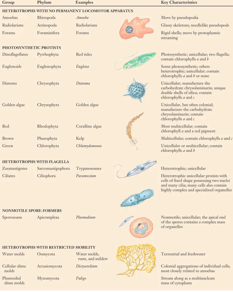
The five criteria we have chosen to define groups are not the only ones that might be chosen, and there is no broad agreement among biologists as to which set of crite-ria is preferable. As molecular analysis gives us a clearer picture of the phylogenetic relationships among the pro-tists, more evolutionarily suitable groupings will without a doubt replace the one represented here. Table 35.1 sum-marizes some of the general characteristics and groupings of the 15 major phyla of protists. It is important to remem-ber that while the phyla of protists discussed here are gen-erally accepted taxa, the larger groupings of phyla pre-sented are functional groupings.
The 15 major protist phyla can be conveniently categorized into five groups according to major shared characteristics.
698
35.3
Protists can be categorized into five groups.
Rhizopoda Actinopoda Foraminifera Pyrrhophyta Euglenophyta Chrysophyta Rhodophyta Phaeophyta Chlorophyta Sarcomastigophora Ciliophora Apicomplexa Oomycota Acrasiomycota Myxomycota Protists
Heterotrophs with no permanent
locomotor apparatus
Photosynthetic protists
Heterotrophs with flagella Nonmotile spore-formers
Heterotrophs with restricted mobility
FIGURE 35.6
Table 35.1 Kinds of Protists Typical
Group Phylum Examples Key Characteristics
HETEROTROPHS WITH NO PERMANENT LOCOMOTOR APPARATUS
Amoebas Rhizopoda Amoeba Move by pseudopodia
Radiolarians Actinopoda Radiolarians Glassy skeletons; needlelike pseudopods
Forams Foraminifera Forams Rigid shells; move by protoplasmic
streaming
PHOTOSYNTHETIC PROTISTS
Dinoflagellates Pyrrhophyta Red tides Photosynthetic; unicellular; two flagella; contain chlorophylls aand b
Euglenoids Euglenophyta Euglena Some photosynthetic; others
heterotrophic; unicellular; contain chlorophylls aand b or none
Diatoms Chrysophyta Diatoma Unicellular; manufacture the
carbohydrate chrysolaminarin; unique double shells of silica; contain chlorophylls aand c
Golden algae Chrysophyta Golden algae Unicellular, but often colonial; manufacture the carbohydrate chrysolaminarin; contain chlorophylls aand c
Red Rhodophyta Coralline algae Most multicellular; contain
chlorophyll aand a red pigment
Brown Phaeophyta Kelp Multicellular; contain chlorophylls aand c
Green Chlorophyta Chlamydomonas Unicellular or multicellular; contain
chlorophylls aand b
HETEROTROPHS WITH FLAGELLA
Zoomastigotes Sarcomastigophora Trypanosomes Heterotrophic; unicellular
Ciliates Ciliophora Paramecium Heterotrophic unicellular protists with
cells of fixed shape possessing two nuclei and many cilia; many cells also contain highly complex and specialized organelles
NONMOTILE SPORE-FORMERS
Sporozoans Apicomplexa Plasmodium Nonmotile; unicellular; the apical end of the spores contains a complex mass of organelles
HETEROTROPHS WITH RESTRICTED MOBILITY
Water molds Oomycota Water molds, Terrestrial and freshwater
rusts, and mildew
Cellular slime Acrasiomycota Dictyostelium Colonial aggregations of individual cells;
molds most closely related to amoebas
Plasmodial Myxomycota Fuligo Stream along as a multinucleate
Heterotrophs with No
Permanent Locomotor
Apparatus
The largest of the five general groups of protists are primarily unicellular or-ganisms with amoeboid forms. There are three principle phyla: the forams and the radiolarians have carbonate shells and the rhizopods lack shells.
Rhizopoda: The Amoebas
Hundreds of species of amoebas are found throughout the world in both fresh and salt waters. They are also abundant in soil. Many kinds of amoe-bas are parasites of animals. Reproduc-tion in amoebas occurs by fission, or the direct division into two cells of equal volume. Amoebas of the phylum Rhizopoda lack cell walls, flagella, meiosis, and any form of sexuality. They do undergo mitosis, with a spin-dle apparatus that resembles that of other eukaryotes.
Amoebas move from place to place by means of their pseudopods, from the Greek words for “false” and “foot” (figure 35.7). Pseudopods are flowing projections of cytoplasm that extend and pull the amoeba forward or engulf food particles, a process called cyto-plasmic streaming. An amoeba puts a pseudopod forward and then flows into it. Microfilaments of actin and myosin similar to those found in muscles are associated with these movements. The pseudopodia can form at any point on the cell body so that it can move in any direction.
Some kinds of amoebas form resis-tant cysts. In parasitic species such as
Entamoeba histolytica, which causes
amoebic dysentery, cysts enable the amoebas to resist di-gestion by their animal hosts. Mitotic division takes place within the cysts, which ultimately rupture and release four, eight, or even more amoebas within the digestive tracts of their host animals. The primary infection takes place in the intestine, but it often moves into the liver and other parts of the body. The cysts are dispersed in the feces and may be transmitted from person to person in in-fected food or water, or by flies. It is estimated that up to 10 million people in the United States have infections of parasitic amoebas, and some 2 million show symptoms of the disease, ranging from abdominal discomfort with
slight diarrhea to much more serious conditions. In some tropical areas, more than half of the population may be infected. The spread of amoebic dysentery can be limited by proper sanitation and hygiene.
Actinopoda: The Radiolarians
The pseudopodia of amoeboid cells give them truly amorphous bodies. One group, however, have more dis-tinct structures. Members of the phy-lum Actinopoda, often called radiolari-ans, secrete glassy exoskeletons made of silica. These skeletons give the uni-cellular organisms a distinct shape, ex-hibiting either bilateral or radial sym-metry. The shells of different species form many elaborate and beautiful shapes and its pseudopodia extrude outward along spiky projections of the skeleton (figure 35.8). Microtubules support these cytoplasmic projections.
Foraminifera: Forams
Members of the phylum Foraminifera are heterotrophic marine protists. They range in diameter from about 20 micrometers to several centimeters. Characteristic of the group are pore-studded shells (called tests) composed of organic materials usually reinforced with grains of inorganic matter. These grains may be calcium carbonate, sand, or even plates from the shells of echin-oderms or spicules (minute needles of calcium carbonate) from sponge skele-tons. Depending on the building mate-rials they use, foraminifera—often in-formally called “forams”—may have shells of very different appearance. Some of them are brilliantly colored red, salmon, or yellow-brown.
Most foraminifera live in sand or are attached to other organisms, but two families consist of free-floating plank-tonic organisms. Their tests may be single-chambered but more often are multichambered, and they sometimes have a spiral shape resembling that of a tiny snail. Thin cytoplas-mic projections called podia emerge through openings in the tests (figure 35.9). Podia are used for swimming, gath-ering materials for the tests, and feeding. Forams eat a wide variety of small organisms.
The life cycles of foraminifera are extremely complex, involving an alternation between haploid and diploid gen-erations (sporic meiosis). Forams have contributed massive 700
FIGURE 35.7
Amoeba proteus. This relatively large amoeba is commonly used in teaching and for research in cell biology. The projections are pseudopods; an amoeba moves by flowing into them. The nucleus of the amoeba is plainly visible.
Heterotrophs with no permanent
locomotor apparatus
Photosynthetic protists
Heterotrophs with flagella Nonmotile spore-formers
accumulations of their tests to the fossil record for more than 200 million years. Because of the excellent preserva-tion of their tests and the often striking differences among them, forams are very important as geological markers. The pattern of occurrence of different forams is often used as a guide in searching for oil-bearing strata. Limestones all over the world, including the famous white cliffs of Dover in southern England, are often rich in forams (fig-ure 35.10).
Amoebas, radiolarians, and forams are unicellular, heterotrophic protists that lack cell walls, flagella, meiosis, and sexuality. Amoebas move from place to place by means of extensions called pseudopodia. The pore-studded tests, or shells, of the forams have openings through which podia extend that are used for locomotion.
FIGURE 35.8
Actinosphaerium,a protist of the phylum Actinopoda (300×).
This amoeba-like radiolarian has striking needlelike pseudopods.
FIGURE 35.9
A representative of the Foraminifera (90×). A living foram with
podia, thin cytoplasmic projections that extend through pores in the calcareous test, or shell, of the organism.
FIGURE 35.10
White cliffs of Dover. The limestone that forms these cliffs is
composed almost entirely of fossil shells of protists, including coccolithophores (a type of algae) and foraminifera.
Photosynthetic Protists
Pyrrhophyta: The Dinoflagellates
The dinoflagellates consist of about 2100 known species of primarily uni-cellular, photosynthetic organisms, most of which have two flagella. A majority of the dinoflagellates are ma-rine, and they are often abundant in the plankton, but some occur in fresh water. Some planktonic dinoflagel-lates are luminous and contribute to the twinkling or flashing effects that we sometimes see in the sea at night, especially in the tropics.
The flagella, protective coats, and biochemistry of dinoflagellates are distinctive, and they do not appear to be directly related to any other phy-lum. Plates made of a cellulose-like material encase the cells. Grooves form at the junctures of these plates and the flagella are usually located within these grooves, one encircling the body like a belt, and the other perpendicular to it. By beating in their respective grooves, these fla-gella cause the dinoflafla-gellate to ro-tate like a top as it moves. The di-noflagellates that are clad in stiff cellulose plates, often encrusted with silica, may have a very unusual ap-pearance (figure 35.11). Most have chlorophylls a and c, in addition to carotenoids, so that in the biochem-istry of their chloroplasts, they re-semble the diatoms and the brown algae, possibly acquiring such chloro-plasts by forming endosymbiotic re-lationships with members of those groups.
Some dinoflagellates occur as sym-bionts in many other groups of or-ganisms, including jellyfish, sea anemones, mollusks, and corals. When dinoflagellates grow as sym-bionts within other cells, they lack their characteristic cellulose plates and flagella, appearing as spherical, golden-brown globules in their host cells. In such a state they are called zooxanthellae. Photosynthetic zooxanthellae provide their hosts with nutrients. It is the photosynthe-sis conducted by zooxanthellae that
makes coral reefs one of the most pro-ductive ecosystems on earth. Corals primarily live in warm tropical seas that are typically extremely low in nu-trients; without the aid of their photo-synthetic endosymbionts, they would not be able to form large reefs in the nutrient-poor environment. Most of the carbon that the zooxanthellae fix is translocated to the host corals.
The poisonous and destructive “red tides” that occur frequently in coastal areas are often associated with great population explosions, or “blooms,” of dinoflagellates. The pigments in the in-dividual, microscopic cells of the di-noflagellates are responsible for the color of the water. Red tides have a pro-found, detrimental effect on the fishing industry in the United States. Some 20 species of dinoflagellates are known to produce powerful toxins that inhibit the diaphragm and cause respiratory failure in many vertebrates. When the toxic di-noflagellates are abundant, fishes, birds, and marine mammals may die in large numbers.
More recently, a particularly dan-gerous toxic dinoflagellate called Pfies-teria piscicida is reported to be a carniv-orous, ambush predator. During blooms, it stuns fish with its toxin and then feeds on the prey’s body fluids.
Dinoflagellates reproduce primarily by asexual cell division. But sexual re-production has been reported to occur under starvation conditions. They have a unique form of mitosis in which the permanently condensed chromo-somes divide longitudinally within the confines of a permanent nuclear enve-lope. After the numerous chromo-somes duplicate, the nucleus divides into two daughter nuclei. Also the di-noflagellate chromosome is unique among eukaryotes in that the DNA is not complexed with histone proteins. In all other eukaryotes, the chromoso-mal DNA is complexed with histones to form nucleosomes, which represents the first order of DNA packaging in the nucleus. How dinoflagellates are able to maintain distinct chromosomes without histones and nucleosomes re-mains a mystery.
702
Noctiluca Ptychodiscus
Ceratium Gonyaulax
Heterotrophs with no permanent
locomotor apparatus
Photosynthetic protists
Heterotrophs with flagella Nonmotile spore-formers
Heterotrophs with restricted mobility
FIGURE 35.11
Some dinoflagellates: Noctiluca,
Ptychodiscus, Ceratium,andGonyaulax. Noctiluca,which lacks the heavy cellulose armor characteristic of most dinoflagellates, is one of the bioluminescent organisms that causes the waves to sparkle in warm seas. In the other three genera, the shorter, encircling flagellum is seen in its groove, with the longer one projecting away from the body of the dinoflagellate. (Not drawn to scale.)
Euglenophyta: The Euglenoids
Most of the approximately 1000 known species of eugle-noids live in fresh water. The members of this phylum clearly illustrate the impossibility of distinguishing “plants” from “animals” among the protists. About a third of the ap-proximately 40 genera of euglenoids have chloroplasts and are fully autotrophic; the others lack chloroplasts, ingest their food, and are heterotrophic. These organisms are not significantly different from some groups of zoomastigotes (see next section), and many biologists believe that the two phyla should be merged into one.
Some euglenoids with chloroplasts may become het-erotrophic if the organisms are kept in the dark; the chloroplasts become small and nonfunctional. If they are put back in the light, they may become green within a few hours. Normally photosynthetic euglenoids may sometimes feed on dissolved or particulate food.
Individual euglenoids range from 10 to 500 micrometers long and are highly variable in form. Interlocking proteina-ceous strips arranged in a helical pattern form a flexible structure called the pellicle, which lies within the cell membrane of the euglenoids. Because its pellicle is flexible, a euglenoid is able to change its shape. Reproduction in this phylum occurs by mitotic cell division. The nuclear en-velope remains intact throughout the process of mitosis. No sexual reproduction is known to occur in this group.
In Euglena (figure 35.12), the genus for which the phy-lum is named, two flagella are attached at the base of a flask-shaped opening called the reservoir,which is located at the anterior end of the cell. One of the flagella is long and has a row of very fine, short, hairlike projections along one side. A second, shorter flagellum is located within the reservoir but does not emerge from it. Contractile vacuoles collect excess water from all parts of the organism and empty it into the reservoir, which apparently helps regulate the osmotic pressure within the organism. The stigma,an organ that also occurs in the green algae (phylum Chloro-phyta), is light-sensitive and aids these photosynthetic or-ganisms to move toward light.
Cells of Euglena contain numerous small chloroplasts. These chloroplasts, like those of the green algae and plants, contain chlorophylls aand b,together with carotenoids. Al-though the chloroplasts of euglenoids differ somewhat in structure from those of green algae, they probably had a common origin. It seems likely that euglenoid chloroplasts ultimately evolved from a symbiotic relationship through ingestion of green algae.
(a) Reservoir Pellicle Basal body Contractile vacuole Second flagellum Stigma Flagellum Nucleus Chloroplast Paramylon granule (b) FIGURE 35.12
Euglenoids. (a) Micrograph of individuals of the genus Euglena
(Euglenophyta). (b) Diagram of Euglena.Paramylon granules are areas where food reserves are stored.
Chrysophyta: The Diatoms and Golden Algae
The Diatoms. Diatoms, members of the phylum Chrys-ophyta, are photosynthetic, unicellular organisms with unique double shells made of opaline silica, which are often strikingly and characteristically marked. The shells of di-atoms are like small boxes with lids, one half of the shell fit-ting inside the other. Their chloroplasts, with chlorophylls
aand c,as well as carotenoids, resemble those of the brown algae and dinoflagellates. In other respects, however, there are few similarities between these groups, and they proba-bly do not share an immediate common ancestor. Another member of the phylum Chrysophyta is the golden algae. Diatoms and golden algae are grouped together because they both produce a unique carbohydrate called chrysolam-inarin.
There are more than 11,500 living species of diatoms, with many more known in the fossil record. The shells of fossil diatoms often form very thick deposits, which are sometimes mined commercially. The resulting “diatoma-ceous earth” is used as an abrasive or to add the sparkling quality to the paint used on roads, among other purposes. Living diatoms are often abundant both in the sea and in fresh water, where they are important food producers. Di-atoms occur in the plankton and are attached to submerged objects in relatively shallow water. Many species are able to move by means of a secretion that is produced from a fine groove along each shell. The diatoms exude and perhaps also retract this secretion as they move.
There are two major groups of diatoms, one with radial symmetry (like a wheel) and the other with bilateral (two-sided) symmetry (figure 35.13). Diatom shells are rigid, and the organisms reproduce asexually by separating the two halves of the shell, each half then regenerating another half shell within it. Because of this mode of reproduction, there is a tendency for the shells, and consequently the individual diatoms, to get smaller and smaller with each asexual re-production. When the resulting individuals have dimin-ished to about 30% of their original size, one may slip out of its shell, grow to full size, and regenerate a full-sized pair of new shells.
Individual diatoms are diploid. Meiosis occurs more fre-quently under conditions of starvation. Some marine di-atoms produce numerous sperm and others a single egg. If fusion occurs, the resulting zygote regenerates a full-sized individual. In some freshwater diatoms, the gametes are amoeboid and similar in appearance.
The Golden Algae. Also included within the Chryso-phyta are the golden algae, named for the yellow and brown carotenoid and xanthophyll accessory pigments in their chloroplasts, which give them a golden color. Unicel-lular but often colonial, these freshwater protists typically have two flagella, both attached near the same end of the cell. When ponds and lakes dry out in summer, golden algae form resistant cysts. Viable cells emerge from these cysts when wetter conditions recur in the fall.
704
FIGURE 35.13
Rhodophyta: The Red Algae
Along with green algae and brown algae, red algae are the seaweeds we see cast up along shores and on beaches. Their characteristic colors re-sult from phycoerythrin, a type of phy-cobilin pigment. Phyphy-cobilins are re-sponsible for the colors of the cyanobacteria. Chlorophyll a also oc-curs with the phycobilins in red algae, just as it does in cyanobacteria. These similarities with cyanobacteria make it likely that the rhodophyta evolved when their heterotrophic eukaryotic ancestor developed an endosymbiotic relationship with a cyanobacteria which eventually gave rise to their chloroplasts.
The great majority of the estimated 4000 species of red algae occur in the sea, and almost all are multicellular.
Red algae have complex bodies made up of interwoven filaments of cells. In the cell walls of many red algae are sulfated polysaccharides such as agar and carrageenan, which make these algae important economically. Agar is used to make gel capsules, as material for dental impressions, and as a base for cosmetics. It is also the basis of the laboratory media on which bac-teria, fungi, and other organisms are often grown. In addition, agar is used to prevent baked goods from drying out, for rapid-setting jellies, and as a temporary preservative for meat and fish in warm regions. Carrageenan is used mainly to stabilize emulsions such as paints, cosmetics, and dairy products such as ice cream. In addition to these uses, red algae such as Por-phyra, called “nori,” are eaten and, in Japan, are even cultivated as a human food crop.
The life cycles of red algae are com-plex but usually involve an alternation of generations (sporic meiosis). None of the red algae have flagella or cilia at any stage in their life cycle, and they may have descended directly from an-cestors that never had them, especially as the red algae also lack centrioles. To-gether with the fungi, which also lack flagella and centrioles, the red algae may be one of the most ancient groups of eukaryotes.
Phaeophyta: The Brown Algae
The phaeophyta, or brown algae, con-sist of about 1500 species of multicel-lular protists, almost exclusively ma-rine. They are the most conspicuous seaweeds in many northern regions, dominating rocky shores almost every-where in temperate North America. In habitats where large brown algae known as kelps (order Laminariales) occur abundantly in so-called kelp forests (figure 35.14), they are respon-sible for most of the food production through photosynthesis. Many kelps are conspicuously differentiated into flattened blades, stalks, and grasping basal portions that anchor them to the rocks.
Among the larger brown algae are genera such as Macrocystis, in which some individuals may reach 100 me-ters in length. The flattened blades of this kelp float out on the surface of the water, while the base is anchored tens of meters below the surface. An-other ecologically important member of this phylum is sargasso weed, Sar-gassum, which forms huge floating masses that dominate the vast Sar-gasso Sea, an area of the Atlantic Ocean northeast of the Caribbean. The stalks of the larger brown algae often exhibit a complex internal dif-ferentiation of conducting tissues analogous to that of plants.
The life cycle of the brown algae is marked by an alternation of genera-tions between a sporophyte and a ga-metophyte. The large individuals we recognize, such as the kelps, are sporophytes. The gametophytes are often much smaller, filamentous indi-viduals, perhaps a few centimeters across. Sporangia, which produce haploid, swimming spores after meio-sis, are formed on the sporophytes. These spores divide by mitosis, giving rise to individual gametophytes. There are two kinds of gametophytes in the kelps; one produces sperm, and the other produces eggs. If sperm and eggs fuse, the resulting zygotes grow into the mature kelp sporophytes, provided that they reach a favorable site.
FIGURE 35.14
Brown algae (Phaeophyta). The massive
“groves” of giant kelp that occur in relatively shallow water along the coasts of the world provide food and shelter for many different kinds of organisms.
Chlorophyta: The Green Algae
Green algae are an extremely varied group of more than 7000 species. The chlorophytes have an extensive fossil record dating back 900 million years. They are mostly aquatic, but some are semiterrestrial in moist places, such as on tree trunks or in soil. Many are microscopic and uni-cellular, but some, such as sea let-tuce, Ulva (see figure 35.16), are tens of centimeters across and easily visible on rocks and pilings around the coasts.
Green algae are of special inter-est, both because of their unusual di-versity and because the ancestors of the plant kingdom were clearly mul-ticellular green algae. Many features of modern green algae closely re-semble plants, especially their chloroplasts which are biochemically similar to those of the plants. They contain chlorophylls aand b,as well as carotenoids. Green algae include a very wide array of both unicellular and multicellular organisms.
Among the unicellular green algae, Chlamydomonas (figure 35.15) is a well-known genus. Individuals are microscopic (usually less than 25 micrometers long), green, rounded, and have two flagella at the anterior
end. They move rapidly in water by beating their flagella in opposite directions. Each individual has an eyespot, which contains about 100,000 molecules of rhodopsin, the same pigment employed in vertebrate eyes. Light received by this eyespot is used by the alga to help direct its swimming. Most individuals of Chlamydomonas are haploid. Chlamy-domonas reproduces asexually (by cell division) as well as sexually. In sexual reproduction, two haploid individuals fuse to form a four-flagellated zygote. The zygote ulti-mately enters a resting phase, called the zygospore, in which the flagella disappear and a tough protective coat is formed. Meiosis occurs at the end of this resting period and results in the production of four haploid cells.
Chlamydomonasprobably represents a primitive state for green algae and several lines of evolutionary specialization have been derived from organisms like it.The first is the evolution of nonmotile, unicellular green algae. Chlamy-domonas is capable of retracting its flagella and settling down as an immobile unicellular organism if the ponds in which it lives dry out. Some common algae of soil and bark, such as Chlorella, are essentially like Chlamydomonasin this
trait, but do not have the ability to form flagella. Chlorellais widespread in both fresh and salt water as well as soil and is only known to reproduce asexually. Recently, Chlorellahas been widely investigated as a possible food source for hu-mans and other animals, and pilot farms have been estab-lished in Israel, the United States, Germany, and Japan.
Another major line of specialization from cells like
Chlamydomonasconcerns the formation of motile, colonial organisms. In these genera of green algae, the Chlamy-domonas-like cells retain some of their individuality. The most elaborate of these organisms is Volvox(see figure 35.1), a hollow sphere made up of a single layer of 500 to 60,000 individual cells, each cell with two flagella. Only a small number of the cells are reproductive. The colony has defi-nite anterior and posterior ends, and the flagella of all of the cells beat in such a way as to rotate the colony in a clockwise direction as it moves forward through the water. The repro-ductive cells of Volvoxare located mainly at the posterior end of the colony. Some may divide asexually, bulge inward, and give rise to new colonies that initially remain within the parent colony. Others produce gametes. In some species of
706 – Strain – Strain – Gamete + Gamete SYNGAMY MEIOSIS Zygospore (diploid) Asexual reproduction + Strain + Strain n 2n Pairing of positive and negative strains FIGURE 35.15
Life cycle of Chlamydomonas(Chlorophyta). Individual cells of this microscopic,
biflagellated alga, which are haploid, divide asexually, producing identical copies of themselves. At times, such haploid cells act as gametes—fusing, as shown in the lower right-hand side of the diagram, to produce a zygote. The zygote develops a thick, resistant wall, becoming a
zygospore; this is the only diploid cell in the entire life cycle. Within this diploid zygospore, meiosis takes place, ultimately resulting in the release of four haploid individuals. Because of the segregation during meiosis, two of these individuals are called the (+) strain, the other two the (–) strain. Only + and – individuals are capable of mating with each other when syngamy does take place, although both may divide asexually to reproduce themselves.
Volvox,there is a true division of labor among the different types of cells, which are specialized in relation to their ulti-mate function throughout the development of the organism. In addition to these two lines of specialization from
Chlamydomonas-like cells, there are many other kinds of green algae of less certain derivation. Many filamentous genera, such as Spirogyra, with its ribbon-like chloro-plasts, differ substantially from the remainder of the green algae in their modes of cell division and reproduction. Some of these genera have even been placed in separate phyla. The study of the green algae, involving modern methods of electron microscopy and biochemistry, is be-ginning to reveal unexpected new relationships within this phylum.
Ulva,or sea lettuce (figure 35.16), is a genus of marine green algae that is extremely widespread. The glistening individuals of this genus, often more than 10 centimeters across, consist of undulating sheets only two cells thick. Sea lettuce attaches by protuberances of the basal cells to rocks or other substrates. The reproductive cycle of Ulva
involves an alternation of generations (sporic meiosis; figure 35.16) as is typical among green algae. Unlike most organisms that undergo sporic meiosis, however,
the gametophytes (haploid phase) and sporophytes (diploid phase) resemble one another closely.
The stoneworts, a group of about 250 living species of green algae, many of them in the genera Charaand Nitella,
have complex structures. Whorls of short branches arise regularly at their nodes, and the gametangia (structures that give rise to gametes) are complex and multicellular. Stoneworts are often abundant in fresh to brackish water and are common as fossils.
Dinoflagellates are primarily unicellular,
photosynthetic, and flagellated. Euglenoids (phylum Euglenophyta) consist of about 40 genera, about a third of which have chloroplasts similar biochemically to those of green algae and plants. Diatoms and golden algae are unicellular, photosynthetic organisms that produce a unique carbohydrate. Diatoms have double shells made of opaline silica. Nonmotile, unicellular algae and multicellular, flagellated colonies have been
derived from green algae like Chlamydomonas—a
biflagellated, unicellular organism. The life cycle of brown algae is marked by an alternation of generations between the diploid phase, or sporophyte, and the haploid phase, or gametophyte.
Spores MEIOSIS Sporangia Sporophyte (2n) Germinating zygote Zygote SYNGAMY Gametes + – + Gametangia + Gametophyte (n) – Gametangia – Gametophyte (n) 2n n Gametes fuse FIGURE 35.16
Life cycle of Ulva.In this green alga, the
gametophyte and the sporophyte are identical in appearance and consist of flattened sheets two cells thick. In the haploid (n) gametophyte, gametangia give rise to haploid gametes, which fuse to form a diploid (2n) zygote. The zygote germinates to form the diploid sporophyte. Sporangia within the sporophyte give rise to haploid spores by meiosis. The haploid spores develop into haploid gametophytes.
Heterotrophs with
Flagella
The phylum Sarcomastigophora con-tains a diverse group of protists com-bined into one phylum because they all possess a single kind of nucleus and use flagella or pseudopodia (or both) for locomotion. We will focus on the class Zoomastigophora.
Zoomastigophora: The Zoomastigotes
The class Zoomastigophora is com-posed of unicellular, heterotrophic or-ganisms that are highly variable in form (figure 35.17). Each has at least one flagellum, with some species hav-ing thousands. They include both free-living and parasitic organisms. Many zoomastigotes apparently reproduce only asexually, but sexual reproduction occurs in some species. The members of one order, the kinetoplastids, in-clude the genera Trypanosoma (figure 35.17c) and Crithidia,pathogens of hu-mans and domestic animals. The eug-lenoids could be viewed as a special-ized group of zoomastigotes, some of which acquired chloroplasts during the course of evolution.
Trypanosomes cause many serious human diseases, the most familiar of which is trypanosomiasis also known as African sleeping sickness (figure 35.18). Trypanosomes cause many other diseases including East Coast fever, leishmaniasis, and Chagas’ dis-ease, all of great importance in tropical areas where they afflict millions of people each year. Leishmaniasis, which is transmitted by sand flies, af-flicts about 4 million people a year. The effects of these diseases range from extreme fatigue and lethargy in sleeping sickness to skin sores and deep eroding lesions that can almost obliterate the face in leishmaniasis. The trypanosomes that cause these diseases are spread by biting insects, including tsetse flies and assassin bugs.
A serious effort is now under way to produce a vaccine for trypanosome-caused diseases. These diseases make it
impossible to raise domestic cattle for meat or milk in a large portion of Africa. Control is especially difficult because of the unique attributes of these organisms. For example, tsetse fly-transmitted trypanosomes have evolved an elaborate genetic mecha-nism for repeatedly changing the anti-genic nature of their protective glyco-protein coat, thus dodging the antibodies their hosts produce against them (see chapter 57). Only a single one out of some 1000 to 2000 variable antigen genes is expressed at a time. Rearrangements of these genes during the asexual cycle of the organism allow for the expression of a seemingly end-less variety of different antigen genes that maintain infectivity by the
try-panosomes.
When the trypanosomes are in-gested by a tsetse fly, they embark on a complicated cycle of development and multiplication, first in the fly’s gut and later in its salivary glands. It is their position in the salivary glands that al-lows them to move into their verte-brate host. Recombination has been observed between different strains of trypanosomes introduced into a single fly, thus suggesting that mating, syn-gamy, and meiosis occur, even though they have not been observed directly. Although most trypanosome repro-duction is asexual, this sexual cycle, re-ported for the first time in 1986, af-fords still further possibilities for recombination in these organisms.
In the guts of the flies that spread them, trypanosomes are noninfective. When they are ready to transfer to the skin or bloodstream of their host, try-panosomes migrate to the salivary glands and acquire the thick coat of glycoprotein antigens that protect them from the host’s antibodies. When they are taken up by a fly, the try-panosomes again shed their coats. The production of vaccines against such a system is complex, but tests are under-way. Releasing sterilized flies to im-pede the reproduction of populations is another technique used to try to con-trol the fly population. Traps made of dark cloth and scented like cows, but 708 Trypanosoma (c) Codosiga (a) Trichonympha (b)
Heterotrophs with no permanent
locomotor apparatus
Photosynthetic protists
Heterotrophs with flagella Nonmotile spore-formers
Heterotrophs with restricted mobility
FIGURE 35.17
Three genera of zoomastigotes (Zoomastigophora), a highly diverse
group. (a) Codosiga,a colonial
choanoflagellate that remains attached to its substrate; other colonial choanoflagellates swim around as a colony, resembling the green alga Volvoxin this respect. (b) Trichonympha,one of the zoomastigotes that inhabits the guts of termites and wood-feeding cockroaches and digests cellulose there. Trichonymphahas rows of flagella in its anterior regions. (c) Trypanosoma,which causes sleeping sickness, an important tropical disease. It has a single, anterior flagellum.
poisoned with insecticides, have like-wise proved effective. Research is pro-ceeding rapidly because the presence of tsetse flies with their associated try-panosomes blocks the use of some 11 million square kilometers of potential grazing land in Africa.
Some zoomastigotes occur in the guts of termites and other wood-eat-ing insects. They possess enzymes that allow them to digest the wood and thus make the components of the wood available to their hosts. The re-lationship is similar to that between certain bacteria and protozoa that function in the rumens of cattle and related mammals (see chapter 51).
Another order of zoomastigotes, the choanoflagellates, is most likely
the group from which the sponges (phylum Porifera) and probably all animals arose. Choanoflagellates have a single emergent flagellum surrounded by a funnel-shaped, con-tractile collar composed of closely placed filaments, a unique structure that is exactly matched in the sponges. These protists feed on bacteria strained out of the water by the collar.
Hiker’s Diarrhea. Giardia lamblia is a flagellate protist (belonging to a small order called diplomonads) found throughout the world, including all parts of the United States and Canada (figure 35.19). It occurs in water, includ-ing the clear water of mountain streams and the water sup-plies of some cities. It infects at least 40 species of wild and domesticated animals in addition to humans. In 1984 in Pittsburgh, 175,000 people had to boil their drinking water for several days following the appearance of Giardiain the city’s water system. Although most individuals exhibit no symptoms if they drink water infested with Giardia,many suffer nausea, cramps, bloating, vomiting, and diarrhea. Only 35 years ago, Giardia was thought to be harmless; today, it is estimated that at least 16 million residents of the United States are infected by it.
Giardia lives in the upper small intestine of its host. It occurs there in a motile form that cannot survive outside the host’s body. It is spread in the feces of infected individ-uals in the form of dormant, football-shaped cysts—some-times at levels as high as 300 million individuals per gram of feces. These cysts can survive at least two months in cool water, such as that of mountain streams. They are relatively resistant to the usual water-treatment agents such as chlo-rine and iodine but are killed at temperatures greater than about 65°C. Apparently, pollution by humans seems to be the main way Giardiais released into stream water. There are at least three species of Giardia and many distinct strains; how many of them attack humans and under what circumstances are not known with certainty.
In the wilderness, good sanitation is important in pre-venting the spread of Giardia. Dogs, which readily contract and spread the disease, should not be taken into pristine wilderness areas. Drinking water should be filtered—the filter must be capable of eliminating particles as small as 1 micrometer in diameter—or boiled for at least one minute. Water from natural streams or lakes should never be con-sumed directly, regardless of how clean it looks. In other regions, good sanitation methods are important to prevent not only Giardiainfection but also other diseases.
FIGURE 35.18
Trypanosomais the zoomastigote that causes sleeping sickness. (a) Trypanosomaamong red blood cells. The nuclei (dark-staining bodies), anterior flagella, and undulating, changeable shape of the trypanosomes are visible in this photograph (500×). (b) The tsetse
FIGURE 35.19
Giardia lamblia. Giardiaare flagellated unicellular parasites that infect the human intestine. Giardiaare very primitive, having only a rudimentary cytoskeleton and lacking mitochondria and chloroplasts. Sequencing of ribosomal RNA suggests that Giardia and Pelomyxa,the eukaryotes most closely related to prokaryotes, should be grouped together. The name Archezoa (Greek arkhaios, “ancient”) has been suggested for the group, stressing its early divergence from bacteria as long as 2 billion years ago.
20 µm
Ciliophora: The Ciliates
As the name indicates, most members of the Ciliophora feature large numbers of cilia. These heterotrophic, unicel-lular protists range in size from 10 to 3000 micrometers long. About 8000 species have been named. Despite their unicellularity, ciliates are extremely complex organisms, in-spiring some biologists to consider them organisms without cell boundaries rather than single cells.
Their most characteristic feature, cilia, are usually arranged either in longitudinal rows or in spirals around the body of the organism (figure 35.20). Cilia are anchored to microtubules beneath the cell membrane, and they beat in a coordinated fashion. In some groups, the cilia have specialized locomotory and feeding functions, becoming fused into sheets, spikes, and rods which may then function as mouths, paddles, teeth, or feet. The ciliates have a tough but flexible outer covering called the pellicle that enables the organism to squeeze through or move around many kinds of obstacles.
All ciliates that have been studied have two very differ-ent types of nuclei within their cells, small micronuclei and larger macronuclei (figure 35.21). The micronuclei, which contain apparently normal diploid chromosomes, di-vide by meiosis and are able to undergo genetic recombina-tion. Macronuclei are derived from certain micronuclei in a complex series of steps. Within the macronuclei are multi-ple copies of the genome, and the DNA is divided into small pieces—smaller than individual chromosomes. In one group of ciliates, these are equivalent to single genes. Macronuclei divide by elongating and constricting and play an essential role in routine cellular functions, such as the production of mRNA to direct protein synthesis for growth and regeneration.
Ciliates form vacuoles for ingesting food and regulating their water balance. Food first enters the gullet, which in the well-known ciliate Paramecium is lined with cilia fused into a membrane (figure 35.21). From the gullet, the food passes into food vacuoles, where enzymes and hydrochloric acid aid in its digestion. After the digested material has been completely absorbed, the vacuole empties its waste contents through a special pore in the pellicle known as the cytoproct. The cytoproct is essentially an exocytotic vesi-cle that appears periodically when solid partivesi-cles are ready to be expelled. The contractile vacuoles, which function in the regulation of water balance, periodically expand and contract as they empty their contents to the outside of the organism.
Ciliates usually reproduce by transverse fission of the parent cell across its short axis, thus forming two identical individuals (figure 35.22a). In this process of cell division, the mitosis of the micronuclei proceeds normally, and the macronuclei divide as just described.
In Paramecium,the cells divide asexually for about 700 generations and then die if sexual reproduction has not oc-curred. Like most ciliates, Parameciumhas a sexual process called conjugation, in which two individual cells remain
attached to each other for up to several hours (figure 35.22b,c). Only cells of two different genetically determined mating types, oddand even,are able to conjugate. Meiosis in the micronuclei of each individual produces several haploid micronuclei, and the two partners exchange a pair of these micronuclei through a cytoplasmic bridge that appears be-tween the two partners.
In each conjugating individual, the new micronucleus fuses with one of the micronuclei already present in that in-dividual, resulting in the production of a new diploid mi-cronucleus in each individual. After conjugation, the macronucleus in each cell disintegrates, while the new diploid micronucleus undergoes mitosis, thus giving rise to two new identical diploid micronuclei within each individ-ual. One of these micronuclei becomes the precursor of the future micronuclei of that cell, while the other micronu-cleus undergoes multiple rounds of DNA replication, be-coming the new macronucleus. This kind of complete seg-regation of the genetic material is a unique feature of the 710
Anterior contractile vacuole
Micronucleus Macronucleus Pellicle Posterior contractile vacuole Food vacuole Gullet Cilia Cytoproct FIGURE 35.20 A ciliate (Ciliophora). Stentor,a funnel-shaped ciliate, showing spirally arranged cilia (120×). FIGURE 35.21
ciliates and makes them ideal organisms for the study of certain aspects of genetics.
Progeny from a sexual division in Parameciummust go through about 50 asexual divisions before they are able to conjugate. When they do so, their biological clocks are restarted, and they can conjugate again. After about 600 asexual divisions, however, Paramecium loses the protein molecules around the gullet that enable it to recognize an appropriate mating partner. As a result, the individuals are unable to mate, and death follows about 100 generations later. The exact mechanisms producing these unusual events are unknown, but they involve the accumulation of a protein, which is now being studied.
The zoomastigotes are a highly diverse group of flagellated unicellular heterotrophs, containing among their members the ancestors of animals as well as the
very primitive Giardia. Ciliates possess characteristic
cilia, and have two types of nuclei. The macronuclei contain multiple copies of certain genes, while the micronuclei contain multigene chromosomes.
Two Paramecium individuals of different mating types come into contact.
The diploid
micronucleus in each divides by meiosis to produce four haploid micronuclei.
Three of the haploid micronuclei degenerate. The remaining micronucleus in each divides by mitosis. Mates exchange micronuclei.
In each individual, the new micronucleus fuses with the micronucleus already present, forming a diploid micronucleus. The macronucleus disintegrates, and the diploid micronucleus divides by mitosis to produce two identical diploid micronuclei within each individual.
One of these micronuclei is the precursor of the micronucleus for that cell, and the other eventually gives rise to the macronucleus. Macronucleus Micronucleus (2n) (c) Diploid micronucleus (2n) Haploid micronucleus (n) (a) (b) FIGURE 35.22
Life cycle of Paramecium. (a) When Parameciumreproduces
asexually, a mature individual divides, and two complete individuals result. (b,c) In sexual reproduction, two mature cells fuse in a process called conjugation (100×).