EUKARYOTICCELL, Apr. 2007, p. 682–692 Vol. 6, No. 4 1535-9778/07/$08.00⫹0 doi:10.1128/EC.00340-06
Copyright © 2007, American Society for Microbiology. All Rights Reserved.
Developmental Regulation of an Adhesin Gene during Cellular
Morphogenesis in the Fungal Pathogen
Candida albicans
䌤
†
Silvia Argimo
´n,
1‡ Jill A. Wishart,
1§ Roger Leng,
1¶ Susan Macaskill,
1Abigail Mavor,
1Thomas Alexandris,
1Susan Nicholls,
1Andrew W. Knight,
2Brice Enjalbert,
1Richard Walmsley,
2Frank C. Odds,
1Neil A. R. Gow,
1and Alistair J. P. Brown
1*
School of Medical Sciences, University of Aberdeen, Foresterhill, Aberdeen AB25 2ZD, United Kingdom,1andGentronix Limited, CTF Building, 46 Grafton Street, Manchester M13 9NT, United Kingdom2
Received 25 October 2006/Accepted 14 December 2006
Candida albicansexpresses specific virulence traits that promote disease establishment and progression.
These traits include morphological transitions between yeast and hyphal growth forms that are thought to contribute to dissemination and invasion and cell surface adhesins that promote attachment to the host. Here, we describe the regulation of the adhesin geneALS3, which is expressed specifically during hyphal development
inC. albicans. Using a combination of reporter constructs and regulatory mutants, we show that this regulation
is mediated by multiple factors at the transcriptional level. The analysis ofALS3promoter deletions revealed that this promoter contains two activation regions: one is essential for activation during hyphal development,
while the second increases the amplitude of this activation. Further deletion analyses using the Renilla
reniformisluciferase reporter delineate the essential activation region between positionsⴚ471 andⴚ321 of the
promoter. Further 5ⴕ or 3ⴕ deletions block activation.ALS3 transcription is repressed mainly by Nrg1 and Tup1, but Rfg1 contributes to this repression. Efg1, Tec1, and Bcr1 are essential for the transcriptional activation ofALS3, with Tec1 mediating its effects indirectly through Bcr1 rather than through the putative Tec1 sites in theALS3promoter.ALS3transcription is not affected by Cph2, but Cph1 contributes to fullALS3
activation. The data suggest that multiple morphogenetic signaling pathways operate through the promoter of this adhesin gene to mediate its developmental regulation in this major fungal pathogen.
Candida albicans is a major opportunistic pathogen of hu-mans (54). This fungus is a frequent cause of superficial oral and vaginal infections, and in immunocompromised patients, C. albicans can disseminate via the bloodstream to invade internal organs, thereby causing deep-seated, systemic infec-tions that are often fatal (54).
Various factors are thought to contribute to the virulence of C. albicans. These include adhesion to host tissue, the ability to undergo reversible morphogenetic transitions between bud-ding (yeast) and filamentous (hyphae and pseudohyphae) growth forms, the secretion of extracellular hydrolases, and rapid switching between different phenotypic forms (30, 42, 44, 65). The contribution of yeast-hypha morphogenesis toC. al-bicansvirulence has been hotly debated (21, 29, 71). However, it is clear that hyphal development is closely associated with tissue invasion (21, 61, 71, 83).
Adherence plays a key role in fungal colonization (27, 68,
70).C. albicansexpresses an array of adhesin genes including HWP1, which encodes a cell surface glycoprotein that acts as a target for mammalian transglutaminases. These enzymes are thought to generate covalent cross-links between Hwp1 on the fungal hyphal surface and proteins on the mammalian cell surface (68, 72). TheALSgene gamily encodes a set of differ-entially regulated cell surface glycosylphosphatidylinositol-an-chored glycoproteins that promote fungal adherence (27, 55). ALS3was initially identified as a member of this gene family that is expressed specifically during hyphal development (28). A second hypha-specificALSgene (ALS8) (40) was later iden-tified as an allele of theALS3gene (81).C. albicans als3/als3 cells are defective in biofilm formation (53, 82). Furthermore, Als3 is involved in adhesion to endothelial and epithelial cells (55), andals3/als3cells display an almost total lack of epithelial destruction in a reconstituted buccal human epithelium model (81). ALS3 expression has been detected in clinical vaginal fluid specimens and in a vaginal candidiasis model (13). These observations indicate a role forALS3in the pathogenicity ofC. albicans.
A complex network of signaling pathways regulates yeast-hypha morphogenesis (10). Following exposure to serum, hy-phal development is activated by a cyclic AMP-protein kinase A pathway that regulates the activity of the-helix-loop-helix transcription factor Efg1 (42, 69). In addition, a mitogen-acti-vated protein kinase pathway, which includes the Ste12-like transcription factor Cph1, activates hyphal development under starvation conditions (38, 41). Additional regulatory factors contribute to the activation of hyphal development, but their relationship to these main signaling pathways remains to be * Corresponding author. Mailing address: School of Medical
Sci-ences, University of Aberdeen, Institute of Medical SciSci-ences, Forester-hill, Aberdeen AB25 2ZD, United Kingdom. Phone: 44-1224-555883. Fax: 44-1224-555844. E-mail: [email protected].
† Supplemental material for this article may be found at http://ec .asm.org/.
‡ Present address: Department of Microbiology, Columbia Univer-sity, 701 West 168th St., New York, NY 10032.
§ Present address: Michael Smith Building, University of Manches-ter, Manchester M13 9PT, United Kingdom.
¶ Present address: Department of Laboratory Medicine and Pathol-ogy, University of Alberta, 87 Ave. and 112 Street, Edmonton, AB T6G 2S2, Canada.
䌤Published ahead of print on 2 February 2007.
682
on September 8, 2020 by guest
http://ec.asm.org/
established (11, 16, 39, 57). These include the transcription factors Tec1 and Cph2, the inactivation of which causes defects in hyphal development (37, 62). Tec1 is a TEA (TEF-1, Tec1p, and AbaAp)/ATTS (AbaAp, TEF-1, Tec1p, and Scalloped) motif transcription factor that is also required forC. albicans virulence (62). It was previously suggested thatTEC1is regu-lated both by Cph2 and Efg1 (36), but their precise roles in gene regulation during hyphal development are not known.
Hyphal development is negatively regulated by the transcrip-tional repressors Tup1, Nrg1, and Rfg1 (6, 9, 31, 46). In the absence of hypha-inducing signals, the global repressor Tup1 inhibits the transcription of hypha-specific genes. This repres-sion is dependent upon Nrg1, which binds to Nrg1 response elements (NREs) in the promoters of these genes and targets Tup1 to these promoters (9, 18, 46). Current models suggest that Rfg1 is a second DNA-binding protein that targets Tup1 to the promoters of hypha-specific genes, although Nrg1 ap-pears to make the major contribution to the repression of hyphal development (31, 32).
The prevailing view is that these morphogenetic signaling pathways combine to regulate the transcription of hypha-spe-cific genes. Genome-wide and gene-spehypha-spe-cific studies have re-vealed only a small number of hypha-specific genes inC. albi-cans. These includeALS3,ECE1,HGC1,HWP1,HYR1,RBT1, andRBT4(2, 5, 8, 28, 48, 67, 83). As described above,ALS3 and HWP1 encode adhesins. The inactivation of RBT1 or RBT4attenuates C. albicans virulence (9).HGC1encodes a hypha-specific cyclin required for hyphal development and vir-ulence (83). With the exception ofHGC1, all known hypha-specific genes appear to encode secreted or cell wall proteins. These observations reinforce the tight link between the forma-tion of hyphae, the cell surface, andC. albicansvirulence.
In this paper, we have examined the organization of the ALS3promoter and determined the relative contributions of key morphogenetic transcription factors to the regulation of this hypha-specific gene. We find that, relative to other C. albicansgenes, the promoter regions ofALS3and other hypha-specific genes are unusually large. We show that the ALS3 promoter is complex, requiring a 150-bp region for hypha-specific activation. This promoter integrates inputs from mul-tiple activators and repressors. Related observations have been made for a second hypha-specific gene (HWP1) by Kim et al. in the accompanying paper (33).
MATERIALS AND METHODS
Strains and growth conditions.C. albicansstrains (Table 1) were grown in YPD at 30°C (63). Hyphal development was induced using 20% bovine calf serum (73).
Strain construction.To generate theC. albicansstrains carrying in situ
ALS3-GFPpromoter fusions, yeast enhanced green fluorescent protein (GFP) was first
integrated immediately downstream of the start codon at theALS3locus to
create SAC500 (Table 1) (14). This was done by PCR amplifying a GFP-URA3
cassette (19) (primers ALS3-F1 and ALS3-F2) (see Table S1 in the supplemental
material) and transforming it intoC. albicansRM1000 (77). The correct
inte-gration at theALS3locus in Ura-positive or -negative transformants was
con-firmed by whole-cell PCR (60) and Southern blotting (not shown). Promoter
deletions were created at theALS3-GFPallele by PCR amplifyingHIS1(15) with
primers designed to target this marker to specific regions of theALS3upstream
region (see Table S1 in the supplemental material). The correct integration of
HIS1upstream of theALS3-GFPallele in His-positive or -negative and
Ura-positive or -negative transformants was confirmed by PCR and Southern
blot-ting.C. albicansstrains with mutated Tec1 sites in theALS3promoter were made
in the same way by using 5⬘ PCR primers that contain mutations in these
sequences (see Table S1 in the supplemental material). These mutations in the
C. albicansgenome were confirmed by PCR amplification of the corresponding regions followed by DNA sequence analysis (not shown).
The first set ofALS3-Renilla reniformisluciferase (RrLUC) promoter fusions
was constructed by PCR amplifying portions of theALS3promoter up to
posi-tion⫹4 of theALS3coding region (see Table S1 in the supplemental material)
and cloning these portions between the ClaI and PstI sites in pCRW3 (66). To
create the next sets of RrLUCpromoter fusions, new BstEII, NdeI, SpeI, NotI,
and MluI sites were introduced into pCRW3 to make pCRW3N (oligonucleo-tides KpnSal and SalKpn) (see Table S1 in the supplemental material). A basal
ALS3promoter region (positions⫺306 to⫹4) was then PCR amplified and
inserted between the PstI and MluI sites in pCRW3N, upstream of the RrLUC
open reading frame (ORF). Various promoterALS3fragments were cloned as
oligonucleotides or PCR fragments (see Table S1 in the supplemental material)
upstream of this basalALS3promoter region. The STRE5-, YRE5-, and GCRE5
-RrLUC fusions were made by cloning oligonucleotides with each sequence
element upstream of a basal RrLUCreporter containing part of theADH1
promoter region (51, 74) (see Table S1 in the supplemental material).
pCRW3-based plasmids were linearized with HindIII and transformed intoC. albicans
CAI8 (Table 1) selecting for theADE2marker. Single-copy integration at the
ade2locus was confirmed by PCR diagnosis.
To test the roles of Bcr1 and Tec1 in C. albicans, a nonrevertible Ura3⫺
segregant of CJN688 (52) was selected (SAC518) (Table 1). SAC518 was
trans-formed with aGFP-HIS1cassette (19), as described above, to generate the in situ
ALS3-GFPreporter in thisbcr1strain (SAC521). Meanwhile, theTEC1ORF was PCR amplified (primers TEC1-F4 and TEC1-R) (see Table S1 in the
sup-plemental material) and cloned into pPYK1-GFP (4). This placedTEC1under
the control of thePYK1promoter. pPYK1-TEC1 and the empty control vector
pPYK1 were linearized with StuI and transformed into SAC520 (BCR1) and
SAC521 (bcr1) (Table 1). Single-copy integration atRPS1was confirmed by PCR
diagnosis (45).
DNA and RNA analysis.DNA was prepared and analyzed by Southern blot-ting as described previously (25, 78). RNA was isolated and Northern analysis
was performed as described previously (24, 47). TheALS3-specific probe was
PCR amplified using primers ALS3-F and ALS3-R, which were described
pre-viously by Hoyer et al. (28).GFPandACT1sequences were analyzed using
probes corresponding to the PCR-amplified ORFs. Primers are specified in Table S1 in the supplemental material.
Reporter assays.GFP fluorescence in wholeC. albicanscells was quantified in 96-well, black, clear-bottomed microplates (Matrix Technologies, Wilmslow, United Kingdom) using a Tecan Ultra 384 Microplate reader (Tecan Trading AG, Switzerland) running XFluor 4 software. Fluorescence polarization was used to distinguish GFP fluorescence from background autofluorescence (34, 35). The method exploits the high fluorescence anisotropy of GFP compared to other autofluorescing species. The difference between the fluorescence that polarized parallel to the excitation light and that which polarized perpendicular to the excitation light was used as the analytical signal. This measurement is relatively large for GFP and small for autofluorescing molecules. Fluorescence and fluorescence polarization measurements were made at 485-nm excitation and 535-nm emission wavelengths, as described previously (35). Means (in “FP [fluorescence polarization] brightness” units) and standard deviations from two to eight independent transformants are presented. Observations were
reproduc-ible in at least two independent experiments. UntransformedC. albicanscells
were used as the background control.
Luciferase assays (relative light units/20g protein/20 s) were performed using
freshC. albicansprotein extracts with a Lumat LB9507 luminometer (EG&G
Berthold) as described previously (46). Means and standard deviations from quadruplicate assays are presented, and similar data were obtained in three experiments using independent transformants.
Microscopy.Cell morphology was monitored using an Olympus BX50 micro-scope and recorded with an Olympus DP11-P digital video camera. Cell numbers were counted using an Improved Neubauer hemocytometer.
Phase-contrast microscopy and fluorescence microscopy were performed using an Axioplan 2 microscope (Carl Zeiss, United Kingdom) with filter sets XF66 (blue emission), XF67 (red emission), and XF77 (green emission) from Omega Optical Inc. (Brattleboro, VT). Images were generated using a Hamamatsu charge-coupled-device camera and analyzed using Openlab 3.0.9 (Improvision,
Coventry, United Kingdom).C. albicanscells were mounted onto
polylysine-coated glass slides and covered with Vectashield immunofluorescence mounting medium (Vector Laboratories, Peterborough, United Kingdom) (3).
In silico promoter analysis.Promoter sequences were analyzed for the pres-ence of putative regulatory elements using MatInspector (12, 56) (http://www
on September 8, 2020 by guest
http://ec.asm.org/
TABLE 1. C. albicansstrains
Strain Genotype or descriptiona
Parent strain Reference or source
SC5314 Wild-type clinical isolate 20
CAI4 ura3::imm434/ura3::imm434 SC5314 17
CAI8 ura3::imm434/ura3::imm434 ade2::hisG/ade2::hisG CAI4 17
RM1000 ura3::imm434/ura3::imm434 his1::hisG/his1::hisG CAI4 50
BWP17 ura3::imm434/ura3::imm434 arg4::hisG/arg4::hisG his1::hisG/his1::hisG RM1000 79
Ca90 ura3::imm434/ura3::imm434 als3::hisG/als3::hisG CAI4 This study
Ca107 ura3::imm434/ura3::imm434 ade2::hisG/ADE2-ALS3696-RrLUC CAI8 This study
Ca108 ura3::imm434/ura3::imm434 ade2::hisG/ADE2-ALS3496-RrLUC CAI8 This study
Ca109 ura3::imm434/ura3::imm434 ade2::hisG/ADE2-ALS3306-RrLUC CAI8 This study
Ca110 ura3::imm434/ura3::imm434 ade2::hisG/ADE2-ALS3129-RrLUC CAI8 This study
Ca111 ura3::imm434/ura3::imm434 ade2::hisG/ADE2-ALS3100-RrLUC CAI8 This study
Ca176 ura3::imm434/ura3::imm434 ade2::hisG/ADE2-ALS3496-307-RrLUC CAI8 This study
Ca167 ura3::imm434/ura3::imm434 ade2::hisG/ADE2-ALS3496-461-RrLUC CAI8 This study
Ca619 ura3::imm434/ura3::imm434 ade2::hisG/ADE2-ALS3483-445-RrLUC CAI8 This study
Ca168 ura3::imm434/ura3::imm434 ade2::hisG/ADE2-ALS3467-421-RrLUC CAI8 This study
Ca620 ura3::imm434/ura3::imm434 ade2::hisG/ADE2-ALS3450-405-RrLUC CAI8 This study
Ca169 ura3::imm434/ura3::imm434 ade2::hisG/ADE2-ALS3429-417-RrLUC CAI8 This study
Ca170 ura3::imm434/ura3::imm434 ade2::hisG/ADE2-ALS3420-386-RrLUC CAI8 This study
Ca621 ura3::imm434/ura3::imm434 ade2::hisG/ADE2-ALS3388-352-RrLUC CAI8 This study
Ca171 ura3::imm434/ura3::imm434 ade2::hisG/ADE2-ALS3374-307-RrLUC CAI8 This study
Ca622 ura3::imm434/ura3::imm434 ade2::hisG/ADE2-ALS3471-321-RrLUC CAI8 This study
Ca623 ura3::imm434/ura3::imm434 ade2::hisG/ADE2-ALS3471-338-RrLUC CAI8 This study
Ca624 ura3::imm434/ura3::imm434 ade2::hisG/ADE2-ALS3448-321-RrLUC CAI8 This study
Ca625 ura3::imm434/ura3::imm434 ade2::hisG/ADE2-ALS3448-338-RrLUC CAI8 This study
SAC500 ura3::imm434/ura3::imm434 his1::hisG/his1::hisG ALS3/ALS3-GFP-URA3 RM1000 This study
SAC501 ura3::imm434/ura3::imm434 his1::hisG/his1::hisG ALS3/HIS1-ALS31449-GFP-URA3 RM1000 This study
SAC502 ura3::imm434/ura3::imm434 his1::hisG/his1::hisG ALS3/HIS1-ALS31449t-GFP-URA3 RM1000 This study
SAC503 ura3::imm434/ura3::imm434 his1::hisG/his1::hisG ALS3/HIS1-ALS31438-GFP-URA3 RM1000 This study
SAC504 ura3::imm434/ura3::imm434 his1::hisG/his1::hisG ALS3/HIS1-ALS31438t-GFP-URA3 RM1000 This study
SAC505 ura3::imm434/ura3::imm434 his1::hisG/his1::hisG ALS3/HIS1-ALS31049-GFP-URA3 RM1000 This study
SAC506 ura3::imm434/ura3::imm434 his1::hisG/his1::hisG ALS3/HIS1-ALS31049t-GFP-URA3 RM1000 This study
SAC507 ura3::imm434/ura3::imm434 his1::hisG/his1::hisG ALS3/HIS1-ALS3885-GFP-URA3 RM1000 This study
SAC508 ura3::imm434/ura3::imm434 his1::hisG/his1::hisG ALS3/HIS1-ALS3885t-GFP-URA3 RM1000 This study
SAC509 ura3::imm434/ura3::imm434 his1::hisG/his1::hisG, ALS3/HIS1-ALS3842-GFP-URA3 RM1000 This study
SAC510 ura3::imm434/ura3::imm434 his1::hisG/his1::hisG ALS3/HIS1-ALS3842t-GFP-URA3 RM1000 This study
SAC511 ura3::imm434/ura3::imm434 his1::hisG/his1::hisG ALS3/HIS1-ALS3696-GFP-URA3 RM1000 This study
SAC512 ura3::imm434/ura3::imm434 his1::hisG/his1::hisG ALS3/HIS1-ALS3471-GFP-URA3 RM1000 This study
SAC513 ura3::imm434/ura3::imm434 his1::hisG/his1::hisG ALS3/HIS1-ALS3306-GFP-URA3 RM1000 This study
SAC530 ura3::imm434/ura3::imm434 ALS3/ALS3-GFP-URA3 CAI4 This study
MMC4 ura3::imm434/ura3::imm434 nrg1::hisG/nrg1::hisG CAI4 46
SAC531 ura3::imm434/ura3::imm434 nrg1::hisG/nrg1::hisG ALS3/ALS3-GFP-URA3 MMC4 This study
DK158 ura3::imm434/ura3::imm434 rfg1::hisG/rfg1::hisG CAI4 31
SAC532 ura3::imm434/ura3::imm434 rfg1::hisG/rfg1::hisG ALS3/ALS3-GFP-URA3 DK158 This study
BCa2-10 ura3::imm434/ura3::imm434 tup1::hisG/tup1::hisG CAI4 6
SAC533 ura3::imm434/ura3::imm434 tup1::hisG/tup1::hisG ALS3/ALS3-GFP-URA3 BCA2-10 This study
SGC124 ura3::imm434/ura3::imm434 ade2::hisG/ade2::hisG ssn6::hisG/ssn6::hisG CAI8 18
SAC534 ura3::imm434/ura3::imm434 ade2::hisG/ade2::hisG ssn6::hisG/ssn6::hisG ALS3/ALS3-GFP-URA3 SGC124 This study
CHY257 ura3::imm434/ura3::imm434 a1 a2::hisG CAI4 43
SAC535 ura3::imm434/ura3::imm434 a1 a2::hisG ALS3/ALS3-GFP-URA3 CHY257 This study
JCK18 ura3::imm434/ura3::imm434 cph1::hisG/cph1::hisG CAI4 41
SAC536 ura3::imm434/ura3::imm434 cph1::hisG/cph1::hisG ALS3/ALS3-GFP-URA3 This study
HLY1927 ura3::imm434/ura3::imm434 his1::hisG/his1::hisG cph2::ARG4/cph2::HIS1 CAI4 37
SAC537 ura3::imm434/ura3::imm434 his1::hisG/his1::hisG cph2::ARG4/cph2::HIS1 ALS3/ALS3-GFP-URA3 This study
HLC67 ura3::imm434/ura3::imm434 efg1::hisG/efg1::hisG CAI4 42
SAC538 ura3::imm434/ura3::imm434 efg1::hisG/efg1::hisG ALS3/ALS3-GFP-URA3 HLC67 This study
AS18 ura3::imm434/ura3::imm434 tec1::hisG/tec1::hisG(pVEC) CAI4 62
SAC540 ura3::imm434/ura3::imm434 tec1::hisG/tec1::hisG(pVEC), post-5-FOA selection AS18 This study
SAC539 ura3::imm434/ura3::imm434 tec1::hisG/tec1::hisG(pVEC), post-5-FOA selection,ALS3/ALS3-GFP-URA3 AS18 This study
CJN702 ura3::imm434/ura3::imm434 arg4::hisG/arg4::hisG his1::hisG/his1::hisG::pHIS1 bcr1::ARG4/bcr1::URA3 BWP17 52
SAC519 ura3::imm434/ura3::imm434 arg4::hisG/arg4::hisG his1::hisG/his1::hisG::pHIS1 bcr1::ARG4/bcr1::URA3,
post-5-FOA selection
CJN702 This study
SAC528 ura3::imm434/ura3::imm434 arg4::hisG/arg4::hisG his1::hisG/his1::hisG::pHIS1 bcr1::ARG4/bcr1::URA3,
post-5-FOA selection,ALS3/ALS3-GFP-URA3
CJN702 This study
CJN688 ura3::imm434/ura3::imm434 arg4::hisG/arg4::hisG, his1::hisG/his1::hisG bcr1::ARG4/bcr1::URA3 BWP17 52
SAC518 ura3::imm434/ura3::imm434 arg4::hisG/arg4::hisG his1::hisG/his1::hisG bcr1::ARG4/bcr1::URA3,
post-5-FOA selection
CJN688 This study
SAC520 ura3::imm434/ura3::imm434 arg4::hisG/arg4::hisG his1::hisG/his1::hisG ALS3/ALS3-GFP-HIS1 BWP17 This study
SAC521 ura3::imm434/ura3::imm434 arg4::hisG/arg4::hisG his1::hisG/his1::hisG bcr1::ARG4/bcr1::URA3,
post-5-FOA selection,ALS3/ALS3-GFP-HIS1
BWP17 This study
SAC522 ura3::imm434/ura3::imm434 arg4::hisG/arg4::hisG his1::hisG/his1::hisG ALS3/ALS3-GFP-HIS1
RPS1-PYK1-URA3
BWP17 This study
SAC523 ura3::imm434/ura3::imm434 arg4::hisG/arg4::hisG his1::hisG/his1::hisG ALS3/ALS3-GFP-HIS1
RPS1-PYK1-TEC1-URA3
BWP17 This study
SAC524 ura3::imm434/ura3::imm434 arg4::hisG/arg4::hisG his1::hisG/his1::hisG bcr1::ARG4/bcr1::ura3⫺ALS3/
ALS3-GFP-HIS1 RPS1-PYK1-URA3
BWP17 This study
SAC525 ura3::imm434/ura3::imm434 arg4::hisG/arg4::hisG his1::hisG/his1::hisG bcr1::ARG4/bcr1::ura3⫺ALS3/
ALS3-GFP-HIS1 RPS1-PYK1-TEC1-URA3
BWP17 This study
a5-FOA, 5-fluoroorotic acid.
684
on September 8, 2020 by guest
http://ec.asm.org/
.genomatix.de/products/MatInspector/index.html) or Regulatory Sequence Analysis Tools (76) (http://www. flychip.org.uk/rsa-tools/).
RESULTS
ALS3 transcription is activated specifically during hyphal
development. To confirm that the ALS3 gene is expressed
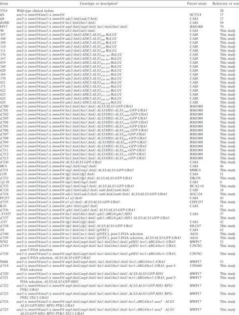
specifically during hyphal development inC. albicans, we ex-amined ALS3 mRNA levels by Northern blotting following exposure to three distinct types of morphogenetic signals: se-rum (Fig. 1A), neutral pH, and N-acetylglucosamine (not shown). The ALS3 mRNA was induced in C. albicans cells growing at 37°C and strongly induced in cells exposed to serum at 37°C (Fig. 1A). This transcript was undetectable in a C. albicans als3/als3null mutant.ALS3mRNA levels correlated strongly with the extent of hyphal development in these
cul-tures. The same was true when hyphal development was in-duced by neutral pH orN-acetylglucosamine (not shown). Our data confirm data from a previous report by Hoyer et al. showing thatALS3is a hypha-specific gene (28).
To test whether the developmental expression pattern of ALS3 is mediated at a transcriptional or posttranscriptional level, we generated anALS3-GFPpromoter fusion. This pro-moter fusion was integrated into theC. albicansgenome in situ at theALS3locus (SAC530) (Table 1). The expression of this reporter was monitored by assaying fluorescence levels inC. albicans SAC530 cells growing in the presence and absence of serum (Fig. 1B and C). The ALS3-GFP promoter fusion displayed an expression pattern that was similar to that of wild-typeALS3 mRNA, indicating thatALS3transcription is induced specifically during hyphal development.
Hypha-specific promoters are unusually long.Having estab-lished that the developmental regulation ofALS3is mediated at the transcriptional level, we performed an in silico compar-ison of the ALS3 promoter region and other hypha-specific promoters (ECE1,HGC1, HWP1, HYR1, RBT1, and RBT4). Our aim was to identify common sequence elements that might contribute to the coordinate regulation of these genes during hyphal development. To achieve this, we analyzed the inter-genic regions that lie upstream of these genes (Fig. 2). Two main observations were made. First, the 5⬘-intergenic regions for hypha-specific genes are unusually long compared to C. albicans genes in general. The estimated average length of intergenic regions for divergently transcribedC. albicansgenes is 1,088 bp, that for convergently transcribed genes is 521 bp, and that for tandemly transcribed genes is 770 bp (26). In contrast, the average length of the upstream intergenic regions for these seven hypha-specific genes is 4.5 kbp (based on the latest genome assembly available in theCandidaGenome Da-tabase) (http://www.candidagenome.org/ [accessed October 2006]). TheALS3intergenic region is 3.0 kbp, andHCG1has the longest region at 9.0 kbp. This provided our first clue that morphogenetically regulated promoters inC. albicansmight be relatively complex. This view is consistent with observations of budding yeast. For example, the developmentally regulated FLO11and HOgenes inSaccharomyces cerevisiaeboth have unusually long and complex promoters (49, 58).
Our second observation was that hypha-specific promoters contain putative binding sites for many known transcription factors inC. albicans. These include putative sites for Efg1, FIG. 1.ALS3transcription is activated during hyphal development.
(A) Northern analysis ofALS3mRNA levels inC. albicansafter 3 h of growth in YPD at 25°C, in YPD containing serum at 25°C, in YPD at 37°C, or in YPD containing serum at 37°C.ALS3, SC5314;als3⌬, Ca90 (Table 1). The proportion of filamentous (as opposed to yeast) cells in each culture is indicated. (B) Fluorescence microscopy ofC. albicans SAC500 cells containing the in situALS3-GFPreporter under equiv-alent conditions. (C) Quantification of GFP fluorescence inC. albicans SAC500 cells under the same conditions.
FIG. 2. In silico analysis of hypha-specific promoters. The lengths of the intergenic regions of hypha-specific genes and the organization of specific sequence elements in their 5⬘regions are presented. Asterisks, Tec1 sites (CATTCY); open squares, E box (CANNTG); gray circles, Nrg1 sites (MVCCCT); closed triangles, Rfg1 sites (YYYATTGTTCTC). The lengths of the intergenic regions were calculated from assembly 20 of the C. albicansgenome sequence (see the CGD website at www.candidagenome.org/ [accessed September 2006]).
on September 8, 2020 by guest
http://ec.asm.org/
Tec1, Nrg1, Rfg1, Cph1, Cph2, Rim101, Cap1, and Gcn4. (Cap1 and Gcn4 are transcription factors that play key roles in responses to oxidative stress and amino acid starvation, respec-tively [1, 74].) However, only a small number of these sites are conserved in all the hypha-specific genes analyzed (Fig. 2). These include putative Efg1, Tec1, and Nrg1 sites. It should be noted, however, that the Efg1 consensus site (E box) is likely to occur by chance in sequences of this length.
Contribution of morphogenetic transcriptional factors to
ALS3 regulation. The ALS3 promoter region contains Nrg1
sites but no obvious Rfg1 sites on the basis of the Rfg1 con-sensus sequence (Fig. 2). However, genome-wide transcrip-tional profiling studies have suggested thatALS3is transcrip-tionally repressed by Rfg1 as well as by Nrg1 and Tup1 (32, 46). Therefore, we compared the influence of these transcription factors upon ALS3 directly by using the in situ ALS3-GFP reporter (Fig. 3). InS. cerevisiae, Tup1 acts in concert with Ssn6, forming a Tup1-Ssn6 corepressor complex that represses the expression of many target genes (64). However, inC. al-bicans, Ssn6 is not thought to play a role in the Tup1-mediated repression of hypha-specific genes largely on the basis of tran-script profiling (18). Therefore, we tested this further by ex-amining the role of Ssn6 inALS3gene regulation. TheALS3 promoter also contains a putative site for the a1/␣2 repressor, which involved in the repression of “haploid-specific” genes in C. albicans(75). Therefore, we included the a1/␣2 repressor in this analysis.
The ALS3-GFP-URA3cassette was transformed into wild-type,nrg1,rfg1,tup1,ssn6, andmtla1 mtla2cells. GFP fluores-cence levels were measured in theseC. albicansstrains during growth in the yeast form (Fig. 3B). As expected, the ALS3-GFPreporter was repressed in wild-type yeast cells. Nrg1 acts through two NREs in theALS3promoter at positions⫺330 and⫺80 (46). Hence, the derepression of theALS3-GFP re-porter in nrg1 and tup1 cells was also expected (Fig. 3B). However, this reporter was only partially derepressed inrfg1 cells and was not derepressed in ssn6 or mtla1 mtla2 cells. These data reinforce the idea thatALS3is repressed mainly by Nrg1 and Tup1 in an Ssn6-independent fashion and that Rfg1 plays a minor role in the regulation ofALS3(18, 32). The data also suggest that although theALS3promoter contains a pu-tative a1/␣2 site, this repressor is not required forALS3 reg-ulation under these conditions.
TheALS3promoter also contains putative sites for several transcription factors that are known to contribute to the acti-vation of hyphal development: Efg1, Cph1, Cph2, and Tec1. Therefore, we examined the contributions of these factors to the activation ofALS3expression during hyphal development (Fig. 3). The activity of the in situ ALS3-GFP reporter was compared in wild-type,efg1,cph1,cph2, andtec1cells following serum induction (Fig. 3C). Both Efg1 and Tec1 were required for the full activation of theALS3-GFPreporter. In contrast, Cph2 was not essential for activation, although Cph2 has been reported to regulateTEC1(36). We did observe considerable variation inALS3-GFPexpression levels in thecph2mutant, and this is reflected in relatively large error bars even though this experiment was performed five times with up to eight independent transformants (Fig. 3C). Decreased ALS3-GFP expression was observed in cph1 cells, suggesting that this mitogen-activated protein kinase pathway does contribute to ALS3 activation following serum stimulation, although this pathway is not required for hyphal development under these conditions (10, 42). We also examined the impact of Bcr1 upon ALS3-GFP; this is discussed below. Taken together, the data indicate that the transcription factors Efg1, Tec1, Nrg1, and Tup1 play important roles in regulatingALS3expression and that Rfg1 and Cph1 contribute toALS3regulation.
TheALS3promoter contains two main activation regions.A
set of mutations was generated at theALS3locus to examine the organization of its promoter. These mutations were gen-erated by inserting aHIS1cassette at a range of positions in the 5⬘intergenic region of theALS3-GFPallele inC. albicans strain SAC500 (Table 1). Essentially, this created a set of promoter mutations in situ at theALS3locus, the activities of which were monitored during hyphal development by measur-ing GFP fluorescence followmeasur-ing serum stimulation.
The removal of sequences between positions ⫺1438 and
⫺1049 (with respect to the first base of the coding region) from the promoter caused a twofold decrease in the activity of the ALS3-GFPallele (Fig. 4). The further removal of sequences between positions⫺1049 and ⫺471 had no significant effect upon expression. However, the removal of sequences between positions⫺471 and⫺306 blockedALS3-GFPactivation com-pletely. We conclude that the full activation ofALS3depends upon two promoter regions. One region (A1 [positions⫺471 to⫺306]) is essential for activation, while a second region (A2 [positions⫺1438 to⫺1049]) enhances this activation. FIG. 3. Contribution of transcriptional regulators to the regulation
ofALS3. (A) Cartoon illustrating the putative impact of transcriptional activators and repressors upon hyphal development. MAP, mitogen-activated protein; cAMP, cyclic AMP. (B) Effect of repressor muta-tions upon the expression of theALS3-GFPreporter after growth for 2 h in YPD at 25°C. (C) Effect of inactivating transcriptional activators on theALS3-GFPreporter after 90 min of growth in YPD containing serum at 37°C. wt, wild type.
686 ARGIMO´ N ET AL. EUKARYOT. CELL
on September 8, 2020 by guest
http://ec.asm.org/
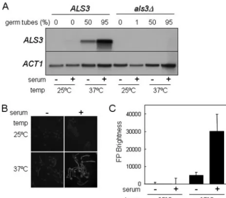
We examined the kinetics of induction ofALS3-GFP tran-scripts to further investigate the contributions of the A1 and A2 activation regions to the hypha-specific induction of ALS3. Northern analysis was performed on C. albicans SAC501, SAC505, and SAC513 cells following serum stim-ulation. SAC501 contains both the A1 and A2 activation regions (ALS31499-GFP), and SAC505 lacks A2 but contains A1 (ALS31049-GFP), whereas SAC513 lacks both A1 and A2 (ALS3306-GFP). All three strains developed hyphae at similar rates following serum stimulation, as expected. However, no induction ofGFPmRNA was observed for the negative control containing theALS3306-GFPfusion (Fig. 5). In contrast,GFP mRNA was strongly induced from the positive control contain-ing both activation regions, reachcontain-ing a maximum at 60 min. Similar kinetics ofGFPmRNA induction were observed for the ALS3-GFP construct that contains only the A1 region. However, theGFPmRNA levels reached only about one-third of those in the positive control (Fig. 5), which correlates well with GFP fluorescence levels from these and related constructs (Fig. 4). This reproducible observation was consistent with the idea that the A1 region is essential for transcriptional activa-tion during hyphal development, while the A2 region increases the amplitude of this activation.
The ALS3 promoter is complex.To examine the essential
activation region (A1) in more detail, we turned to the sensi-tive RrLUCreporter (66). First, we tested the robustness of this approach for the dissection of theALS3promoter. A set of ALS3-RrLUC promoter fusions containing or lacking the A1 region were integrated into the genome ofC. albicansCAI8, and their expression was examined in yeast and hyphal cells. As expected, all of these constructs were inactive in yeast cells (not shown), and only those containing the A1 region (positions
⫺496 to ⫺306) were induced in hyphal cells (Fig. 6A). This indicated that the ALS3-RrLUC fusions accurately reflected the behavior of in situALS3-GFPfusions and confirmed the
presence of an essential activation region in this part of the promoter.
AdditionalALS3-RrLUCconstructs were generated to fur-ther define the 5⬘and 3⬘ends of the A1 region. Hypha-specific activation was lost if 5⬘sequences between positions⫺471 and
⫺448 were deleted (not shown). Activation was retained if 3⬘ FIG. 4. Effect of in situ promoter mutations uponALS3-GFPexpression. GFP fluorescence was quantified in eachC. albicansSAC strain (Table 1) after 90 min of growth in YPD containing serum at 37°C. The coordinate of each promoter deletion endpoint is provided. Wild-type Tec1 sites are indicated by black boxes, and mutated Tec1 sites are indicated by gray boxes.
FIG. 5. Kinetic analysis ofALS3-GFPtranscript levels during se-rum-induced hyphal development. (A) Northern analysis of ALS3-GFPtranscripts at various times (minutes) after the serum induction of C. albicansstrains carrying different promoter deletions (Table 1). A1⫹ A2⫹, SAC501 cells in which theALS3-GFPfusion contains both acti-vation regions; A1⫹A2⫺, SAC505 cells in which theALS3-GFPfusion contains only the A1 activation region; A1⫺ A2⫺, SAC513 cells in which theALS3-GFPfusion lacks both activation regions. PCR-am-plifiedALS3andACT1probes were used (see Materials and Meth-ods). (B) Quantification ofALS3-GFPtranscript levels relative to the internalACT1 mRNA control. Similar results were obtained when quantifying relative to 26S rRNA. Also, similar results were obtained in a second independent experiment.
on September 8, 2020 by guest
http://ec.asm.org/
sequences between positions⫺321 and⫺307 were removed, but further 3⬘deletions to position⫺331 resulted in reduced levels of expression in hyphal cells and the derepression of RrLUCexpression in yeast cells. This was consistent with the disruption of activating sequences and the loss of Nrg1-medi-ated repression through the deletion of the NRE at position
⫺330. We concluded that the A1 activation region lies between positions ⫺471 and ⫺321. This activation region does not
correlate well with an in silico analysis of putative regulatory elements in theALS3promoter (Fig. 2), reinforcing the view that in isolation, in silico analyses of promoter elements are a poor predictor of regulatory function.
In an attempt to define the A1 region more precisely, we generated a further set of RrLUCconstructs containing short overlapping fragments from the A1 region. None of these constructs displayed expression levels equivalent to those of FIG. 6. Analysis of the A1 activation region in theALS3promoter. Various RrLUCpromoter-RrLUCfusions were constructed and trans-formed intoC. albicansCAI8 (Table 1). The expression levels of these luciferase fusions were assayed after 3 h of growth in YPD containing serum at 37°C. (A) The expression ofALS3promoter deletions that target the A1 activation region was assayed. (B) Fragments of the A1 activation region were cloned upstream of theALS3306-RrLUCfusion, and the expression of these constructs was assayed. Black boxes, putative YRE; gray
boxes, putative GCRE. (C) Oligonucleotides containing multiple STREs, YREs, or GCREs were cloned upstream of a basal RrLUCreporter, and the luciferase levels generated by these constructs were assayed.
688 ARGIMO´ N ET AL. EUKARYOT. CELL
on September 8, 2020 by guest
http://ec.asm.org/
the control (Fig. 6B), indicating that no single enhancer ele-ment within the A1 region was sufficient to confer hypha-specific activation. Weak activation (⬍20% of the control) was observed for some fragments. This might have suggested that multiple copies of a weak element could combine to provide strong activation. However, none of these fragments shared any obvious sequence elements.
Putative binding sites for the transcription factors Msn4/ Msn2 (STRE [C4T]), Cap1 (YRE [TTA[G/C]TAA]), and
Gcn4 (GCRE [TGACTC]) do exist in the promoters of hypha-specific genes, and these elements are present inALS3 pro-moter fragments that provide weak transcriptional activation. Therefore, we tested whether STRE, YRE, or GCRE ele-ments can activate transcription in response to serum induc-tion (Fig. 6C). The YRE- and GCRE-RrLUC reporters dis-played weak activation compared with the ALS3-RrLUC control, suggesting that these elements might contribute to the weak activation seen for the short ALS3 promoter fusions examined in Fig. 6B. However, the YRE element mediates transcriptional activation inC. albicansyeast cells in response to oxidative stress (51), and the GCRE activates transcription in yeast cells in response to amino acid starvation (74). Neither Cap1 nor Gcn4 is required for serum-induced morphogenesis. Hence, these elements cannot account for the hypha specificity of the A1 promoter region. Nevertheless, it is conceivable that YRE and GCRE elements might contribute to the transcrip-tional activation of hypha-specific genes in the context of the natural promoters.
Taken together, the data suggest that the A1 promoter re-gion is complex. Sequence elements close to the 5⬘and 3⬘ends of this region are required for the transcriptional activation of ALS3during hyphal development. These elements appear to function in combination to mediate hypha-specific activation.
Tec1 acts indirectly through Bcr1 to regulate ALS3
tran-scription. Putative Tec1 sites exist in all hypha-specific pro-moter regions (Fig. 2). Five such sites are present in theALS3 promoter at positions⫺1499,⫺1438,⫺1049,⫺885, and⫺842. Furthermore, Tec1 is required for the morphogenetic activa-tion of ALS3 (Fig. 3C). Therefore, we reasoned that Tec1 might act directly upon theALS3promoter via (some of) the putative Tec1 sites. To test this, we generated a set of in situ ALS3promoter mutants in which the Tec1 sites were sequen-tially inactivated and compared them to a parallel set of con-trol mutations containing the Tec1 sites (Fig. 4 and Table 1). No significant difference in expression level was observed be-tween each Tec1 site mutation (Fig. 4, gray bars) and its cor-responding control (black bars). This indicated that the puta-tive Tec1 sites are not required for the hypha-specific activation ofALS3 and hence that Tec1 might act indirectly upon this gene.
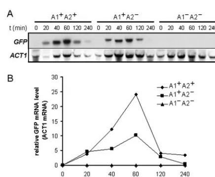
Recently, Nobile and Mitchell (52) identifiedBCR1as being a regulator of biofilm formation in C. albicans. During the course of that work, they showed thatALS3mRNA levels are reduced inbcr1cells and thatBCR1expression is reduced in a tec1mutant. This raised the possibility that Tec1 might regu-lateALS3indirectly via Bcr1. We tested this idea by first asking whetherBCR1is required for the transcriptional activation of theALS3-GFPreporter.ALS3-GFPexpression was lost inbcr1 cells, indicating that Bcr1 is essential for the transcriptional activation ofALS3during hyphal development (Fig. 3C). We
then tested whetherTEC1overexpression enhancesALS3 ex-pression and whether this effect is dependent upon BCR1. TEC1overexpression was engineered by transforming a PYK1-TEC1fusion intoC. albicansSAC520 cells and growing them on glucose-containing medium to activate thePYK1promoter (4). This led to the significant overexpression of ALS3-GFP (Fig. 7). This overexpression was blocked in a bcr1 mutant background, confirming that Tec1 acts indirectly uponALS3 transcription via Bcr1.
DISCUSSION
Yeast-hypha morphogenesis has been studied intensively in C. albicansbecause of its likely contribution to the pathoge-nicity of this fungus (21, 22, 29, 61, 71, 83). A complex network of signaling pathways has been shown to control hyphal devel-opment, but the mechanistic relationships between these path-ways remain obscure (10). These signaling pathpath-ways are thought to converge on the promoters of those genes that respond specifically during hyphal development (7, 10).ALS3 is one of a small set of hypha-specific genes inC. albicansthat includesALS3,ECE1,HGC1,HWP1,HYR1,RBT1, andRBT4 (2, 5, 8, 28, 40, 48, 67, 83). In this study, we have confirmed that the hypha-specific activation ofALS3is mediated at the tran-scriptional level (Fig. 1). Clearly, a complete understanding of morphogenetic signaling depends upon the dissection of hy-pha-specific promoters and the mechanisms by which these pathways regulate these promoters.
We have shown thatALS3is regulated by a complex array of transcription factors: Efg1, Cph1, Tec1, Bcr1, Nrg1, Rfg1, and Tup1 (Fig. 3). WhenC. albicanscells grow in the yeast form, ALS3transcription is repressed mainly by Nrg1, which binds to NREs located at positions⫺330 and⫺80 in the promoter (46). Rfg1 also contributes toALS3repression (Fig. 3) (32), but the promoter element(s) through which Rfg1 operates inC. albi-canshas not been experimentally defined. Both Nrg1 and Rfg1 are thought to act by interacting with the global repressor Tup1, which mediates transcription through direct interactions FIG. 7. Effect of ectopicTEC1expression andBCR1inactivation onALS3-GFPexpression. The in situALS3-GFPreporter was intro-duced into wild-type (BCR1) andbcr1cells, and these strains were transformed with the emptyPYK1expression vector (v) or the PYK1-TEC1 plasmid (TEC1) to generate strains SAC522 (v) (BCR1), SAC523 (TEC1) (BCR1), SAC524 (v) (bcr1), and SAC525 (TEC1) (bcr1) (Table 1). GFP fluorescence levels were assayed in these strains after 90 min of growth in YPD-containing serum at 37°C.
on September 8, 2020 by guest
http://ec.asm.org/
with the transcription complex, by positioning nucleosomes on the promoter, or by a combination of both mechanisms (23, 80). InS. cerevisiae, interactions between Tup1 and its cognate DNA binding proteins often depend on Ssn6 (64). However, this does not appear to be the case for Nrg1 in the context of hypha-specific genes. It has been suggested that the repression of hypha-specific genes by Nrg1 and Tup1 does not depend upon Ssn6 (18), and we have confirmed this forALS3in this study (Fig. 3).
Cph1 and the A2 region of the promoter are required only for full ALS3 activation. This might suggest that Cph1 en-hancesALS3transcription via the A2 region. However, there are no obvious occurrences of the putative Cph1 consensus site in theALS3 promoter, and therefore, Cph1 might act indi-rectly to regulateALS3transcription (Fig. 8).
In contrast, Efg1 is essential for the transcriptional activa-tion ofALS3during hyphal development (Fig. 3C). Efg1 has been shown to bind an E box in vitro (40), and hypha-specific promoters do contain this type of sequence element (Fig. 2). However, the degenerate E-box consensus is likely to occur frequently by chance (1/256), and to date, there are no reports confirming that Efg1 regulates transcription via the E box inC. albicans.
Although Tec1 is essential for the activation ofALS3(Fig. 4) and putative Tec1 sites exist in theALS3 promoter (Fig. 2), these sites do not contribute significantly toALS3 activation (Fig. 4). Instead, Tec1 regulatesALS3transcription indirectly through Bcr1 (Fig. 7), which is also essential forALS3 activa-tion (Fig. 3). These observaactiva-tions are entirely consistent with recent data from Nobile et al. They showed that Tec1 and Bcr1 are required for the formation of biofilms inC. albicansand that Bcr1 acts downstream of Tec1 to regulate the expression of adhesin genes required for biofilm formation, such asALS3 andHWP1(52, 53).
The transcriptional activation of ALS3 is dependent upon the A1 promoter region (Fig. 4) as well as upon Efg1, Tec1,
and Bcr1 (Fig. 3). The A1 promoter region is complex: no single sequence element within this 150-bp region was capable of driving hypha-specific expression, and the trimming of se-quences at either the 5⬘ or 3⬘ end of this A1 region blocked hypha-specific activation (Fig. 6). This is consistent with the idea that several different regulatory factors converge upon the A1 region to cooperate inALS3activation. Hence, Tec1-Bcr1 and Efg1 might regulateALS3cooperatively via the A1 pro-moter region (Fig. 8). An NRE lies at the 3⬘border of the A1 region at position⫺330. It has been reported that Nrg1 might act as a transcriptional activator under some circumstances (47, 59). Hence, it is conceivable that Nrg1 might also contrib-ute to the hyphal activation ofALS3.
In parallel studies, Kim and coworkers (33) made similar observations about the regulation of a second hypha-specific gene,HWP1. TheHWP1promoter also contains two activation regions. One region, which binds an array of chromatin remod-eling proteins, is essential forHWP1 activation, whereas the second distal region increases the amplitude of this activation (33). Hence, this class of developmentally regulated genes ap-pears to be controlled by complex interactions between several critical transcription factors at the level of their promoters. It has long been recognized thatC. albicansresponds to an ex-tremely disparate range of environmental conditions by form-ing hyphae (54). The unusual length of promoters of hypha-specific genes and the complexity and diversity of factors regulating their transcription not only are compatible with the diversity of conditions known to favor hypha formation but also suggest that morphogenetic changes inC. albicansmay be affected by events in several regulatory pathways whose stim-ulation may not always be specifically or directly related to cell shape.
ACKNOWLEDGMENTS
We thank Susan Budge for excellent technical assistance. We also thank Paula Sundstrom for making data available to us prior to pub-lication.
This work was supported by funding from the Wellcome Trust (063204 and 068143), the BBSRC (1/CEL 4563 and 1/P17124), and the EC (QLK2CT-2000-00795 and MRTN-CT-2003-504148).
REFERENCES
1.Alarco, A. M., and M. Raymond.1999. The bZip transcription factor Cap1p
is involved in multidrug resistance and oxidative stress response inCandida
albicans. J. Bacteriol.181:700–708.
2.Bailey, D. A., P. J. F. Feldmann, M. Bovey, N. A. R. Gow, and A. J. P. Brown.
1996. TheCandida albicans HYR1gene, which is activated in response to
hyphal development, belongs to a gene family encoding yeast cell wall
pro-teins. J. Bacteriol.178:5353–5360.
3.Barelle, C. J., C. L. Manson, D. M. MacCallum, F. C. Odds, N. A. R. Gow, and A. J. P. Brown.2004. GFP as a quantitative reporter of gene regulation inCandida albicans. Yeast21:333–340.
4.Barelle, C. J., C. L. Priest, D. M. MacCallum, N. A. R. Gow, F. C. Odds, and A. J. P. Brown.2006. Niche-specific regulation of central metabolic pathways
in a fungal pathogen. Cell. Microbiol.8:961–971.
5.Birse, C. E., M. Y. Irwin, W. A. Fonzi, and P. S. Sypherd.1993. Cloning and
characterization ofECE1, a gene expressed in association with cell
elonga-tion of the dimorphic pathogenCandida albicans. Infect. Immun.61:3648–
3655.
6.Braun, B. R., and A. D. Johnson.1997. Control of filament formation in
Candida albicansby the transcriptional repressor TUP1. Science277:105– 109.
7.Braun, B. R., and A. D. Johnson.2000.TUP1,CPH1andEFG1make
independent contributions to filamentation inCandida albicans. Genetics
155:57–67.
8.Braun, B. R., W. S. Head, M. X. Wang, and A. D. Johnson.2000.
Identifi-cation and characterization ofTUP1-regulated genes inCandida albicans.
Genetics156:31–44.
FIG. 8. Working model illustrating the effects of morphogenetic regulators on the transcriptional regulation ofALS3. As described in the text, theALS3promoter has two activation regions (A1 and A2), with A1 being essential for hypha-specific activation (Fig. 4, 5, and 6). ALS3activation is dependent upon Efg1, Bcr1, and Tec1 (Fig. 3), with the latter acting through Bcr1 (Fig. 7) (53). These factors might act through the A1 region, but no direct interaction with this region has been demonstrated. Like the A2 promoter region, Cph1 contributes to ALS3activation but is not essential for this activation (Fig. 3 and 5). It is not known whether Cph1 acts directly or indirectly upon theALS3 promoter (dotted line). Nrg1 represses transcription in a Tup1-depen-dent fashion (9) by binding to NREs in theALS3promoter (46). Rfg1 contributes to this repression, but the ALS3promoter contains no obvious Rfg1 sites (Fig. 2), and it is not known whether Rfg1 acts directly upon theALS3promoter (dotted line).
690 ARGIMO´ N ET AL. EUKARYOT. CELL
on September 8, 2020 by guest
http://ec.asm.org/
9.Braun, B. R., D. Kadosh, and A. D. Johnson.2001.NRG1, a repressor of
filamentous growth inCandida albicans, is down-regulated during filament
induction. EMBO. J.20:4753–4761.
10.Brown, A. J. P.2002. Morphogenetic signalling pathways inCandida
albi-cans, p 95–106.InR. Calderone (ed.),Candidaand candidiasis. ASM Press,
Washington, DC.
11.Brown, D. H., A. D. Giusani, X. Chen, and C. A. Kumamoto.1999.
Filamen-tous growth ofCandida albicansin response to physical environmental cues
and its regulation by the uniqueCZF1gene. Mol. Microbiol.34:651–662.
12.Cartharius, K., K. Frech, K. Grote, B. Klocke, M. Haltmeier, A. Klingenhoff, M. Frisch, M. Bayerlein, and T. Werner.2005. MatInspector and beyond: promoter analysis based on transcription factor binding sites. Bioinformatics
21:2933–2942.
13.Cheng, G., K. Wozniak, M. A. Wallig, P. L. Fidel, S. R. Trupin, and L. L. Hoyer.2005. Comparison betweenCandida albicansagglutinin-like sequence gene expression patterns in human clinical specimens and models of vaginal
candidiasis. Infect. Immun.73:1656–1663.
14.Cormack, B., G. Bertram, M. Egerton, N. A. R. Gow, S. Falkow, and A. J. P. Brown.1997. Yeast-enhanced green fluorescent protein (yEGFP): a reporter
of gene expression inCandida albicans. Microbiology143:303–311.
15.Dennison, P. M. J., M. Ramsdale, C. L. Manson, and A. J. P. Brown.2005.
Gene disruption inCandida albicansusing a synthetic codon-optimised
Cre-loxPsystem. Fungal Genet. Biol.42:737–748.
16.Doedt, T., S. Krishnamurthy, D. P. Bockmu¨hl, B. Tebarth, C. Stempel, C. L. Russell, A. J. P. Brown, and J. F. Ernst.2004. APSES proteins regulate
morphogenesis and metabolism inCandida albicans. Mol. Biol. Cell15:
3167–3180.
17.Fonzi, W. A., and M. Y. Irwin.1993. Isogenic strain construction and gene
mapping inCandida albicans. Genetics134:717–728.
18.Garcı´a-Sa´nchez, S., A. Mavor, C. L. Russell, S. Argimo´n, P. Dennison, B. Enjalbert, and A. J. P. Brown.2005. Global roles of Ssn6 in Tup1- and Nrg1-dependent gene regulation in the fungal pathogen, Candida albicans.
Mol. Biol. Cell16:2913–2925.
19.Gerami-Nejad, M., J. Berman, and C. A. Gale.2001. Cassettes for PCR-mediated construction of green, yellow, and cyan fluorescent protein fusions inCandida albicans. Yeast18:859–864.
20.Gillum, A. M., E. Y. Tsay, and D. R. Kirsch.1984. Isolation of theCandida albicansgene for orotidine-5⬘-phosphate decarboxylase by complementation ofS. cerevisiae ura3andE. coli pyrFmutations. Mol. Gen. Genet.198:179– 182.
21.Gow, N. A. R., A. J. P. Brown, and F. C. Odds.2002. Fungal morphogenesis
and host invasion. Curr. Opin. Microbiol.5:366–371.
22.Gow, N. A. R., Y. Knox, C. A. Munro, and W. D. Thompson.2003. Infection of chick chorioallantoic membrane (CAM) as a model for invasive hyphal
growth and pathogenesis of Candida albicans. Med. Mycol.41:331–338.
23.Green, S. R., and A. D. Johnson.2004. Promoter-dependent roles for the Srb10 cyclin-dependent kinase and the Hda1 deacetylase in Tup1-mediated
repression inSaccharomyces cerevisiae. Mol. Biol. Cell15:4191–4202.
24.Hauser, N. C., M. Vingron, M. Scheideler, B. Krems, K. Hellmuth, K.-D. Entian, and J. D. Hoheisel. 1998. Transcriptional profiling on all open
reading frames ofSaccharomyces cerevisiae. Yeast14:1209–1221.
25.Hoffman, C. S., and F. Winston.1987. A ten-minute DNA preparation from
yeast efficiently releases autonomous plasmids for transformation ofE. coli.
Gene57:267–272.
26.Holton, N. J., T. J. D. Goodwin, M. I. Butler, and R. T. M. Poulter.2001. An
active retrotrasposon inCandida albicans. Nucleic Acids Res.29:635–647.
27.Hoyer, L. L.2001. The ALS gene family ofCandida albicans. Trends
Micro-biol.9:176–180.
28.Hoyer, L. L., T. L. Payne, M. Bell, A. M. Myers, and S. Scherer.1998.
Candida albicans ALS3and insights into the nature of the ALS gene family.
Curr. Genet.33:451–459.
29.Hube, B.2004. From commensal to pathogen: stage- and tissue-specific gene
expression ofCandida albicans. Curr. Opin. Microbiol.7:336–341.
30.Hube, B., and J. Naglik.2002. Extracellular hydrolases, p. 107–122.InR.
Calderone (ed.),Candidaand candidiasis. ASM Press, Washington, DC.
31.Kadosh, D., and A. D. Johnson.2001. Rfg1, a protein related to theS. cerevisiaehypoxic regulator Rox1, controls filamentous growth and virulence inC. albicans. Mol. Cell. Biol.21:2496–2505.
32.Kadosh, D., and A. D. Johnson.2005. Induction of theCandida albicans
filamentous growth program by relief of transcriptional repression: a
ge-nome-wide analysis. Mol. Biol. Cell16:2903–2912.
33.Kim, S., M. J. Wolyniak, J. F. Staab, and P. Sundstrom.2007. A
368-base-paircis-actingHWP1promoter region, HCR, ofCandida albicansconfers
hypha-specific gene regulation and binds architectural transcription factors
Nhp6 and Gcf1p. Eukaryot. Cell6:693–709.
34.Knight, A. W., N. J. Goddard, P. R. Fielden, M. G. Barker, N. Billinton, and R. M. Walmsley.1999. Fluorescence polarisation of green fluorescent pro-tein (GFP). A strategy for improved wavelength discrimination for GFP
determinations. Analyt. Commun.36:113–117.
35.Knight, A. W., N. J. Goddard, N. Billinton, P. A. Cahill, and R. M. Walmsley.
2002. Fluorescence polarization discriminates green fluorescent protein from
interfering autofluorescence in a microplate assay for genotoxicity. J.
Bio-chem. Biophys. Methods51:165–177.
36.Lane, S., C. Birse, S. Zhou, R. Matson, and H. P. Liu.2001. DNA array studies demonstrate convergent regulation of virulence factors by Cph1,
Cph2, and Efg1 inCandida albicans. J. Biol. Chem.276:48988–48996.
37.Lane, S., S. Zhou, T. Pan, Q. Dai, and H. P. Liu.2001. The basic
helix-loop-helix transcription factor Cph2 regulates hyphal development inCandida
albicanspartly via Tec1. Mol. Cell. Biol.21:6418–6428.
38.Leberer, E., D. Harcus, I. D. Broadbent, K. L. Clark, D. Dignard, K. Ziegelbauer, A. Schmidt, N. A. R. Gow, A. J. P. Brown, and D. Y. Thomas.
1996. Homologs of the Ste20p and Ste7p protein kinases are involved in
hyphal formation ofCandida albicans. Proc. Natl. Acad. Sci. USA93:13217–
13222.
39.Leng, P., P. E. Sudbery, and A. J. P. Brown.2000. Rad6p represses yeast-hypha morphogenesis in the human fungal pathogen, Candida albicans. Mol.
Microbiol.35:1264–1275.
40.Leng, P., P. Lee, H. Wu, and A. J. P. Brown.2001. Efg1, a morphogenetic
regulator inCandida albicans, is a sequence-specific DNA binding protein. J.
Bacteriol.183:4090–4093.
41.Liu, H., J. R. Kohler, and G. R. Fink.1994. Suppression of hyphal formation inCandida albicansby mutation of aSTE12homolog. Science266:1723– 1726.
42.Lo, H. J., J. R. Kohler, B. DiDomenico, D. Loebenberg, A. Cacciapuoti, and G. R. Fink.1997. NonfilamentousC. albicansmutants are avirulent. Cell
90:939–949.
43.Miller, M. G., and A. D. Johnson.2002. White-opaque switching in Candida albicans is controlled by mating-type locus homeodomain proteins and
al-lows efficient mating. Cell110:293–302.
44.Mitchell, A. P.1998. Dimorphism and virulence inCandida albicans. Curr.
Opin. Microbiol.1:687–692.
45.Murad, A. M. A., P. R. Lee, I. D. Broadbent, C. J. Barelle, and A. J. P. Brown.
2000. CIp10, an efficient and convenient integrating vector forCandida
albicans. Yeast16:325–327.
46.Murad, A. M. A., P. Leng, M. Straffon, J. Wishart, S. Macaskill, D. MacCallum, N. Schnell, D. Talibi, D. Marechal, F. Tekaia, C. d’Enfert, C. Gaillardin, F. C. Odds, and A. J. P. Brown.2001.NRG1represses
yeast-hypha morphogenesis and yeast-hypha-specific gene expression inCandida
albi-cans. EMBO J.20:4742–4752.
47.Murad, A. M. A., C. d’Enfert, C. Gaillardin, H. Tournu, F. Tekaia, D. Talibi, D. Marechal, V. Marchais, J. Cottin, and A. J. P. Brown.2001. Transcript
profiling inCandida albicansreveals new cellular functions for the
transcrip-tional repressors, CaTup1, CaMig1 and CaNrg1. Mol. Microbiol.42:981–
993.
48.Nantel, A., D. Dignard, C. Bachewich, D. Harcus, A. Marcil, A.-P. Bouin, C. W. Sensen, H. Hogues, M. van het Hoog, P. Gordon, T. Rigby, F. Benoit, D. C. Tessier, D. Y. Thomas, and M. Whiteway.2002. Transcript profiling of
Candida albicanscells undergoing the yeast-to-hyphal transition. Mol. Biol.
Cell13:3452–3465.
49.Nasmyth, K.1985. At least 1400 base pairs of 5⬘-flanking DNA is required
for the correct expression of theHOgene in yeast. Cell42:213–223.
50.Negredo, A., L. Monteoliva, C. Gil, J. Pla, and C. Nombela.1997. Cloning
analysis and one-step disruption of theARG5,6gene ofCandida albicans.
Microbiology143:297–302.
51.Nicholls, S., M. Straffon, B. Enjalbert, A. Nantel, S. Macaskill, M. Whiteway, and A. J. P. Brown.2004. Msn2/4-like transcription factors play no obvious
roles in the stress responses of the fungal pathogenCandida albicans.
Eu-karyot. Cell3:1111–1123.
52.Nobile, C. J., and A. P. Mitchell.2005. Regulation of cell-surface genes and
biofilm formation by theC. albicanstranscription factor Bcr1p. Curr. Biol.
15:1150–1155.
53.Nobile, C. J., D. R. Andes, J. E. Nett, F. J. Smith, F. Yue, Q.-T. Phan, J. E. Edwards, S. G. Filler, and A. P. Mitchell.2006. Critical role of
Bcr1-depen-dent adhesins inC. albicansbiofilm formationin vitroandin vivo. PLoS
Pathog.2:636–649.
54.Odds, F. C.1988.Candidaand candidosis, 2nd ed. Bailliere Tindall, London, United Kingdom.
55.Oh, S. H., G. Cheng, J. A. Nuessen, R. Jajko, K. M. Yeater, X. Zhao, C. Pujol, D. R. Soll, and L. L. Hoyer.2005. Functional specificity ofCandida albicans
Als3p proteins and clade specificity ofALS3alleles discriminated by the
number of copies of the tandem repeat sequence in the central domain.
Microbiology151:673–681.
56.Quandt, K., K. Frech, H. Karas, E. Wingender, and T. Werner.1995. MatInd and MatInspector: new fast and versatile tools for detection of consensus
matches in nucleotide sequence data. Nucleic Acids Res.23:4878–4884.
57.Ramon, A. M., A. Porta, and W. A. Fonzi.1999. Effect of environmental pH
on morphological development ofCandida albicans is mediated via the
PacC-regulated transcription factor encoded byPRR2. J. Bacteriol.181:
7524–7530.
58.Rupp, S., E. Summers, H.-J. Lo, H. Madhani, and G. R. Fink.1999. MAP kinase and cAMP filamentation signaling pathways converge on the
unusu-ally large promoter of the yeastFLO11gene. EMBO. J.18:1257–1269.
59.Russell, C. L., and A. J. P. Brown.2005. Expression of one-hybrid fusions
on September 8, 2020 by guest
http://ec.asm.org/
withStaphylococcus aureuslexA inCandida albicansconfirms that Nrg1 is a transcriptional repressor and that Gcn4 is a transcriptional activator. Fungal
Genet. Biol.42:676–683.
60.Sathe, G. M., S. O’Brien, M. M. McLaughlin, F. Watson, and G. P. Livi.
1991. Use of polymerase chain reaction for rapid detection of gene insertions
in whole yeast cells. Nucleic Acids Res.19:4775.
61.Saville, S. P., A. L. Lazzell, C. Monteagudo, and J. L. Lopez-Ribot.2003. Engineered control of cell morphology in vivo reveals distinct roles for yeast
and filamentous forms ofCandida albicansduring infection. Eukaryot. Cell
2:1053–1060.
62.Schweizer, A., S. Rupp, B. N. Taylor, M. Rollinghoff, and K. Schro¨ppel.2001. The TEA/ATTS transcription factor CaTec1p regulates hyphal development
and virulence inCandida albicans. Mol. Microbiol.38:435–445.
63.Sherman, F.1991. Getting started with yeast. Methods Enzymol.194:3–21. 64.Smith, R. L., and A. D. Johnson.2000. Turning off genes by Ssn6-Tup1: a conserved system of transcriptional repression in eukaryotes. Trends
Bio-chem. Sci.25:325–330.
65.Soll, D. R.2002. Phenotypic switching, p. 123–142.InR. Calderone (ed.),
Candidaand candidiasis. ASM Press, Washington, DC.
66.Srikantha, T., A. Klapach, W. W. Lorenz, L. K. Tsai, L. A. Laughlin, J. A. Gorman, and D. R. Soll.1996. The sea pansyRenilla reniformisluciferase serves as a sensitive bioluminescent reporter for differential gene expression inCandida albicans. J. Bacteriol.178:121–129.
67.Staab, J. F., C. A. Ferrer, and P. Sundstrom.1996. Developmental expres-sion of a tandemly repeated, proline and glutamine-rich amino acid motif on
hyphal surfaces ofCandida albicans. J. Biol. Chem.271:6298–6305.
68.Staab, J. F., S. D. Bradway, P. L. Fidel, and P. Sundstrom.1999. Adhesive
and mammalian transglutaminase substrate properties ofCandida albicans
Hwp1. Science283:1535–1538.
69.Stoldt, V. R., A. Sonneborn, C. E. Leuker, and J. F. Ernst.1997. Efg1p, an
essential regulator of morphogenesis of the human fungal pathogenCandida
albicans, is a member of a conserved class of bHLH proteins regulating
morphogenetic processes in fungi. EMBO J.16:1982–1991.
70.Sundstrom, P.2002. Adhesion inCandidaspp. Cell. Microbiol.4:461–469. 71.Sundstrom, P.2006.Candida albicanshyphal formation and virulence, p.
45–47.InJ. Heitman, S. G. Filler, J. E. Edwards, Jr., and A. P. Mitchell (ed.),
Molecular principles of fungal pathogenesis. ASM Press, Washington, DC. 72.Sundstrom, P., E. Balish, and C. M. Allen. 2002. Essential role of the
Candida albicanstransglutaminase substrate, hyphal wall protein 1, in lethal
oroesophageal candidiasis in immunodeficient mice. J. Infect. Dis.185:521–
530.
73.Swoboda, R. K., G. Bertram, S. Delbruck, J. F. Ernst, N. A. R. Gow, G. W. Gooday, and A. J. P. Brown.1994. Fluctuations in glycolytic mRNA levels
during the yeast-to-hyphal transition inCandida albicansreflect underlying
changes in growth rather than a response to cellular dimorphism. Mol.
Microbiol.13:663–672.
74.Tripathi, G., C. Wiltshire, S. Macaskill, H. Tournu, S. Budge, and A. J. P. Brown.2002. CaGcn4 co-ordinates morphogenetic and metabolic responses
to amino acid starvation inCandida albicans. EMBO J.21:5448–5456.
75.Tsong, A. E., M. G. Miller, R. M. Raisner, and A. D. Johnson.2003. Evo-lution of a combinatorial transcriptional circuit: a case study in yeasts. Cell
115:389–399.
76.van Helden, J., B. Andre´, and J. Collado-Vides.2000. A website for the
computational analysis of yeast regulatory sequences. Yeast16:177–187.
77.Walther, A., and J. Wendland.2003. An improved transformation protocol
for the human fungal pathogenCandida albicans. Curr. Genet.42:339–343.
78.Wicksteed, B. L., I. Collins, A. Dershowitz, L. I. Stateva, R. P. Green, S. G. Oliver, A. J. P. Brown, and C. S. Newlon.1994. A physical comparison of
chromosome III in six strains ofSaccharomyces cerevisiae. Yeast10:39–57.
79.Wilson, R. B., D. Davis, and A. P. Mitchell.1999. Rapid hypothesis testing withCandida albicansthrough gene disruption with short homology regions.
J. Bacteriol.181:1868–1874.
80.Zhang, Z., and J. C. Reese.2004. Redundant mechanisms are used by
Ssn6-Tup1 in repressing chromosomal gene transcription inSaccharomyces
cerevisiae. J. Biol. Chem.279:39240–39250.
81.Zhao, X., S.-H. Oh, G. Cheng, C. B. Green, J. A. Nuessen, K. Yeater, R. P. Leng, A. J. P. Brown, and L. L. Hoyer.2004.ALS3andALS8represent a
single locus that encodes aCandida albicansadhesion; functional
compari-sons between Als3p and Als1p. Microbiology150:2415–2428.
82.Zhao, X., K. J. Daniels, S.-H. Oh, C. B. Green, K. M. Yeater, D. R. Soll, and L. L. Hoyer.2006.Candida albicansAls3p is required for wild-type biofilm
formation on silicone elastomer surfaces. Microbiology152:2287–2299.
83.Zheng, X., Y. Wang, and Y. Wang.2004. Hgc1, a novel hypha-specific G1
cyclin-related protein regulates Candida albicans hyphal morphogenesis.
EMBO J.23:1845–1856.
692 ARGIMO´ N ET AL. EUKARYOT. CELL