Vol. 46, No. 5, pp. 673 - 678, 2005
This study aimed to investigate the relationship between bladder trabeculation, urinary function, and the stage of pelvic organ prolapse (POP). The medical records of 104 patients with POP who underwent cystoscopies and urodynamic studies were reviewed retrospectively. Age, incidence of detrusor instability, stage and site of POP, and the parameters of urody-namic studies of patients with and without bladder trabecula-tion were compared. The difference in the incidence of bladder trabeculation was estimated between patients with and without a suspected bladder outlet obstruction. There were significant differences in the patients' age, stage of POP, and maximal voiding velocity. Patients with a suspected bladder outlet obstruction had a significantly higher incidence of bladder trabeculation. In addition, patients with advanced stages of POP were also found to have a higher incidence of bladder trabeculation.
Key Words:Bladder trabeculation, urodynamic studies, pelvic organ prolapse
INTRODUCTION
Bladder trabeculation, which is detected by a cystoscope, is the secondary result of a bladder outlet obstruction and is known to be caused by morphological and histological changes due to hypertrophy and hyperplasia of the bladder
muscle and the infiltration of the connective tis-sue, as confirmed through animal testing.1-4 Some studies have suggested that bladder trabeculation is generated by the aging process. However, a principal pathological mechanism was reported to be connected with changes of the bladder muscle in order to compensate for the increased urethral resistance in the lower bladder, which is caused by physiological or anatomical reasons such as a bladder outlet obstruction. The degree of the obstruction differs according to the site or degree of hypertrophy. Increased total volume of the prostate can mechanically accompany the obstruc-tion of the lower urinary tract and result in an increase of intravesical pressure (Pdet) and a de-crease of the maximal voiding velocity (Qmax) in the urodynamic study.5-7
Pelvic organ prolapse (POP) was first reported in 15218 with a vaginal hysterectomy. According to a 1963 study in Switzerland, the incidence of POP was 30.8% in women between 20 and 59 years old, and the study reported an 11.1% risk of undergoing surgical treatment for this disease in women up to 80 years old.9 POP is generated by the weakening of the support tissue of the urethra, bladder, and pelvis, which is caused by dysfunction of the fibromuscular tissue that fixes the pelvic structure to the pelvic cavity.10 POP is related to delivery injury, neurosis, pelvic surgery, estrogen deficiency, constipation, chronic cough, myopathy, connective tissue disorder, etc.11-14 Dys-function of the bladder, urethra, or prolapsed organ can be associated with the anatomical
rea-The Significance of Bladder Trabeculation in the Female
Lower Urinary System: An Objective Evaluation by
Urodynamic Studies
Sang Wook Bai, Soo Hyeon Park, Da Jung Chung, Joo Hyun Park, Jong Seung Shin, Sei Kwang Kim, and Ki Hyun Park
Department of Obstetrics and Gynecology, Institute of Women's Life Science, Yonsei University College of Medicine, Seoul, Korea.
Received January 27, 2005 Accepted June 28, 2005
son that the female genitals are located close to the lower urinary tract, colon, and rectum. POP can induce the symptoms related to organ func-tion, urinafunc-tion, bowel movement, sexual life, and local symptoms caused by the prolapsed organ. The local symptoms related to urination are ex-emplified by urinary incontinence, frequency, nocturia, urgency, residual urine, etc.15 POP is known to be related to urinary tract obstruction, which causes recurrent urinary tract infection and hydronephrosis, in addition to disturbances of urination.11
In the male population, there is a high preva-lence of prostatic hypertrophy and, consequently, acute dysuria, which are both capable of in-ducing urinary obstruction. Subsequently, numer-ous studies have made progress in the diagnosis and treatment of patients with prostatic hyper-trophy.5-7 In cases of continuing dysuria, the secondary changes of hyperplasia or hypertrophy of the detrusor layer were observed.1-4 There have also been several studies investigating bladder trabeculation as a secondary change of the detrusor by a bladder outlet obstruction in men. However, only a few objective studies on female bladder outlet obstructions and its related symp-toms, diagnosis, treatment, and prognosis have progressed because symptoms of stress, urinary incontinence, urge incontinence, and infection, rather than those of urinary obstruction, are more prevalent in women due to differences in ana-tomical structures. Other reasons could include the limitations of urodynamic study for diag-nosing and treating dysuria12,16-18 and patients' reluctance in visiting the hospital for urinary dis-turbances due to the privacy of female urination and the lack of recognition of and interest in the symptoms.
The prolapse of the urethra or bladder and the mechanical obstruction of the urinary system caused by the uterine prolapse can lead to the increase in urinary resistance and secondary changes of the detrusor. Accordingly, this study was designed to verify the correlation and sig-nificance between bladder trabeculation and a bladder outlet obstruction in patients with POP through the objective analyses of the bladder trabeculation and urodynamic studies.
MATERIALS AND METHODS
This study was approved by the Institutional Review Board at Yonsei University, College of Medicine. The medical records of the patients who visited the outpatient clinic of Yonsei University Medical Center for symptoms of POP and under-went cystoscopy and urodynamic study from March 1, 1999 to March 30, 2003 were reviewed retrospectively. This study compared the patients' age, prevalence of detrusor instability, stage and site of POP, and the parameters of the urodyna-mic study, such as Qmax (mL/sec), Pdet (cmH2O), postvoidal residual volume (PVR, voided vol-ume/residual volume, mL), and maximal capacity (mL), between the patients with and without blad-der trabeculation on the cystoscopy. The stage and site of POP were detected and classified using the Pelvic Organ Prolapse-Quantification (POP-Q) system.19After restoration of the prolapsed organ, the urodynamic study was performed using Dantec-5000 (Menuet, Copenhagen, Denmark), including multi-channel cystometry, urethral pressure pro-filometry, and uroflowmetry. The ratio of the residual urine was recorded (using the lower value of the results from the voiding cystometry and the uroflowmetry), and the presence or absence of the detrusor instability was determined by the urodynamic study. The difference in the incidence of trabeculation between the patients with and without a suspicious bladder outlet ob-struction was evaluated. A suspicious bladder outlet obstruction was defined on the urodynamic study as a Qmax less than 12 mL/sec and a Pdet higher than 20 cmH2O. The statistical methods used in this study were the Student's t-test and the chi square test, with a level of significance at p< 0.05.
RESULTS
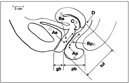
in the incidence of detrusor among the two groups. However, the mean age was significantly higher in the group with bladder trabeculation (64.65 ± 2.64 vs 57.786 ± 0.79, p< 0.05) (Table 2). The mean values of the stages in the groups with and without bladder trabeculation were 3.13 ± 0.40 and 2.86 ± 0.31 (p= 0.023), respectively, which means that the stage of POP was significantly more advanced in the group with bladder trabe-culation. In addition, according to the POP sites Aa, Ba, C, Bp, and Ap, substituted with 1, 2, 3, 4, and 5 (Fig. 1), the mean values of the sites were 2.19 ± 0.47 and 1.92 ± 0.86 (p= 0.095) in the groups with and without the trabeculation, respectively, which means that there was no significant differ-ence according to the POP site (Table 2). The para-meters of the urodynamic study, such as Qmax, Pdet, residual urine ratio, and maximal capacity, in the groups with and without bladder
trabecula-Table 1. Comparison of Epidemiologic Characteristics
Trabeculation
p Yes (n = 54) No (n = 50)
Gravidity (median)(year) 5.4 ± 1.2 5.0 ± 0.8 0.0858*
Parity (median) 3.5 ± 0.7 3.8 ± 0.5 0.124*
Previous pelvic surgery 5 3
Difficult labor 20 15 0.278*
Hypertension 4 5
Diabetes mellitus 8 4
Asthma 3 4
Chronic obstructive pulmonary disease 1 0
[image:3.595.65.536.118.267.2]*not significant.
[image:3.595.65.536.311.453.2]Fig. 1. Schematic presentation of a female pelvic organ prolapse (POP-Q classification) and substituted number for each point. Aa, point on the anterior vagina 3 cm proximal to the external urethral meatus. Ba, most distal or dependent point of any portion of the anterior vaginal wall from point Aa to just anterior to the vaginal cuff or anterior lip of the cervix. C, most dependent edge of the cervix or vaginal cuff. Bp, corresponding point to Ba on the posterior vaginal wall. Ap, corresponding point to Aa on the posterior vaginal wall.
Table 2. Comparison between Patients with and without Trabeculation
Trabeculation No trabeculation p
Age (years) 57.786 ± 0.79 64.65 ± 2.64 0.0013
Stage of POP 2.86 ± 0.31 3.13 ± 0.40 0.011
Site of POP 1.92 ± 0.86 2.19 ± 0.47 0.095
DI prevalence (%) 7.69 9.62 0.727
Qmax (mL/sec) 24.628 ± 119.18 20.206 ± 79.99 0.026
Pdet (cmH2O) 21.788 ± 199.18 28.538 ± 411.94 0.051
Max. capacity (mL) 468.068 ± 445.78 482.947 ± 694.17 0.402
PVR (%) 7.827 ± 196.04 22.429 ± 2728.95 0.054
[image:3.595.316.536.491.633.2]tion were 24.625 ± 119.18 mL/sec vs 20.206 ± 79.99 mL/sec (p< 0.05), 28.538 ± 411.94 cm H2O vs 21.788 ± 199.89 cmH2O (p= 0.052), 22.429 ± 2728.95% vs 7.827 ± 196.04% (p= 0.05), and 482.94 ± 7694.17 mL vs 468.06 ± 8445.78 mL (p= 0.402), respectively. Although Qmax was more significantly decreased in the group with trabeculation than in the group without, Pdet, residual urine ratio, and maximal capacity were not significantly different between the two groups (Table 2). Comparison of the two groups showed that the incidence of trabeculation was significantly higher in patients with a sus-picious bladder outlet obstruction than without an obstruction (80% vs 46.81%, p= 0.046) (Table 3).
DISCUSSION
As the average life span of humans increases, the interest in geriatric diseases has also increased and numerous studies on POP, which commonly occurs in older women, have been undertaken. It was reported that POP accounted for appro-ximately 0.09-0.3% of gynecological disease.15 However, this estimation is inaccurate because the symptoms of POP are quite diverse and most women do not visit the hospital owing to the prolapse. POP is a condition in which intrapelvic cavity structures, such as the uterus, bladder, urethra, or rectum, are prolapsed outside the pel-vic cavity because of an incomplete pelpel-vic sup-porting structure. This is caused by structural and anatomical changes in the nervous and muscular systems due to the aging process and a history of delivery or pelvic surgery. In this study, the risk factors for developing this condition were com-pared in the two groups and the incidences of pregnancy, delivery, urinary incontinence, and pelvic surgery, and the medical histories of dia-betes mellitus, hypertension, asthma, chronic ob-structive pulmonary disease, and difficult delivery were not significantly different between the groups.
Patients with POP could present with
symp-toms of vaginal hemorrhage, abdominal pain, lumbar pain, constipation or tenesmus, and ure-thral or vesical dysfunctions which can cause nary disorders, such as frequency, nocturia, uri-nary incontinence, urgency, dysuria, etc. In addi-tion, complications, such as chronic urinary tract infection or hydronephrosis, might develop, pro-longing the period of urinary disorders.15Dietz et al.20 reported that if the neck of the bladder is prolapsed over 2 cm, then the urethra is twisted. Thus, the urine flow will be significantly reduced, and when coexisting with enterocele, the maximal voiding velocity and the average urine flow will decrease.
In evaluating female patients complaining of symptoms of a lower urinary system disorder, there were not adequate examination guidelines or methods, other than a detailed history, diag-nosis, and a urochemistry examination. The most commonly utilized methods were urodynamic study, endoscopy, or radiologic methods such as MRI (magnetic resonance imaging).21 Costantiniet al. stated that the first method for evaluating the female patients with a urinary disorder was urodynamic study because it demonstrated over 70% of specificity, 50-100% of sensitivity, and a high negative predictive value in female patients with dysuria.22 Since subjective symptoms alone by themselves are not reliable for evaluating patients with disturbances of urination, a simple and non-invasive urodynamic study can be help-ful as a basic test to diagnose those female patients19,23 and the necessity of pressure-flow study was suggested for determining the causes.24,25 However, in spite of its usefulness for diagnosing male patients with prostatic hyper-trophy and a bladder outlet obstruction, there are limitations in applying this method to female patients, due to the gender-related differences in anatomic structures and causative factors of men.26 Therefore, it is necessary to study the ap-propriate method for examining female patients with disturbances of urination caused by POP.27 Although the incidence of a bladder outlet
ob-Table 3. Relationship between Bladder Trabeculation and Bladder Outlet Obstruction
Qmax < 12 mL/sec & Pdet > 20 cmH2O Qmax > 12 mL/sec or Pdet < 20 cmH2O p
struction in women is not well known, some studies have reported an incidence of 2.7-23% incidence in women with lower urinary system symptoms.28,29 Blaivas et al.26 diagnosed bladder outlet obstructions in 50 cases (8.3%) of a total 600 cases using urodynamic study performed on the women showing lower urinary system symptoms. On the urodynamic study, the diagnosis of blad-der outlet obstruction was restricted by three conditions: Qmax below 12 mL/sec and over 20 cmH2O of Pdet; low Qmax and evidence of an outlet obstruction on radiologic tests with the maintenance of Pdet of at least 20 cmH2O; and the maintenance of Pdet 20 cmH2O under the transu-rethral insertion, but the inability to urinate. In this study, the group with a suspicious bladder outlet obstruction had below 12 mL/sec of Qmax and over 20 cmH2O of Pdet. Trabeculation was detected in 80% of this group and in 40% of the control group. Regardless of several studies reporting a correlation between bladder trabe-culation and an outlet obstruction,30 the relation-ship is not established yet. To do so, it is neces-sary to construct an objective method for eval-uating bladder trabeculation and a system capable of reducing the errors in evaluation methods, the examiners, and the test period. It is also essential to confirm the differences in the urodynamic study results according to the degrees of trabe-culation in the patients with a suspected of a bladder outlet obstruction.
Through this study, a possible relationship between bladder trabeculation, the stage of POP and bladder outlet obstruction was suggested, but there were several drawbacks. For example, the mean age in the trabeculation group was signi-ficantly higher and this should not be overlooked because trabeculation genesis is related to aging. Therefore, confounding factors, such as age, ac-companying disease, history of surgery, and stage of POP in the control group, should be studied under the same conditions. The subjects had detrusor instability on the urodynamic study and cystoscopy. Since several examiners participated in this study, interobserver variation could have existed. Additionally, the accompanying urinary disturbances should also be classified and the existence or absence of the structural or functional factors and accompanying gynecological diseases
should be confirmed by tests.
Until now, an adequate method for evaluating the urinary function of patients with POP has not been suggested. Urinary dysfunction caused by POP could be improved by correcting the pro-lapse through surgical treatment. Results from this study indicate that a bladder outlet obstruc-tion can be suitably diagnosed by evaluating the urinary function of POP patients through urody-namic study. Additionally, the objective evalua-tion of bladder trabeculaevalua-tion by cystoscopy could provide useful information in the diagnosis and treatment of patients with POP and concurrent urinary disturbances.
REFERENCES
1. Brierly RD, Hindley RG, McLarty E, Harding DM, Thomas PJ. A prospective controlled quantitative study of ultrastructural changes in the underactive detrusor. J Urol 2003;169:1374-8.
2. Kokcu A, Yanik F, Cetinkaya M, Alper T, Kandemir B, Malatyalioglu E. Histopathological evaluation of the connective tissue of the vaginal fascia and the uterine ligaments in women with and without pelvic relxation. Arch Gynecol Obstet 2002;266:75-8.
3. Kojima M, Inui E, Ochiai A, Naya Y, Kamoi K, Ukimura O, et al. Reversible changes of bladder hypertrophy due to benign prostatic hyperplasia after surgical relief of obstruction. J Urol 1997;158:89-93. 4. Saito M, Ohmura M, Kondo A. Effects of long-term
partial outflow obstruction on bladder function in the rat. Neurourol Urodyn 1996;15:157-65.
5. Witjes WP, Aarnink RG, Ezz-el-Din K, Wijkstra H, Debruyne EM, de la Rosette JJ. The correlation between prostate volume, transition zone volume, transition zone index and clinical and urodynamic investigations in patients with lower urinary tract symptoms. Br J Urol 1997;80:84-90.
6. Reynard JM, Yang Q, Donovan JL, Peters TJ, Schafer W, de la Rosette JJ, et al. The ICS-'BPH' study: uro-flowmetry, lower urinary tract symptoms and bladder outlet obstruction. Br J Urol 1998;82:619-23.
7. Gomes CM, Trigo-Rocha FE, Arap MA, Arap S. Bladder outlet obstruction and urodynamic evaluation in patients with benign prostatic hyperplasia. Braz J Urol 2001;27:575-88.
8. Emge LA, Durfee RB. Pelvic organ prolapse: four thou-sand years of treatment. Clin Obstet Gynecol 1966;9: 997-1032.
10. Porges RF, Smilen SW. Long-term analysis of the surgi-cal management of pelvic support defects. Am J Obstet Gynecol 1994;171:1518-26.
11. Benassi L, Bocchialini E, Bertelli M, Kaihura CT, Ricci L, Siliprandi V. Risk of genital prolapse and urinary incontinence due to pregnancy and delivery. A pro-spective study. Minerva Ginecol 2002;54:317-24. 12. Goh JT. Biomechanical properties of prolapsed vaginal
tissue in pre- and postmenopausal women. Int Urogynecol J Pelvic Floor Dysfunct 2002;13:76-9. 13. Deval B, Rafii A, Poilpot S, Aflack N, Levardon M.
Prolapse in the young woman: study of risk factors. Gynecol Obstet Fertil 2002:30;673-6.
14. Sze EH, Sherard GB 3rd, Dolezal JM. Pregnancy, labor, delivery and pelvic organ prolapse. Obstet Gynecol 2002;100:981-6.
15. Bai SW, Kang SH, Kim SK, Kim JY, Park KH. The effect of pelvic organ prolapse on lower urinary tract func-tion. Yonsei Med J 2003;44:94-8.
16. Lemack GE, Zimmern PE. Pressure flow analysis may aid in identifying women with outflow obstruction. J Urol 2000;163:1823-8.
17. Chassagne S, Bernier PA, Haab F, Roehrborn CG, Reisch JS, Zimmern PE. Proposed cutoff values to define bladder outlet obstruction in women. Urology 1998;51;408-12.
18. Bump RC, Mattiason A, Bo K, Brubaker LP, DeLancey JO, Klarskov P, et al. The standardization of termi-nology of female pelvic organ prolapse and pelvic floor dysfunction. Am J Obstet Gynecol 1996;175:10-7. 19. Blaivas JG, Groutz A. Bladder outlet obstruction
nomo-gram for women with lower urinary tract sympto-matology. Neurourol Urodyn 2000;19:553-64.
20. Dietz HP, Haylen BT, Vancaillie TG. Female pelvic organ prolapse and voiding function. Int Urogynecol J
Pelvic Floor Dysfunct 2002;13:284-8.
21. Rovner ES, Wein AJ. Evaluation of lower urinary tract symptoms in females. Curr Opin Urol 2003;13:273-8. 22. Costantini E, Mearini E, Pajoncini C, Biscotto S, Bini V,
Porena M. Uroflowmetry in female voiding distur-bances. Neurourol Urodyn 2003;22:569-73.
23. Minardi D, Garofalo F, Yehia M, Cristalli AF, Giammarco L, Galosi AB,et al.Pressure-flow studies in men with benign prostatic hypertrophy before and after treatment with transurethral needle ablation. Urol Int 2001;66:89-93.
24. Nitti VW, Tu LM, Gitlin J. Diagnosing bladder outlet obstruction in women. J Urol 1999;161:1535-40. 25. Farrar DJ, Osborne JL, Stephenson TP, Whiteside CG,
Weir J, Berry J, et al. A urodynamic view of bladder outflow obstruction in the female: factors influencing the results of treatment. Br J Urol 1976;47:815-22. 26. Massey JA, Abrams PH. Obstructed voiding in the
femal. Br J Urol 1988;61:36-9.
27. Rees DL, Whitfield HN, Islam AK, Doyle PT, Mayo ME, Wickham JE. Urodynamic findings in adult females with frequency and dysuria. Br J Urol 1976;47: 853-60.
28. Groutz A, Blaivas JG, Fait G, Sassone AM, Chaikin DC, Gordon D. The significance of the American Urological Association symptom index score in the evaluation of women with bladder outlet obstruction. J Urol 2000; 163:207-11.
29. Pang MW, Yip SK. An overview of pelvic floor recon-structive surgery for pelvic organ prolapse. J Pediatr Obstet Gynaecol 2003;15:35-9.