REVIEW
Direct neuronal reprogramming: learning from and for
development
Giacomo Masserdotti1,2,*, Sergio Gascón1,2,* and Magdalena Götz1,2,3,‡
ABSTRACT
The key signalling pathways and transcriptional programmes that instruct neuronal diversity during development have largely been identified. In this Review, we discuss how this knowledge has been used to successfully reprogramme various cell types into an amazing array of distinct types of functional neurons. We further discuss the extent to which direct neuronal reprogramming recapitulates embryonic development, and examine the particular barriers to reprogramming that may exist given a cell’s unique developmental history. We conclude with a recently proposed model for cell specification called the‘Cook Islands’model, and consider whether it is a fitting model for cell specification based on recent results from the direct reprogramming field.
KEY WORDS: Conversion, Direct reprogramming, Neurogenesis, Neuron, Neuronal subtype, Transdifferentiation, Transcription factor
Introduction
During development, specific cell fates are programmed by extrinsic and intrinsic cues. Whereas loss-of-function experiments can test for the requirement of a gene, gain-of-function experiments aim to demonstrate the sufficiency of a gene in instructing developmental fate decisions. For example, MyoD was identified as a crucial regulator of muscle fate for its ability to convert mouse embryonic fibroblasts into muscle cells (Tapscott et al., 1988), and ectopic expression ofeyeless, the homologue of Pax6 in Drosophila melanogaster, was sufficient to redirect the fate of antennae cells towards an eye phenotypein vivo (Halder et al., 1995; Ypsilanti and Rubenstein, 2016). Thus, the field of transcription factor-based reprogramming (see Glossary) emerged from testing the potential of key fate determinants that are active during development, and was employed to elucidate when and where such key factors may reach the limit of their function. Indeed, gain-of-function experiments moved from development to postnatal and adult cell types: for example, probing the role of Pax6 in neurogenesis led to the discovery that postnatal glial cells
in vitroas well as some glial cells in the adult brainin vivocould be converted into neurons by this single transcription factor (Berninger et al., 2007; Buffo et al., 2008; Heins et al., 2002; Kronenberg et al., 2010; Ninkovic et al., 2013).
Initial attempts at direct reprogramming (see Glossary) were based on the assumption that cells derived from the same lineage
or the same germ layer might be easier to convert compared with less closely related cells. This concept derives from and is depicted in the famous Waddington’s landscape model, which describes development as a unidirectional process that over time restricts the potential of a totipotent cell downwards towards a highly specialized cell through the establishment of epigenetic barriers, represented by high mountain peaks (Waddington, 1957). As a consequence of these barriers, conversion between cells derived from different lineages is prevented (Sieweke, 2015). However, over the years this notion has been challenged (Gurdon, 1962; Takahashi and Yamanaka, 2006), and direct reprogramming has proven that cells from various lineages can be directly converted into cells of other lineages, even across germ layers – for example, fibroblasts or hepatic cells into neurons (Marro et al., 2011; Vierbuchen et al., 2010). Indeed, this has been shown to occur naturally in vivo, a process known as transdifferentiation (see Glossary; Zuryn et al., 2014). Thus, lineage boundaries established during cellular specification and differentiation can be overcome, depending on the potency of the factors employed. This raises two fundamental and related questions: (1) to what extent does reprogramming recapitulate a developmental trajectory; and (2) is the developmental origin and, more specifically, the epigenetic history of the starting cell type negligible? The underlying rationale of reprogramming is based on the expression of factors that are essential during development; however, their action during direct reprogramming can be rather different. This is because the factors are active in a very different context, which may or may not provide the proper environment for them to fulfil their potential. Indeed, studying how certain factors function in various reprogramming contexts may bring new insight that can feed back into developmental studies.
In this Review, we provide an overview of the main concepts that have emerged from recent studies in direct neuronal reprogramming, including the lessons this field has learnt from developmental biology, as well as new ideas for future research in neural development. Specifically, we discuss neural specification during development and provide a summary of how direct reprogramming can instruct specific neuronal subtypes, and the remaining challenges for each cell type. We further consider the mechanistic basis for these direct reprogramming approaches, which includes not only transcriptional and epigenetic events, but also regulation of the cell cycle and metabolic state. Sieweke recently proposed a model of cell fate acquisition based on Captain Cook’s voyage through the Pacific islands, hereafter termed the ‘Cook Islands’ model (Sieweke, 2015), which is a more comprehensive and accurate model of direct reprogramming between different cell types, encompassing many variables such as cell cycle and metabolic state. We conclude this Review by highlighting the close interplay between reprogramming and developmental biology, and discuss how the two related areas can inform and advance each other.
1
Institute of Stem Cell Research, Helmholtz Center Munich, Ingolstädter Landstrasse 1, Neuherberg/Munich D-85764, Germany.2Physiological Genomics, Biomedical Center, Ludwig-Maximilians University Munich, Großhadernerstrasse 9, Martinsried 82154, Germany.3Excellence Cluster of Systems Neurology, Großhadernerstrasse 9, Martinsried 82154, Germany.
*These authors contributed equally to this work
‡Author for correspondence ([email protected])
M.G., 0000-0003-1551-9203
DEVEL
O
Regional specificity and neuron specification in development
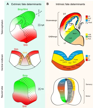
During brain development, neural stem and progenitor cells (NSCs and NPCs; see Glossary) located in the ventricular zone produce a variety of neurons at different stages and in different regions, thereby generating the immense neuronal heterogeneity and complexity of the adult brain. The biggest challenge in neuronal reprogramming is not to reliably generate only some types of neurons, but rather one amongst hundreds (or thousands) of specific types of neurons (Kodama et al., 2012; Markram et al., 2004; Masland, 2004). This is a prerequisite for sustainable cell-based therapies for brain repair (Heinrich et al., 2015; Kriks et al., 2011), and is also of conceptual interest because it could help to decipher the molecular logic behind the array of highly specific neuronal subtypes generated during development (for a review, see Hobert, 2016). Neuronal and glial fate and diversity are established very early during embryogenesis, when the crosstalk between multiple signalling pathways defines the initial regionalization of the embryonic brain (for recent reviews, see Briscoe and Small, 2015; Rowitch and Kriegstein, 2010). This regionalization is fundamental for generating the immense neuronal diversity found in the adult brain. Early signalling organizers specify anterior/ posterior as well as dorsal/ventral coordinates, thereby defining major brain compartments, typically by instructing cross-regulatory transcription factor networks (Fig. 1A). For example, delineation of the forebrain from the mid/hindbrain occurs through the reciprocal inhibition of Otx2 and Gbx2 (Inoue et al., 2012). Within the forebrain, sonic hedgehog (Shh) and bone morphogenetic protein (Bmp) signalling instruct the ventral and dorsal telencephalon, respectively (Fig. 1A). The transcription factor Pax6 is expressed in NSCs and NPCs in the dorsal telencephalon, where it antagonizes the transcription factors Gsx1 and Gsx2, which are expressed in the
NSCs and NPCs of the ventral telencephalon (Fig. 1A) (Hébert and Fishell, 2008).
Extrinsic signalling events not only pattern distinct regions of the developing brain, but they also initiate transcriptional cascades that eventually lead to the generation of different neuronal and glial subtypes. In the dorsal telencephalon, Pax6+ and Emx1/2+
NSCs upregulate the proneural (see Glossary) basic helix-loop-helix (bHLH) transcription factors Neurog1 (also known as Ngn1) and Neurog2 (also known as Ngn2) in regions where the signalling factor wingless (Wnt) is highly expressed (Fig. 1B) (Ohtaka-Maruyama and Okado, 2015). Neurog1 and Neurog2 instruct the fate of glutamatergic neurons in the dorsal telencephalon through the induction of several downstream targets, such as NeuroD and the T-box brain genes Tbr2
(Eomes) and Tbr1 (Fig. 1B) (Fode et al., 2000; Guillemot, 2007). In this region, glutamatergic projection neurons are generated in successive waves forming distinct horizontal layers of neurons projecting to different targets and receiving distinct inputs (Toma and Hanashima, 2015). Each of these projection neuron subtypes is specified by a set of transcription factors (Lodato and Arlotta, 2015; Molyneaux et al., 2008), some of which have the hallmarks of master regulators or neuronal selector genes (see Glossary) (Hobert, 2011). For instance, the transcription factor Fezf2 specifies cortico-spinal glutamatergic projection neurons by directly binding to and regulating genes that govern glutamatergic transmitter identity and axonal pathfinding, while it represses genes of the GABAergic fate (Lodato et al., 2014). Thus, transcription factor cascades regulate both early and late aspects of neuronal identity.
The mechanisms that specify glutamatergic neurons differ along the neuraxis, such that the transcription factors Tlx1 and Tlx3 are postmitotic selectors for glutamatergic fate in the spinal cord (Cheng et al., 2004), whereas in the brain they are not expressed and Tbr1/2 take on their roles (Fig. 1B). Likewise, GABAergic neurons are specified by Dlx genes in the ventral telencephalon (Casarosa et al., 1999; Guillemot et al., 1993; Poitras et al., 2007), by Gata2 and Helt (also known as Megane) in the di- and mesencephalon, and by Ptf1a in the spinal cord, cerebellum and retina (Achim et al., 2014). In the dorsal midbrain, the generation of GABAergic neurons requires the expression of Ascl1 and Helt (Guimera et al., 2006; Wende et al., 2015).
Cross-repressive mechanisms ensure the generation of one (glutamatergic) or the other (GABAergic) neuron identity. For instance, deletion of Neurog1 and Neurog2 in the telencephalon causes de-repression of Ascl1 and the aberrant generation of GABAergic neurons in the dorsal telencephalon (Casarosa et al., 1999; Fode et al., 2000; Nieto et al., 2001). Conversely, Neurog2 expression inAscl1 null embryos rescues ventral neurogenesis but does not induce ectopic expression of glutamatergic markers (Parras et al., 2002), demonstrating the key role of Dlx and Olig2 transcription factors in governing GABAergic neuron fate in this region (Petryniak et al., 2007). Many of the key transcription factors that regulate neuronal identity have been identified (Fig. 1B), and it is clear that they act in a highly region-specific context and as part of specific transcriptional networks.
The knowledge gained from studies on embryonic neurogenesis raised several questions with regard to direct neuronal reprogramming. First, to what extent can one transcription factor induce the correct neuronal subtype if expressed in cells of the same regional specification, for example glial cells of the same region? Second, to what extent does the neuronal reprogramming process recapitulate the programme elicited by a particular Glossary
Direct reprogramming.The conversion of one cell into another without passing through an intermediate multipotent state.
Master regulator. A transcription factor that is involved in cell fate decisions and is sufficient to reprogramme cells of one lineage into another (e.g. Pax6; Halder et al., 1995).
Neural stem or progenitor cell.The name given to a self-renewing cell typically close to the ventricle and capable of giving rise to neurons and glia. The terms neural stem cell and neural progenitor cell are often used interchangeably, as properties such as self-renewal and the range of progeny are limited. For a recent review, see Götz et al. (2015). Pioneer factor.A transcription factor that is capable of engaging silent, unmarked chromatin and initiating the recruitment of other factors to activate genes specific to a new fate (Iwafuchi-Doi and Zaret, 2016). Proneural factor.A transcription factor that is sufficient to instruct non-neural cells towards a neuronal fate, for example Ascl1 (Bertrand et al., 2002).
Reprogramming.The process of changing one cell type into a different cell type. In this Review, we use this term specifically to describe the forced conversion as instructed by experimental manipulation, in contrast to endogenous cases of transdifferentiation.
Terminal selector. A protein necessary to maintain a specific phenotype, also known as a terminal selector factor; its removal causes the loss of subtype specification, but not loss of the cell type (see Hobert, 2016).
Transdifferentiation.The transition of a differentiated cell into a distinct differentiated cell type. This term is often used for reprogramming and encompasses very similar events. We use this term to discriminate transdifferentiation occurring naturally, such as when a hindgut cell turns into a motor neuron inC. elegans(Zuryn et al., 2014), rather than forced fate conversion in direct reprogramming.
DEVEL
O
transcription factor in the same region during development, and would the same factor, when expressed in cells with a different regional background, elicit a different neuronal identity, as observed during development? Third, would additional factors be required to convert developmentally more distant cell types, for example fibroblasts into neurons? Finally, given the role of major signalling pathways in establishing regionalization and neuronal specificity during development, to what extent is the signalling information required for specifying neuronal identity during reprogramming? Throughout the remainder of this Review, we will examine each of these questions closely and attempt to answer them based on experimental data.
Direct neuronal reprogramming: learning from development The first evidence for direct neuronal reprogramming was obtained when it was shown that Pax6, the deletion of which impairs neurogenesis in the developing forebrain, was sufficient to convert postnatal glial cells isolated from cerebral cortex into neurons (Heins et al., 2002). Neurog2, a downstream target of Pax6, proved to be even more efficient in converting postnatal astrocytes into fully functional glutamatergic pyramidal-like neurons (Fig. 2) (Berninger et al., 2007; Heinrich et al., 2010). Interestingly, forced expression
of Ascl1, together with Dlx2, has been shown to elicit the generation of GABAergic neurons in agreement with its role in the developing telencephalon (Casarosa et al., 1999; Heinrich et al., 2010). In essence, a single transcription factor is thus able to reprogramme non-neuronal cells into the respective type of fully functional neuron of the same brain region, thus addressing the first question above. Other factors, however, are not necessarily sufficient on their own to instruct a full neuronal identity, and are therefore not considered to be master regulators (see Glossary). Only factors that are sufficient to achieve direct fate conversion entirely on their own qualify as such, even though they may not necessarily act as pioneer factors (see Glossary) with the capacity to open closed chromatin.
The ability of these proneural factors to impose a specific neuronal phenotype in glial cells paved the way for addressing the second question: namely, to examine the transcriptional events that occur during the conversion process and compare them with those identified during development. The fact that different transcription factors could be used to reprogramme the same cells (astrocytes) from the same region (cerebral cortex) into distinct neuronal subtypes also provided an unprecedented opportunity to examine the transcriptional networks induced by different factors in the same cellular context.
Lmx1a Lmx1b
Ascl1 FoxA2 Otx2
Neurog2
Lmx1a Lmx1b
FoxA2
Otx2
Nurr1 Pitx3
Vmat2
FoxA2 Nurr1 Wnt
Shh
Wnt5a
Wnt1 Wnt5a
Wnt1
Intrinsic fate determinants Extrinsic fate determinants
GABAergic Glutamatergic
DT
VT
NeuroD Tbr2 Tbr1
Nkx2-1
Lhx6 Dlx2
Ascl1 Pax6
Neurog2 VZ
SVZ CP
Pax6
T
elencephalon
V
entral midbrain
Neural tube
A P D
V
MGE LGE
PoA
VZ IZ MZ Fgf8
Bmp/Wnt
Shh
RA
Bmp
Shh
B
A
Pax3/7
Pax6
Nkx6-1
Sensory
Motor Evx1/2
En1 Lhx3 Gata3
Sim1 Hb9
Lhx2
Foxd3
Pax2
Lmx1b Tlx3
Tlx1/3
Pax2
Isl1/2
M
afA
/B
Oc3
Nurr1 Pt
f1a
Brn3a
Lhx1
F
[image:3.612.45.410.56.481.2]ox P2 Bcl11B
Fig. 1. Extrinsic and intrinsic developmental fate determinants. (A,B) During nervous system development, extrinsic factors set specific regional identities: the cortex, ventral midbrain and neural tube. Morphogen gradients (A) instruct progenitor cells to express neural specific transcription factors (B) in a hierarchical manner, from fate
determinants in the ventricular zone (VZ), through the subventricular zone [SVZ in the cortex and intermediate zone (IZ) in the ventral midbrain] towards the cortical plate (CP, in the cortex) or mantle zone (MZ, in the ventral midbrain). Glutamatergic neurons are generated from dorsal telencephalon (DT), GABAergic neurons from the ventral telencephalon (VT). A, anterior; Bmp, bone morphogenic protein; D, dorsal; Fgf, fibroblast growth factor; LGE, lateral ganglionic eminence; MGE, medial ganglionic eminence; P, posterior; PoA, preoptic area; RA, retinoic acid; Shh, sonic hedgehog; V, ventral; Wnt, wingless. (Reviewed by Rowitch and Kriegstein, 2010.) Vmat2 (Slc18a2); Brn3a (Pou4f1); Oc3 (Onecut3).
DEVEL
O
To begin to understand the transcriptional cascade triggered during reprogramming, unbiased transcriptome analysis was performed on astrocytes reprogrammed with either Ascl1 or Neurog2. Many genes showed dynamic changes in expression during the time course analysed, suggesting the rapid acquisition of a neuronal fate (Masserdotti et al., 2015). Surprisingly, Neurog2-and Ascl1-induced transcriptional changes showed little overlap, suggesting that Neurog2 and Ascl1 regulate largely different neurogenic cascades in the same cellular background. However, some, albeit few, genes were regulated by both transcription factors, such as common neuronal components of the cytoskeleton likeDcx
and Tubb3(βIII-tubulin), and also a few common transcriptional regulators such asInsm1,Hes6,Neurod4,Prox1,Sox11andTrnp1
(Bae et al., 2000; Farkas et al., 2008; Inoue et al., 2002; Lavado et al., 2010; Mu et al., 2012; Ninkovic et al., 2013; Stahl et al., 2013). Notably, not only are some of the identified downstream targets required for Neurog2- and Ascl1-induced neuronal conversion, but NeuroD4 alone was also sufficient to generate functional neurons, although apparently not sufficient to elicit a neuronal subtype identity. NeuroD4 is a close family member of NeuroD1, which is also a downstream target of Neurog2 and also appears to be sufficient to reprogramme astrocytes into functional glutamatergic neuronsin vitro andin vivo(Fig. 2; Table 1) (Guo et al., 2014). Importantly,in vitrotranscriptional analysis revealed that the hierarchy in gene activation during reprogramming was similar to that observed duringin vivodevelopment (Masserdotti et al., 2015). For example,Tbr2andTbr1, although direct targets of Neurog2 (Schuurmans et al., 2004), were not induced early during reprogramming, but at later time points, suggesting that their activation requires other events, such as chromatin remodelling and/ or expression of other factors, to occur earlier (Berninger et al.,
2007; Englund et al., 2005; Heinrich et al., 2010; Masserdotti et al., 2015). Despite these commonalities, however, overall there was little overlap between Neurog2-regulated genes during reprogramming and development in cerebral cortex progenitors (Gohlke et al., 2008; Masserdotti et al., 2015), indicating that embryonic neurogenesis is not fully recapitulated in direct neuronal reprogramming, but rather that different transcriptional circuits may be at play.
Reprogramming of atsrocytes is particularly useful for understanding the role of regional identity in influencing neuronal subtypes, as astrocytes appear to inherit and maintain their regional identity (Hochstim et al., 2008). For instance, expression of Ascl1 in cerebral cortex-derived astrocytes induces a GABAergic programme (Masserdotti et al., 2015), whereas it leads to the production of both GABAergic and glutamatergic neurons in dorsal midbrain astrocytes in vitro, consistent with its role during embryonic development in that region (Liu et al., 2015; Achim et al., 2014). Interestingly, the efficiency of neuronal conversion is lower when instructing a type of neuron not normally generated from cells of this region. For example, expression of Neurog2 in cortical astrocytes induces neurons with 70-80% efficiency, whereas Ascl1 generates neurons with only about 40% efficiency (Heinrich et al., 2010). These data suggest an important role for regional specification of the starter cell in defining the identity of the induced neurons.
Reprogramming of fibroblasts highlights the relevance of the starting population in influencing the neuronal subtype obtained by expression of the same transcription factor. In terms of their developmental origins, fibroblasts are more distantly related to neurons than are astrocytes, and so an important question is whether and how this affects direct reprogramming. Forced expression of Ascl1 in cultured fibroblasts generates
OB TH
STR CC CX
HY HP
CB MB
P
MY PAL
LV
Mixed neurons Glutamatergic neurons GABAergic neurons
Peripheral sensory neurons Retinal photoreceptor neurons
Fibroblast
Astrocyte
Pericyte
Medium-size spiny neurons
Mouse
Human
Mouse
Human
Motor neurons
Dopaminergic neurons
[image:4.612.52.467.54.381.2]Key
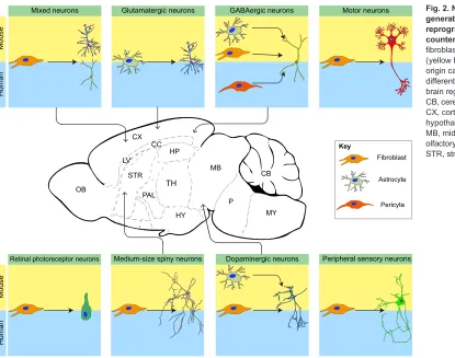
Fig. 2. Neuronal subtypes generated via direct
reprogramming and theirin vivo counterparts.Cultured cells (glia, fibroblasts or pericytes) of mouse (yellow background) or human (blue) origin can be reprogrammed into different neuronal subtypes of the brain regions indicated in the centre. CB, cerebellum; CC, corpus callosum; CX, cortex; HP, hippocampus; HY, hypothalamus; LV, lateral ventricle; MB, midbrain; MY, medulla; OB, olfactory bulb; P, pons; PAL, pallium; STR, striatum; TH, thalamus.
DEVEL
O
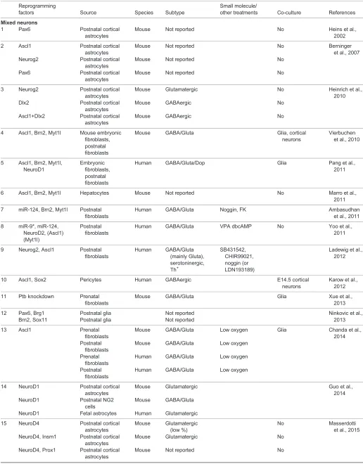
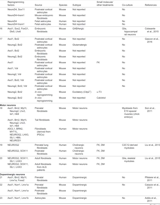
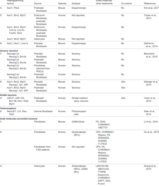
Table 1. Summary of direct neuronal reprogramming factors and outcome
Reprogramming
factors Source Species Subtype
Small molecule/
other treatments Co-culture References
Mixed neurons
1 Pax6 Postnatal cortical
astrocytes
Mouse Not reported No Heins et al.,
2002
2 Ascl1 Postnatal cortical astrocytes
Mouse Not reported No Berninger
et al., 2007 Neurog2 Postnatal cortical
astrocytes
Mouse Not reported No
Pax6 Postnatal cortical astrocytes
Mouse Not reported No
3 Neurog2 Postnatal cortical astrocytes
Mouse Glutamatergic No Heinrich et al.,
2010 Dlx2 Postnatal cortical
astrocytes
Mouse GABAergic No
Ascl1+Dlx2 Postnatal cortical astrocytes
Mouse GABAergic No
4 Ascl1, Brn2, Myt1l Mouse embryonic fibroblasts, postnatal fibroblasts
Mouse GABA/Gluta Glia, cortical
neurons
Vierbuchen et al., 2010
5 Ascl1, Brn2, Myt1l, NeuroD1
Embryonic fibroblasts, postnatal fibroblasts
Human GABA/Gluta/Dop Glia Pang et al.,
2011
6 Ascl1, Brn2, Myt1l Hepatocytes Mouse Not reported No Marro et al.,
2011
7 miR-124, Brn2, Myt1l Postnatal fibroblasts
Human GABA/Gluta Noggin, FK Ambasudhan
et al., 2011
8 miR-9*, miR-124, NeuroD2, (Ascl1) (Myt1l)
Postnatal fibroblasts
Human GABA/Gluta VPA dbcAMP No Yoo et al.,
2011
9 Neurog2, Ascl1 Postnatal fibroblasts
Human GABA/Gluta (mainly Gluta), serotoninergic, Th+
SB431542, CHIR99021, noggin (or LDN193189)
Ladewig et al., 2012
10 Ascl1, Sox2 Pericytes Human GABAergic E14.5 cortical
neurons
Karow et al., 2012
11 Ptb knockdown Prenatal fibroblasts
Mouse GABA/Gluta Glia Xue et al.,
2013
12 Pax6, Brg1 Postnatal glia Not reported Ninkovic et al.,
2013 Brn2, Sox11 Postnatal glia Not reported
13 Ascl1 Prenatal
fibroblasts
Mouse GABA/Gluta Low oxygen Glia Chanda et al.,
2014 Postnatal
fibroblasts
Mouse GABA/Gluta Low oxygen
Prenatal fibroblasts
Human GABA/Gluta Low oxygen
Postnatal fibroblasts
Human GABA/Gluta Low oxygen
14 NeuroD1 Postnatal cortical astrocytes
Mouse Glutamatergic Guo et al.,
2014 NeuroD1 Postnatal NG2
cells
Mouse GABA/Gluta
NeuroD1 Fetal astrocytes Human Glutamatergic
15 NeuroD4 Postnatal cortical astrocytes
Mouse Glutamatergic (low %)
No Masserdotti
et al., 2015 NeuroD4, Insm1 Postnatal cortical
astrocytes
Mouse Glutamatergic No
NeuroD4, Prox1 Postnatal cortical astrocytes
Mouse Not reported No
Continued
DEVEL
O
Table 1. Continued
Reprogramming
factors Source Species Subtype
Small molecule/
other treatments Co-culture References
NeuroD4, Sox11 Postnatal cortical astrocytes
Mouse Not reported No
NeuroD4+Insm1 Mouse embryonic fibroblasts
Mouse Not reported No
NeuroD4 Fetal astrocytes Human Not reported No
NeuroD4+Insm1 Fetal astrocytes Human Not reported No
16 Ascl1, Sox2, FoxG1, Dlx5, Lhx6
Embryonic fibroblasts
Mouse GABAergic Glia,
hippocampal rat neurons
Colasante et al., 2015
17 Ascl1, Bcl2 Postnatal cortical astrocytes
Mouse Not reported No Gascon et al.,
2016 Neurog2, Bcl2 Postnatal cortical
astrocytes
Mouse Glutamatergic No
Ascl1, Bcl2 Embryonic fibroblasts
Mouse Not reported No
Neurog2, Bcl2 Embryonic fibroblasts
Mouse Not reported No
Ascl1 Postnatal cortical astrocytes
Mouse Not reported FK No
Ascl1, Vdr Postnatal cortical astrocytes
Mouse Not reported No
Neurog2, Vdr Postnatal cortical astrocytes
Mouse Not reported No
Ascl1, Bcl2, Vdr Postnatal cortical astrocytes
Mouse Not reported No
Neurog2, Bcl2, Vdr Postnatal cortical astrocytes
Mouse Not reported No
Neurog2, Bcl2 In vivo
reprogramming
Mouse Excitatory (Ctip2+) α-T3
Neurog2, Bcl2 In vivo
reprogramming
Mouse Not reported Calcitriol
Motor neurons
18 Ascl1, Brn2, Myt1l, Neurog2, Lhx3, Isl1, Hb9
Prenatal fibroblasts
Mouse Motor neurons Myoblasts from
E10 epaxial muscles (chick embryo)
Son et al., 2011
Ascl1, Brn2, Myt1l, Neurog2, Lhx3, Isl1, Hb9
Tail fibroblasts Mouse Motor neurons
ASCL1, BRN2, MYT1L,
NEUROG2, LHX3, ISL1, HB9, NEUROD1
Fibroblasts (derived from iPSCs)
Human Motor neurons
19 NEUROG2 Prenatal lung fibroblasts
Human Cholinergic neurons
FK, DM C2C12-derived myotubes
Liu et al., 2013
NEUROG2, SOX11 Postnatal fibroblasts
Human Cholinergic neurons
FK, DM
20 NEUROG2, SOX11, ISL1, LHX3
Adult fibroblasts Human Motor neurons FK, DM Glia, skeletal myotubes
Liu et al., 2015
NEUROG2, SOX11, ISL1, LHX3
Adult fibroblasts from ALS patients
Human Motor neurons FK, DM
Dopaminergic neurons 21 Ascl1, Brn2, Myt1l,
Lmx1a, Foxa2
Prenatal fibroblasts
Human Dopaminergic No Pfisterer et al.,
2011
22 Ascl1, Nurr1, Lmx1a Prenatal fibroblasts
Mouse Dopaminergic No Caiazzo et al.,
2011 Ascl1, Nurr1, Lmx1a Postnatal
fibroblasts
Mouse Dopaminergic
23 Ascl1, Nurr1, Lmx1b Astrocytes Mouse Dopaminergic No Addis et al.,
2011
Continued
DEVEL
O
Table 1. Continued
Reprogramming
factors Source Species Subtype
Small molecule/
other treatments Co-culture References
24 Ascl1, Pitx3 Postnatal fibroblasts
Mouse Dopaminergic No Kim et al., 2011
25 Ascl1, Brn2, Myt1l Embryonic fibroblasts, postnatal fibroblasts
Human Not reported No Torper et al.,
2013
Ascl1, Brn2, Myt1l, Lmx1a, Lmx1b, FoxA2, Otx2
Embryonic fibroblasts, postnatal fibroblasts
Human Dopaminergic No
Ascl1, Brn2, Myt1l Astrocytes Mouse Not reported No
26 Ascl1, Nurr1, Lmx1a Prenatal fibroblasts
Mouse Dopaminergic No Dell’Anno
et al., 2014
Sensory neurons 27 Neurog2 (or
Neurog1), Brn3a
Prenatal fibroblasts
Mouse Sensory No Blanchard
et al., 2015 Neurog2 (or
Neurog1), Brn3a
Postnatal fibroblasts
Mouse Sensory No
Neurog2 (or Neurog1), Brn3a
Fibroblasts (derived from iPSCs)
Human Sensory No
Neurog2 (or Neurog1), Brn3a
Postnatal fibroblasts
Human Sensory No
28 Ascl1, Brn2, Myt1l, Neurog1, Isl1, Klf7
Prenatal fibroblasts
Mouse Sensory Glia Wainger et al.,
2015 Ascl1, Brn2, Myt1l,
Neurog1, Isl1, Klf7
Postnatal fibroblasts
Human Sensory Glia
Striatal neurons 29 miR-9*, miR-124,
Bcl11B, Dlx1, Dlx2, Myt1l
Postnatal fibroblasts
Human Striatal medium spiny neurons
Glia Victor et al., 2014
Photoreceptors
30 NeuroD1, Crx, Rax, Otx2
Dermal fibroblasts Human Photoreceptor cells
Seko et al., 2014
Small molecule-converted neurons
31 Fibroblasts Mouse GABA/Gluta FK, ISX9,
CHIR99021, I-BET151
Li et al., 2015
32 Fibroblasts Human Glutamatergic
(80%)
VPA, CHIR99021, Repsox, FK, SP600625, GO6983, Y-27632
Hu et al., 2015
Fibroblasts from FAD patients
Human Not reported VPA, FK, CHIR99021, Repsox, SP600625, GO6983, Y-27632
33 Astrocytes Human Glutamatergic
(90%), GABA (8%)
LDN193189, SB431242, TTNPB, thiazovivin, CHIR99021, DAPT, SAG, Purmo
Zhang et al., 2015
DAPT, N-[N-(3,5-difluorophenacetyl)-L-alanyl]-S-phenylglycine t-butyl ester; DM, dorsomorphin; dbcAMP, dibutryl-cyclic AMP; E, embryonic day; FAD, familial Alzheimer’s disease; FK, forskolin; GABA/Gluta, mixed population of GABAergic and glutamatergic neurons; GABA/Gluta/Dop, mixed population of GABAergic, glutamatergic and dopaminergic neurons; iPSCs, induced pluripotent stem cells; ISX9, isoxazole 9; Ptb, polypyrimidine tract binding protein (Ptbp1); Purmo, purmorphamine; SAG, smoothened agonist; Th, tyrosine hydroxylase; TTNPB, arotinoid acid; VPA, valproic acid.
DEVEL
O
glutamatergic neurons (Chanda et al., 2014), whereas it induces a mix of glutamatergic and GABAergic neurons from midbrain astrocytes (Achim et al., 2014; Liu et al., 2015). Likewise, fibroblasts transduced with Neurog2 and treated with small molecules (see next section) do not turn into glutamatergic neurons but rather acquire a cholinergic/motor neuron identity (Liu et al., 2013). Thus, it is clear that the starting cell identity influences the neuronal subtype obtained (Table 1); however, the extent to which this is linked to the developmental memory rather than the specific transcriptome or proteome of the starting cell remains to be understood. Initial attempts to reprogramme human fibroblasts into neurons relied on four factors, a finding that many believed was owing to their relative developmental distance from neurons (Fig. 2) (Pang et al., 2011). However, as we will discuss below, manipulation of key signalling pathways can lead to the efficient conversion of human cells with relatively few transcription factors, and thus careful determination of the correct reprogramming environment seems to be equally important as, if not more important than, using multiple factors for reprogramming.
The role of region-specific signalling pathways in direct reprogramming: links to development
Given the importance of signalling pathways in specifying and patterning neural tissue during development, one may anticipate that small molecule agonists or antagonists influencing these pathways could also contribute to reprogramming cells into neurons. Strikingly, manipulation of these and other pathways is indeed sufficient to convert fibroblasts (mouse and human) and astrocytes (human) into neurons (Hu et al., 2015; Li et al., 2015; Zhang et al., 2015).
Neuronal conversion of human astrocytes and fibroblasts mediated by small molecules seems to require sequential phases of reprogramming, or a first phase of neuronal fate induction followed by a second phase of neuronal maturation (Table 1; Hu et al., 2015; Zhang et al., 2015). This strict order suggests some degree of organization in the molecular cascades involved in reprogramming that may recapitulate a developmental programme. Remarkably, most of these small molecules influence key developmental pathways. For example, each chemical-mediated reprogramming paradigm contains at least one compound that interferes with the activation of the transforming growth factorβ(TGFβ) family of proteins (Table 2; Ladewig et al., 2012; Liu et al., 2013; Zhang et al., 2015; Hu et al., 2015; Li et al., 2015), or their immediate downstream effectors, the SMAD family of transcription factors (Ladewig et al., 2012; Li et al., 2015). As these pathways abolish neural induction during embryogenesis (Chang and Harland, 2007; Yaguchi et al., 2007) and regulate epithelial-to-mesenchymal transition (EMT) (Dunn et al., 2004; Yang and Weinberg, 2008), their inhibition may contribute to the repression of alternative cell fates, in particular in cells of mesenchymal origin such as fibroblasts. Interestingly, inhibition of the TGFβ pathway has also been found to enhance neuronal conversion of astrocytes (Zhang et al., 2015), which is consistent with the role of TGFβin restricting neuronal differentiation of NSCs and promoting their progression towards the astrocytic lineage (Stipursky and Gomes, 2007).
Another approach for small molecule-based neuronal reprogramming has been the use of inhibitors to block the activity of Rho-associated protein kinase, otherwise known as ROCK inhibitors (Hu et al., 2015; Zhang et al., 2015). During neural development, Rho family molecules regulate neuronal differentiation, migration (Cappello, 2013; Cappello et al., 2012) and neurite development (Govek et al., 2005). In vitro, RhoA inhibits nerve growth factor (NGF)-induced formation of the actin-rich filaments that initiate neurite growth in neurons and prevents neurite outgrowth promoted by neurotrophin 3 (NT-3; Ntf3) and brain-derived neurotrophic factor (BDNF) (Da Silva et al., 2003; Schwamborn and Püschel, 2004; Yamaguchi et al., 2001). Accordingly, ROCK inhibitors promote differentiation of embryonic stem cells into neurons and induce neurite outgrowth of reprogrammed neurons (Hu et al., 2015). In agreement with the role of Rho kinase pathways in cell death and survival during embryogenesis, ROCK inhibition also improved survival in direct neuronal reprogramming (Shi and Wei, 2007; Zhang et al., 2015).
[image:8.612.47.297.431.737.2]Shh specifies neuronal subtypes in a concentration-dependent mannerin vivo(Briscoe and Small, 2015), and agonists of Shh have also been used in reprogramming of astrocytes in vitro (Fig. 1; Table 2; Zhang et al., 2015). It would be interesting to test whether exposure to different concentrations of Shh could convert astrocytes into different neuronal subtypes, either by re-specifying dorso-ventral patterning or by co-regulating neuron-specific genes (Fig. 1). This could also contribute to uncovering the molecular mechanisms underlying the specification of neuronal identities during development. Another fundamental cascade involved in the maintenance of progenitor cells and inhibition of neuronal differentiation during embryogenesis is Notch signalling (Louvi and Artavanis-Tsakonas, 2006), which also promotes astrogliogenesis (Namihira et al., 2009) besides many other roles in neural development. Treatment with DAPT, a γ-secretase inhibitor that blocks the proteolytic cleavage of the activated Notch receptor and promotes neuronal differentiation in competent cells (Pierfelice et al., 2011), has been shown to facilitate the conversion of human astrocytes towards induced neurons (Zhang
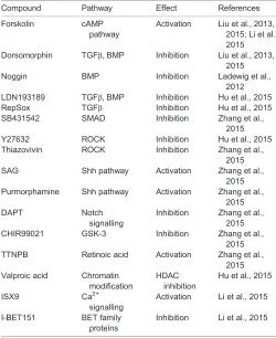
Table 2. Summary of the small molecules employed in direct neuronal reprogramming, and their action on the known target pathways
Compound Pathway Effect References
Forskolin cAMP pathway
Activation Liu et al., 2013, 2015; Li et al., 2015 Dorsomorphin TGFβ, BMP Inhibition Liu et al., 2013,
2015 Noggin BMP Inhibition Ladewig et al.,
2012 LDN193189 TGFβ, BMP Inhibition Hu et al., 2015 RepSox TGFβ Inhibition Hu et al., 2015 SB431542 SMAD Inhibition Zhang et al.,
2015 Y27632 ROCK Inhibition Hu et al., 2015 Thiazovivin ROCK Inhibition Zhang et al.,
2015 SAG Shh pathway Activation Zhang et al.,
2015 Purmorphamine Shh pathway Activation Zhang et al.,
2015
DAPT Notch
signalling
Inhibition Zhang et al., 2015 CHIR99021 GSK-3 Inhibition Zhang et al.,
2015 TTNPB Retinoic acid Activation Zhang et al.,
2015 Valproic acid Chromatin
modification
HDAC inhibition
Hu et al., 2015
ISX9 Ca2+
signalling
Activation Li et al., 2015
I-BET151 BET family proteins
Inhibition Li et al., 2015
DEVEL
O
et al., 2015). In summary, these observations clearly demonstrate some similarities between the processes that regulate normal neuronal differentiation during development and direct neuronal reprogramming mediated by small molecules.
Despite the examples discussed above, the link between well-known signalling pathways active in development and reprogramming is not always so clear. For instance, all reprogramming paradigms include GSK-3 (Gsk3b) inhibitors, which seem to activate neuronal genes during changes in cell fate, such as Ncam1, Dcx, Map2 and several neurogenic fate determinants, such as Ascl1 or Neurog2 (Hu et al., 2015). However, during development, GSK-3 activity enhances neuronal differentiation of radial glia and reduces progenitor expansion (Kim et al., 2009). In agreement with these observations, GSK-3 inhibitors significantly improve the reprogramming efficiency of fibroblasts towards pluripotency (Kirby et al., 2012; Lyssiotis et al., 2009) and facilitate self-renewal of mouse embryonic stem cells (Knockaert et al., 2002), probably through activation of Wnt signalling (Marson et al., 2008). These observations suggest that the downregulation of the GSK-3 pathway opposes neuronal differentiation and promotes a progenitor-like stage. Perhaps a reversion to a more undifferentiated state may have some beneficial effect in direct reprogramming, although progenitor genes such as
Sox2,Pax6orOlig2are induced very little, if at all (Hu et al., 2015; Zhang et al., 2015).
Overall, these recent approaches relying entirely on small molecules uncover the possibility of instructing a neuronal fate without any genetic manipulation, and demonstrate the power of these versatile pathways to specify cell fate. However, more work is needed to understand how the interplay among such pathways mediates fate conversion and to what extent this mimics their activity during development.
Neuronal subtype specification: from dopaminergic neurons to photoreceptors
Rather than recapitulating the exact developmental signalling environment required for the activation of specific transcription factor networks (Achim et al., 2014; Briscoe and Small, 2015), a more direct approach to neuronal subtype specification could be to overexpress the respective transcriptional regulators of a core specification programme active during development. Indeed, this
approach has been used for inducing many other neuronal subtypes lost in traumatic injury or neurodegenerative diseases, and generation of which in vitrocould be potentially useful for cell-based therapies as well as for disease modelling (Blanchard et al., 2015; Caiazzo et al., 2011; Gascón et al., 2016; Rouaux and Arlotta, 2013; Son et al., 2011; Victor et al., 2014; Wainger et al., 2015). Especially for the latter, direct reprogramming has the advantage of maintaining the age of the starter cell (Mertens et al., 2015), in contrast to the re-setting that occurs when generating induced pluripotent stem cells (Lapasset et al., 2011). In the next section, we will review successful attempts to generate very distinct neuronal subtypes (Fig. 2) with the aim of deciphering the molecular logic involved in establishing their identity (Fig. 3).
Dopaminergic neurons
Midbrain dopaminergic neurons are of great interest owing to their loss in Parkinson’s disease. They are generated in the floor plate of the mesencephalon (Fig. 1) and several genes have been implicated in their generation and specification. These include Otx2, which is involved in early patterning; FoxA1/2, which instructs the commitment of the progenitor cells; Lmx1a/b, which is important for progenitor cell differentiation; and Pitx3 and Nurr1 (Nr4a2), which are involved in the maturation and long-term survival of midbrain dopaminergic neurons (Fig. 1; Arenas et al., 2015). Accordingly, many of these transcription factors have been successfully used to induce dopaminergic neurons from fibroblasts or astrocytes (Figs 2, 3; Addis et al., 2011; Jang et al., 2011; Pfisterer et al., 2011; Torper et al., 2013), with some of the resulting cells even capable of surviving when transplantedin vivo
(Kim et al., 2011).
The molecular logic behind the different transcription factor combinations for reprogramming is such that factors active at different stages of specification and differentiation are usually combined. As mentioned above, Ascl1, Lmx1a and Nurr1 are active at different stages in the dopaminergic lineage and together can reprogramme fibroblasts into dopamine-releasing neurons (Caiazzo et al., 2011; Dell’Anno et al., 2014). However, none of these studies has been able to generate dopaminergic neurons belonging to the specific A8-10 clusters (Hegarty et al., 2013). Thus, it appears that a deeper level of understanding is required regarding the key regulators that specify the A8-10 clusters in order to generate
NeuroD1
Neurog2
NeuroD4
Dlx2
Prox1 Sox11
Insm1
Retinal neurons
Striatal medium spiny neurons
miR-124; miR-9/9*
Sensory neurons
Glutamatergic neurons
Motor neurons
Dopaminergic neurons
Neuronal cells
GABAergic neurons Rest
Ptb 11
30
21 22 23 24
3 16
10 15
15 15
3 14
29
27
Ascl1 15
25 26 20
[image:9.612.53.375.535.730.2]28 18
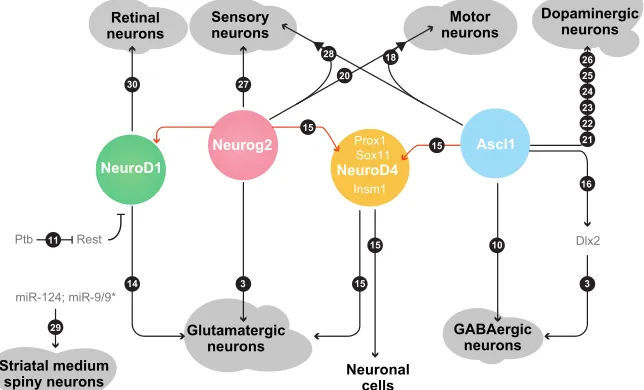
Fig. 3. Molecular cascades in direct neuronal reprogramming.To date, several different types of neurons can be generated upon expression of specific molecules, most of which are transcription factors. Among them, Neurog2 and Ascl1 are the most widely employed. Red lines connect either Neurog2 or Ascl1 with their direct downstream targets. Numbers in black circles refer to transcription factor reprogramming combinations as listed in Table 1.
DEVEL
O
distinct functional subtypes of midbrain dopaminergic neurons via direct reprogramming (Kriks et al., 2011).
Striatal neurons
A further important neuronal subtype recently produced by direct neuronal reprogramming are the medium-sized spiny neurons (MSNs). These are the main output neurons of the striatum and the ones that degenerate in Huntington’s disease (Zuccato et al., 2010). They have been induced from human fibroblasts by combining pan-neurogenic factors such as miR-124, miR-9/9* (Yoo et al., 2011) and Myt1l (Vierbuchen et al., 2010), with Dlx1/2, a key patterning factor of the lateral ganglionic eminence (Fig. 1A) and Bcl11B (also known as Ctip2), a late postmitotic differentiation factor from this region (Arlotta et al., 2008). Interestingly, some transplanted induced MSNs survived for more than 6 months and projected to their correct targets, the globus pallidus and substantia nigra (Victor et al., 2014). Interestingly, Ctip2 is required for the normal development of striatal patch neurons, but so far the patch identity has not yet been detected in the induced MSNs.
Cerebral cortex projection neuron subtypes
Although glutamatergic pyramidal-like neurons have already been generatedin vitro(Heinrich et al., 2010; Masserdotti et al., 2015), the crucial next step is to instruct distinct neuronal subtypes located in different layers and projecting to distinct target sites, either within the cerebral cortex or to subcortical targets (Lodato and Arlotta, 2015). Seminal work has demonstrated the conversion of one type of cortical projection neuron, the callosal projection neurons of layer II/III, into layer-V/VI subcortical projection neurons via the forced expression of the transcription factor Fezf2 at an early stage of differentiation (Lodato and Arlotta, 2015; Rouaux and Arlotta, 2013). Interestingly, direct reprogramming from non-neuronal cells using Neurog2 and Bcl2 generated deep layer pyramidal neurons, based on the expression of Ctip2 and FoxP2 and absence of the upper layer markers Cux1 and Satb2 (Gascón et al., 2016). Although much more work is required to identify the efferent projections and afferent innervation of these neurons, this is yet another example of how a transcription factor important for cell-type specification during development can also specify that cell type during reprogramming, as Neurog2 is involved in deep layer neuron generationin vivo(Schuurmans et al., 2004).
Motor neurons
Direct reprogramming to spinal cord motor neurons has also relied on the expression of key developmental regulators (Figs 1, 3; Table 1; Son et al., 2011), again by combining common neurogenic factors such as Ascl1, Neurog2, Myt1l and Brn2 (Pou3f2) with transcription factors specific to spinal cord motor neuron development, such as Lhx3 (Cho et al., 2014), Isl1 (Cho et al., 2014; Ericson et al., 1992; Thaler et al., 2002) and Hb9 (Mnx1) (Lee et al., 2009, 2005). The combination of these seven factors generated functional motor neurons from mouse embryonic fibroblasts, which were capable of forming functional neuromuscular junctions with co-cultured myotubes and could survive when transplantedin vivo(Son et al., 2011).
An alternative approach to generate induced motor neurons has been the expression of two pan-neurogenic transcription factors, Neurog2 and Sox11, together with forskolin, dorsomorphin and Fgf2 treatment. This combination was sufficient to reprogramme postnatal and adult human fibroblasts into cholinergic (as marked by choline acetyltransferase, ChAT) Hb9+ induced motor
neurons (Fig. 2) (Liu et al., 2013). However, these neurons
were only partially specified, as they failed to express Isl1 and
Lhx3. Indeed, the co-expression of these two factors together with Neurog2 and Sox11 significantly increased the proportion of reprogrammed motor neurons: up to 84-95% of cells expressing βIII-tubulin were also HB9 and ChAT positive (Liu et al., 2015). These data therefore further support the requirement of key region-specific transcription factors for the specification of distinct neuronal subtypes. Of note, even the poorly specified neurons formed functional synapses with muscle cells (Liu et al., 2013), highlighting that more stringent assays are needed to ensure correct neuronal identity. Genome- and proteome-wide analysis combined with electrophysiological analysis will be the gold standard to determine unequivocally the identity of the induced neuron. This is also important for future studies that aim to generate specific subtypes of motor neurons, such as branchial, visceral or somatic motor neurons of different subtypes – alpha, beta or gamma. Correct specification of neuronal subtypes is also crucial for disease modelling, particularly for neurodegenerative diseases, which largely affect only specific subtypes. Indeed, induced motor neurons generated from fibroblasts of amyotrophic lateral sclerosis (ALS) patients recapitulated some aspects of ALS pathology, like the mislocalization of the ribonucleoprotein FUS, smaller soma, lower firing frequency and higher susceptibility to cell death over time (Lai et al., 2011; Liu et al., 2015).
Retinal photoreceptors
Photoreceptors are sensory neurons of the central nervous system that develop after induction of the eye anlage as part of the diencephalon. Their induction from human dermal fibroblasts has been achieved by combining the neurogenic factor NeuroD1, which plays key role in photoreceptor development (Hatakeyama and Kageyama, 2004), with the patterning factor of the eye anlage retinal homeobox protein RAX (also known as RX) (Zagozewski et al., 2014) and the photoreceptor fate determinants Otx2 and Crx (Zagozewski et al., 2014). The induced photoreceptors comprise cells with rod or cone characteristics as indicated by the presence of rhodopsin, S and M opsin, and significant responses to light stimulus detected in some of the reprogrammed cells (Seko et al., 2014). Although these results are encouraging, more work is required to improve the efficiency of reprogramming and to ensure their survivalin vivo.
Peripheral sensory neurons
Peripheral sensory neurons are the afferent neurons that receive external stimuli and transmit them to the central nervous system. From a developmental perspective, these neurons differentiate from neural crest cells (Pavan and Raible, 2012), and hence a promising approach for generating them has been to reprogramme human fibroblasts into neural crest cells, which can then be further differentiated into peripheral neurons (Kim et al., 2014). However, direct neuronal reprogramming has proven to be more successful in obtaining specific subtypes of peripheral neurons than the neural crest cell route (Blanchard et al., 2015; Wainger et al., 2015). As peripheral sensory neurons are different to those of the central nervous system, an important question is how similar or different the molecular requirements are for inducing these cells. Interestingly, the same pan-neurogenic factors employed to generate central nervous system neurons–namely Ascl1, Myt1l and Neurog1–are sufficient to induce an A-δ nociceptor phenotype in 14% of transduced cells when combined with the terminal differentiation factors Isl1 and Klf7 (Wainger et al., 2015). These latter two factors,
DEVEL
O
Isl1 and Klf7, are important in maintaining the expression of TrkA (Ntrk1) in peripheral sensory neurons (Marmigere and Ernfors, 2007). Of the resulting induced nociceptor cells, about half were functional, as evidenced by their response to capsaicin stimulation and sensitization by prostaglandin E2 (Wainger et al., 2015).
A second pool of factors, Neurog1/2 and Pou4f1 (also known as Brn3), was successful in converting fibroblasts into sensory neurons, albeit with a low efficiency (4-6%; Blanchard et al., 2015). Single-cell expression analysis revealed non-overlapping expression of TrkA, TrkB (Ntrk2) and TrkC (Ntrk3), suggesting that the reprogramming protocol generated different sensory subtypes. This could imply that the relative expression of each of the two factors could determine the subtype of nociceptor generated, or alternatively that other factors are required to refine terminal differentiation further. Indeed, tuning the level and the length of the expression of Neurog1/2 and Pou4f1 has hardly been tested at all in reprogramming and will be crucial to investigate in order to understand the exact molecular logic of the underlying transcriptional events.
In summary, the identified combinations of factors tested so far uncover the instructive role of some common reprogramming factors, particularly Ascl1 and Neurog1/2, in direct neuronal conversion of different cell types. They also highlight the requirement for lineage-specific factors to generate different neuronal subtypes (Fig. 3). However, determination of neuronal subtype identity is still in its infancy and naturally limitedin vitro, where neither the adequate input nor selective projection and output of the respective neuron can be identified. Thus, much more work is requiredin vivo, with a particular focus on the connectivity of the reprogrammed neurons. In addition, specific transcriptional signatures are becoming available for specific neuronal subtypes, as shown recently in pioneering work identifying molecular signatures of neurons at the single-cell level (Bikoff et al., 2016; Fuzik et al., 2016; Zeisel et al., 2015). These transcriptional signatures could provide a comprehensive molecular map of specific neuronal identities that could be used to classify reprogrammed neurons. These studies could also uncover novel selector factors for direct neuronal conversion. This would allow assessment of whether direct reprogramming can generate truly subtype-specific neuronsin vitro, or whether the environment–that is the embryonic or adult brain, or organoids (Lancaster et al., 2013; Qian et al., 2016)–is required for complete phenotype maturation.
Learning from neuronal reprogramming: transcriptional dynamics, the cell cycle and metabolic hurdles
The ability of key developmental transcription factors to specify different neuronal subtypes highlights their instructive role towards a neuronal fate. In addition, direct neuronal reprogramming may also be used to understand better the molecular function of these factors in defined transcriptional networks. For example, Ascl1 can trigger different neuronal identities when expressed in either astrocytes or fibroblasts. How does the presence or absence of other transcription factors in the starting cell type affect the outcome of Ascl1 overexpression? Co-expression of Sox2, FoxG1, Ascl1, Dlx5 and Lhx6 (Figs 1, 3) converts murine and human fibroblasts into telencephalic, mainly parvalbumin-positive GABAergic interneurons (Colasante et al., 2015). Interestingly, Dlx2, a key regulator of GABAergic neuronal fate (Petryniak et al., 2007), was induced upon Sox2 and FoxG1 co-expression (Colasante et al., 2015). Sox2 was shown to interact with Ascl1, while FoxG1 was essential for Dlx2 upregulation both in vitro and in vivo. The identification of this regulatory network might explain why Ascl1
reprogrammes astrocytes into GABAergic neurons, as Sox2 and FoxG1 are already expressed in postnatal astrocytes and thus their exogenous expression is not required (Heinrich et al., 2010; Masserdotti et al., 2015; Zhang et al., 2014). Characterizing which factors are already present in the starting cell population may help to decipher the transcriptional networks required for neuronal specification in development and reprogramming. Indeed, proteomic changes not reflected in the transcriptome contribute significantly to the fate changes in other paradigms of direct reprogramming (Di Stefano et al., 2016).
Key components of the proteome include chromatin-modifying complexes, which can also affect the reprogramming process (Di Stefano et al., 2016). During reprogramming of mouse embryonic fibroblasts to neurons, for instance, Ascl1 occupies most of the genomic loci bound in NSCs and NPCs, even though in mouse embryonic fibroblasts these are covered by nucleosomes and therefore not easily accessible (Wapinski et al., 2013). A specific histone signature correlates with Ascl1-binding sites in mouse embryonic fibroblasts and NPCs that consists of monomethylation of histone 3 lysine 4, acetylation of histone 3 lysine 27 and trimethylation of histone 3 lysine 9 (Wapinski et al., 2013). Removing trimethylation of histone 3 lysine 9 via the expression of JmJD2d (Kdm4d) resulted in impaired efficiency of neuronal conversion. The ability of Ascl1 to act as a pioneer transcription factor (Iwafuchi-Doi and Zaret, 2016; Zaret and Mango, 2016) seems to be associated with this epigenetic signature, because in cells with low levels of these marks, such as human keratinocytes and osteoblasts, Ascl1 occupies almost completely different, off-target sites lacking the E-box motif. These cells, as well as adult human pericytes and human liver cells, can be poorly, if at all, reprogrammed by Ascl1 alone (Karow et al., 2012; Marro et al., 2011; Wapinski et al., 2013). Interestingly, co-transduction of adult human brain-derived pericytes with Ascl1 and Sox2 was sufficient to induce functional neurons (Karow et al., 2012). A possible scenario for the functional interaction between Ascl1 and Sox2 could be that their biochemical interaction is required for the activation of the key neuronal genes in cells otherwise refractory to neuronal reprogramming (see also Colasante et al., 2015). Chromatin remodelling is also required for Pax6-mediated reprogramming, as Brg1 (Smarca4)-deficient glia cannot be reprogrammed into neurons (Ninkovic et al., 2013), and the above-mentioned miR-9/9* and miR-124 regulate the composition of the Brg1-associated factors (BAF) complex during neural development (Yoo et al., 2009).
Data such as these prove the importance of chromatin status in direct reprogramming, and bring a fresh perspective to neurodevelopmental biology. One question is whether Ascl1 requires the same histone modifications to initially activate its
in vivo targets as in reprogramming. It will also be interesting to understand whether other transcription factors require a different chromatin signature to work efficiently, and, conversely, if a given reprogramming cocktail could work in different cell types upon proper chromatin manipulation. To this end, direct reprogramming contributes essential new information about the mechanisms that stabilize cell fate, which also represent hurdles in reprogramming. For example, the histone chaperone LIN-53 limits the reprogramming of non-neuronal cells in larvae or adult worms (Caenorhabditis elegans) into neurons (Tursun et al., 2011) and the mammalian orthologue Rbbp4 is part of the histone chaperone complex CAF-1 that has recently been shown to limit reprogramming towards several cell types, including neurons
(Cheloufi et al., 2015).
DEVEL
O
It has been suggested that mitotic transition during the cell cycle provides a ‘window of opportunity’ during which the gene expression profile, and hence the fate, of a cell can be changed (Halley-Stott et al., 2014; Holtzer et al., 1975). However, direct reprogramming has clearly demonstrated that proliferation is not a prerequisite for the cell fate switch, as many cell types can convert into neurons without cell division (Fishman et al., 2015; Gascón et al., 2016; Heinrich et al., 2010; Karow et al., 2012; Marro et al., 2011). In fact, the overexpression of the cell cycle activator Myc actually reduced the efficiency of fibroblast conversion to neuronal cells (Fishman et al., 2015). This supports the observation that BET-bromodomain inhibitors enhance neuronal reprogramming mediated by small molecules (Hu et al., 2015; Li et al., 2015; Zhang et al., 2015), as these compounds cause cell cycle arrest and inhibit Myc expression at transcriptional levels (Cheng et al., 2007; Delmore et al., 2011; Gallagher et al., 2014).
Cell cycle and Myc expression are both strongly linked to the metabolic and redox state of cells (Bretones et al., 2015). In nervous system development, however, very little is known about the metabolic changes that occur during the transition from neural stem and progenitor cells to neurons or distinct glial cells (Diaz-Castro et al., 2015). Embryonic fate transitions such as the differentiation of NSCs into neurons often include progenitor intermediates, which affords the opportunity to have better control of all aspects of commitment and differentiation. Conversely, in direct reprogramming the starter cell fate and newly imposed cell fate transiently coexist, and this may create stressful conditions for the cell undergoing transdifferentiation. In line with this, cells undergoing reprogramming suffer from an overshoot in oxidative stress (Gascón et al., 2016). This may be due to the need to change from the metabolism of astrocytes and fibroblasts, which utilize primarily anaerobic glycolysis andβ-oxidation (McKay et al., 1983; Tsacopoulos and Magistretti, 1996), to an oxidative metabolism, which is required by neurons. During this metabolic transition, the upregulation of the protective machinery may be delayed, thereby leading to excessive oxidative stress (Gascón et al., 2016; Quadrato et al., 2016), with subsequent death by ferroptosis (Gascón et al., 2016). Accordingly, protection from oxidative stress and cell death greatly improved reprogramming efficiency after brain injuryin vivo
(Gascón et al., 2016).
These findings prompt the question of why NSCs do not run into the same problem when generating neurons. A possible explanation is that their gradual differentiation, through successive rounds of proliferation and often an intermediate progenitor state, could allow them to acquire the most appropriate metabolism slowly. Indeed, recent RNA-seq data revealed that adult NSCs start to change their metabolism when they become activated and enter cell cycle (Llorens-Bobadilla et al., 2015; Shin et al., 2015). Even the recent identification of NSCs turning into neurons in the adult zebrafish forebrain without any apparent division seems to occur rather slowly (Barbosa et al., 2015), over the course of weeks, which would allow time for metabolic transition. Although endogenous glial cells in the adult neurogenic niches regulate metabolic transition apparently well, a key question is whether the metabolic switch is a consequence of the fate change or contributes to regulate it. Reprogramming experiments suggest that metabolic change is required for fate change, as neuronal conversion cannot occur when oxidative phosphorylation is blocked by oligomycin-A (Gascón et al., 2016). Strikingly, in this condition astrocytes do not die, but rather remain in their astrocytic state despite the constant expression of the neurogenic fate determinant (Gascón et al., 2016). In this
context, as well as in fibroblasts, Myc may represent a major hurdle, as it is a metabolic regulator promoting a glycolytic programme (Dang, 2013; Wang et al., 2011). Indeed, mounting evidence suggests that changes in metabolism may play an instructive role in fate acquisition and conversion (Folmes and Terzic, 2016), and are not just a mere consequence of fate change. For example, during development there is a strict regulation of metabolic pathways along sequential phases of stem cell proliferation and differentiation, with glycolysis as the preferred metabolic state for rapid proliferation (Vander Heiden et al., 2009). Cells in the early embryo prior to the blastocyst stage rely almost entirely on glycolysis (Barbehenn et al., 1978; Shyh-Chang et al., 2013). When embryonic stem cells differentiate there is a dramatic decrease of glycolytic flux (Cho et al., 2006; Chung et al., 2007; Facucho-Oliveira et al., 2007; Wang et al., 2009) by enrichment of unsaturated lipids (Yanes et al., 2010). The oxidation of these unsaturated lipids by reactive oxygen species (ROS) generated during the emerging oxidative metabolism improved embryonic stem cell differentiation (Yanes et al., 2010). Thus, it is clear that dynamic changes in cellular metabolism can have a direct impact on fate decisions.
During adult neurogenesis, metabolism and related ROS signalling play a crucial role in cell fate decisions (Prozorovski et al., 2015). For example, maintenance of progenitors requires
de novolipogenesis (Knobloch et al., 2013), while commitment and differentiation are associated with reduced expression of glycolytic genes (Llorens-Bobadilla et al., 2015; Shin et al., 2015). This in turn is influenced by the activity of mTOR (Amiri et al., 2012; Kim et al., 2009), a metabolic rheostat involved in the integration of nutrients and regulation of growth factor signalling (Mihaylova et al., 2014). The extent to which metabolic components regulate the fate and activation of NSCs, both during development and in the adult brain, remains not only a fascinating area of research but also has great relevance for understanding NSC behaviour in disease when metabolic pathways are often altered, for example by inflammation. Identification of the causal relationships between metabolic pathways and fate change is crucial for understanding both endogenously programmed fate decisionsin vivoand forced fate changes by reprogramming.
Conclusions
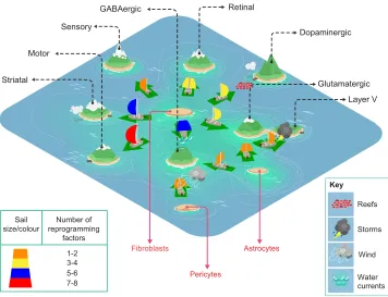
Accumulating evidence has made it clear that cells do not revert back to an earlier developmental stage during direct neuronal reprogramming. Thus, the Waddington’s landscape model no longer accommodates many of the observations made in direct reprogramming. In addition, it makes further assumptions that are not necessarily met: it suggests that a cell is naturally fated to differentiate, like a marble rolling down a mountain to the valley because of gravity; it does not consider direct lineage conversions, unless one introduces‘tunnels’below the surface of the hills and valleys; and, last, it does not represent the energy required for cell fate specification and conversions to happen. A new model has been recently proposed that reconciles these aspects by incorporating the concept that a cell can move from one stable state to another with little preferred directionality (Sieweke, 2015). Called the Cook Islands model (Fig. 4), a boat, which represents a cell, can sail from island to island rather freely (Huang, 2009). In this case, the islands represent different stable cell fates: however, major constraints, such as high waves, reefs, shallow passages or storms, block certain routes. These constraints represent actual barriers to cell fate acquisition, such as different germ layer origin between the starter cell and the reprogrammed entity (and thus a different epigenetic
DEVEL
O
state), or different energetic demands during and after the conversion [due to transcriptional changes, synthesis of new proteins, as well as a profound metabolic switch (Mathieu et al., 2014)]. Each of these constraints influences the probability that a given cell will change fate, sending the sailing boat towards different shores, depending on the type of constraint that is encountered. Identifying such constraints, investigating how they are established during development, and how they may be overcome eitherin vivoorin vitroduring reprogramming are essential tasks for the future.
Reprogramming studies highlight the state of our knowledge of developmental biology, profiting from what is known and casting light on the still rather dark spots. Generation of the diverse array of neurons by direct reprogramming has relied on key findings from development; but, at the same time, generating precise neuronal subtypes is still in its infancy and shows us the limitations of our knowledge in this regard. As revealed by recent single-cell transcriptome analysis, neuronal diversity is as large or even larger at the transcriptional level than expected from the diversity of morphology, connectivity and electrophysiological properties (Bikoff et al., 2016; Telley et al., 2016; Zeisel et al., 2015). However, it is still not well understood how this level of neuronal complexity is generated during development. These issues of neuronal subtype diversification relate back to the key issue of fate maintenance–are there common mechanisms that stabilize the fate of these thousands of distinct cell types or is there a hierarchy in safe-guarding somatic cell identity? The close interplay between
knowledge from development and reprogramming holds many promises for answering these crucial questions.
Acknowledgements
We thank Beatriz Gascón for providing support for the figures, and Olof Torper, Stefan Stricker, Jovica Ninkovic, Kalina Draganova, Sven Falk, Vidya Ramesh, Gianluca Russo and Pia Johansson for insightful comments on the manuscript and discussions.
Competing interests
The authors declare no competing or financial interests.
Funding
This work was supported by an Advanced European Research Council grant [ChroNeuroRepair GA no. 340793]; Sonderforschungsbereich (SFB) 870; the Bavarian Research Network ForIPs; the Helmholtz Association; and the Deutsche Forschungsgemeinschaft.
References
Achim, K., Salminen, M. and Partanen, J. (2014). Mechanisms regulating
GABAergic neuron development.Cell. Mol. Life Sci.71, 1395-1415.
Addis, R. C., Hsu, F.-C., Wright, R. L., Dichter, M. A., Coulter, D. A. and
Gearhart, J. D. (2011). Efficient conversion of astrocytes to functional
midbrain dopaminergic neurons using a single polycistronic vector.PLoS ONE 6, e28719.
Ambasudhan, R., Talantova, M., Coleman, R., Yuan, X., Zhu, S., Lipton, S. A.
and Ding, S. (2011). Direct reprogramming of adult human fibroblasts to
functional neurons under defined conditions.Cell Stem Cell9, 113-118.
Amiri, A., Cho, W., Zhou, J., Birnbaum, S. G., Sinton, C. M., McKay, R. M. and
Parada, L. F.(2012). Pten deletion in adult hippocampal neural stem/progenitor
cells causes cellular abnormalities and alters neurogenesis.J. Neurosci.32, 5880-5890.
Sensory
Striatal Motor
Retinal
Dopaminergic
Glutamatergic GABAergic
Fibroblasts Astrocytes
Pericytes
Layer V
Water currents
Wind Storms Reefs
7-8 5-6 3-4 1-2 Sail
size/colour
Number of reprogramming
factors
[image:13.612.127.483.56.329.2]Key
Fig. 4. The Cook Islands model applied to neuronal reprogramming.In the Cook Islands model, each island represents a different stable state. In essence, a boat, namely a cell, can move from island to island freely and in reprogramming such transitions are instructed by the combinatorial expression of specific transcription factors (depicted in the model as different colours in the sails of the boats). More complicated routes need bigger sails with several transcription factors, for example the reprogramming of fibroblasts to striatal or dopaminergic neurons. By contrast, small sails, representing fewer transcription factors, are sufficient to push boats to more closely related neuronal subtypes, for example from astrocytes to glutamatergic or GABAergic neurons. Certain routes are easier than others due to various factors such as distance and/or the relative water level, representing the various barriers to reprogramming as they occur bothin vitro
andin vivo. Other routes are particularly difficult due to the presence of strong opposing sea currents (see fibroblasts to GABAergic neurons), sudden storms (see glutamatergic to Layer V) or reefs (fibroblasts to dopaminergic). The presence of these hurdles may require novel routes to be taken, or more potent and equipped boats; that is, new strategies for reprogramming may be required to overcome cell type-specific barriers to reprogramming.