STUDIES ON THE STRUCTURE AND FUNCTION OF MAMMALIAN CHROMOSOMES
thesis by Joel A. Huberman
In Partial Fulfillment of the Requirements For the Degree of
Doctor of Philosophy
California Institute of Technology Pasadena, California
1968
iii
ACKNOWLEDGMENTS
Many people have contributed, both directly and indirectly, to the work contained in this thesis. I am deeply grateful to all of them. Special thanks are due to:
Giuseppe Attardi for providing careful and constructive criticism, for encourag:ing me to keep working even when success seemed unlikely, and for being a friend as well as an advisor.
Ray Owen for writing the letters that attracted me to Caltech, and for handling my occasional requests in a warm and intelligent manner.
William Dreyer for providing me with experience in protein chem-istry and for encouraging unorthodox thought,
James Bonner for providing me with numerous opportunities to be a 11local world1s authority" and "colleague".
Jerome Vinograd and Norman Davidson for demonstrating the power of physical techniques and for helpful discussions.
Art Riggs for introducing me to DNA autoradiography and for sharing with me the excitement of the experiments described in Part II.
Don Robberson for his assistance in my attempts at electron micro-scopy,
Roger Radloff for his assistance in my attempts at analytical centrifugation,
cuss ion.
Benneta Keeley for keeping the HeLa cells healthy and abundant. Laverne Wenzel for providing assistance of all sorts on a moment's notice,
All the other members of the Attardi group.
Robbi Hunt for staying up late to type this thesis.
My wife, Anne, for being a constant source of encouragement and
understanding.
v
ABSTRACT
At levels of organization between the Watson-Crick model of DNA
on the one hand, and the microscopically visible mitotic or meiotic
chromosome on the other, very little is known about the structure or
function of chromosomes in eukaryotic organisms . The studies reported
in this thesis.were an attempt to learn more about the arrangement
of certain DNA sequences in mammalian chromosomes, about the size of
the DNA molecules in such chromosomes, and about the replication of
these DNA molecules.
Part I contains the results of experiments designed to determine
the distribution of the hundreds of genes (DNA sequences) for ribosomal
RNA among the chromosomes of HeLa cells. In the course of these
experi-ments, methods were developed for isolating metaphase chromosomes on
a large scale from HeLa cells and for fractionating them on the basis
of sedimentation velocity, Hybridization experiments between
ribo-somal RNA and DNA from the various fractions of isolated chromosomes
showed that the genes for ribosomal RNA are confined entirely to small
HeLa cell chromosomes.
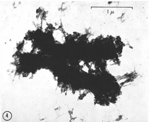
In Part II are reported the results of autoradiographic
experi-ments intended to help determine the size and manner of replication of
the DNA in mammalian chromosomes. All the experiments described in
Part II are the result of collaboration with Dr, Arthur D. Riggs. We
used a modification (by Dr. Riggs) of the technique for autoradiography
PART
ACKNOWLEDGMENTS ABSTRACT
vii
TABLE OF CONTENTS
TITLE
I THE ISOLATION AND FRACTIONATION OF METAPHASE CHROMOSOMES FROM HELA CELLS
Introduction
Chapter I: Isolation of Metaphase Chromosomes from
PAGE iii
v
1 2
HeLa Cells 4
Publication
Further Discussion References
Chapter II: Studies of Fractionated HeLa Cell Metaphase Chromosomes. I. The Chromosomal Distribution of DNA Complementary to 28S and 18S Ribosomal RNA and to Cyto-plasmic Messenger RNA.
Publication
II AUTORADIOGRAPHIC STUDIES OF THE STRUCTURE AND REPLICATION OF MAMMALIAN CHROMOSOM...t\L DNA
Introduction
Chapter I: Autoradiography of Chromosomal DNA Fibers from Chinese Hamster Cells
Publication
Further Discussion
Mammalian Chromosomes
Publication
Further Discussion
References
83
84
141
1
PART I
THE ISOLATION AND FRACTIONATION OF METAPHASE CHROMOSOMES FROM
suggested the project which eventually resulted in the studies des -cribed in this Part. He had conducted preliminary hybridization experiments between DNA and ribosomal RNA from HeLa cells. These experiments had suggested that each HeLa cell contains hundreds of genes for both 28S and 18S ribosomal RNA. The manner in which these hundreds of ribosomal RNA genes are distributed among the 60-70 chro-mosomes of the HeLa cell was completely unknown at the time, and Dr. Attardi suggested that this would be an interesting question to investigate.
My first approach was to attempt autoradiographic detection of hybridization between ribosomal RNA and whole chromosomes (isolated on a microscopic scale by the method of Prescott and Bender, 1). Unfortunately, this approach proved to be completely unsuccessful.
'
However, the report of Chor~zy, Bendich, Borenfreund
&
Hutchison (2) on large-scale isolation of metaphase chromosomes from mouse 11210 cells had just appeared, and this suggested an alternative approach to the problem, It should be quite simple, we reasoned, to isolate meta-phase chromosomes from HeLa cells by the same method (2), to fractionate them on the basis of sedimentation velocity, and then to test DNA from each of the fractions for capacity to hybridize with ribosomal RNA.3 '
Chor~zy et al. (2) proved unsatisfactory for isolation of chromosomes from HeLa cells. Therefore, I developed a different method of chromo-some isolation which would work for HeLa cells. This method is pre-sented in Chapter I, along with several observations on the properties of the isolated chromosomes.
Additional difficulties were encountered when I tried to
frac-tionate the isolated chromosomes. Tlese difficulties, and the methods used to eliminate or minimize them, are discussed in Chapter II.
The results of the hybridization experiments are also reported in Chapter II. Although some question about the interpretation of the hybridization experiments remains, the results suggest that ribo-somal RNA genes are found only in small HeLa cell chromosomes, perhaps only in those chromosomes which carry nucleolar organizers. This con-dusion is completely consistent with data from other laboratories
(reviewed in Chapter II), obtained after this project was started, which suggest that ribosomal RNA genes are always found exclusively
Isolation of Metaphase Chromosomes from HeLa Cells
The material in the first part of this chapter is reprinted by
5
ISOLATION OF lVIETAPHASE
CHROlVIOSOlVIES FROM HELA CELLS
JOEL A. HUBERMAN and GIUSEPPE ATTARD!
From the Division of Biology, California Institute of Technology, Pasadena
ABST.RACT
The authors have developed a method for large-scale isolation of metaphase chromosomes from HeLa cells. The distinguishing feature of this method is the use of a pH sufficiently low (about 3) to stabilize the chromosomes against mechanical damage. Many milligrams
of fairly pure, morphologically intact chromosomes can be isolated in 8 hr or less of total
working time. The isolated chromosomes contain about 2.0 mg of acid-soluble protein, 2.7 mg of acid-insoluble protein and 0.66 mg of RNA for each milligram of DNA. The RNA bound to the isolated chromosomes consists mainly of ribosomal RNA, but there is also a
significant amount of 45S RNA.
INTRODUCTION
Many possible biochemical and biophysical ap-proaches to the study of chromosomes in higher
organisms have been hindered, until recently, by the lack of suitable procedures for large-scale isola-tion of chromosomes. Although the methods for isolation of interphase chromosomes, or " chroma-tin," which have been developed in recent years ( 1, 2) are satisfactory for certain purposes, a
definite need still exists for a procedure which will allow large-scale isolation of morphologically in-tact metaphase chromosomes. Metaphase
chromo-somes are an indispensable complement to inter
-phase chromosomes for the general study of chromosome structure. In addition, metaphase
chromosomes have the unique advantage of being so condensed that they can be distinguished micro -scopically both from each other and from con
-taminating nonchromosomal material. Conse-quently, one is not limited to studying the average properties of all chromosomes; one can also
examine single types of chromosomes.
According to our experience, in the isolation of metaphase chromosomes by most previously
pub-lished methods (3-5), morphological damage to
some of the chromosomes cannot be avoided and
only partial purification of the chromosomes from cell debris can be achieved. \11/e report here a method for the rapid preparation, in milligram
quantities, of fairly pure, morphologically intact metaphase chromosomes from HeLa cells. \11/e
also report the results of studies on the chemical composition of isolated chromosomes.
i\'IATERIAL AND METHODS
Cultivation of Cells
HeLa S3 cells (6) were grown in suspension
cul-ture in a modified Eagle's medium (7) supplemented with 53 calf serum. For accumulation of metaphase cells, partial synchrony was induced by lowering
the culture temperature to 4°C for I hr and then returning it to 37°C (8). Ten to 11 hr later, colch
i-cine was added to a final concentration of 0.5 to
I X 10-5 M. The cells were harvested by centrifu ga-tion 9 to IO hr after colchicine addition and washed
3 times in 0.137M NaCl, 0.005M KC!, 0.007M NaH2P04, 0.025 M Tris, pH 7.4. This procedure routinely produced about 303 metaphase cells.
Reprinted from THE JouRNAL OF CELL B10LOGY, 1966, Vol. 31, No. 1, pp. 95-105 Printed in U.S.A.
each other. Five min later, 3 vol of 0.1 M sucrose, 7 X 10⁻⁴ M CaC12, 3 X 10-< M MgCl2, 3.3 X 10-a M
I-IC! were added slowly, with stirring, to each volume of cell suspension. Slow addition of the acid solution was necessary to prevent clumping of the c hromo-somes in metaphase cells. The measured final pH was about 3.0. Higher pH values (up to 3.3) allowed satisfactory breakage of cells and conservation of chromosome morphology, but separation of the
chromosomes from cytoplasmic debris was more difficult.
A phase-contrast microscope was used to check
the result of acid addition. Cells suspended in hy -potonic medium appeared grey, with little internal contrast. The chromosomes in metaphase cells were barely visible. After the pH had been adjusted to
3.3-3.0, the chromosomes, evenly distributed through-out the cytoplasm of metaphase cells, appeared dis-tinct and bright.
After adjustment of pH, a Potter-Elvehjem glass
homogenizer with a motor-driven Teflon pestle
was used to homogenize the cells. The course of homogenization was checked with a microscope.
As an end point for homogenization, the time was chosen when all interphase cells were broken (us-ually after less than l min). At this stage the great
majority of metaphase cells were also broken.
The released chromosomes were usually single and free of obvious attached debris. The following steps separated these chromosomes from the nuclei
and cytoplasmic debris which were also produced by homogenization.
The homogenate was centrifuged at 900 g (2000 RPM in the International PR2 centrifuge, head No. 269, International Equipment Co., Needham Heights, Massachusetts) for 30 min. The resulting pellet contained nuclei, chromosomes, and the larger
cytoplasmic debris. Most debris remained in the su -pernatant.
The supernatant was discarded and the pellet resuspended in HCM (1 X 10⁻³ M HCl, 7 X 10-·1 M CaCl2, 3 X 10⁻⁴ M MgCl)2, using about 40 ml of
I-ICM for each milliliter of pellet. The suspension
was rehomogenized briefly with a Potter-Elvehjem
homogenizer to break up any clumps that might have formed as a result of pelleting.
Up to 20 ml of suspension at a time were then
gently layered onto 200 ml of a 0.1 to 0.8 M linear sucrose gradient in I-ICM (final pl-I adjusted to 3.0) which had been formed in a 250 ml glass centrifuge
and some clustered chromosomes were pelleted at the bottom. A crude fractionation of chromosomes on the basis of sedimentation velocity was also pro-duced; most large chromosomes were found near the bottom, while most small chromosomes remained near the top.
The top 20 ml of the gradient were discarded and the rest was sucked off, leaving a small amount (about 10 ml) in the bottom of the centrifuge bottle so as not to disturb the pelleted nuclei. The s uper-natant was then mixed until the sucrose was evenly distributed, and the chromosomes were collected by centrifugation at 850 g (2000 RPM in the International PR-2 centrifuge, head No. 284) for 90 min. The pellet contained very few nuclei (less than 3% of the total DNA in the pellet was from whole nuclei if the initial proportion of metaphase cells was 15% or greater). There was, however, still considerable con-tamination by debris.
Most of the debris was removed by the following procedure. The pellet was resuspended in a small volume of HCM with brief rehomogenization to break up clumps. Ten ml of 2.2 M sucrose in I-ICM were placed in a Spinco S\IV-25 plastic tube (Beck-man Instruments, Inc., Palo Alto, California) and 15 to 20 ml of chromosome suspension were layered on top. The upper three-fourths of the tube contents were gently stirred to form a rough gradient. After centrifugation at 20,000 RPM for 1 hr the chromo-somes were found in a pellet at the bottom of the tube, while most cytoplasmic debris remained float-ing above the 2.2 M sucrose layer. The yield of chro-mosomes at this point, as determined by DNA deter-mination (see below) or by direct counting in a Petroff-Hausser counting chamber (C. A. Hausser and Son, Philadelphia, Pennsylvania), was about one-third of the chromosomes from all cells scored as in metaphase before homogenization.
Chrornosorne Storage
Chromosomes stored in HCM at 2° to 4°C retained their morphological integrity for many months. They could also be stored frozen in I-ICM at - 70°C.
Chernical Analysis
Acid-soluble proteins were extracted from chromo
-somal or nuclear suspensions with 0.2 M HCI at 0°C for ),-2 hr. The residue was removed by centrifugation and extracted once more with another portion of
7
d
~ .w.rw•--,..,,.- -:
"
r
~"'
•
• 'ir ....~l .... #
...
.
'
.
,"
.
..
., . ~ ~....
~...
..
..
.
..
.
.
.
~..
~..
..
FIGURE l Isolated HeLa metaphase chl'Omosomes suspended in HCM. Phase contrast. X llOO.
0.2 M HCI. Trichloroacetic acid was added to the pooled supernatants to a final concentration of 203 .
The acid-soluble proteins were allowed to precipitate
overnight at 0°C and were then collected by ce n-trifugation, dissolved in 1 M NaOH, and determined by the method of Lowry et al. (9). Vacuum-dried
calf thymus histone was used as a standard.
The residue left after I-IC! extraction was washed once with ethanol-ether (3: I), then resuspended in
103 trichloroacetic acid and heated at 100°C for
20 min to hydrolyze nucleic acids. After one more
wash with 103 trichloroacetic acid the residue was
dissolved in 1 M NaOH, and acid-insoluble proteins were determined by the method of Lowry et al. (9)
using vacuum-dried bovine serum albumin as a
standard.
For nucleic acid determinations, the general
pro-cedure of Schmidt and Thannhauser (IO) was fol-lowed. Chromosomal or nuclear suspensions were
precipitated with 103 trichloroacetic acid, washed once with ethanol ether (3: 1), then dissolved in 0. 3
M KOH. RNA was hydrolyzed by incubation at 37°C for 18 hr. Perchloric acid was then added to a final concentration of 0.5 M, and the samples were kept at 0°C for at least Yz hr. The precipitate of
DNA, protein, KC104, and other materials was
washed once with a small volume of 0.5 M perchloric
acid. The wash was combined with the RNA
hy-drolysate, and RNA in this pooled solution was de-termined by the orcinol method (I I) using n-ribose as a standard.
DNA in the precipitate was determined, after hy-drolysis in 0.5 M perchloric acid at 70°C for 15 min,
by the diphenylamine procedure as described by Burton (12), using n-deoxyribose as a standard.
RN A Purification
RNA was purified from isolated chromosomes or nuclei by a procedure described in detail elsewhere
(13) which involves cold phenol-sodium dodecylsul
-fate extraction of total nucleic acids, followed by di-gestion of DNA with RN ase-free DN ase.
Acridine Orange Staining
Samples were air-dried on clean glass slides, fixed in 953 ethanol-ether ( 1 : I) and stained according to the procedure of von Bertalanffy et al. (14). A Zeiss fluorescence microscope equipped with an HBO 20ow· mercury light source, a Schott BG 12 excitation filter, and an Sp Orange 2 barrier filter was used to
examine the slides .
T c A G '( cc Pn Pyr
Exp. I
Chromosomal 30.0 20.0 29.3 20.7 40.7 1.00
Whole cell 30.2 20.1 29.5 20.2 40.3 0.99
Exp. 2
Chromosomal 30.0 20.0 30.1 19.9 39.9 .uo
Whole cell 30.1 19.9 30. I 19.9 39.3 .OU
10µ.
0
}'1Gt:RE 2 The metaphase chromosomes of a single IleLa cell. Bright field. Cells \\"ere blocked in rneta
-phase ll"ith colchicine, u ·pended in 1':'~ sodium citrate for 10 min, fixed in acetic acid-ethanol (3:'2) for 10 min and then stained in I'/~ orcein in lactic acid-acetic acid (I: 1). Cells suspended in stain solution "·e1·c squashed by tlnnnb pressure bet\\"een a slide and a coyer slip. X 1100.
Base Composition
DNA was purified from isolated chromosomes or from whole HeLa cells by the Marmur procedure
(15). About 400 µg of DNA were dissolved in 0.5
ml of 88 to 90% formic acid and hydrolyzed in a
scaled tube under nitrogen at 175°C for I hr (16). The hydrolysatc was evaporated to dryness and redissolved in 25 µ1 of I M HCI. Two 10 µI portions were used for chromatography. Descending chrom
a-tography was carried out on \-Vhatman No. l filter paper, using methanol :concentrated HCI: H 20
9
10µ.
•
•
•
Fwu1m 3 Isolated I-IcLa rnetaphasc chroinoso1ncs. Ilright field. A s111all quantity of chro1nosome suspension in TlCM "·as spread on a glass slide and alloll'ed to dry. The slide 'ms treated ll'ith 1% sodium
citrate for 10 min, fixed in acetic acid-etlrn11ol (3: '2) for 10 111in and then stained in l c/c, orccin in lactic
acid-acetic acid (1: 1). X 1100.
(70:20:10 by vol) as solvent (17). The chromato-grams were dried, and the bases were located with a
short wavelength UV light. The bases were eluted in small volumes of 0.1 M HCI and determined sp
ec-trophotometrically. The extinction coefficients given
by Bendich (18) were used.
HESULTS AND DISCUSSION
Ejj'ecls of Low pll
A distinguishing feature of the chromosome i so-lation procedure presented here is the use of a pH sufficiently low (about 3) to stabilize the chromo
-somes against mechanical damage and to weaken
the cytoplasm so that the cells break easily and
aggregation of cytoplasmic debris is minimized.
Low pH (303 acetic acid; pH 1.8) has also been
used by Somers et al. ( 4) for chromosome isolation.
However, under their conditions histones were
completely extracted. A third isolation method
employing low pH (pH 3.7) has recently been reported ( 19).
Lmvering the pH has the eITect of increasing the
contraction of the chromosomes. As viewed in the
phase-contrast microscope, the chromosomes b
e-come smaller and also brighter. The bright
appear-ance of acid-treated chromosomes is evident in
Fig. 1. It is caused by an increase in the refractive index of the chromosomes as they contract. This
extreme contraction is partly responsible for the
increased resistance of the chromosomes to m
e-chanical damage at low pH. However, contraction
alone cannot completely explain low pH stabili za-tion: although chromosomes can be made to con-tract equally well at higher pH (5-7) by the use of sufficiently large (ca. 3 X IQ-3M) concentrations of divalent cations, they still remain susceptible to mechanical damage. The unique strengthening achieved at low pH may be a result of the denatur -ation and precipitation of some chromosomal proteins.
Low pH was also found to be critical for
success-ful liberation of chromosomes from metaphase cells. At pH values higher than about 3.3, chroma
•
.p·
t
©
'\ ,,3
•.
.
FIGURE 4 Electron micrograph of a typical isolated HeLa metaphase chromosome. Grids were p1·e -pared by touching the carbon-Fonuva.r film to the surface of a suspension of chromosomes in HCM, then loading immediately into a grid holder under 303 ethanol. The rest of the procedure has been described by DuPraw (21~. (Courtesy of Dr. E. J. DuPraw.) X 33,000.
somes were only partially released during ho-mogenization, and they tended to aggregate with cytoplasmic debris during pelleting.
The use of such a low pH introduces the possi
-bility of undesirable side effects. Certainly, low pH causes denaturation of some chromosomal pro-teins, but this would not be a drawback for most applications of isolated chromosomes. Low pH
might also extract histones. This possibility has been examined, and it has been found that most histones are not extracted under the conditions of
our isolation procedure (20). However, some
lysine-rich histones found in samples of HeLa
chromatin prepared without use of low pH are extracted (20).
In addition, low pH might cause depurini
za-tion of nucleic acids. To test this possibility, we
determined the base composition of DNA purified from isolated chromosomes and compared it with the base composition of DNA purified from whole HeLa cells. The results are presented in Table I.
No loss of purines was detected in chromosomal DNA. If depurinization occurs, it must be less
extensive than the experimental error, estimated to be about 1 3.
Morphology and Purity of
Isolated Chromosomes
The metaphase chromosomes from a typical
colchicine-treated HeLa cell prepared by the standard squash technique are shown in Fig. 2. They should be compared to the isolated
chromo-somes shown in Fig. 3. It is evident that the iso -lated chromosomes are very similar to the chromo
-somes prepared by the standard squash technique. Indeed, when the pH was kept below 3.3, we found no examples of morphological distortion during isolation.
Dr. E. J. DuPraw has been kind enough to
examine our isolated chromosomes with the elec
-tron microscope, using his whole-mount technique
,.
11
..
Iµ.,.
I
J.
I ~1
.,..,
) . <
r
..
,
©
•F1aunE 5 Electron n1icrogrnphs of well presc1Tcd, isolated I leLa 1netaphase ch1·0111oso1i1es, Grids \\'ere
prepared as in Fig. 4. (Courtesy of Dr. E. J. DuPrnll'.) X 28,000.
Each value for chromatin represents the aver
-age of triplicate determinations on one prepa
-ration. Chromosomes were isolated as d
e-scribed in the Materials and Methods section. I nterphase nuclei were isolated from the same cell homogenates used in chromosome prepa-rations. The nuclear pellet from the sucrose
gradient centrifugation was collected and freed from any· contaminating cytoplasm by
centrifugation through 2.2 M sucrose (in the
same manner as chromosomes). Chromatin
was isolated from whole HeLa cells (l, 20).
mg acid- mg acid-soluble insoluble
mg R1 A protein protein
mg DNA mg DNA mg DNA
Chromosomes 0.66 2.0 2.7
Nuclei 0.38 l .9 2. l
Chromatin 0.15 l. l 1.0
(21). He found that typical isolated chromosomes
had the extremely condensed appearance shown
in Fig. 4. The thin fibers, which he has found in
honey bee (21) and human (22) chromosomes, if
present, seemed fused together. However, in a
small proportion of isolated chromosomes, such
thin fibers could be readily observed (Fig. 5). The chromosomes used for these pictures were su
s-pended in HCM. The "fusion" of fibers evident in Fig. 4 is probably the manifestation, at the electron microscope level, of the extreme
chromo-some contraction observed in HCM at the light
microscope level. However, the contraction
ob-served in HCM has been found to be a reversible
phenomenon. All isolated chromosomes are
capable of expanding at the light microscope level.
For example, the chromosomes in Fig. 3 have been
expanded (relative to those in Fig. 1) by the treatment described in the legend to Fig. 3. It is
possible that all expanded, isolated chromosomes
would reveal fibers like those in Fig. 5.
In the absence of reliable information on the
chemical composition of metaphase chromosomes
(see below), purity of the chromosome
prepara-tions must also be determined morphologically. Unfortunately the morphological criterion is not a
contamination can be made by using acridine orange staining and fluorescence microscopy. After acridine orange staining, red-fluorescing cytoplasm shows a sharp contrast to the yello w-green-fluorescing chromosomes. When this method is applied to our isolated chromosome prep ara-tions, a small amount of RNA-containing
con-tamination in the form of isolated debris or of
bodies apparently attached to the chromosomes
can be recognized. DNA-containing debris is not
apparent, however.
Chemical Composition of
Isolated Chromosomes
Despite the presence of a certain amount of
contamination in our chromosome preparations,
we felt that a chemical composition study would
be valuable, both to provide an indication of the actual chemical composition of purified c
hromo-somes and as a reference for further chromosome
purification. We have also studied the chemical
composition of whole interphase HeLa nuclei and interphase HeLa chromatin. Our results are
presented in Table II.
The large amount of RNA in metaphase c
hro-mosomes relative to interphase chromatin and even
to whole nuclei suggests, at first, that cytoplasmic contamination may be extensive. There are several
reasons, however, for thinking that the RNA
con-tent of metaphase chromosomes may really be
unusually large. First, we have some evidence that
a large fraction of the RNA in our chromosome preparations is actually bound to the chr omo-somes; isolated chromosomes which have been
extensively pretreated with DNase fluoresce
orange-red rather than yellow-green after acridine
orange staining. The amount of red staining due
to chromosomes after DNase treatment seems, by
visual estimate, to be considerably greater than that due to debris. Subsequent RNase treatment
shows that the red staining of DNase-treated
chromosomes (and of debris) is probably due to RNA and not to denatured DNA; only a barely
visible greenish fluorescence remains.
Second, cytological studies (23-26) have shown that during the course of mitosis the amount of
13
0.800
NUCLEAR RNA
0.600 0 tO N 0 0.400 0 0.200
10 20 30 40
Fraction number
0.800
CHROMOSOMAL RNA
0.600
0 tO
N
8
0.4000.200
~
10 20 30 40
Fraction number
Fwmrn 6 RNA was purified (as described in the Materials and Methods section) from a quantity
of isolated chromosomes containing about 0.5 mg of DNA and from a quantity of nuclei, isolated as described in Table Tr, containing about 1.5 mg of DNA. The RNA was dissolved in 0.5 ml of acetate
buffer (0.1 M NaCl, 0.01 M sodium acetate buffer, pH 5.0) and layered on top of 25 ml linear 5 to 20%
sucrose gradients in the same buffer. The grndients were centrifuged at 25,000 RPM at 2°C in the Spinco Model L ultracentrifuge for 7 hr.
RNA bound to the chromosomes increases, reach
-ing a maximum at metaphase; it then gradually
decreases during anaphase and telophase. These
changes in chromosomal RNA content during
mitosis have been termed the '"chromosomal RNA
cycle" (27).
Finally, investigators in other laboratories, using metaphase chromosomes isolated by different procedures, have also found very high RNA co
n-tents in metaphase chromosomes. Lin and
Chargaff (5) have found an RNA to DNA ratio of
0.64 for HeLa metaphase chromosomes, while Cantor and Hearst (19) have reported an RNA to DNA ratio of 1.0 for mouse ascites tumor m eta-phase chromosomes. Maio and Schildkraut, in a
recently published abstract (28), have reported an
RNA to DNA ratio of 0.8 for HeLa metaphase
chromosomes.
Our findings for the protein content of
meta-phase chromosomes also require comment. First,
acid-soluble proteins in metaphase chromosomes than in interphase chromatin. The protein result:' may also be misleading because of the unknown
extent of contamination and because of variation in the color values for different proteins in the test of Lowry et al. (9).
Sedimentation Profile of RiY A
from Isolated Chromosomes
vVe have taken a first step toward elucidation of the nature of the RNA bound to meta phase chromo-somes by purifying RNA from isolated metaphase
chromosomes and comparing it to RNA from
inter-phase nuclei. The sedimentation profile of RNA from these sources is shown in Fig. 6. The se
di-mentation profile of HeLa nuclear RNA is similar
to that found by Penman (29) for the same
material, and by Steele et al. (30) for rat liver
nuclear RNA. One recognizes two peaks,
cor-responding to the two ribosomal RNA species, and
a faster component with a sedimentation constant
of about 45S. The latter presumably represents the large size ribosomal RNA precursor described in different types of animal cells (3 l-33). The
pres-ence in the nucleus of l BS RNA in amounts
con-siderably smaller, relative to the major ribosomal RNA component, than found in cytoplasmic ribosomal RNA is in agreement with Penman's
observations (29), suggesting that there are no mature ribosomes, but only precursors, in the
nucleus: according to this author, the 45S RNA is cleaved into l BS RNA, which is immediately transferred to the cytoplasm, and 35S RNA, which remains in the nucleus to be transformed into 2BS RNA. In addition to the ribosomal RNA species and their large precursors, one can see in the
sedimentation profile of nuclear RNA small
amounts of 4S RNA, and a polydisperse RNA
with sedimentation constants between 6S and REFERENCES
l. BONNER, J., CHALKLEY, R. G., DAHMUS, M.,
FAMBROUGH, D., FUJIMURA, F., HUANG, R. C.,
HUBERMAN, j., JENSEN, R., MARUSHIGE, K., OHLENBUSCH, H., OLIVERA, B., and VVrn-HOLM, J., J\!fethod Enzymol. in press.
ribosomal RNA components and the 45S RNA
species. The amount of ribosomal RNA relative to DNA is about three times as large as in nuclear RNA; there is, on the contrary, relatively less
polydisperse RNA and only a very small amount of 4S RNA. As concerns the significance and origin
of the chromosomal associated RNA, only specula
-tions are possible at present. Evidence has been presented that ribosomal RNA precursors are
localized in the nucleoli (30, 31). Hence the
pres-ence of a 45S component in chromosomal RNA is consistent with the hypothesis that, during pro-phase, at least some of the materials from the
disintegrating nucleoli are bound to the condensing chromosomes. More difficult to interpret is the
presence of the two ribosomal RNA species. The fact that the ratio of major to minor component is similar to that observed in cytoplasmic ribosomal RNA may be indicative of a cytoplasmic origin for these species (either as a result of accidental contamination during extraction or of an ass
ocia-tion of physiological significance occurring during mitosis). On the other hand, one cannot exclude
the possibility that some of these ribosomal
com-ponents were still intranuclear at the end of pro-phase and became associated with the condensing chromosomes. Further experiments will be required
to determine the origin and significance of the
ribo-somal RNA present in the preparations of meta-phase chromosomes.
This work was supported by United States Public
Health Service grants GM-11726 and 5 -Fl-GM-21,622.
The authors gratefully acknowledge the help of Mr. John Elberfeld in part of this work and the valuable technical assistance of Mrs. Benneta Keeley and Mrs. LaVerne VVenzel.
Received for jmblication 4 April 1966.
2. FRENSTER, J. H., ALLFREY, V. G., and MIRSKY, A. E., Proc. Nat. Acad. Sc., 1963, 50, 1026. 3. C1mR~zv, M., BENDICH, A., BoRENFREUND, E.,
and HUTCHISON, D. J., J. Cell Biol., 1963, 19,
59.
15
4. SOMERS, C. E., COLE, A., and Hsu, T. C., Exp. Cell Research, Suppl. 9, l 963, 220.
5. LIN, H. J., and CHARGAFF, E., Biochim. et
Bio-j1hy;ica Acta, l 964, 91, 69 I.
6. PucK, T. T., and F1s1-1ER, H. W., J. Exj1. ivied.,
l 956, 10,t, 427.
7. LEVINTOW, L., and DARNELL, J. E., J. Biol.
Chem., l 960, 235, 70.
8. NEWTON, A. A., in Synchrony in Cell Division and Growth, (E. Zeuthen, editor), New York,
Interscience Publishers, Inc., l 964, 441.
9. LOWRY, 0. H., ROSEBROUGH, N. ]., FARR, A. L., and RANDALL, R. J., J. Biol. Clum.,
1951, 193, 265.
l 0. SCHMIDT, G., and T1-1ANNHAUSER, S. J., J. Biol. Chem., l 945, 161, 83.
l I. SCHNEIDER, \Iv. C., in Methods in Enzymology, (S. P. Colowick and N. 0. Kaplan, editors),
New York, Academic Press Inc., 1957, 3, 680.
12. BURTON, K., Biochem. J., 1956, 62, 315.
13. ATTARD!, G., PARNAS, H., J-lwANG, M. J-1., and
ATTARD!, B., J. 1\!lol. Biol., in press.
14. VON BERTALANFFY, L., MASIN, M., and MASIN,
F., Cancer, l 958, 11, 873.
15. MARMUR, J., J. Mol. Biol., 1961, 3, 208. 16. WYATT, G. R., in The Nucleic Acids, (E.
Char-gaff and J. N. Davidson, editors), New York,
Academic Press Inc., l 955, 1, 243.
17. KIRBY, K. S., Biochim. et Biophysica Acta, 1955,
18, 575.
18. BENDICH, A., in Methods in Enzymology, (S. P. Colowick and N. 0. Kaplan, editors), New York, Academic Press Inc., 1957, 3, 715.
19. CANTOR, K. P., and HEARST, J. E., Proc. Nat.
Acad. Sc., 1966, 55, 642.
20. HUBERMAN, j. A., FAMBROUGH, D. 1\1., DAHMUS, M., and SADGOPAL, A., in preparation.
21. DuPRAW, E. J., Proc. Nat. Acad. Sc., 1965, 53,
16 I.
22. DuPRAW, E . .J., Nature, 1966, 209, 577. 23. KAUFMANN, B. P., McDONALD, M., and GAY,
I-I., Nature, 1948, 162, 814.
24. JACOBSON, VV., and \-VEBB, M., Exp. Cell Research, 1952, 3, 163.
25. Boss, J., Exp. Cell Research, 1955, B, 181.
26. LoVE, R., Nature, 1957, 180, 1338.
27. MAZIA, D., in The Cell, (J. Brachet and A. E.
Mirsky, editors), New York, Academic Press Inc., 1961, 3, 181.
28. MAIO, J. J., and ScHILDKRAUT, C. L., Fed. Proc.,
1966, 25, 707.
29. PENMAN, S., J. Mo!. Biol., 1966, li, 117. 30. STEELE, \-V. ]., OKAMURA, N., and Busc1-1, H., J.
Biol. Chem., 1965, 240, I 742.
31. PERRY, R. P., Proc. Nat. Acad. Sc., 1962, 48, 2179.
32. Sc1-IERRER, K., LATHAM, I-I., and DARNELL, J. E.,
Proc. Nat. Acad. Sc., 1963, 49, 240.
33. GEORGIEV, G. P., SAMARINA, o. P., LERMAN,
M. J., SMIRNOV, M. N., and SEVERTZOV, A. N.,
Nature, 1963, 200, 129 l.
34. HoussAIS, J. F., and ATTARD!, G., Proc. Nat.
Acad. Sc., in press.
35. SrnATANI, A., DE KLOET, S. R., J\LLFREY, V. G.,
and MIRSKY, A. E., Proc. Nat. Acad. Sc., 1962,
't8, 471.
36. BRAWERMAN, G., GOLD, L., and EISENSTADT, J.,
Proc. Nat. Acad. Sc., 1963, 50, 630.
new developments have occurred which are relevant to the results con-tained in the report. First, the method which I have developed for fractionating isolated chromosomes (see Chapter II) has also proved capable of providing considerable further purification. That is, most of the microscopically visible debris which contaminates
chroma-somes isolated by the method given in this chapter can be removed
when such chromosomes are fractionated by the method given in Chapter
II. Compare the amounts of debris visible in Figure 3 of Chapter I and in Plate I of Chapter II. Unfortunately, the chemical composition of chromosomes purified by the fractionation procedure has not yet been tested.
•
Second, the puzzling observation reported in this chapter of un-expectedly large amounts of ribosomal RNA bound to the chromosomes may now be explained. Salzman, Moore
&
Mendelsohn (3) have recently published yet another method for isolation of chromosomes from HeLa cells. They, too, found th~t the RNA bound to the chromosomes was mostlyribosomal. In order to determine whether this ribosomal RNA was naturally
bound to the metaphase chromosomes in living cells or was bound only after the cells were disrupted for chromosome isolation, they mixed 14
c-uridine-labeled HeLa cell ribosomes with unlabeled metaphase HeLa
cells and then carried out their usual chromosome isolation. They found
17
quantity of radioactive ribosomes bound suggested that all or nearly all of the ribosomal RNA bound to isolated metaphase chromosomes must be the result of unnatural attachment of cellular ribosomes to the chromosomes during or after cell disruption.
Finally, recent observations by Anil Sadgopal (personal communi-cation) have helped to clarify the nature of the "acid-soluble" protein component of isolated metaphase chromosomes. Note in Table II of this chapter that isolated HeLa cell metaphase chromosomes have a mass ratio of acid-soluble protein: DNA of 2.0 (when HCl is used as acid) . This value is nearly twice that observed for interphase chromatin (1.1; see Table II), and it raises the question of whether the histone: DNA ratio is actually higher for metaphase than for interphase chromosomes.
However, Anil Sadgopal has found that if H
2
so
4 is used as acid rather than HCl, the mass ratio of acid-soluble protein: DNA is approxi-mately 1 for both interphase and metaphase chromosomes. Furthermore, by means of acrilamide gel electrophoresis, he has been able to showthat while 70% of the proteins extracted from metaphase chromosomes by H
2
so
4 are true histones, only 35% of the HCl-extractable proteins are histones. Thus the histone: DNA ratio in metaphase chromosomes is approximately the same as that in interphase chromatin, and isolated metaphase chromosomes contain a great deal of HCl-soluble non-histone'
2. Chortzy, M.j Bendich, A., Borenfreund, E.
&
Hutchison, D.J. (1963). J. Cell Biol.12.,
59.3. Salzman, N.P., Moore, D.E. & Mendelsohn, J. (1966). Proc. Nat.
19
CHAPTER II
Studies of Fractionated HeLa Cell Metaphase Chromosomes I. The Chromosomal Distribution of DNA Complementary to 28S and 18S Ribosomal RNA and to Cytoplasmic Messenger RNA.
by
Joel A. Huberman and Giuseppe Attardi
metaphase chromosomes on the basis of sedimentation velocity. The
method employs low-speed centrifugation of isolated chromosome
pre-parations through glycerol-sucrose density gradients. We have
purified DNA from the various chromosome fractions and tested the
ability of this DNA to form hybrids with ribosomal RNA and with
cytoplasmic messenger RNA. The results show that DNA complementary
to ribosomal RNA is confined to the smaller HeLa cell chromosomes
(which include those carrying a nucleolar organizer), while DNA
complementary to cytoplasmic messenger RNA is distributed among
21
1. INTRODUCTION
One of the principal obstacles to more rapid expansion of our
knowledge of mammalian cells has been the difficulty or, in some cases,
impossibility of performing genetic experiments, either with whole animals or with cells in tissue culture. Limited classical genetic
experiments have been possible with short-lived mammals such as mice.
In addition, considerable human genetic information has been obtained
from studies of family trees. Another approach to mammalian genetics is offered by the possibility of fusing cells of different genotypes
in tissue culture to produce hybrid cells. Recently some progress
has been made, by using cell hybridization, in our understanding of
genetic regulation in mammalian cells (Harris, Watkins, Ford
&
Schoefl,1966; Davidson, Ephrussi
&
Yamamoto, 1966). In view of the vast amount that remains to be learned about mammalian g~netics> however, thesuc-cesses of classical genetics and of cell hybridization have been quite
limited.
We introduce a thi~d experimental approach to mammalian genetics.
Although this approach, too, has severe limitations, it is capable of
providing information not easily obtainable by the other approaches and
therefore should prove a valuable complement to them. This new approach
is based on the possibility, recently demonstrated in many laboratories,
of isolating intact metaphase chromosomes from mammalian cells. We
have fractionated such isolated chromosomes on the basis of their
approach can provide a crude "mapping" of certain types of genes.
In further discussion we shall use the word "site" to mean a stretch of DNA capable of hybridizing specifically with one molecule of one RNA species.
Previous experiments using hybridization between whole cell DNA and ribosomal RNA have demonstrated a large multiplicity of rRNA1 sites in eukaryotic cells (for review see Perry, 1967). For example, at least 200 sites have been found for 288 and 18S rRNA in HeLa cells (Mcconkey
&
Hopkins, 1964; Attardi, Huang&
Kabat, 1965b). The pre-sence of so many rRNA sites in the DNA of each cell raises the question of how these sites are distributed among the chromosomes of the cell.In the case of both Drosophila melanogaster (Ritossa
&
Spiegelman, 1965) and Xenopus laevis (Wallace&
Birnst:l~l, 1966) strong evidence now exists for the nucleolar location of rRNA sites. However, there is some question about the location of rRNA sites in HeLa cells. In an earlier attempt to localize 28S rRNA sites in HeLa cells, McConkey&
Hopkins (1964) did indeed find a large enrichment of such sites in DNA associated with isolated nucleolio However, the amount of nucleolus-assoc.iated DNA was only large enough to account for about one-seventh of the total 28S rRNA sites in whole cells. The possibility that the remaining 28S rRNA sites were randomly distributed among the chromosomes could not be excluded. The results which we present in this paper
1
23
demonstrate, hcwever, that all 28S and 18S rRNA sites are clustered on the smaller chromosomes of the HeLa cell and may well be confined entirely to those chromosomes bearing nucleolar organizers.
Our experiments on the chromosomal distribution of sites for pooled cytoplasmic mRNA were undertaken as a control for the results we
had obtained with rRNA. We found, as we had anticipated, that mRNA sites are located to about the same extent in all the chromosome frac-tions.
2. MATERIALS AND METHODS (a) Chromosome isolation
Chromosomes were isolated by a modification of the method we have described previously (Huberman
&
Attardi, 1966). HeLa S3 cells growing in suspension culture were exposed for 15 hr to 0.01 ug/ml. of vinblastine sulfate (Maio&
Schildkraut, 1966b) and then harvested. By this treatment 50 to 80% of the cells were blocked in metaphase.Further steps were carried out at 0-4°C. The cells were washed twice in buffered isotonic saline (pH. 7.4). The pellet of washed cells was gently resuspended in 15 volumes of 0.1
£1
sucrose, 0.000711 CaC12, 0.0003 ~ MgC1
2 (Somers, Cole
& Hsu, 1963).
Five min later, 3 volumes of 0,1J1
sucrose, 00007li
CaC12, 0,0003
li
MgC12, 0,0033 J:1 HCl were added slowly, with stirring, to each volume of cell suspension" The final pH was about 3,0, A Potter-Elvehjem glass homogenizer with athe resulting pellet, which contained metaphase chromosomes, inter
-phase nuclei, and some cytoplasmic debris, was resuspended in at least
30 ml. of 0.001
li
HCl, 0.0007li
CaC12, 0.0003
li
MgC12, with brief rehomogenization to break up any clumps of chromosomes that might haveformed as a result of pelleting. Ten ml. quantities of 2.2 M sucrose in
0.0007
li
CaC12, 0.0003
li
MgC12 (adjusted to pH 3.0 with HCl) were placed in plastic ultracentrifuge tubes, and 15 to 20 ml. quantities ofchromo-some suspension were layered on top. The upper three-fourths of the tube
contents were then gently stirred to form a rough density gradient.
The tubes were centrifuged at 80,000
R
for 1 hr. Nearly all chromosomesand nuclei sedimented to the bottom, while most cytoplasmic debris
floated above the 2.2
li
sucrose layer.(b) Chromosome fractionation
The pellets of chromosomes and nuclei were resuspended in a small
volume of 0.001
li
HCl, 0.0007li
CaC12, 0.0003
li
MgC12 (about 5 ml. per pellet). One-ninth volume of 0.2li
tris, pH 7.4, 0.01li
CaC12, 0.5%
saponin was added to the suspension and mixed, The suspension was then
further diluted with at least one volume of 0.02
li
tris, pH 7.0, 0.002 M CaC12, 0.05% saponin (FM or fractionation medium) and centrifuged at
25
Each ml. of the resulting pellet was resuspended in at least 10 ml. of FM and homogenized briefly by hand with a tight-fitting Dounce
homogenizer (Kontes Glass Co., Vineland, N.J.) to break up clumps. Use of a Potter-Elvehjem homogenizer rather than a Dounce homogenizer may result in morphological damage to some of the chromosomes. To each
10 ml. of suspension 3.3 ml. of glycerol were then added and mixed thoroughly,
Ten to fifteen ml. of suspension were gently layered onto 140 ml. of a linear gradient from 30% (w/w) sucrose in FM (on the bottom) to 30% (w/w) glycerol in FM (on the top) which had been formed in a 250 ml.
glass centrifuge bottle. The gradient was centrifuged at 450
K
in aswinging bucket type rotor for 40 min (4°C) with careful acceleration and deceleration to prevent mixing. Fractions of a convenient size
(usually 4 ml.) were collected through a thin glass tube inserted to the bottom of the centrifuge bottle.
In most experiments the fractions were pooled into classes contain-ing approximately equal amounts of material and the chromosomes from each class were collected by centrifugation at 50,000
K
for 10 min.(c) Nucleic acid preparations
DNA was prepared from whole HeLa cells, chromosomes, or other cell fractions by the Marmur procedure (Marmur, 1961),
&
Attardi, 1966) as has the method for RNA extraction from polysomes(Attardi, Parnas, Hwang
&
Attardi, 1966).(d) Hybridization procedures
DNA and rRNA were hybridized under conditions described previously
(Attardi, Huang
&
Kabat, 1965a, b) with the following modifications:Mixtures containing heat-denatured DNA (20 µg/ml.) and 32P-labeled 28S
or 18S rRNA (1 pg/ml.) in 2X SSC (0.3 ~NaCl, 0.03 ~Na citrate) were
incubated at 70°c for 2.5 hr, then cooled slowly to room temperature.
After ribonuclease treatment and passage through a column of Sephadex
G-100, the hybridized material was collected on membrane filters and
0
washed at 60 C. In some experiments the hybridized RNA was eluted from
the filters and then centrifuged through a sucrose gradient at 24,500
rev./min (SW-25.1 rotor, Spinco Model. L centrifuge) for 24 to 27 hr in
the case of 28S RNA or for 38 hr in the case of 18S RNA.
Cytoplasmic mRNA was hybridized with DNA under conditions identical
to those for rRNA, except that the incubations were performed with
27
3. RESULTS
(a) Chromosome fractionation
(i) The method.
Although preliminary reports of metaphase chromosome fraction-ation have appeared previously (Maio
&
Schildkraut, 1966a; Huberman&
Attardi, 1966), this is the first detailed account of the procedures involved and the results obtained. The reader may be interested, there-fore, in a brief description of the advantages and drawbacks of thefractionation method we have chosen - sedimentation velocity fractionation in a density gradient.
The overwhelming advantage of sedimentation velocity fractionation is that it works. None of the other techniques we have tried so far has produced a convincing and practical chromosome fractionation. A list of these techniques follows:
1. Equilibrium centrifugation in a buoyant sucrose density gradient. In our experiments, which were all carried out at pH 3, the vast majority of chromosomes formed a thin band at a density of about 1.31 g/ml.
A few chromosomes were found, for unknown reasons, at positions of slightly higher density. Small numbers of chromosomes were also found at
po-sitions of lower density, ranging from the chromosome band to the regions of cytoplasmic debris, In most cases pieces of cytoplasmic debris were obviously attached to these 11
light11
in mobility could be demonstrated for chromosomes of different
morpho-logical types. Buffers ranging from pH 3.2 to pH 5.2 were tried. The
isoelectric pH of the chromosomes was measured to be about 4.6.
3. Filtration. Selective filtration experiments were attemptef
with Millipore filters (Millipore Corp., Bedford, Y.iass.) and Nuclepor2
filters (General Electric Co., Pleasanton, Calif.) of pore sizes ranging
from about 2 to 10
p.
Although both filter types prevented the passageof most chromosomes larger than their pore sizes, both also became
rapidly clogged.
Although sedimentation velocity fractionation proved, even in our
initial experiments, to be much more successful than the techniques
listed above, we soon learned that its resolution is limited by several
factors. First, the differences in sedimentation velocity between the
various morphological groups of HeLa cell chromosomes have turned out
to be small, while variations in the state of contraction of the isolated
chromosomes have proved undesirably large. These variations in
contrac-tion produce sufficient spread in the sedimentacontrac-tion velocity of the
various chromosome groups to effectively prevent complete resolution of
groups of similar size,
However, the most serious limitation is the tendency of the
chromo-somes to aggregate and form clusters. We have encountered two types
29
in metaphase cells before homogenization are dispersed incompletely or
not at all during homogenization, leaving clusters of two or more
chromo-somes. The chromosomes in these clusters are bound to each other in
some manner that is very resistant to further homogenization. We have
found no effective way to remove clusters of this type; fortunately
?nly a small proportion of chromosomes is involved.
The second type of clustering, which can be much more frequent, is
the result of aggregation occurring after homogenization. Some clusters
of this type are always formed by pelleting. In addition such
aggrega-tion can occur spontaneously in suspension. Although clusters of this
type can easily be redispersed by mild homogenization, they present a
problem in sedimentation velocity fractionations because they can form
during the process of sedimentation itself.
Although we have not been able to eliminate the problem of such
clustering completely, we have been able to minimize it in two ways.
First, we use low chromosome concentrations during fractionation to
reduce the frequency of chance contacts. Good results are obtained
when the chromosome concentration in the suspension layered over the
9
glycerol-sucrose gradients is 10 per ml. or less. Second, we use a
medium in which the probability of aggregation as a result of chance
contact is minimized, For this purpose we have found that FM (0.02 ~
tris, pH 7.0, 0.002 l:1 CaC1
2, 0.05% saponin),a modification of a medium
for chromosome studies suggested by Maio and Schildkraut (1966b), gives
best results. The saponin in this medium helps to reduce the probability
We have also attempted to minimize the limitation on resolution
due to the small differences in sedimentation velocity between the
various chromosome groups. In conventional sucrose gradients, a strong
viscosity gradient is also present. Since the viscosity increases with
distance from center of rotation, the sedimentation of large particles
is impeded more than that of small particles. and the final separation
between large and small particles is reduced. We hare considerably
decreased this viscosity gradient, while maintaining a good density
20°/4° 20°
gradient, by using 30% glycerol
<e
=
1.0727 g/ml., 'I)=
2.50 cp)20°/4° 20°
rather than water (~ = 0. 9982 g/ml.,
Y/
1.00 cp) or sucrose20°/4° 20°
of a comparable density (18%; ~ = 1.0721 g/ml., l)
a density gradient, with 30% sucrose
= 1. 79 cp)
20°/4°
(
f>
as light solution in
20°
1.1270 g/ml., )1
=
3.19 cp) as heavy solution. We found considerableimprovement in the separation of chromosome groups with such
glycerol-sucrose gradients. In addition we have found that the increased overall
viscosity of such gradients greatly improves their stability against
convection.
Another problem of the sedimentation velocity fractionation method
should be mentioned: there is a selective loss of large chromosomes.
Part of this loss results from selective impact of large chromosomes
against the walls of the centrifuge bottle. Since sedimentation is
31
probability that a chromosome will hit a wall of the bottle is greater
for chromosomes which tend to move further down the bottle. The rest
of the large chromosome loss is due to aggregation. Large chromosomes
aggregated with other chromosomes of any size sediment so rapidly that
they are usually lost to the bottom of the gradient while small
chromo-somes are usually lost only if they have aggregated with large chromochromo-somes.
(ii) Description of fractionation
The results of a typical fractionation, in terms of chromosome
distribution, are shown in Fig. 1. Notice that different chromosome
types are not resolved into discrete peaks. Instead, a rather smooth
distribution is obtained. In all the experiments described here, this
smooth distribution was arbitrarily divided into four parts. A typical
division is shown in Fig. 1. Here chromosome class (1) was intended
to include the largest chromosomes while class (4) was intended to
include the smallest chromosomes. The chromosomes found below class
(1) were mostly in clusters. The chromosomes found above class (4)
were predominantly ones removed accidentally from the bottom and sides
of the centrifuge bottle during collection of fractions.
Rather large quantities of DNA were required from each chromosome
class for the hybridization experiments described in this article. To
provide sufficient DNA, the corresponding chromosome classes from six
to nine separate fractionations were pooled for DNA extraction. No
appreciable loss of resolution resulted from pooling. Plate 1 shows
photographs of the pooled chromosome classes which provided DNA for the
Figure 1. Distribution of chromosomes after centrifugation through a glycerol-sucrose density gradient.
8
Chromosomes were isolated from about 3 x 10 HeLa cells and suspended in 10 ml. of FM plus 3.3 ml. of glycerol as described in Materials and Methods. The chromosome suspension was layered onto 140 ml. of a linear gradient from 30% glycerol to 30% sucrose in FM
0
and centrifuged 40 min at 450 .& (4 C). Fractions were collected as described in Materials and Methods. The chromosome concentration in
even-numbered fractions was determined by counting in a bacterial counting chamber. Standard deviations are indicated by the error bars.
33
7
.
0
6.0
I'-I 0
x
5.0
E
...
4
.
0
Q) a.Cfl
\
Q)
E
3.0
0
Cfl
0 ,.
c
2
.
0
0
... .c
u
1
.
0
t
(I)t
(2)
t
(
3)
t
(4)t
.--/
0
4 8
12
16
20
24
28
32
36
Bottom
Fraction number Top
Plate I. Isolated and fractionated HeLa metaphase chromosomes.
A small amount of a suspension in FM containing 30% glycerol of
each pooled chromosome class from chromosome preparation I (see text)
was spread on a clean glass slide and allowed to dry for 60 min. The
half-wet chr'omosomes were then fixed in acetic acid-ethanol (3:2 by vol) for 10 min and stained in 1% orcein in lactic acid-acetic acid
..
~
~..
•
'
1•
~•
~-
.,._
,.
1.
..
•
..
...
,.
..
'
.
..
.
-
"""'
Ila+
~~
~
....
,.
,,
~
~·
•
#)/'•
•
•tc•
,
.
4
•
:r
.
..
-~
..
~
'f
•
4! ..
•
.,
,
..
..
..
.
...
,
•
....
.
....
35"
.
"'
~'
~•
,.
•
"
-
-·
.,
'I
...
•
..
....
4
• #•
~•,
"
..
~
'
•
.
..
•.t Iclasses was not a trivial task. Our method for characterizing the
chromosome classes was based on the assumption that, since HeLa cells
are of human fema1eorigin, the aneuploid HeLa karyotype could be
des-cribed in terms of the "Denver system" (BCH:lk et al., 1960) for the human karyotype. In Plate II are shown karyotypes for two typical HeLa cells with the chromosomes arranged as well as possible according to
the "Denver system", The difference in state of contraction between the
two chromosome sets will be discussed below. Note that although the identification of a few chromosomes in the k~ryotypes shown may be dubious, most chromosomes can be placed unambiguously in one of the
Denver groups (as will be clear later on, the general pattern of our
results is not affected by minor. uncertainties in classification). Note also that the number of chromosomes from the two HeLa cells in each of the Denver groups is fairly close to the number expected for a triploid human female cell. Since the number of chromosomes in each
of the Denver groups varies from HeLa cell to HeLa cell (compare the
two cells shown in Plate II), we made the approximation that the average
number of HeLa cell chromosomes in each Denver group is the number
expected for a triploid human female cell, so that the average
37
Plate II. Karyotypes of two individual HeLa cells
-6
Cells were blocked in metaphase with colchicine (5 x 10 M),
suspended in 1% sodium citrate for 10 min to allow spreading of
chroma-sames, fixed in acetic acid-ethanol (3:2 by vol) for 10 min and then
stained with 1% orcein in lactic acid-acetic acid (1:1 by vol). Cells
suspended in stain solution were squashed by thumb pressure between
a slide and a cover slip. Photographic enlargements were made of the
metaphase chromosome sets of single cells. The images of individual
chromosomes were cut out of the enlargements, arranged as well as
pos-sible into morphological groups according to the Denver system (B~~k
et al., 1960), and rephotographed. Bright field (x 1300).
"Group symbols" are the letter symbols commonly applied to the
morphological groups of the Denver system. The numbers assigned_ by
the Denver system to the human chromosomes in different groups are
corre s ponding group human eel I a eel I b symbol chromosomes A 1-3