ScholarWorks @ Georgia State University
ScholarWorks @ Georgia State University
Biology Dissertations Department of Biology
5-5-2012
LIV-1 Promotes Prostate Cancer Epithelial-to-Mesenchymal
LIV-1 Promotes Prostate Cancer Epithelial-to-Mesenchymal
Transition and Metastasis Through HB-EGF Shedding and
Transition and Metastasis Through HB-EGF Shedding and
EGFR-mediated ERK Signaling
mediated ERK Signaling
Hui-wen Lue
Hui-wen Lue, Georgia State University
Follow this and additional works at: https://scholarworks.gsu.edu/biology_diss
Recommended Citation Recommended Citation
Lue, Hui-wen, "LIV-1 Promotes Prostate Cancer Epithelial-to-Mesenchymal Transition and Metastasis Through HB-EGF Shedding and EGFR-mediated ERK Signaling." Dissertation, Georgia State University, 2012.
https://scholarworks.gsu.edu/biology_diss/115
TION AND METASTASIS THROUGH HB-EGF SHEDDING AND EGFR-MEDIATED ERK
SIGNALING
by
HUI-WEN LUE
Under the Direction of Dr. Leland W.K. Chung
ABSTRACT
LIV-1, a zinc transporter, is an effector molecule downstream from soluble growth factors. This
protein has been shown to promote epithelial-to-mesenchymal transition (EMT) in human
pan-creatic, breast, and prostate cancer cells. Despite the implication of LIV-1 in cancer growth and
metastasis, there has been no study to determine the role of LIV-1 in prostate cancer progression.
Moreover, there is no clear delineation of the molecular mechanism underlying LIV-1 function
in cancer cells. In this study, we found increased LIV-1 expression in a progresssive manner in
benign, PIN, primary and bone metastatic human prostate cancer. We characterized the
mecha-nism by which LIV-1 drives prostate cancer EMT in an androgen-refractory human prostate
(deriva-pressed ARCaPE cells had elevated levels of HB-EGF and matrix metalloproteinase (MMP) 2
and MMP 9 proteolytic enzyme activities, without affecting intracellular zinc concentration. The
activation of MMPs resulted in the shedding of heparin binding-epidermal growth factor
(HB-EGF) from ARCaPE cells, eliciting constitutive epidermal growth factor receptor (EGFR)
phos-phorylation and its downstream extracellular signal regulated kinase (ERK) signaling. Further
investigation of the HB-EGF promoter revealed that both Stat3 and AP-1 controlled HB-EGF
promoter activity. Ectopic LIV-1 overexpression induced AP-1 and Stat3 activation. Blockade of
both Stat3 and AP-1 by specific inhibitors or dominant negative expression vectors diminished
the HB-EGF promoter activity induced by 1 overexpression. These results suggest that
LIV-1 is involved in prostate cancer progression as an intracellular target of growth factor receptor
signaling which promotes EMT and cancer metastasis. LIV-1 could be an attractive therapeutic
target for the eradication of pre-existing human prostate cancer and bone and soft tissue
metasta-ses.
TION AND METASTASIS THROUGH HB-EGF SHEDDING AND EGFR-MEDIATED ERK
SIGNALING
by
HUI-WEN LUE
A Dissertation Submitted in Partial Fulfillment of the Requirements for the Degree of
Doctor of Philosophy
in the College of Arts and Sciences
Georgia State University
Copyright by Hui-wen Lue
TION AND METASTASIS THROUGH HB-EGF SHEDDING AND EGFR-MEDIATED ERK
SIGNALING
by
HUI-WEN LUE
Committee Chair: Dr. Zhi-Ren Liu
Committee Co-Chair: Dr. Leland W.K. Chung
Committee: Dr. Ritu Aneja
Electronic Version Approved:
Office of Graduate Studies
College of Arts and Sciences
Georgia State University
ACKNOWLEDGEMENTS
I would like to especially thank Dr. Leland Chung for accepting me into his laboratory.
From him, I learned what a great scientist should be. Thanks for giving me this great project and
also giving me lots of freedom to do my research. Thank you and Haiyen for your dedication and
encouragement. I would like to thank Dr. Zhi-Ren Liu for mentoring me and for always being so
kind to me. Whenever I have experimental questions, you always guide me to a right direction. I
would like to acknowledge my committee, Dr. Ritu Aneja forher continuous support and helpful
suggestions on the improvement of this dissertation work.
I would like to thank LaTesha Warren. I could not have done this without you. Thank you
for helping me with all the issues for my graduate career. Special thanks to the members of the
Chung and Liu lab. Lillian, Willian and Yinwei, thank you all for helping and supporting me
when I was in need.
Finally, I would like to thank my family: Dad, Mom, and my brother Wilson, for their
encouragement and support throughout my graduate career. I would like to especially thank my
husband Howard for his strongest support and love. Without him, I could not have done this.
Last, special thanks to all the people I loved and who have supported me along the way. Thank
TABLE OF CONTENTS
ACKNOWLEDGEMENTS ... iv
LIST OF TABLES ... xi
LIST OF FIGURES ... xii
1 INTRODUCTION ... 1
1.1 Prostate cancer ... 1
1.1.1 Tumorigenic signaling pathways in prostate cancer ... 3
1.1.2 Tumor microenvironment ... 6
1.2 EMT ... 7
1.2.1 The concept of EMT ... 7
1.2.2 EMT during embryonic development ... 9
1.2.3 EMT during tissue regeneration and fibrosis ... 10
1.2.4 EMT during cancer progression ... 10
1.2.5 Molecular mechanism of EMT ... 12
1.2.6 Extracellular signals and intracellular networks regulating EMT ... 15
1.3 LIV-1... 17
1.3.1 LIV-1 belongs to LIV-1 subfamily of ZIP transporters ... 17
1.3.2 LIV-1 is associated with EMT and cancer progression ... 19
1.4 EGFR (Epidermal growth factor receptor) signaling in cancer ... 20
1.4.1 Receptor activation ... 20
1.4.2 EGFR function in normal development and in cancer ... 21
1.4.3 EGFR in angiogenesis ... 23
1.4.5 EGFR as a therapeutic target ... 25
1.5 The Ras/Raf/MEK/ERK cascade in cancer progression ... 27
1.5.1 ERK1/2 is necessary for cell proliferation ... 28
1.5.2 The ERK pathway in cancer... 29
1.5.3 ERK in cell migration and invasion ... 30
1.6 Signal transducer and activator of transcription 3 (Stat3) ... 31
1.6.1 The mechanisms of STAT activation ... 32
1.6.2 Stat3 in cancer development and progression ... 33
1.7 Matrix metalloproteinase 2/9 (MMP2/9) ... 34
1.7.1 MMP2/9 in cancer progression ... 34
1.7.2 The regulation of MMP2/9... 36
1.8 Heparin-binding epidermal growth factor-like growth factor (HB-EGF) ... 37
1.8.1 HB-EGF belongs to EGF-like growth factor family... 37
1.8.2 Ectodomain shedding of HB-EGF and EGFR transactivation ... 38
1.8.3 Expression of HB-EGF is high in human cancer ... 40
1.8.4 HB-EGF promoter regulation ... 40
1.8.5 HB-EGF is involved in the malignant phenotype of tumors ... 41
1.8.6 HB-EGF as a therapeutic target... 42
2 LIV-1 PROMOTES HUMAN PROSTATE CANCER EPITHELIAL-TO-MESENCHYMAL TRANSITION AND SKELETAL AND SOFT TISSUE METASTASES ... 50
2.1 Abstract ... 50
2.3 Material and Methods ... 53
2.3.1 Ethics statement. ... 53
2.3.2 Cell lines and cell culture. ... 53
2.3.3 Antibodies and reagents. ... 54
2.3.4 Transfection. ... 54
2.3.5 siRNA knockdown. ... 55
2.3.6 RNA extraction. ... 55
2.3.7 Semiquantitative expression analysis with reverse transcription-polymerase chain reaction (RT-PCR). ... 56
2.3.8 Western blotting. ... 57
2.3.9 Scratch wound healing assay. ... 58
2.3.10 Trans-well migration and invasion assays... 58
2.3.11 Assessment of tumorigenic and metastatic potentials. ... 59
2.3.12 Immunohistochemistry (IHC) of tissue microarray (TMA). ... 59
2.3.13 Statistical analysis. ... 60
2.4 Results... 61
2.4.1 LIV-1 was involved in promoting EMT in ARCaP cell model. ... 61
2.4.2 Production and characterization of polyclonal antibodies to human LIV-1... …63
2.4.3 Stable LIV-1 overexpression induced EMT in ARCaPE cells. ... 63
2.4.4 LIV-1 overexpression promoted in vivo prostate tumor formation and distant metastases... 65
3 LIV-1 PROMOTES HUMAN PROSTATE CANCER EMT AND
METASTASIS THROUGH HB-EGF SHEDDING AND CONSTITUTIVE
EGFR-MEDIATED ERK SIGNALING... 87
3.1 Abstract ... 87
3.2 Introduction ... 88
3.3 Material and Methods ... 91
3.3.1 Cell lines and cell culture. ... 91
3.3.2 Antibodies and reagents. ... 91
3.3.3 Transfection. ... 92
3.3.4 Semiquantitative expression analysis with RT-PCR. ... 92
3.3.5 Western blotting. ... 93
3.3.6 Measuring intracellular zinc concentration. ... 94
3.3.7 Trans-well migration and invasion assays... 94
3.3.8 Gelatin zymography. ... 95
3.3.9 Enzyme-linked immunosorbent assay (ELISA) for HB-EGF. ... 95
3.4 Results... 96
3.4.1 LIV-1 overexpression activates EGFR and downstream ERK signaling.96 3.4.2 Block of EGFR and ERK signaling reduce migratory and invasive abilities of the LIV-1 overexpressing cells. ... 97
3.4.3 LIV-1 overexpression did not enhance intracellular zinc concentration of LIV-1 overexpressing cells. ... 97
3.4.5 Increase of MMP2 and MMP9 activity caused an increase of soluble
HB-EGF, which in turn activates EGFR signaling. ... 99
4 LIV-1 UPREGULATES HB-EGF PROMOTER ACTIVITY THROUGH AP-1 AND STAT3 TRANSCRIPTION FACTORS ... 113
4.1 Abstract ... 113
4.2 Introduction ... 114
4.3 Material and Methods ... 116
4.3.1 Cell lines and cell culture. ... 116
4.3.2 Antibodies and reagents. ... 116
4.3.3 Transfection. ... 117
4.3.4 Drug treatment of cells. ... 117
4.3.5 Dual Luciferase Reporter assay. ... 118
4.3.6 Semiquantitative expression analysis with RT-PCR. ... 118
4.3.7 Chromatin Immunoprecipitation (ChIP). ... 119
4.3.8 Real-time Quantitative PCR. ... 120
4.3.9 Site-directed Mutagenesis. ... 121
4.4 Results... 122
4.4.1 LIV-1 overexpressing cells up-regulates the transcription of HB-EGF through a phosphor-ERK and phosphor-Stat3 dependent pathway. ... 122
4.4.2 Stimulation of the HB-EGF promoter is mediated by AP-1 and phosphor-Stat3 transcription factors. ... 123
4.4.4 Proximal AP-1 and Stat3 binding elements are required for HB-EGF
activity in LIV-1 overexpressing cells. ... 126
4.4.5 AP-1 and phosphor-stat3 bind to HB-EGF promoter in vivo. ... 127
5 DISCUSSION ... 151
LIST OF TABLES
LIST OF FIGURES
Figure 1.1 Triple antibody staining (AMACR, p63 and HMWCK). …..………..……….43
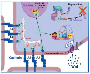
Figure 1.2 Wnt/β-catenin signaling in cancer. ………..……….44
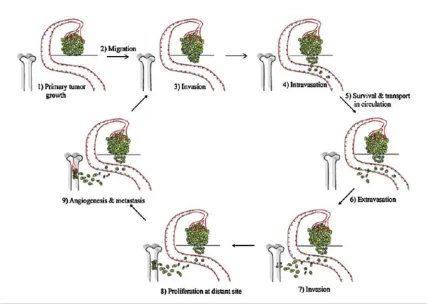
Figure 1.3 Steps of metastasis. ……….45
Figure 1.4 Secondary structure analysis of LIV-1.………...……….46
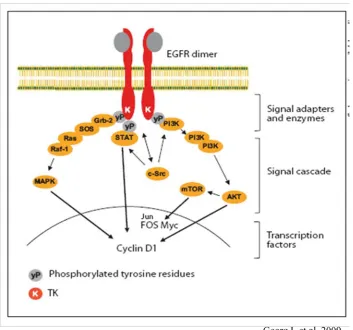
Figure 1.5 EGFR signaling pathways.………...……….………….47
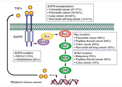
Figure 1.6 Ras/Raf/MEK/ERK signaling in tumors....……….……….48
Figure 1.7 Multiple ways lead to activate Stat3.………...……..……..……….49
Figure 2.1 LIV-1 is a mediator in ARCaPE cell EMT.………...……….69
Figure 2.2 Treatment of growth factors induced EMT...70
Figure 2.3 Knockdown of LIV-1 induced MET...71
Figure 2.4 Knockdown of LIV-1 reduced migratory capability...72
Figure 2.5 Knockdown of LIV-1 suppressed invasive ability. ...73
Figure 2.6 Transient overexpression of LIV-1 induced EMT...74
Figure 2.7 The produced antibodies to LIV-1 were subjected to validation for specificity...75
Figure 2.8 Specificity of LIV-1 antibody was comfirmed by IHC...76
Figure 2.9 LIV-1 overexpression induced EMT...77
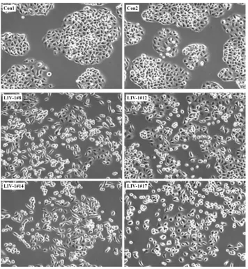
Figure 2.10 ARCaPE clones overexpressing LIV-1 displayed EMT-like changes in cellular mor-phology. ……….78
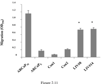
Figure 2.11 Overexpression of LIV-1 exhibited increased migratory ability...79
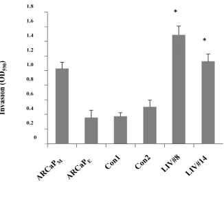
Figure 2.12 Overexpression of LIV-1 exhibited enhanced invasive ability...80
Figure 2.13 LIV-1 overexpression promoted subcutaneous tumor growth...81
Figure 2.15 Histopathologic confirmations of bone and soft tissue metastasis...83
Figure 2.16 LIV-1 expression is associated with human prostate cancer progression. ...84
Figure 2.17 LIV-1 expression was correlated with prostate cancer progression...85
Figure 2.18 Statistical analysis of LIV-1 expression correlated with prostate cancer progres-sion. ...86
Figure 3.1 LIV-1 overexpressing cells (LIV#8 and LIV#14) showed increased phosphorylated EGFR (p-EGFR) and ERK (p-ERK)...100
Figure 3.2 EGFR inhibitor (AG1478) treatment reduced phosphorylated EGFR and ERK in LIV-1 overexpressing cells...LIV-10LIV-1
Figure 3.3 Inhibition of EGFR suppressed migratory ability and invasive ability of LIV-1 over-expressing clones...102
Figure 3.4 Inhibition of ERK signaling resulted in similar suppression of cellular motility to the EGFR inhibition. ...103
Figure 3.5 Intracellular labile Zn...104
Figure 3.6 Total intracellular zinc concentration………...105
Figure 3.7 Treatment of EGF and EGFR inhibitors in LIV-1 overexpressing cells...106
Figure 3.8 RT-PCR showed increased HB-EGF, MMP2 and MMP9 expression in LIV-1 overex-pressing cells...107
Figure 3.9 MMP2/9 activity was higher in LIV-1 overexprssing cells. ...108
Figure 3.10 The effect of MMP2/9 enzymatic activity on HB-EGF shedding was evaluated by ELISA...109
Figure 3.12 Treatment of MMP2/9 inhibitors decreased migratory and invasive ability of LIV-1
overexpressing cells...111
Figure 3.13 Diagram depicts the proposed role of LIV-1 in prostate cancer cell EMT and
metas-tasis...112
Figure 4.1 MMP 2/9 inhibitors reduced the EGFR and downstream ERK and Stat3
ing. ………128
Figure 4.2 HB-EGF mRNA expression was increased in LIV-1 overexpressing cells. …...129
Figure 4.3 HB-EGF promoter activity is significantly increased in LIV-1 overexpressing
cells. ……….130
Figure 4.4 The signaling pathways in LIV-1 overexpressing cells. ...131
Figure 4.5 Different drugs were used to examine the importance of each signaling
way. ...132
Figure 4.6 Inhibition of ERK and Stat3 signalings suppressed HB-EGF mRNA expression in
vi-vo. ...133
Figure 4.7 AP-1 is one of the major transcription factors downstream of ERK
signal-ing. ...134
Figure 4.8Inhibition of AP-1 reduced HB-EGF promoter activity in LIV-1 overexpressing
cells...135
Figure 4.9 ERK and AP-1 acted in the same pathway. …...136
Figure 4.10 Overexpression of constitutive active Stat3 stimulated HB-EGF promoter activity in
ARCaPE cells. ...137
Figure 4.11 Blockade of AP-1 and Stat3 suppressed HB-EGF promoter activity in LIV-1
Figure 4.12 Treatment of AP-1 and Stat3 inhibitors inhibited HB-EGF mRNA expression. ....139
Figure 4.13 HB-EGF promoter deletion constructs. …...140
Figure 4.14 Stat3 reponse elements lied between -1022 to -681...141
Figure 4.15 Only full length HB-EGF responded to the treatment of Ap-1 and Stat3 inhibi-tors. ……….142
Figure 4.16 Site-derected mutagenesis of HB-EGF promoter reporter constructs. ...143
Figure 4.17 DNA sequencing results of site-directed mutagenic constructs. …...144
Figure 4.18 Mutation of AP-1 or Stat3 binding sites decreased HB-EGF promoter ty. ...145
Figure 4.19 Mutation of both AP-1 and Stat3bind sites abrogated HB-EGF promoter activi-ty. ...146
Figure 4.20 Stat3 and c-Jun interact with HB-EGF promoter in vivo. ...147
Figure 4.21 Stat3 inhibitor decreases of Stat3 interacting with HB-EGF promoter. …...148
Figure 4.22 AP-1 inhibitor decreases of AP-1 interacting with HB-EGF promoter. ...149
1 INTRODUCTION
1.1 Prostate cancer
Prostate cancer is the most commonly diagnosed cancer and second leading cause of
can-cer-related death in men in the United States. The American Cancer Society estimated that more
than 217,730 new cases of prostate cancer were diagnosed and over 32,050 men died of the
dis-ease in 2010 (Bubendorf et al., 2000; Keller et al., 2001). Roughly 1 man in 6 will be diagnosed
with prostate cancer during his life time and 1 in 36 will die of prostate cancer. If prostate cancer
is detected at an early stage, curative treatment by radical prostatectomy or radiotherapy is
possi-ble (Bagshaw et al., 1994; Zincke et al., 1994). However, once prostate cancer metastasizes the
mortality rate is extremely high.
The prostate is a gland which belongs to the male reproductive system and produces fluid
for semen. The main cell types of the prostate epithelium are the basal, secretory glandular and
neuroendocrine cells. The glandular cells secret PSA and prostatic acid phosphatase into the
glandular lumina and are the major cell type in normal and hyperplastic epithelium. The
secreto-ry glandular cells have high AR expression and thus are androgen-dependent for their growth. In
contrast, basal cells express low or undetectable AR and locate on the basement membrane.
Neu-roendocrine cells also locate on the basement membrane and both basal and neuNeu-roendocrine cells
are androgen insensitive. Almost all prostate cancers develop from secretory epithelial cells of
the prostate gland and often grow slowly within the gland. It is believed that high grade prostatic
intraepithelial neoplasia (HGPIN) is a precursor of prostate cancer. PIN, first described in 1969
cells begin uncontrolled proliferation to form a tumor. Eventually, the tumor cells may gain
mi-gratory and invasive ability and invade surrounding tissue, circulate in the bloodstream and
lym-phatic system, and finally colonize at metastatic sites.
PIN glands characteristically contain basal cells around their periphery (Figure 1.1). The
presence of basal cells is an indicator differentiating PIN from prostatic adenocarcinoma in
which the basal cells are absent. Normal prostate, PIN, and prostatic carcinoma can be easily
dis-tinguished using specific basal cell marker (p63) and the prostate cancer marker
Alpha-methylacyl-CoA racemase (AMACR), since AMACR is expressed at a much higher level in
ad-enocarcinoma than in non-neoplastic prostatic glands. Typically, normal prostate only shows
ba-sal cell staining, whereas prostate carcinoma only shows AMACR marker staining. However,
HGPIN exhibits both markers, which is different from normal prostate or adenocarcinoma
(Zynger and Yang, 2009).
Early prostate cancer usually has no symptoms and is usually diagnosed by a PSA test. If
the prostate cancer is confined within the gland, radical prostatectomy is potentially curative.
However, prostate cancers that have spread outside the gland are typically treated with hormone
therapy. Hormone therapy, also called androgen deprivation therapy, aims to reduce levels of
testosterone and dihydrotestosterone. Castration induces apoptosis of the majority of prostate
cancer cells and causes tumors to shrink or grow more slowly. Most prostate cancers will have
an initial favorable response to hormone therapy, hobut over time prostate cancer cells adapt to
the low androgen environment and start to grow again. As the cancer progresses, prostate cancer
cells gradually become androgen-independent and stop responding to hormone therapy.
Chemo-therapy is given if prostate cancer has already metastasized and hormone Chemo-therapy fails. However,
understanding of the mechanisms underlying cancer metastasis is vital for developing new
thera-peutic drugs.
1.1.1 Tumorigenic signaling pathways in prostate cancer
Androgen signaling. Androgen and androgen receptor (AR) both play critical roles in
normal prostate development as well as prostate cancer(Suzuki et al., 2003). For example,
trans-genic mice engineering express high levels of the AR in the prostate tend to develop
PIN(Stanbrough et al., 2001). AR is a ligand-activated transcription factor which belongs to a
steroid hormone receptor family. AR controls the expression of numerous mitotic gene products,
such as PSA, c-fos, Drg-1 and caveolin-1, which are important for the normal and neoplastic
de-velopment of the prostate. In vitro studies showed that AR activation leads to stimulation of the
survival signals and metastatic potential in LNCaP cells treated with androgen (Li et al., 2001;
Torring et al., 2003). As cancer progresses, prostate cancer changes from androgen-dependent to
androgen-independent. Many mechanisms have been proposed and changes of AR signaling are
believed to play a crucial role. In androgen-independent prostate tumors, the aberrant AR
activa-tion may be due to AR amplificaactiva-tion, AR mutaactiva-tion, ligand-independent receptor activaactiva-tion, and
an increase of co-activator expression or decrease of co-repressor expression. In fact, AR
ampli-fication has been found in 20-30% of hormone refractory patients(Koivisto et al., 1998). Increase
of AP expression allows cancer cells to survive in a low or depleted androgen environment. Over
80 mutations of AR have been identified, and most of them are mutated in the transactivation
domain or ligand-binding domain, thus causing gain-of-function mutations(Gottlieb et al., 2004).
prostate(Han et al., 2005). Only AR mutants caused development of prostate cancer, but not wild
type AR, addressing the importance of AR mutations. In addition, AR activity could be activated
in the absence of androgen by several growth factor cascades, including EGF, IGF-1, KGF, IL-6
and PKA pathway (Culig et al., 1994; Grossmann et al., 2001; Ueda et al., 2002). These factors
are ligands for receptor tyrosine kinases and activation of these pathways may stimulate AR
acti-vation and promote growth of cancer cells in a low androgen environments. Furthermore, an
in-crease of coactivator expression is another mechanism which causes AR activation(Gregory et
al., 2001). In vitro studies showed that overexpression of AR coactivator enhance AR activity to
low levels of androgen. In clinical specimens, AR coactivators- transcriptional factor 2, steroid
receptor coactivator 1, and nuclear receptor coactivator amplified in breasrt cancer 1-have been
shown to enhance expressions along with increases of AR expression in androgen-independent
prostate cancer. Thus increases of coactivator expressions enhanced AR reponses similar to AR
mutation.
Wnt/β-catenin signaling cascades. The aberrant activation of the canonical Wnt/β-catenin
signaling pathway also contributes prostate cancer progression. In the absence of Wnt signaling,
free cytoplasmic β-catenin is quickly turned over by a destruction complex consisting of the
ade-nomatous polyposis coli protein (APC), axin, glycogen synthase kinase 3-beta (GSK3β) and c
a-sein kinase Iepsilon(CKI)17, 18. CKI and GSK3β phosphorylate N-terminal serine/threonine
res-idues of β-catenin(Amit et al., 2002; Liu et al., 2002). The phosphorylated β-catenin is targeted
by an E3 ubiquitin ligase called β-TrCP (beta-transducin repeat-contain protein) and then
de-graded. Binding of Wnt molecules to the Frizzled-LRP5-LRP6 receptor complex leads to the
inhibition of this degradation complex. Therefore, free cytoplasmic β-catenin is stabilized,
activator in a complex with the LEF/TCF DNA binding proteins (Behrens et al., 1996; Korinek
et al., 1997; van Noort and Clevers, 2002). TCF/LEF molecules bind to promoter regions of
tar-get genes in a sequence-specific manner by recognizing the consensus sequence motif T/A T/A
CAAAG24. In the absence of nuclear β-catenin, TCF/LEF usually binds to members of the
groucho/TLE proteins which are transcriptional repressors, thus causing inhibition of
transcrip-tion of target genes. When β-catenin enters the nucleus, it displaces groucho/TLE proteins and
binds to LEF/TCF to increase transcription of target genes including regulators of cell cycle, cell
proliferation and metastasis (Daniels and Weis, 2005; Gavert and Ben-Ze'ev, 2007)(Figure 1.2).
Several Wnt ligands have been reported to express at significant levels in prostatic stromal cells,
androgen-dependent and independent cell linesn and tumor tissues (Chen et al., 2004; Zhu et al.,
2004). Moreover, high levels of Wnt-1 and β-catenin were detected in 77% of patients with
lymph node metastasis and 85% in skeletal metastasis, suggesting the significant of this pathway.
Hedgehog signaling cascades. Abnormal hedgehog signaling has also found to cause
can-cer. The expression of hedgehog signaling components were found to be up-regulated in prostate
cancer cells compared to normal prostate tissue. Increases of sonic hedgehog ligand, SHH, lead
to the activation of the GLI-1 transcription factor which controls tumorigenic genes of cyclin D1
and c-Myc, resulting in sustaining growth of prostate cancer cells (Fan et al., 2004; Karhadkar et
al., 2004; Sanchez et al., 2004).
Cytokine signaling cascades. The up-regulation of several cytokines in the serum of
pros-tate cancer patients seems to be associated with the development of more malignant types of
prostate cancer. For instance, higher expression of IL-6 appears in serum and tissues from
pa-tients with high grade prostate cancer, and is associated with poor patient outcome (Culig et al.,
activation of AR in androgen-independent prostate cancer cells, suggesting a role in promoting
androgen-independent progression of prostate cancer (Yang et al., 2003). TGF-β has also been
found to have high levels in serum of patients with advanced prostate cancer. TGF-β has dual
functions in prostate. TGF-β inhibits the growth of normal prostate epithelial cells, but can pr
o-mote EMT and metastasis in advanced prostate cancers (Bhowmick et al., 2004). Thus, targeting
of these tumorigenic signaling pathways may provide a good therapeutic benefit.
1.1.2 Tumor microenvironment
The interactions between epithelial cells and their microenvironment are crucial in
nor-mal prostate development and adult function. Disregulation of stronor-mal-epithelial interactions has
been suggested to contribute to malignant progression and tumorigenesis (Hayward et al., 1996;
Hayward et al., 1998; Hayward et al., 1997). In particular, the interaction of tumor cells with
platelets, lymphocytes, fibroblasts, and marcrophages was proved to be involved in tumor
pro-gression. The interaction between tumor cells and host cells is reciprocal. First, tumor cells may
secret some soluble factors, such as TGF-β and PDGF, to induce stromal fibroblasts to undergo
myofibroblast transition, a process shared by wound healing and tumorigenesis. Tuxhorn et al.
provided evidence that prostate cancer epithelium induced fibroblast-to-myofibroblast transition
with an increase of α-smooth muscle actin, vimentin, and calponin expressions, which are
char-acteristic of the myofibroblast phenotype, in the surrounding stroma (Tuxhorn et al., 2002).
Evi-dence shows that myofibroblasts are also crucial for cancer cell progression. Chung et al.
demon-strated that co-inoculation of prostate cancer cells and normal stromal fibroblasts from the fetal
with cancer-associated myofibroblasts show enhanced growth and metastatic potential,
suggest-ing a role for myofibroblasts in cancer progression (Chung et al., 1989). In addition,
cancer-associated stroma may release cytokines or neuroendocrine factors, such as HGF, VEGF, IGF-1
and IL-6, which may stimulate cancer cell invasiveness, angiogenesis, and tissue remodeling. Cat
et al. showed that myofibroblasts can be induced by tumor cell-derived TGF-β, with an increased
release of HGF, VEGF, and IL-6 (Cat et al., 2006), which have been shown to be involved in
EMT and androgen-independent progression. In vivo studies also demonstrated that tumor
growth and metastasis is significantly reduced in fibroblast-deficient mice. In summary, cancer
cells not only produce some factors which favor their survival and growth, but also secret factors
which may promote host stromal cells produce more effectors which in turn act as tumor
stimu-lators to enhance the invasiveness of tumor cells. Thus, a better understanding of
stromal-epithelial interactions would be expected to reveal novel therapeutic options.
1.2 EMT
1.2.1 The concept of EMT
Epithelial-to-mesenchymal transition (EMT) is a phenomenon by which epithelial cells
acquire migratory and invasive potential during physiological and pathological processes such as
embryonic development, wound healing, and cancer progression. Epithelial cells and
mesenchymal cells are distinguished by their unique visual appearance. Epithelial cells are
ad-herent cells and they attach laterally to each other to form a sheet of cells called an epithelium.
Cell-to-cell junctions and adherent proteins hold neighboring epithelial cells tightly together and
thick and is polarized along an apical-basal axis where the basal surface interacts with basal
membrane. In addition, the intracellular cytoskeleton network maintains the cell structure and
provides rigidity and polarization. In contrast, mesenchymal cells are non-polarized and have a
diffuse network; therefore they are irregular in shape and less rigid, accounting for the increased
migratory ability.
The concept of “Epithelial-Mesenchymal Transformation” was first described by
Eliza-beth Hay in 1995 using the chick primitive streak as a model (Hay, 1995). Hay proposed that
epithelial cells can undergo dramatic changes and transform into mesenchymal cells during
em-bryonic development. Since it is clear that EMT is a reversible process and mesenchymal cells
can revert back to epithelial phenotype, termed “Mesenchymal-Epithelial Transiton” (MET), the
term of Mesenchymal Transformation” has been replaced with
“Epithelial-Mesenchymal Transition”.
Turning epithelial cells into mesenchymal cells requires profound changes in epithelial
cell organization (Kalluri, 2009; Kalluri and Weinberg, 2009). First, epithelial cells need to
dis-assemble the cell-cell junctions and lose their apical-basal polarity. Cell surface proteins like
E-cadherin which mediate epithelial cell to epithelial cell concection are replaced by N-E-cadherin
which provides weaker adhesive properties, allowing cells to adopt the mesenchymal phenotype.
In addition, cytoskeletal networks are reorganized and the cytokeratin intermediate filaments are
replaced by vimentin. These changes convert the cell from a cuboidal to a spindle shape and are
crucial for cells to leave the epithelium and begin to migrate individually. Second, in order to
migrate and invade into the extracellular matrix, cells need to express proteases that degrade
ex-tracellular matrix simultaneously. Thus, upon undergoing EMT, cells lose epithelial cellular
them to migrate through the extracellular matrix (Rorth, 2009). Once arriving at their destination,
these mesenchymal cells may undergo the reverse process of MET and establish themselves as
one of many possible cell types. In summary, the characteristics of EMT are a loss of epithelial
marker expressions, particularly E-cadherin, and an increase in mesenchymal marker expressions,
such as N-cadherin, vimentin and fibronectin.
1.2.2 EMT during embryonic development
EMT happens in many biological and pathological events such as embryonic
develop-ment, normal tissue repair and wound healing, and cancer progression. Primary EMT takes place
during the implantation of the embryo, gastrulation, and organ development. During embryonic
implantation, the trophoectoderm cells undergo EMT to invade into the endometrium and anchor
in the placenta (Pijnenborg et al., 1980).
Gastrulation is a process by which the initial epithelial layer-epiblast forms a three grem
layer, the ectoderm, the endoderm, and mesoderm. During gastrulation, the first EMT is the
breakdown of the basement membrane underlying the epiblast. Cells in the primitive streak
un-dergo EMT, resulting in the ingression of these cells within the primitive streak. The ingressing
cells then either undergo MET to form the endoderm or remain mesenchymal to form the
meso-derm.
Another example of primary EMT happens during neural crest formation. Epithelial cells
of the neuroectoderm undergo EMT and generate a group of migratory neural crest cells (Tucker,
differ-ent cell types including the neurons of the peripheral nervous system, pigmdiffer-ent cells, and the cells
of the adrenal medulla.
1.2.3 EMT during tissue regeneration and fibrosis
Inflammatory cells and fibroblasts mediate the release of inflammatory agents as well as
components of extracellular matrix including collagens, fibronectins, elastin, and tenacins. Under
pathological conditions, these stromal cells release inflammatory signals to stimulate normal
epi-thelial cells undergoing EMT. Such EMT is found to be associated with progressive fibrotic
dis-eases of the kidney, liver, heart, lung, and intestine (Kim et al., 2006; Potenta et al., 2008;
Zeisberg et al., 2007a; Zeisberg et al., 2007b). Many studies used fibroblast-specific protein 1
(FSP1), α-SMA, and collagen I as mesenchymal markers for the EMT that occurs during fibrosis
(Okada et al., 1997; Strutz et al., 1995). The expression of these markers was found to be
corre-lated with the prognosis and extent of fibrosis. Such cells express FSP1 mesenchymal marker
and α-SMA, but these cells still display epithelial morphology as well as E-cadherin. The
behav-ior of these cells indicates that under inflammatory stimuli, epithelial cells can have different
de-grees of EMT, termed “partial EMT”. These cells then leave the epithelial layer and eventually
accumulate in the tissue with a loss of all the epithelial markers and gain of a fully fibroblastic
phenotype (Okada et al., 1996).
1.2.4 EMT during cancer progression
Most cancer deaths are due to metastatic tumors instead of the primary tumor. Metastasis
sites. The process of metastasis in epithelial cancer consists of multiple steps. First, cancer cells
lose cell-cell contact, become motile and gain the ability to invade to surrounding tissue. Once
the cancer cells escape the basement membrane, they intravasate into local blood vessels,
circu-late through the blood stream, extravasate from the blood vessel and finally colonize at the
sec-ondary site (Chambers et al., 2002; Woodhouse et al., 1997). Metastasis will not happen if any of
these steps fail (Figure 1.3).
Accumulated evidence indicates that EMT is associated with cancer progression (Thiery,
2002). EMT phenomena have been observed in many cancers, including breast, pancreatic,
ovar-ian, colon, lung, esophageal and prostate. The characteristics of oncogenic EMT include
disas-sembly of tight junctions and adherent junctions, loss of apical-basal polarity, cytoskeleton
rear-rangement, and a gain of mesenchymal phenotype with increased migratory and invasive
proper-ties. The process of EMT during cancer progression and metastasis closely parallels
develop-mental EMT. Numerous EMT inducers in cancer cell lines have been indentified including
Transforming Growth Factor-β (TGF-β), Wnt, Snail/Slug, Twist and Six1, and these abnormally
expressed EMT inducers are also critical during developmental EMT. Extensive mouse studies
and cell line experiments have demonstrated that cancer cells can undergo EMT and acquire
mi-gratory and invasive properties when treated with EMT inducers. Blocking specific regulators
could revert the mesenchymal phenotype as well as suppress invasive ability. Moreover, cells
exhibiting EMT properties are often seen at the invasive front of primary tumors where EMT is
likely to be induced by exposure to cytokines or extracellular stimuli. These cells are considered
to be the ones that eventually invade the surrounding stroma and spread to distant sites. Thus,
EMT is thought to be a critical mechanism for the metastatic spread of cancer cells. In addition,
out-come and tumor malignancy. However, phenotype changes in EMT are much more difficult to
observe in vivo, since only a subset of tumor cells may undergo EMT at any one time. The main
argument for the lack of a role of EMT in cancer is that from a histopathological point of view,
metastatic tumors seem no different from the primary tumors. Therefore, the significance of
EMT in cancer metastasis is still being debated (Christiansen and Rajasekaran, 2006; Garber,
2008; Thompson et al., 2005).
Recently, circulating tumor cells isolated from patients with progressive metastatic solid
tumors, with a focus on men with castration-resistant prostate cancer (CRPC) and women with
metastatic breast cancer (BC), were found to coexpress both epithelial and mesenchymal markers,
suggesting that EMT processes indeed exist in clinical CTC (Armstrong et al.). In addition, EMT
cells and non-EMT cells both are required for metastasis (Tsuji et al., 2009). When only EMT
cells or only non-EMT cells were subcutaneously injected, no metastasis was observed. EMT
cells could be found in the blood stream, but failed to colonize a secondary site. Metastatic tumor
was observed only when both EMTand non-EMT cells were co-injected. Moreover, metastasis
was observed when non-EMT cells were i.c injected into mouse tail vein, but not EMT cells.
These findings suggest that EMT cells are responsible for invasion into the circulation, and only
non-EMT cells are able to colonize at secondary sites. EMT cells and non-EMT cells need to
co-operate in order to metastasize. Thus, EMT is now considered to play a critical role in the
inva-sive steps of the metastatic cascade, causing invainva-sive metastatic spread of tumors.
E-cadherin regulation. One of the most important characteristics of EMT is the loss of
cell-cell adhesion with down regulation of epithelial cadherin (E-cadherin). E-cadherin, a
calci-um-dependent transmembrane protein, is the most important adhesion molecule in epithelial cells.
The intracellular part of E-cadherin is linked to the actin cytoskeleton through interaction with β
-catenin. The extracellular part of E-cadherin binds to the binding sites of other E-cadherin on
ad-jacent cells. Cadherin-cadherin binding is strong and serves as a major cell-cell adhesion force.
The cadherin-catenin complex is essential for cell architectural integrity. Disruption of either of
the complex components causes significant changes in cellular behavior and often results in
tumorigenesis. Cadherin-mediated cell-cell adhesion is highly dynamic, enabling the
reorganiza-tion and dispersal of cells. E-cadherin has been extensively studied in human epithelial cancers
and loss of E-cadherin expression results in tumor progression, metastasis, and poor prognosis in
various human cancers (Chan et al., 2003; Dorudi et al., 1993; Gould Rothberg and Bracken,
2006; Kowalski et al., 2003).
Transcription factors. Multiple mechanisms of E-cadherin loss have been demonstrated,
including transcriptional, genetic or epigenetic changes that cause a functional loss of E-cadherin.
The loss of E-cadherin expression at the transcriptional level was first identified in several cancer
cell lines as well as human cancers, including prostate, breast, colorectal, lung, pancreatic, and
thyroid cancers (Strumane et al., 2004; Van Aken et al., 2001). Later, the E-box response
ele-ments in the proximal E-cadherin promoter which determine epithelium-specific expression and
sites of repressor binding were identified (Behrens et al., 1991; Rodrigo et al., 1999). Importantly,
several developmental important transcription factors that induce EMT also repress E-cadherin
during tumor progression. These transcription factors includes the snail family of zinc finger
directly bind to E-cadherin promoter to repress E-cadherin expression (Batlle et al., 2000; Cano
et al., 2000; Comijn et al., 2001; Eger et al., 2005). Among them, Snail is one of the key
regula-tors of EMT. Overexpression of Snail reduces not only E-cadherin and other adhesion molecules
but also induces mesenchymal markers to promote EMT and mesenchymal phenotype (Cano et
al., 2000). In addition, Snail expression could be up-regulated by many known oncogenic
signal-ing pathways, such as transformsignal-ing growth factor-β (TGF-β) receptor pathway, epidermal
growth factor (EGF) receptor pathway, and fibroblast growth factor (FGF) pathway. Furthermore,
abnormal expressions of these transcriptional repressors have been found in many human cancers.
During EMT, these transcriptional repressors not only suppress E-cadherin expression but also
repress other adhesion molecules and induce mesenchymal phenotype (Aigner et al., 2007; De
Craene et al., 2005; Vandewalle et al., 2005).
Genetic and epigenetic control. Besides transcriptional control, E-cadherin can be
regu-lated by genetic or epigenetic mechanisms. Mutations of E-cadherin are found in gastric cancer
(Becker et al., 1994; Brooks-Wilson et al., 2004) and lobular breast cancer (Sarrio et al., 2003).
E-cadherin promoter polymorphism is potentially a good marker for the risk of bladder cancer
recurrence (Lin et al., 2006). In addition, hypermethylation of the E-cadherin promoter region is
found in various human cancers, resulting in loss of e-cadherin expression (Graff et al., 1995;
Yoshiura et al., 1995). Furthermore, the extent of methylation of the E-cadherin region during
cancer progression is unstable and heterogeneous, suggesting that methylation may induce EMT
through downregulation of E-cadherin expression to promote metastatic progression (Graff et al.,
1995).
microRNAs. Recently, small non-coding RNAs of 20-22-nucelotides (microRNAs) that
regulation of EMT. The miR-141, miR-200b, and miR-205 families are critical controls in ZEB1
and ZEB2 expression, leading to EMT regulation (Gregory et al., 2008; Park et al., 2008).
Selec-tive knockdown of miR-141, miR-200b, and miR-205 family miRNAs was sufficient to suppress
E-cadherin expression and induce EMT in MDCK3 and HCT116 cells. Additionally,
overex-pression of these miRNAs results in E-Cadherin re-exoverex-pression and MET in mesenchymal cells.
The mechanisms of microRNA involvment in EMT need to be elucidated in more detail.
1.2.6 Extracellular signals and intracellular networks regulating EMT
Interaction between cancer cells and the tumor microenvironment profoundly influences
the behavior of cancer cells. The tumor microenvironment is composed of ECM,
cancer-associated fibroblast, myofibroblast, and immune cells. EMT is observed particularly at the
inva-sive front, suggesting that the microenvironment plays a critical role in regulating EMT(Le et al.,
2008). EMT can be induced by growth factors, cytokines, or ECM proteins secreted by the
mi-croenvironment. For example, conditioned media from cancer-associated fibroblasts induce
EMT in breast cancer cells (Lebret et al., 2007). In addition, TGF-β signaling by stromal
myofibroblasts can induce secretion of hepatocyte growth factor which promotes cancer cell
pro-liferation and invasion (Lewis et al., 2004).
EMT regulators. Cell signaling pathways are critical inducers of EMT through
transcrip-tional or post-transcriptranscrip-tional induction of several EMT transcription factors, including Snail,
Slug, twist, ZEB1, and ZEB2. A variety of extracellular signals have been shown to induce EMT
in cancers. TGF-β, EGF family members, FGF, HGF, and IGF have all been shown to trigger
studied EMT inducers is TGF-β. The TGF-β family of cytokines binds to transmembrane
recep-tor serine/threonine kinases which in turn activate cytoplasmic Smads (Massague et al., 2005).
Activated Smads translocate to the nucleus and activate e-cadherin repressors of the Snail family.
TGF-β has dual roles. TGF-β inhibits the growth of normal epithelial cells, but can promote ca
n-cer progression at n-certain stages through its ability to induce EMT (Shi and Massague, 2003;
Siegel and Massague, 2003). In vitro studies showed that TGF-β induced EMT in many types of
cancer cells with a loss of cell-cell adhesion and cell polarity, and gain of mesenchymal
pheno-type (Ozdamar et al., 2005; Peinado et al., 2003; Zavadil and Bottinger, 2005; Zhao et al., 2008).
In early stage breast cancer, TGF-β is a tumor suppressor through its growth inhibitory effect
(Reynisdottir et al., 1995). In contrast, TGF-β promotes metastasis in later stages of breast cancer,
at least in part through its ability to induce EMT(Muraoka-Cook et al., 2006; Muraoka et al.,
2002). In vivo, TGF-β also enhanced tumor aggressiveness in a mouse model (Muraoka-Cook et
al., 2006) and blockade of TGF-β reduced metastasis and primary tumor growth in a mouse
model (Siegel et al., 2003). In addition, clinical studies also support a positive correlation
be-tween expression of TGF-β ligands and poor prognosis (Ghellal et al., 2000; Mu et al., 2008).
Wnts, a large family of cysteine-rich, secreted lipid-modified signaling proteins, are
po-tent regulators of cell proliferation and differentiation (Willert et al., 2003). The Wnt/β-catenin
pathway is implicated in EMT during development and cancer (Logan and Nusse, 2004; Reya
and Clevers, 2005). Loss of function in Wnt signaling leads to developmental abnormalities,
whereas constitutively active Wnt signaling causes tumorigenesis (Polakis, 2007; Taipale and
Beachy, 2001). In vitro studies showed that activation of the Wnt pathway induces EMT by
Furthermore, active Wnt signaling also clinically correlates with poor outcome in breast cancer
patients (Logullo et al., ; Prasad et al., 2009).
1.3 LIV-1
1.3.1 LIV-1 belongs to LIV-1 subfamily of ZIP transporters
Zinc is an essential metal for all cells and plays an important role in a variety of
physio-logical and biochemical processes, including gene expression, growth, metabolism, development,
and differentiation (Vallee and Falchuk, 1993). Zinc deficiency is associated with diverse
disor-ders, such as impaired immunity, retarded growth, brain development disordisor-ders, delayed wound
healing, retarded skeletal development, and development of osteoporosis (Andrews and
Gal-lagher-Allred, 1999; Eberle et al., 1999; Hall et al., 1999; Nishi, 1996; Rink and Gabriel, 2000).
Therefore zinc homeostasis needs to be tightly controlled. Zinc cannot passively diffuse across
cell membranes because of its charges, so two families of mammalian zinc transporters are
re-quired to transport zinc across cell membranes: ZnT (Zinc transporter) proteins and the Zip (Zrt
and Irt-like) proteins. They have opposite functions in cellular zinc transportation. ZnT
trans-porters are responsible for transporting zinc out of cells to reduce intracellular zinc, whereas ZIP
transporters promote zinc uptake to increase intracellular zinc. Normal prostate gland
accumu-lates high levels of citrate and zinc compared to other tissues. Zinc accumulation by the prostate
epithelial cells is achieved through the ZIP family of zinc uptake transporters. The ZIP family
contains four subfamilies(Guerinot, 2000). Subfamily I is mainly fungal and plant sequences,
while subfamily II consists of mammalian, nematode and insect genes. The gufA subfamily is
subfamily. 15 members of ZIP family proteins have been identied, but only a few of them have
been functionally characterized. Among them, hZIP1 has been proposed to be the major zinc
transporter for many tissues (Gaither and Eide, 2001; Guerinot, 2000). In addition, hZIP1 has
been demonstrated to have constitutive expression in normal prostate cells and function in the
uptake and accumulation of zinc in prostate cells (Franklin et al., 2003). Clinical and
experi-mental studies showed that hZIP1 gene expression is down regulated and zinc is depleted in
maglignant prostate compared to normal prostate (Franklin et al., 2005a). Furthermore,
overex-pression of hZIP results in decrease of malignancy of prostate cancer cells in vitro and in vivo
(Golovine et al., 2008; Huang et al., 2006a).
LIV-1, which was originally identified as an estrogen-regulated gene in metastatic breast
cancer, has been characterized as a new subfamily of ZIP zinc transporters termed
LZT(el-Tanani and Green, 1995). Based on a computer analysis of secondary structure, LIV-1 contains
6-8 transmembrane domains, a long extracellular N terminus, a short extracellular C terminus,
and a consensus sequence for a catalytic zinc-binding site of metalloproteases (HEXPHE) with a
molecular mass of 84 kDa (Taylor et al., 2003; Taylor and Nicholson, 2003) (figure 1.4). The
catalytic zinc is required for the proteolytic metalloprotease activity (Massova et al., 1998). Of
interest, no other zinc transporters contain this potential metalloprotease motif, suggesting that
LIV-1 may have different function other than transporting zinc. In addition, by using V5-tag
LIV-1 exression vector, LIV-1 has been shown to locate on the plasma membrane, especially
concentrating on lamellipodiae. Moreover, LIV-1 has been demonstrated to be able to transport
1.3.2 LIV-1 is associated with EMT and cancer progression
LIV-1 was first identified in the breast cancer cell line ZR-75-1 as an estrogen-regulated
gene (el-Tanani and Green, 1995), and is predominately expressed in hormonal controlled tissues
with high levels in breast, prostate, pituitary gland and brain (Taylor et al., 2003). In clinical
samples, LIV-1 expression correlates with ERα in breast tumor biopsies (Dressman et al., 2001;
Tozlu et al., 2006) and is associated with the spread of breast cancer to the regional lymph nodes
(Manning et al., 1994), suggesting a role in metastasis.
EMT has been implicated in the progression of many solid tumors, including prostate
cancer (Whitbread et al., 2006; Xu et al., 2006; Zhau et al., 2008), and is considered a key
mo-lecular event in cancer progression (Thiery, 2003). LIV-1 was reported to be a downstream target
of STAT3 and essential for the nuclear localization of Snail in zebrafish gastrula organizing cells
for their migration (Yamashita et al., 2004). LIV-1 cooperates with Snail by binding to
E-cadherin promoter and repressing its transcription (Batlle et al., 2000). LIV-1 mRNA was
re-cently shown to be higher in cervical cancer in situ than in normal tissues (Zhao et al., 2007a).
RNAi mediated suppression of LIV-1 in HeLa cells significantly inhibited their proliferation,
colony formation, migratory, and invasive ability (Zhao et al., 2007b). LIV-1 has also been
re-ported to be elevated in clinical pancreatic carcinoma and induced EMT in pancreatic cancer
cells (Unno et al., 2009). However, LIV-1 expression was also reported to correlate with
E-cadherin expression(Shen et al., 2009) and to be associated with better outcome in breast cancer
patients (Kasper et al., 2005). Although results are conflicting, LIV-1 is still thought to be an
ob-ligatory co-factor regulating EMT-associated genes. Since the potential diagnostic and
is to investigate the function of LIV-1 in cancer progression and evaluate the therapeutic
poten-tial of LIV-1.
1.4 EGFR (Epidermal growth factor receptor) signaling in cancer
1.4.1 Receptor activation
Epidermal growth factor receptor (EGFR; ErbB1), a 170kDA membrane protein, belongs
to the ErbB family of receptor tyrosine kinases (RTKs), which includes HER2 (ErbB2), ErbB3,
and ErbB4. These ErbB receptors share a common structure with an extracellular ligand-binding
domain, a single transmembrane domain, and a cytoplasmic tyrosine kinase domain. Under
un-stimulated conditions, the ErbB receptors are present as a monomer. Upon binding of
receptor-specific ligands, the receptors undergo dimerization and cause transactivation of the intracellular
tyrosine kinase domain in which specific residues are phosphorylated. Subsequently, intracellular
signal proteins are recruited by the phosphrylated residues, resulting in the activation of
intracel-lular pathways, including the mitogen-activated protein kinase (MAPK) and the
phosphatidylin-ositol-3 kinase (PI-3K) pathways, which ultimately modulate gene transcription (Olayioye et al.,
2000; Yarden and Sliwkowski, 2001).
Another mechanism to cause ErbB receptor activation is known as receptor
transactiva-tion. For example, GPCR agonists, such as endothelin-1, bombesin, and thrombin, could cause
receptor activation through stimulating metalloproteinases which in turn cleave EGF-like ligand
precursors, leading to phosphorylation of receptors (Carpenter, 2000; Gschwind et al., 2002;
Gschwind et al., 2001; Prenzel et al., 1999). In addition, cytokine has been shown to indirectly
Moreo-ver, wnt signaling has also been shown to be able to induce receptor activation through
metallo-proteinase-mediated cleavage of EGF-like ligands (Civenni et al., 2003).
1.4.2 EGFR function in normal development and in cancer
EGFR functions are ubiquitously expressed in multiple cellular processes including
em-bryogenesis, differentiation, survival, proliferation and tumor progression. The importance of
EGFR in developmental processes is supported by knockout mouse experiments. EGFR
knock-out mice are embryonic lethal at day 10.5-13.5 p.c. In addition, null mutation of EGFR causes
developmental defects in the skin, lung, pancreas, GI tract and central nervous system (Miettinen
et al., 1995; Sibilia et al., 1998; Sibilia and Wagner, 1995; Threadgill et al., 1995). EGFR is
im-portant not only because it has essential roles in normal physiological processes during
develop-ment, but also because it is involved in numerous types of human cancers. The ErbB receptors
were found to be disregulated in several malignant tumors including lung, breast, colon,
squa-mous cell cancer of the head and neck, and prostate cancer (Mendelsohn, 2002; Salomon et al.,
1995). Cancer patients whose tumors have increased expression of EGFR or ErbB2 tend to have
a more malignant tumors associated with a poor outcome (Allred et al., 1992; Hynes and Stern,
1994; Nicholson et al., 2001; Salomon et al., 1995; Sjogren et al., 1998; Slamon et al., 1987).
Aberrant receptor signaling is due to receptor overexpression (Hirsch et al., 2003), receptor
mu-tation causing ligand-independent activation (Moscatello et al., 1995), or autocrine activation by
ligand overexpression (Prenzel et al., 1999). Different studies have shown that overexpression of
EGFR or ErbB-2 could induce both in vitro and in vivo transformations. It is thought that high
acti-vation. Many genetic alterations of ErbB receptors have been demonstrated in human cancers.
Among those mutations, the EGFRvIII mutant receptor mutated in the extracellular domain has
been found to occur in breast, ovarian, lung, and glioblastoma cancer and is associated with
lig-and independent EGFR activity (Kuan et al., 2001; Pedersen et al., 2001). Moreover, EGFR
ag-onists, such as EGF, TGF-α, HB-EGF, and amphiregulin, are confirmed by a number of studies
to show overexpression of these proteins in a variety of solid tumors (Normanno et al., 2005a;
Salomon et al., 1995). For example, TGF-α is frequently coexpressed with EGFR in prostate
cancer (Seth et al., 1999), breast cancer (Umekita et al., 2000), lung, ovary, non-small cell lung
cancer (Hsieh et al., 2000), and gastrointestinal stromal tumors (Cai et al., 1999). All these
al-terations contribute to constitutive active ErbB signaling that leads to cancer development.
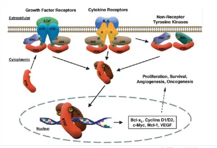
The ErbB receptors are able to activate different intracellular signaling pathways,
includ-ing the ras/raf/MAPK, PI3K/Akt, and STAT pathways (Figure 1.5). Activation of these signalinclud-ing
proteins has been shown to regulate cellular functions involved in cancer development and
pro-gression (Normanno et al., 2006). It is well known that activated MAPK promotes cell migration.
Clinical studies demonstrated that tumors with high levels of active MAPK have a particularly
poor prognosis (Feldkamp et al., 1999). Activation of the MAP kinase pathway is associated
with increasing prostate cancer Gleason score and tumor stage (Gioeli et al., 1999).
Overexpres-sion of Ras enhances androgen hypersensitivity in LNCaP cells (Bakin et al., 2003b). In contrast,
overexpression of dominant negative Ras converted androgen-independent cells back to
andro-gen-dependent status in C4-2 prostate cancer cells (Bakin et al., 2003a). The PI3K/Akt pathway
is very important in mediating cell survival, since activated PI3K may inhibit proapoptotic
mole-cules resulting in cell survival (Datta et al., 1997). An increase of PI3K has been found in
prostate tissue or PIN (Liao et al., 2003). In addition, PTEN, a negative regulator of PI3K
signal-ing, has been identified to frequently mutate and be inactivated in prostate cancers (Li et al.,
1997). Stat3has also been implicated in cell survival. Inhibition of Stat3 in vivo results ina
de-crease of anti-apoptotic Bcl-XL and increased cell death (Grandis et al., 2000). Together,
numer-ous data provide strong evidence that disregulated ErbB signaling plays a crucial role in cancer
development and metastasis.
1.4.3 EGFR in angiogenesis
Angiogenesis is essential for tumor growth and metastasis. It has been demonstrated that
the ErbB receptor/ligand network regulates the process of neovascularization, in which
endothe-lial cells proliferate and undergo differentiation. On the one hand, endotheendothe-lial cells themselves
express ErbBs. On the other hand, EGFR signaling regulates the production of proangiogenic
facors, the most potent being VEGF, in different tumor cells. For example, upon EGF treatment,
glioma cells upregulate the secretion of VEGF (Goldman et al., 1993). Conditioned media from
EGF-stimulated glioma cells induced tube formation of human umbilical vein endothelial cells
(HUVECs), and this effect was blocked by an anti-VEGF antibody. In addition, EGFR activation
was shown to regulate VEGF promoter activity in glioblastoma cells through the MAPK and
PI3K pathways, which were independent of the hypoxia-induced HIF-1 pathway, a potent
induc-er of VEGF (Maity et al., 2000). In prostate cancinduc-er cells, EGFR was also shown to regulate
ex-pressions of angiogenic factors. In particular, EGF significantly increased the secretion of VEGF
sup-pressed EGF-induced VEGF expression both in prostate cancer cells cultured in vitro or
implant-ed in the flank of nude mice (Bianco et al., 2004; Sini et al., 2005).
ErbB ligands also have a direct effect on endothelial cells. Using an orthotopic nude mice
model, TGF-α-expressing tumor cells directly induced EGFR expression in endothelial cells
(Baker et al., 2002). Baker et al. demonstrated that only tumor-associated endothelial cells
ob-tained from EGF/TGF-α positive cancers express functional EGFR, which can be activated upon
stimulation with either EGF or TGF-α (Baker et al., 2002). In addition, stimulation of HUVECs
with EGF or HB-EGF resulted in an increase of EGFR phosphorylation and ERK activation (Sini
et al., 2005). Taken together, evidence supports the idea that tumor cells may induce expression
and activation of the EGFR pathways in endothelial cells to promote tumor-associated
angiogen-esis. Thus, a better understanding the role of the EGFR/ligand network in endothelial cell/tumor
cell interactions will provide strategies to inhibit angiogenesis which in turn could block tumor
proliferation, survival and metastasis.
1.4.4 EGFR in bone metastasis
The most common metastatic site of prostate cancer is the bone (Li et al., 2006). Nearly
80% of advanced prostate cancer cases result in bone metastasis and generate severe pain and
disability (Landis et al., 1999). Bone metastasis results from dysregulation of the bone formation
and bone resorption processes. The cells responsible for bone remodeling are osteoblasts, which
secret new bone, and osteoclasts, which dissolve bone matrix. Two factors necessary and
suffi-cient for osteoclast formation and activation are macrophage colony stimulating factor (M-CSF),
ac-tivates pre-osteoclasts and is secreted by osteoblasts and bone marrow stroma cells (Boyle et al.,
2003). Cancer cells are able to synthesize many growth factors and cytokines which can lead to
the activation of osteoclasts (Roodman, 2001). EGFR signaling involvment in the pathogenesis
of bone metastasis was demonstrated in a clinical trial of the EGFR inhibitor gefitinib in breast
cancer patients (von Minckwitz et al., 2005). A significant improvement in bone pain was
ob-served in patients treated with gefitinib. Evidence showed that both EGF and TGF-α are able to
stimulate bone turnover and osteoclastogenesis in different systems (Guise et al., 1993; Ibbotson
et al., 1983; Ibbotson et al., 1985; Zhu et al., 2007). EGFR inhibitor treatment inhibits M-CSF
and RANKL production in bone marrow stromal cells and thus inhibits osteoclast formation
(Normanno et al., 2005b). These data suggests that EGFR signaling regulates the ability of bone
marrow stroma cells to induce osteoclastogenesis.
Anti-EGFR inhibitors also have an effect on prostate cancer cells. For example, gefitinib
treatment reduced the ability of conditioned media from prostate cancer cells to induce RANKL
expression in osterblasts (Angelucci et al., 2006). In addition, EGFR signaling activates the
ex-pression of proteases which play an important role in metastasis. For instance, the urokinase-type
plasminogen-activator (uPAR) and matrix matalloproteinases (MMPs) are necessary for the
in-vasive ability of tumor cells and metastasis (Guise and Mundy, 1998; Nemeth et al., 2002).
Gefitinib treatment inhibited the expressions of uPAR and MMP-9 in prostate cancer cells, and
reduced the metastatic potential of these cancer cells (Angelucci et al., 2006).
Cancer patients whose tumors show dysregulated EGFR or ErbB-2 tend to have a more
aggressive disease and a poor clinical outcome. Because of the importance of ErbB receptors, a
huge effort has been made to develop therapies that target ErbBs. Two current successful
ap-proaches have been developed: anti-EGFR monoclonal antibodies and EGFR-specific tyrosine
kinase inhibitors (TKIs). Anti-EGFR MAbs, such as cetuximab and panitumumab, bind to the
extracellular domain of EGFR, thus preventing the binding of EGFR ligands and activating the
receptors (Sato et al., 1983). In contrast, TKIs, such as gefitinib and erlotinib, prevent the
bind-ing of adenosine triphosphate to the intracellular tyrosine kinase domain, thus inhibitbind-ing
intracel-lular tyrosine kinase activity and subsequent signaling (Lichtner et al., 2001).
Both MAbs and TKIs result in decreases in the MAPK, PI3K/Akt, and Jak/Stat pathways.
However, anti-EGFR MAbs also cause downregulaiton of EGFR expression. Cetuximab is a
chimeric anti-EGFR MAb which is approved for treating pateients with metastatic colorectal
cancer refractory to irinotecan-based chemotherapy. Cetuximab has higher affinity toward EGFR
compared to TGF-α or EGF and induces EGFR internalization and antibody-dependent
cell-mediated cytotoxicity. In preclinical studies in nude mice bearing irinotecan-resistant colorectal
tumors, cetuximab exerted strong antitumor activity (Prewett et al., 2002). However, cetuximab
only showed a 10-20% response rate in several clinical trials (Cunningham et al., 2004; Saltz et
al., 2004). TKIs are low molecular weight synthetic molecules which block the intracellular
ki-nase activity of EGFR. For example, Gefitinib was approved for the treatment of patients with
locally advanced or metastatic non-small cell lung cancer after failure of both docetaxel and
plat-inum-based chemotherapies. In phase II clinical trials, Gefitinib treatment showed a significant
response rate in patients with advanced non-small cell lung cancer (Fukuoka et al., 2003). In
pro-gression (Giaccone et al., 2004; Herbst et al., 2004). However, Gefitinib did show a significant
response rate in a particular population of patients with specific EGFR mutation (Inoue et al.,
2009; Inoue et al., 2006). Gefitinib treatment showed a 50-70% responsive rate with great
pro-gression-free survival in tthis group of patients and gefitinib was approved in Europe for treating
patients with advanced or metastatic non-small cell lung cancer carrying EGFR mutation.
Anti-EGFR therapy has shown significant responses in some cancers, but no therapeutic
response resulted in the majority of cancer patients. In fact, patients initially responsive to
anti-EGFR treatment develop drug resistance over time. Potential mechanisms of resistance to
EGFR-targeted therapies have been proposed. For example, constitutive activation of
down-stream signaling of EGFR, such as PI3K mutations and K-ras mutations, has been demonstrated
as one of the possible causes (Prenen et al., 2009; Sartore-Bianchi et al., 2009). In addition,
acti-vation of an alternative receptor or EGFR mutation are also possible mechanisms for the
devel-opment of resistance to anti-EGFR therapies. Thus, a better understanding of the molecular
mechanisms of resistance to EGFR-targeted therapies will further increase our understanding of
EGFR signaling and ultimately improve treatment strategies.
1.5 The Ras/Raf/MEK/ERK cascade in cancer progression
The ERK cascade is activated by a large number of extracellular stimuli. Activation of
this cascade controls a variety of cellular processes, including proliferation, differentiation,
de-velopment, cell survival, migration, apoptosis, and oncogenic transformation (Seger and Krebs,
1995; Torii et al., 2004; Viala and Pouyssegur, 2004; Yoon and Seger, 2006). Signaling is
recruit-ing Raf kinase to the plasma membrane (Wellbrock et al., 2004) (Figure 1.6). Once Raf is
acti-vated, Raf transmits signaling by phosphorylating MEK and activates MEK (Ahn et al., 1991).
Upon activation, MEKs act as dual specific kinases and phosphorylate the Tyr and Thr residues
of ERKs, leading to ERKs activation (Seger et al., 1992). Once phosphorylated by MEKs, ERKs
become a potent protein kinase and are able to phosphorylate a large number of downstream
tar-gets (Yoon and Seger, 2006). In particular, ERKs phosphorylate and activate a series of
tran-scription factors, such as c-fos (Murphy et al., 2002), c-jun (Morton et al., 2003), p53 (Milne et
al., 1994), Elk1 (Gille et al., 1992), and Ets1/2(Yang et al., 1996), which play critical roles in the
initiation and regulation of proliferation and oncogenic transformation. The inactivation of ERKs
is mainly mediated by protein Ser/Thr phosphatases or MAPK phosphatases (Sun et al., 1993).
MAPK phosphatases inactivate MAPKs by simultaneously removing phosphates from both Tyr
and Thr residues, thus terminating the signaling cascade.
1.5.1 ERK1/2 is necessary for cell proliferation
ERK1 and ERK2 were originally identified as mitogen-stimulated phosphoproteins of
41-45 kDa (Cooper et al., 1984; Nakamura et al., 1983). The direct involvement of ERK1/2 in the
mitogenic response was shown by the inhibition of ERKs. Overexpression of dominant negative
ERK1 or ERK1 siRNA inhibited fibroblast cell proliferation (Pages et al., 1993). In addition,
treatment with MEK1/2 inhibitors which prevent ERKs from activation was shown to reduce the
proliferation of various cell types, including fibroblasts, T lymphocytes, smooth muscle cells,
hepatocytes and epithelial cell lines (Brunet et al., 1994; DeSilva et al., 1998; Dudley et al., 1995;