R E S E A R C H A R T I C L E
Open Access
Gene expression profiling of flaxseed in mouse
lung tissues-modulation of toxicologically relevant
genes
Floyd Dukes
1, Stathis Kanterakis
1, James Lee
1, Ralph Pietrofesa
1, Emily S Andersen
1, Evguenia Arguiri
1, Sonia Tyagi
1,
Louise Showe
2, Yassine Amrani
3and Melpo Christofidou-Solomidou
1*Abstract
Background:Flaxseed (FS), a nutritional supplement consisting mainly of omega-3 fatty acids and lignan phenolics has potent anti-inflammatory, anti-fibrotic and antioxidant properties. The usefulness of flaxseed as an alternative and complimentary treatment option has been known since ancient times. We have shown that dietary FS supplementation ameliorates oxidative stress and inflammation in experimental models of acute and chronic lung injury in mice resulting from diverse toxicants. The development of lung tissue damage in response to direct or indirect oxidant stress is a complex process, associated with changes in expression levels of a number of genes. We therefore postulated that flaxseed might modulate gene expression of vital signaling pathways, thus interfering with the development of tissue injury.
Methods:We evaluated gene expression in lungs of flaxseed-fed (10%FS) mice under unchallenged, control conditions. We reasoned that array technology would provide a powerful tool for studying the mechanisms behind this response and aid the evaluation of dietary flaxseed intervention with a focus on toxicologically relevant molecular gene targets. Gene expression levels in lung tissues were analyzed using a large-scale array whereby 28,800 genes were evaluated.
Results:3,713 genes (12.8 %) were significantly (p<0.05) differentially expressed, of which 2,088 had a>1.5-fold change. Genes affected by FS include those in protective pathways such as Phase I and Phase II.
Conclusions:The array studies have provided information on how FS modulates gene expression in lung and how they might be related to protective mechanisms. In addition, our study has confirmed that flaxseed is a nutritional supplement with potentially useful therapeutic applications in complementary and alternative (CAM) medicine especially in relation to treatment of lung disease.
Keywords:Antioxidant, CAM, Flaxseed, Genomic profiling, Lignans, Lung disease, Oxidative stress
Background
Flaxseed (FS), a nutritional supplement known since an-cient times with high contents of omega-3 fatty acids and lignans, has recently gained popularity in complementary and alternative (CAM) medicine mostly due to its benefits in cardiovascular diseases [1]. FS oil contains 52 % alpha-linolenic acid (ALA) [2], a precursor of eicosapentaenoic acid (EPA) and docosahexaenoic acid (DHA), and
omega-3 fatty acids–essential fats that are both examples of
poly-unsaturated fatty acids. Omega-3 fatty acids help reduce inflammation and may be helpful in treating a variety of cardiovascular and autoimmune diseases [3-5]. In addition to omega-3 fatty acids (O-FA), FS also contains phenolic botanical agents called lignans. The FS lignan, secoisolari-ciresinol diglucoside (SDG), is metabolized in the mamma-lian intestine to the mammamamma-lian lignans enterodiol (ED) and enterolactone (EL), phytoestrogens demonstrating antioxidant properties [6]. The oxygen free radical
scaven-ging properties of the FS lignans have been shownin vitro
by either direct hydroxyl radical scavenging activity [7,8]
* Correspondence:[email protected]
1Department of Medicine, Pulmonary, Allergy and Critical Care Division,
University of Pennsylvania, 3615 Civic Center Boulevard, Abramson Research Building, Suite 1016C, Philadelphia, PA 19104, USA
Full list of author information is available at the end of the article
or inhibition of lipid peroxidation [9-11]. With its add-itional platelet-activating-factor (PAF) antagonism [12], the lignan SDG may exert antioxidant activity by inhibiting production of reactive oxygen species (ROS) by white blood cells. The antioxidant properties of FS lignans were also verified in animal models of endotoxic shock in dogs [12], diabetes in rats [13], and in carbon tetrachloride-induced oxidative stress in rats [14]. While usefulness of the main bioactive ingredients of FS (O-FA, lignans) has been the focus of several studies, their contribution in modulation of gene expression in various tissues has never been investigated. In this work, we evaluated the effects of dietary wholegrain FS in modulating gene expression changes in lung tissues. In future studies we intend to ex-pand our gene profiling studies to include evaluation of the FS-lignan complex (FLC).
Our group was first to investigate the role of flaxseed in acute and chronic lung injury and our findings suggested a protective role of dietary flaxseed [10,11,15-17] in murine model systems of acute and chronic lung injury. This prompted the current study, wherein the genetic profiling of flaxseed in murine lungs has been evaluated. We specific-ally focused on genetic changes occurring three weeks after
flaxseed supplementation–the time required by lignans to
achieve steady state in murine circulation as confirmed by plasma mass spectrometric analysis [15]. Mouse arrays cov-ering 28,800 genes in the murine genome were evaluated. We first evaluated genes most up- and down-regulated in our dataset, calculated the number of statistically significant genes, and quantified our false positive rates. We then used those genes to run an aggregate pathway analysis, build gene networks according to the interactions between our significant set, and validate the results seen in the individual gene analysis. Finally, we proposed the most significant function of our test set, relative to controls. In this first reported study of genomic profiling of lung tissues in re-sponse to dietary flaxseed supplementation we focused on specific gene groups of interest shown to be relevant to acute lung injury, including antioxidant enzymes, members of the apoptotic pathway, members of the Phase I and Phase II detoxification pathways, pro-fibrogenic cytokines like TGF-beta1, and members of the cell cycle. Findings from this study will provide insight to gene-nutrient interac-tions thus providing scientific evidence for the usefulness of FS as a CAM modality in lung disease.
Results
Dietary flaxseed alters gene expression pattern in mouse lung tissues
Our group has shown that dietary FS supplementation is protective in various mouse models of pulmonary oxida-tive challenge including hyperoxia [15], thoracic radiation-induced injury [11,17], and ischemia/reperfusion injury [10,16]. The current study was designed to evaluate gene
expression changes in lung tissues of unchallenged mice supplemented with dietary FS to elucidate the anti-inflam-matory, anti-fibrotic, and anti-oxidant effects of FS. Gene expression levels from individual lung tissue samples were evaluated on separate arrays. Overall, 3,713 genes (12.9 %)
were significantly (p<0.05) differentially expressed as a
re-sult of the diet; and of those, 2,088 (7.2 %) had>1.5-fold
change.
In hierarchical cluster analysis, as shown in Figure 1, the untreated control and flaxseed-treated samples formed separate hierarchical clusters containing all of the samples from their respective groups. In principle component analysis, the two groups also formed distinct separate clusters containing all of the samples of their re-spective groups (data not shown).
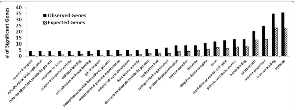
Enriched gene ontology analysis was conducted
wherein significantly (p<0.05) overrepresented
categor-ies were identified. Within the set of genes that were sig-nificantly differentially expressed in the array, 120 ontology categories were significantly overrepresented. Figure 2 compares expected and observed representa-tions for a selected list of ontologies. The included ontologies related to DNA synthesis, autophagy, and cell cycle progression and regulation.
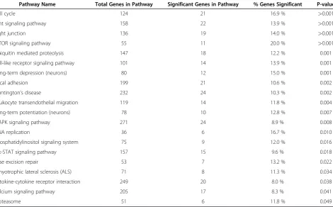
Data analysis by Pathway-Express demonstrated that a number of gene pathways were significantly impacted in the FS-fed group. Table 1 lists selected pathways, including base excision repair pathway, cell cycle pathway, cytokine-cytokine receptor interaction pathway, Janus kinase-signal transducer and activator of transcription signaling (JAK-STAT) pathway, leukocyte transendothelial migration pathway, mTOR signaling pathway, phosphatidylinositol signaling pathway, and Toll-like receptor (TLR) signaling pathway. All genes from these impacted signaling path-ways (many of which were down-regulated) have been pro-vided in a separate table (see Additional file 1). Specifically,
a large decrease in Rbl2 expression (−42.2 fold) suggested
a down-regulation of the cell cycle pathway, as this protein is a known key regulator of activation of cell division. ATM expression was also decreased, suggesting the ab-sence of genotoxic stress to the tissue. Cytokine-cytokine receptor interaction pathway was down-regulated with diminished expression of receptors for interleukin (IL)-2, IL-7, IL-12, IL-21, and TGF-beta.
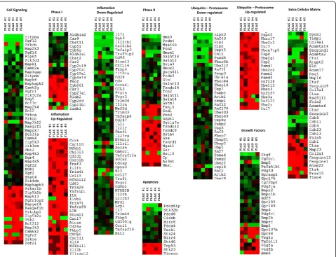
Cluster analysis and heat map of the expression of phase I genes, phase II genes, inflammatory genes, genes involved in cell signaling and apoptosis, ubiquitin-proteasome com-plex genes, growth factors, and extracellular matrix genes are shown in Figure 3. All gene clusters included both up-regulated and down-up-regulated genes, suggesting that the impact of flaxseed lignans was complex. Various growth factors, mitogen activate protein (MAP) kinases,
cyto-chromes P450 (CYPs), glutathione-S-transferases (GSTs),
domain (ADAM), and chemokine (C-X-C motif) receptor (CXC) gene groups were among the set of impacted genes. Importantly, these clusters indicated that gene expression was predominantly down-regulated.
Table 1 provides other examples of important path-ways in the mouse lungs that have been affected by flax-seed treatment (not all genes have been selected). FS efficiently regulated the expression of a number of genes encoding proteins that have a broad spectrum of activity. Based on its intrinsic properties, FS appeared to regulate at least five different groups of molecules essential in the regulation of (i) gene expression (transcription factors), (ii) signal transduction (signaling pathways), (iii) inflam-matory responses (cytokines), (iv) cell proliferation (cell cycle regulation), and (v) cell remodeling (via cytoskel-eton apparatus). These findings demonstrated that FS treatment was undoubtedly effective in driving changes of key genes in the lungs explaining, at least in part, the protective action against lung injury reported in our pre-vious studies [10,11,15-17].
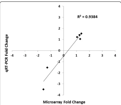
Quantitative validation of microarray gene expression by qRT-PCR and western blot confirmation of protein levels
Reverse transcription polymerase chain reaction (RTPCR) was performed to validate the differential expression of fibroblast growth factor 1 (Fgf1), TGF-beta receptor 1 (Tgfbr1), Tgfbr2, leukemia inhibitory factor (Lif), p21, and
Bcl-2–associated X protein (Bax). The changes in
expres-sion levels for these genes revealed by qRT-PCR were simi-lar to those determined by the microarray (Figure 4).
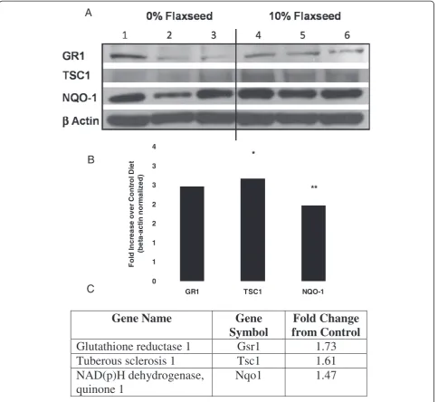
Additionally, we validated some of the microarray data by Western blot analysis of select genes (Figure 5). Flaxseed is known for its antioxidant properties and thus the antioxi-dant and Phase II detoxification enzymes, GR1 and NQO-1, respectively were selected for protein confirmation. We also selected tuberous sclerosis protein 1 (TSC1), a multi-functional protein and member of a key pathway impli-cated in cell growth and metabolism, namely the Akt/ TSC1-TSC2/mTOR pathway [18]. There was good
correl-ation (R2= 0.9384) between the findings of the microarray
[image:3.595.58.541.89.204.2]data and the Western blot.
Figure 2Statistically significant enrichment (p<0.05) of Gene Ontology categories within the significant (p<0.05) genes with a
>1.5-fold change in the FS-fed group.
[image:3.595.57.545.521.702.2]Discussion
Interest in the use of CAM natural products has grown significantly in recent times and FS, a botanical dietary supplement has gained significant popularity due to its
antioxidant, anti-inflammatory and anticarcinogenic
properties. Specifically, several studies have convincingly reported that dietary FS supplementation has a beneficial role in the management of a number of conditions in-cluding diabetes [19], lung ischemia/reperfusion injury [10], atherosclerosis [20], radiation therapy [17] and renal diseases [21] where oxidative stress is thought to be pathogenic. It is therefore important to determine the molecular mechanisms by which dietary flaxseed exerts its therapeutic action. Natural products such as FS are widely used for health purposes. Investigations about their bioactive components, their molecular and cellular targets, as well as markers of potential beneficial or harmful biological effects will provide valuable and much needed information in order to maximize their useful-ness. Our study was conducted to identify natural prod-uct-induced gene regulation and/or expression changes that may identify mechanistic pathways helping to eluci-date biochemical, cellular, or metabolic FS targets.
While our group has shown the functional significance of dietary FS in boosting nuclear factor erythroid 2-related factor 2 (Nrf2)-mediated protective enzyme expression in
lung tissues [11], little is known in unchallenged hosts about gene expression and molecular activation changes
induced by flaxseed’s anti-inflammatory, anti-fibrotic, and
antioxidant properties . To date, no studies have taken a genome wide inventory of genes significantly impacted by a FS diet in unchallenged conditions. Here via gene ex-pression analysis, we observe for the first time significant biological impacts attributed to FS.
An important outcome of this study was the demon-stration that dietary FS supplementation has the poten-tial to either positively or negatively modulate the function of a number of key regulatory proteins in the lungs thus explaining to some extent, the therapeutic value of FS reported in recent literature. Our study pro-vides direct evidence that dietary FS leads to the expres-sion of an array of genes that have an impact in various cellular responses that regulate cell growth and prolifera-tion, extracellular matrix synthesis, inflammaprolifera-tion, and oxidative stress (see Table 1). These findings will serve as the first steps to identify the gene signature by which FS exerts its therapeutic action in various experimental models of human diseases [10,11,15,17]. Of the 2,088 genes that were significantly differentially expressed with
a>1.5-fold change in the FS-fed group, 1,482 (70.9 %) of
[image:4.595.65.536.112.404.2]those were down-regulated. Hierarchal clustering and Principle Component Analysis (data not shown) between
Table 1 Pathway categorization of significantly (p<0.05) differentially expressed genes with a 1.5-fold change in the FS-fed group
Pathway Name Total Genes in Pathway Significant Genes in Pathway % Genes Significant P-value
Cell cycle 124 21 16.9 % >0.001
Wnt signaling pathway 158 22 13.9 % >0.001
Tight junction 136 19 14.0 % >0.001
mTOR signaling pathway 55 11 20.0 % >0.001
Ubiquitin mediated proteolysis 147 18 12.2 % 0.001
Toll-like receptor signaling pathway 101 14 13.9 % 0.001
Long-term depression (neurons) 80 12 15.0 % 0.001
Focal adhesion 199 21 10.6 % 0.002
Huntington’s disease 232 24 10.3 % 0.002
Leukocyte transendothelial migration 119 14 11.8 % 0.004
Long-term potentiation (neurons) 78 10 12.8 % 0.007
MAPK signaling pathway 271 24 8.9 % 0.008
DNA replication 36 6 16.7 % 0.010
Phosphatidylinositol signaling system 75 9 12.0 % 0.016
Jak-STAT signaling pathway 157 15 9.6 % 0.018
Base excision repair 53 7 13.2 % 0.022
Amyotrophic lateral sclerosis (ALS) 71 8 11.3 % 0.034
Cytokine-cytokine receptor interaction 249 20 8.0 % 0.038
Calcium signaling pathway 205 17 8.3 % 0.041
the two groups resulted in a distinct separation between the two, indicating an overall consistency of the expres-sion profile in individual subjects responding to the diet. In the ontology overrepresentation analysis of the
signifi-cant genes (p<0.05 and>1.5-fold change) expressed in
the FS-fed group, several ontologies were identified that related to oxygen transport, the extracellular matrix and genome maintenance processes, specifically those of the mitochondrial genome. In the context of lung disease, these processes could affect the lungs efficiency, its re-sponse to inflammation, and its rere-sponse to ROS.
An important effect of FS treatment is its ability to regulate the expression of a number of molecules, in-cluding signaling molecules, which could impact the ini-tiation and/or perpetuation of inflammatory responses. FS therapy down-regulated the expression of transcrip-tion factor ATF-2, a key target of kinases such as JNK and p38 MAPK. The concept that MAPK pathways is a natural target of FS is further supported by the fact that
additional key enzymes controlling MAPK pathways were strongly down-regulated by FS including MAPK1 (also known as ERK1/2), MAPK kinase 3 (upstream of p38), and MAPK kinase 7 (upstream of JNK). As an ex-ample, MAPK kinase 3 was suppressed greater than six-fold compared to untreated controls. Although downre-gulation by FS of the phospho-MAPK signaling pathway in tumor tissues has been reported [22], this was the first documentation that at least in lung tissues, FS may modulate MAPK activation by downregulating expres-sion of the upstream kinases. Importantly, a potential molecular mechanism for the protection shown by diet-ary FS in a mouse model of ischemia-reperfusion injury reported previously by our group [10] has been eluci-dated. Other studies have indeed confirmed that p38 MAPK plays a crucial role in the development of tissue injury seen in other experimental models of ischemia/ reperfusion such as transplantation or myocardial infarc-tion [23-26]. Recent studies have shown that p38 MAPK
[image:5.595.59.539.88.455.2]FLA X # 1 FLA X # 2 FLA X # 3 FLA X # 4 FLA X # 5 FLA X # 1 FLA X # 2 FLA X # 3 FLA X # 4 FLA X # 5 FLA X # 1 FLA X # 2 FLA X # 3 FLA X # 4 FLA X # 5 FLA X # 1 FLA X # 2 FLA X # 3 FLA X # 4 FLA X # 5 FLA X # 1 FLA X # 2 FLA X # 3 FLA X # 4 FLA X # 5 FLA X # 1 FLA X # 2 FLA X # 3 FLA X # 4 FLA X # 5 FLA X # 1 FLA X # 2 FLA X # 3 FLA X # 4 FLA X # 5 FLA X # 1 FLA X # 2 FLA X # 3 FLA X # 4 FLA X # 5 FLA X # 1 FLA X # 2 FLA X # 3 FLA X # 4 FLA X # 5 FLA X # 1 FLA X # 2 FLA X # 3 FLA X # 4 FLA X # 5
represented novel targets for the treatment of chronic lung diseases including asthma and chronic obstructive pulmonary disease (COPD) [27]. It is possible that diet-ary FS, by its ability to inhibit MAPK pathway activation may be a useful agent in the treatment of related lung diseases.
[image:6.595.57.290.87.289.2]Acute and chronic lung injury induces an inflamma-tory cascade, characterized by the recruitment and acti-vation of inflammatory immune cells in the lung [28]. Our data showed that FS modulated the expression pro-file of several genes encoding proteins implicated in the induction of the inflammatory pathway, as well as a decreased activation of several inflammation-related sig-naling pathways. Among the novel mechanisms capable of mediating the protective effect of FS in lung injury was the down-regulation of proteins called Poly ADP-ribose polymerase (PARP1 or 2). Studies using knockout mice or soluble inhibitors (INO1001, 1,7-dimethylxanthine) found that PARP1 was essential in driving the development of lung injury in response to various noxious stimuli in-cluding mechanical ventilation [29], lipopolysaccharide induced sepsis [30,31], and allergen sensitization in asthma [32-34].
The function and activation of Phase II enzymes in this experimental context left us with numerous questions about the complex nature of these compounds. Phase II enzymes play an important role in eliminating xenobiotics and their metabolites formed in Phase I reactions [35]. Genes within this group were up-regulated and down-regulated about equally, as shown in the heat map analysis (Figure 3). While genes encoding antioxidant enzymes
such as GSTa3 and GSTm7 were down-regulated, other key antioxidant compounds, such as GSTa4, Txnrd1, Txndc12, Txndc9, and Sod1, were up-regulated. GSTs comprise a family of enzymes that catalyze the conjugation of glutathione to a number of endogenous and exogenous electrophilic compounds, as either membrane-bound or cytosolic compounds [35].
The gene E2F3 was up regulated in the FS diet treatment by 3.9-fold, suggesting that it may be important in the cell cycle function of lung tissue. E2F3 is thought to control cell cycle progression and proliferation in neoplastic and non-neoplastic cells [36]. Genes controlled by E2F3 seem to determine the timing of G1/S transition [37-39]. Evi-dence suggests that overexpression of E2F3 represents an oncogenic event during human bladder carcinogenesis and in many cases of prostate cancer [40-42].
The ubiquitin-proteasome pathways process and elim-inate miss-folded or malformed proteins in the respect-ive tissue. A highly actrespect-ive ubiquitin mediated proteolysis system indicates an excess of miss-formed proteins within the cell. While several genes of these pathways were up-regulated, the majority of them were down-regulated. This demonstrated that there were fewer mal-functions within cellular processing and potentially fewer cases of apoptosis. Moreover, the FS diet effectively down-regulated the majority of genes implicated in apoptosis. Down-regulation of such genes under unchal-lenged conditions suggested that FS might prevent apoptosis.
Leukocyte transendothelial migration is a normal part of immune surveillance in the cell. Such cell types are important to heal tissue injury and re-establish the epi-thelial barriers. Matrix metalloproteinases (MMPs) are extracellular endopeptidases that can function to facili-tate the migration of cells by breaking down the ECM barriers, while focal adhesions are important stress fiber anchors that function in the dynamics of cell transloca-tion [43]. Our data showed that these proteins were both up- and down-regulated, but the majority of ECM-related genes were down-regulated (Figure 3). A pre-dominant decrease in ECM activity might mean that FS decreased the turnover and/or generation of ECM in the lung through its anti-inflammatory and anti-apoptotic activity.
Conclusions
In conclusion, this microarray study of lung tissues from mice supplemented with a flaxseed-diet has provided unique insights into the genes that were modulated in the mouse genome secondary to the presence of flaxseed, a botanical wholegrain with potent anti-inflammatory, antioxidant, and anti-fibrotic properties. This global gene expression profile may yield further insights into the pro-tective properties and associated cell signaling attributes
of flaxseed, helping to establish this ancient wholegrain as a useful contemporary modality in complementary and alternative medicine relevant to acute and chronic pulmonary disease.
Methods
Animals
Female C57BL/6 mice of ages six to eight weeks were used throughout this study. All animals were cared for,
handled, and housed at the Children’s Hospital of
Phila-delphia (CHOP) animal facility (PhilaPhila-delphia, PA). All
protocols were performed in accordance with National Institutes of Health guidelines and with the approval of the CHOP and the University of Pennsylvania Animal Use Committees.
Diets and dietary treatments
The semi-purified AIN-93 G diet was used as the base diet and was supplemented with 10 % (w/w) FS as pre-pared by Purina Mills (TestDiet, Bloomsburg, IN). The 10 % (#56906) FS dose was selected based on published reports [44] and from our own work [10,11,15-17].
B
C
Gene Name
Gene
Symbol
Fold Change
from Control
Glutathione reductase 1
Gsr1
1.73
Tuberous sclerosis 1
Tsc1
1.61
NAD(p)H dehydrogenase,
quinone 1
Nqo1 1.47
0 1 1 2 2 3 3 4
GR1 TSC1 NQO-1
Fol
d
In
cr
ea
se over
C
o
nt
ro
l D
iet
(b
e
ta-act
in
nor
m
a
li
z
ed)
*
**
***
[image:7.595.58.543.89.533.2]A
Control and experimental diets were isocaloric and equivalent in terms of the percentage of protein, carbo-hydrate, and fat. The Physiological Fuel Value (PFV) in all diets was kept the same, namely at 3.85 Kcal/g.
While the flaxseed seeds were stored at−80°C, the
for-mulated chow pellets were stored at 4°C and checked regularly for oxidative degradation. Specifically, peroxide content analysis was performed at the North Dakota State University (NDSU, Fargo, North Dakota). Analysis of our diets yielded values ranging from 0.71-2.1 meq/kg reflecting negligible oxidation considering that for most food products, values of 20 meq/Kg peroxide content are considered acceptable,. Additionally, to avoid potential degradation during an experimental procedure, the diets in the cage receptacles were changed completely on a weekly basis. Whole ground yellow FS (Lot# 1012338) was kindly provided by Dr. James Hammond, (NDSU) and the North Dakota Flaxseed Council. Mice were kept on the respective diets for 3 weeks prior tissue harvest as described previously [15].
RNA isolation, amplification, and hybridization
After the mice were sacrificed, the lungs were immediately placed in 4 M guanidine isothiocyanate, 0.5 % N-laurylsar-cosine, 25 mM sodium citrate, and 0.1 M ß-mercaptoetha-nol solution and homogenized. Total lung RNA as described previously [45] was isolated using a modified one-step method of acid guanidinium-thiocyanate phenol-chloroform extraction [46], followed by removal of contam-inating genomic DNA by DNase I treatment (Roche Mo-lecular Biochemicals, Indianapolis, IN). Only RNA with a
260/280 ratio of>1.7 was used.
To check for genomic DNA contamination, 2μg of total
RNA was used as a template in a PCR reaction with the pri-mers for intronic sequences of the mouse PECAM-1 gene. No visible PCR product in total RNA sample was detected after 35 cycles, together with a positive control using as low as 500 pg of genomic DNA as a template in the PCR
reac-tion. 0.5 μg RNA target was labeled with 33P, 3,000–
5,000 Ci/mM using reverse transcriptase. Hybridization was in 2.5 ml Micro-Hyb (Research Genetics) at 42°C for 18 h. The first wash was terminated at 0.5x saline-sodium citrate (SSC)/ 1 % Sodium dodecyl sulfate (SDS). Filters were then exposed to a PhosphorImager screen for 4 days, scanned at
50-μm resolution on a Storm PhosphorImager, and
visua-lized using ImageQuant (Molecular Dynamics). Filters were then further washed with 0.1x SSC/ 0.5 % SDS, exposed to a PhosphorImager screen for 7 days, scanned and analyzed.
cDNA arrays
The cDNA filter arrays were purchased from The Wistar Institute Genomics facility (Philadelphia, PA). Three
2.5x7.5–cm nylon filters, MA-07, -10, and−11, carrying
a total of 28,800 probes for individual genes were used.
Specifically, MA-07 contains the first two thirds of the National Institute of Aging (NIA) 15,000 clone Mouse Developmental Library. cDNA libraries of origin were created from pre- and peri-implantation mouse embryos. MA-10 contains the remaining 5,000 genes from the NIA developmental clone set plus a set equivalent to the immunogene clone set included on MA-02 and 2,100
genes from “BMAP” clone set from Research Genetics
(Carlsbad, CA). MA-11 contains Research Genetics
(Invitrogen) plates 51–79: 6,079 cDNA clones from NIA
mouse 7.4 K cDNA clone set, 665 selected Immunogenes and 5 Leishmania genes. These mouse arrays were used to analyze the 5 samples coming from mice fed for 3 weeks with a 10 % FS diet and 5 samples from mice on control diets. The 10 samples were hybridized as a single batch on sequentially printed arrays. All arrays used in this work were printed from the same PCR preparations.
Array analysis
The data for each array were analyzed with ArrayVision (Imaging Research Inc., Piscataway, NJ.), using the me-dian pixel for each spot and local background correction. Expression values for each array were normalized by the background-corrected signal median spot of the array and transformed to corresponding Z-scores for cluster-ing. Quantile normalization was used to make the overall distribution of values for each array identical (while pre-serving the overall distribution of the values). It consists of two steps: i) Create a mapping between ranks and
values. For rank 1 find the n values, one per array that
are the smallest value on the array, and save their aver-age. Similarly to rank 1, for rank 2, the second smallest
values and on up to the nlargest values (one per array)
was saved and averaged; ii) For each array, we replaced the actual values with these averages [47]. The normal-ized and raw data from all mouse arrays used for this study was uploaded in Gene Expression Omnibus, under the following platform accession numbers: MA07: GPL2900, MA10: GPL2918 and MA11: GPL2921.
Western blotting
Mice were fed control (0 %) or treatment (10 % FS) for 3 weeks as for genomic studies. Lungs were har-vested for immunoblot analysis which was performed on whole lung homogenates as previously described [10]. Primary antibodies used included Glutathione Re-ductase 1 (Gr1) (Abcam, Cambridge, UK), NAD(P)H quinone oxidoreductase-1 (NQO-1) (Novus Biologicals, Littleton, CO), Tuberus sclerosis 1 (TSC1) (Cell Signal-ing, Danvers, MA) and Beta Actin (Sigma, St Louis,
MO). Densitometry of Western blots with β-actin
Quantitative RT-PCR: validation of selected genes
To validate the gene expression differences measured by microarray analysis, six selected genes were assessed with quantitative real-time PCR (qRT-PCR) analysis. As shown in Figure 4 the expression fold change differences of both up-regulated and down-regulated genes mea-sured by qRTPCR were consistent with those meamea-sured by microarray analysis. Since dietary flaxseed has anti-apoptotic, anti-fibrotic and cell cycle regulating proper-ties, we chose to evaluate genes relevant to these afore-mentioned processes. Genes: Fgf1 (AK084481), Tgfbr1 (BQ551162), Lif (BG079564), TGFBR2 (BG085088), p21 (AI852492) and Bax (AI323521) were chosen. Two micrograms of total RNA were reverse transcribed to
cDNA using Oligo(dT)15 primer (Promega, Madison,
WI) and powerscript reverse transcriptase (Clontech). Synthesized cDNA was then submitted to real-time PCR using either the LightCycler System (Roche Molecular Biochemicals) as previously described [11] or the Smart-Cycler System (Cephied, Sunnyvale, CA). The amount of cDNA was normalized using ß-actin levels. A minimum of three samples from control-diet lungs and flaxseed-fed mice were pooled and analyzed in quadruplicate. The relative expression level based on cycle number was compared between groups.
Pathway analysis
To identify pathways modulated in the flaxseed-fed group,
an analysis of significantly (p <0.05) differentially
expressed genes with >1.5-fold change was completed
using Pathway-Express [48]. This software uses pathways present in the Kyoto Encyclopedia of Genes and Genomes (KEGG) database and calculates significance, through hypergeometric distribution testing, based on the relative changes of the contained genes.
Hierarchical clustering analysis
Clustering of the samples by expression of statistically
significant (p <0.05) genes with >1.5-fold change was
completed using the Hierarchical Clustering method in TIGR Multi Experiment Viewer [49]. The complete link-age method was used with Euclidean distance as the dis-tance metric.
Gene ontology enrichment analysis
Statistically significant (p <0.05) genes with >1.5–fold
change were analyzed for enrichment of gene ontology
categories with Webgestalt [50].The number of observed
versus expected genes were compared for selected
cat-egories calculated to have (p<0.05).
Statistics
To assess the significant differences between groups in
the microarray analysis, a >1.5–fold change filter and
permutation basedt-test (p<0.05) were performed using
the TIGR Multi Experiment Viewer [49].
Additional file
Additional file 1:Heat map-Fold Changes.
Abbreviations
ALA: Acid alpha-linolenic acid; COPD: Chronic obstructive pulmonary disease; CYP: Cytochrome P450; DHA: Docosahexaenoic acid; EPA: Eicosapentaenoic acid; ED: Enterodiol, EL, Enterolactone; ECM: Extracellular matrix; FS: Flaxseed; IL: Interleukin; KEGG: Kyoto encyclopedia of genes and genomes;
LA: Linoleum acid; MMP: Metalloproteinases; NQO-1: NADAH quinone oxidoreductase-1; NIEHS: National institute of environmental health sciences; NIA: National institute on aging; NIH: National institutes of health; PAF: Platelet-activating-factor; ROS: Reactive oxygen species; SSC: Saline-sodium citrate; SDG: Secoisolariciresinol diglucoside; SDC: Sodium dodecyl sulfate; CHOP: Children’s Hospital of Philadelphia; TSCI: Tuberus sclerosis 1.
Competing interests
The author(s) declare that they have no competing interests.
Acknowledgements
The authors wish to thank Dr. Jason Turowski and Dr. Ian Blair for critically reviewing the manuscript and for their helpful discussions.
Author details
1Department of Medicine, Pulmonary, Allergy and Critical Care Division,
University of Pennsylvania, 3615 Civic Center Boulevard, Abramson Research Building, Suite 1016C, Philadelphia, PA 19104, USA.2The Wistar Institute, Philadelphia, PA 19104, USA.3Department of Infection, Immunity and
Inflammation, Leicester University, Leicester, UK.
Authors’Contributions
FD and SK carried out the computational part of the study and assisted in drafting the manuscript. JL carried out the animal experimentation and isolated the RNA used for the array work. RP and ESA assisted with the gene validation studies to confirm array findings. EA assisted with the tissue isolation and tissue processing. ST assisted with the western blots conducting protein validation studies to complement array findings. LS supervised the gene array work and provided the data set with gene expression changes. YA assisted with the evaluation of the data. MCS supervised the project, designed the experiment and wrote the manuscript. All authors read and approved the final manuscript.
Received: 20 January 2012 Accepted: 20 April 2012 Published: 20 April 2012
References
1. Bloedon LT, Szapary PO:Flaxseed and cardiovascular risk.Nutr Rev2004,
62(1):18–27.
2. Kurzer MS, Xu X:Dietary phytoestrogens.Annu Rev Nutr1997,17:353–381. 3. Skerrett PJ, Hennekens CH:Consumption of fish and fish oils and
decreased risk of stroke.Prev Cardiol2003,6(1):38–41.
4. Simopoulos AP:Omega-3 fatty acids in inflammation and autoimmune diseases.J Am Coll Nutr2002,21(6):495–505.
5. Kris-Etherton PM, Harris WS, Appel LJ:Omega-3 fatty acids and
cardiovascular disease: new recommendations from the American Heart Association.Arterioscler Thromb Vasc Biol2003,23(2):151–152.
6. Xue JY, Liu GT, Wei HL, Pan Y:Antioxidant activity of two
dibenzocyclooctene lignans on the aged and ischemic brain in rats.Free Radic Biol Med1992,12(2):127–135.
7. Prasad K:Hydroxyl radical-scavenging property of secoisolariciresinol diglucoside (SDG) isolated from flax-seed.Mol Cell Biochem1997,168
(1–2):117–123.
9. Kitts DD, Yuan YV, Wijewickreme AN, Thompson LU:Antioxidant activity of the flaxseed lignan secoisolariciresinol diglycoside and its mammalian lignan metabolites enterodiol and enterolactone.Mol Cell Biochem1999,
202(1–2):91–100.
10. Lee JC, Bhora F, Sun J, Cheng G, Arguiri E, Solomides CC, Chatterjee S, Christofidou-Solomidou M:Dietary flaxseed enhances antioxidant defenses and is protective in a mouse model of lung ischemia-reperfusion injury.Am J Physiol Lung Cell Mol Physiol2008,294(2):L255–265. 11. Lee JC, Krochak R, Blouin A, Kanterakis S, Chatterjee S, Arguiri E, Vachani A,
Solomides CC, Cengel KA, Christofidou-Solomidou M:Dietary flaxseed prevents radiation-induced oxidative lung damage, inflammation and fibrosis in a mouse model of thoracic radiation injury.Cancer Biol Ther 2009,8(1):47–53.
12. Pattanaik U, Prasad K:Oxygen free radicals and endotoxic shock: effect of flaxseed.J Cardiovasc Pharmacol Ther1998,3(4):305–318.
13. Prasad K:Secoisolariciresinol diglucoside from flaxseed delays the development of type 2 diabetes in Zucker rat.J Lab Clin Med2001,138
(1):32–39.
14. MacDonald-Wicks LK, Garg ML:Modulation of carbon tetrachloride-induced oxidative stress by dietary fat in rats(open star).J Nutr Biochem 2002,13(2):87–95.
15. Kinniry P, Amrani Y, Vachani A, Solomides CC, Arguiri E, Workman A, Carter J, Christofidou-Solomidou M:Dietary flaxseed supplementation ameliorates inflammation and oxidative tissue damage in experimental models of acute lung injury in mice.J Nutr2006,136(6):1545–1551.
16. Razi SS, Latif MJ, Li X, Afthinos JN, Ippagunta N, Schwartz G, Sagalovich D, Belsley SJ, Connery CP, Jour G,et al:Dietary flaxseed protects against lung ischemia reperfusion injury via inhibition of apoptosis and inflammation in a murine model.J Surg Res2011,171(1):e113–121.
17. Christofidou-Solomidou M, Tyagi S, Tan KS, Hagan S, Pietrofesa R, Dukes F, Arguiri E, Heitjan DF, Solomides CC, Cengel KA:Dietary flaxseed administered post thoracic radiation treatment improves survival and mitigates radiation-induced pneumonopathy in mice.BMC Cancer2011,
11(1):269.
18. Rosner M, Hanneder M, Siegel N, Valli A, Hengstschlager M:The tuberous sclerosis gene products hamartin and tuberin are multifunctional proteins with a wide spectrum of interacting partners.Mutat Res2008,
658(3):234–246.
19. Haliga R, Mocanu V, Paduraru I, Stoica B, Oboroceanu T, Luca V:Effects of dietary flaxseed supplementation on renal oxidative stress in experimental diabetes.Rev Med Chir Soc Med Nat Iasi2009,
113(4):1200–1204.
20. Fuchs D, Piller R, Linseisen J, Daniel H, Wenzel U:The human peripheral blood mononuclear cell proteome responds to a dietary flaxseed-intervention and proteins identified suggest a protective effect in atherosclerosis.Proteomics2007,7(18):3278–3288.
21. Velasquez MT, Bhathena SJ:Dietary phytoestrogens: a possible role in renal disease protection.Am J Kidney Dis2001,37(5):1056–1068. 22. Truan JS, Chen JM, Thompson LU:Flaxseed oil reduces the growth of
human breast tumors (MCF-7) at high levels of circulating estrogen.Mol Nutr Food Res2010,54(10):1414–1421.
23. Hashimoto N, Takeyoshi I, Yoshinari D, Tsutsumi H, Tokumine M, Totsuka O, Sunose Y, Ohwada S, Matsumoto K, Morishita Y:Effects of a p38 mitogen-activated protein kinase inhibitor as an additive to Euro-Collins solution on reperfusion injury in canine lung transplantation1.Transplantation 2002,74(3):320–326.
24. Kobayashi M, Takeyoshi I, Yoshinari D, Matsumoto K, Morishita Y:P38 mitogen-activated protein kinase inhibition attenuates ischemia-reperfusion injury of the rat liver.Surgery2002,131(3):344–349. 25. Ma XL, Kumar S, Gao F, Louden CS, Lopez BL, Christopher TA, Wang C, Lee
JC, Feuerstein GZ, Yue TL:Inhibition of p38 mitogen-activated protein kinase decreases cardiomyocyte apoptosis and improves cardiac function after myocardial ischemia and reperfusion.Circulation1999,99
(13):1685–1691.
26. Tanno M, Bassi R, Gorog DA, Saurin AT, Jiang J, Heads RJ, Martin JL, Davis RJ, Flavell RA, Marber MS:Diverse mechanisms of myocardial p38 mitogen-activated protein kinase activation: evidence for MKK-independent activation by a TAB1-associated mechanism contributing to injury during myocardial ischemia.Circ Res2003,93(3):254–261.
27. Chung KF:p38 mitogen-activated protein kinase pathways in asthma and COPD.Chest2011,139(6):1470–1479.
28. Rahman I, MacNee W:Role of transcription factors in inflammatory lung diseases.Thorax1998,53(7):601–612.
29. Kim JH, Suk MH, Yoon DW, Kim HY, Jung KH, Kang EH, Lee SY, Suh IB, Shin C, Shim JJ,et al:Inflammatory and transcriptional roles of poly (ADP-ribose) polymerase in ventilator-induced lung injury.Crit Care2008,12(4): R108.
30. Liaudet L, Pacher P, Mabley JG, Virag L, Soriano FG, Hasko G, Szabo C:
Activation of poly(ADP-Ribose) polymerase-1 is a central mechanism of lipopolysaccharide-induced acute lung inflammation.Am J Respir Crit Care Med2002,165(3):372–377.
31. Murakami K, Enkhbaatar P, Shimoda K, Cox RA, Burke AS, Hawkins HK, Traber LD, Schmalstieg FC, Salzman AL, Mabley JG,et al:Inhibition of poly (ADP-ribose) polymerase attenuates acute lung injury in an ovine model of sepsis.Shock2004,21(2):126–133.
32. Boulares AH, Zoltoski AJ, Sherif ZA, Jolly P, Massaro D, Smulson ME:Gene knockout or pharmacological inhibition of poly(ADP-ribose) polymerase-1 prevents lung inflammation in a murine model of asthma.Am J Respir Cell Mol Biol2003,28(3):322–329.
33. Oumouna M, Datta R, Oumouna-Benachour K, Suzuki Y, Hans C, Matthews K, Fallon K, Boulares H:Poly(ADP-ribose) polymerase-1 inhibition prevents eosinophil recruitment by modulating Th2 cytokines in a murine model of allergic airway inflammation: a potential specific effect on IL-5.J Immunol2006,177(9):6489–6496.
34. Suzuki Y, Masini E, Mazzocca C, Cuzzocrea S, Ciampa A, Suzuki H, Bani D:
Inhibition of poly(ADP-ribose) polymerase prevents allergen-induced asthma-like reaction in sensitized Guinea pigs.J Pharmacol Exp Ther2004,
311(3):1241–1248.
35. Castell JV, Donato MT, Gomez-Lechon MJ:Metabolism and bioactivation of toxicants in the lung. The in vitro cellular approach.Exp Toxicol Pathol 2005,57(Suppl 1):189–204.
36. Cooper CS, Nicholson AG, Foster C, Dodson A, Edwards S, Fletcher A, Roe T, Clark J, Joshi A, Norman A,et al:Nuclear overexpression of the E2F3 transcription factor in human lung cancer.Lung Cancer2006,54(2):155– 162.
37. Leone G, DeGregori J, Yan Z, Jakoi L, Ishida S, Williams RS, Nevins JR:E2F3 activity is regulated during the cell cycle and is required for the induction of S phase.Genes Dev1998,12(14):2120–2130.
38. Humbert PO, Verona R, Trimarchi JM, Rogers C, Dandapani S, Lees JA:E2f3 is critical for normal cellular proliferation.Genes Dev2000,14(6):690–703. 39. Wu L, Timmers C, Maiti B, Saavedra HI, Sang L, Chong GT, Nuckolls F,
Giangrande P, Wright FA, Field SJ,et al:The E2F1-3 transcription factors are essential for cellular proliferation.Nature2001,414(6862):457–462. 40. Feber A, Clark J, Goodwin G, Dodson AR, Smith PH, Fletcher A, Edwards S,
Flohr P, Falconer A, Roe T,et al:Amplification and overexpression of E2F3 in human bladder cancer.Oncogene2004,23(8):1627–1630.
41. Oeggerli M, Tomovska S, Schraml P, Calvano-Forte D, Schafroth S, Simon R, Gasser T, Mihatsch MJ, Sauter G:E2F3 amplification and overexpression is associated with invasive tumor growth and rapid tumor cell proliferation in urinary bladder cancer.Oncogene2004,23(33):5616–5623.
42. Foster CS, Falconer A, Dodson AR, Norman AR, Dennis N, Fletcher A, Southgate C, Dowe A, Dearnaley D, Jhavar S,et al:Transcription factor E2F3 overexpressed in prostate cancer independently predicts clinical outcome.Oncogene2004,23(35):5871–5879.
43. Chen P, Parks WC:Role of matrix metalloproteinases in epithelial migration.J Cell Biochem2009,108(6):1233–1243.
44. Chiu PY, Mak DH, Poon MK, Ko KM:In vivo antioxidant action of a lignan-enriched extract of Schisandra fruit and an anthraquinone-containing extract of Polygonum root in comparison with schisandrin B and emodin.Planta Med2002,68(11):951–956.
45. Perkowski S, Sun J, Singhal S, Santiago J, Leikauf GD, Albelda SM:Gene expression profiling of the early pulmonary response to hyperoxia in mice.Am J Respir Cell Mol Biol2003,28(6):682–696.
46. Chomczynski P, Sacchi N:Single-step method of RNA isolation by acid guanidinium thiocyanate-phenol-chloroform extraction.Anal Biochem 1987,162(1):156–159.
47. Ballman KV, Grill DE, Oberg AL, Therneau TM:Faster cyclic loess: normalizing RNA arrays via linear models.Bioinformatics2004,20
(16):2778–2786.
49. Saeed AI, Bhagabati NK, Braisted JC, Liang W, Sharov V, Howe EA, Li J, Thiagarajan M, White JA, Quackenbush J:TM4 microarray software suite.
Methods Enzymol2006,411:134–193.
50. Zhang B, Kirov S, Snoddy J:WebGestalt: an integrated system for exploring gene sets in various biological contexts.Nucleic Acids Research 2005,33:W741–W748.
doi:10.1186/1472-6882-12-47
Cite this article as:Dukeset al.:Gene expression profiling of flaxseed in mouse lung tissues-modulation of toxicologically relevant genes.BMC Complementary and Alternative Medicine201212:47.
Submit your next manuscript to BioMed Central and take full advantage of:
• Convenient online submission
• Thorough peer review
• No space constraints or color figure charges
• Immediate publication on acceptance
• Inclusion in PubMed, CAS, Scopus and Google Scholar
• Research which is freely available for redistribution