Neuroscience Honors Theses Neuroscience Institute
Spring 5-5-2017
Effects of Dietary Emulsifiers on Vasopressin and
Oxytocin in Mice
Krishna Mehta
Follow this and additional works at:https://scholarworks.gsu.edu/neuro_hontheses
This Thesis is brought to you for free and open access by the Neuroscience Institute at ScholarWorks @ Georgia State University. It has been accepted for inclusion in Neuroscience Honors Theses by an authorized administrator of ScholarWorks @ Georgia State University. For more information, please [email protected].
Recommended Citation
EFFECTS OF DIETARY EMULSIFIERS ON VASOPRESSIN AND OXYTOCIN IN MICE
An Honors Thesis
Submitted in Partial Fulfillment of the
Requirements for Graduation with
Undergraduate Research Honors
Georgia State University
2017
by
Krishna Dinesh Mehta
Committee:
_______________________________________
Dr. Geert J. de Vries, Honors Thesis Director
________________________________________
Dr. Sarah Cook, Honors College Associate Dean
__________________________
EFFECTS OF DIETARY EMULSIFIERS ON VASOPRESSIN AND OXYTOCIN IN MICE
by
KRISHNA DINESH MEHTA
Under the Direction of Geert J. de Vries, Ph.D
ABSTRACT
Microbiota are micro-organisms that colonize the internal and external surfaces of the body and
contribute to developmental processes and the microbiota-gut-brain axis, thereby to the high
comorbidity between mental and gastrointestinal diseases. Recent evidence suggests that the gut
microbiota influence the expression of vasopressin (AVP), a neuropeptide that mediates anxiety,
stress and sickness behaviors and oxytocin (OXT), a neuropeptide that modulates social behavior
and energy homeostasis. One way that the gut microbiota can be altered is through diet. Dietary
emulsifiers, ambiphillic molecules used to stabilize emulsions of oil and water, alter the gut
microbiome and increase intestinal inflammation. Recently, we have demonstrated that the
emulsifiers carboxymethylcellulose (CMC) and polysorbate 80 (P80) increase anxiety-like
behavior and reduce social behavior. Here, we investigate the roles of AVP and OXT as potential
neural mechanisms by which emulsifiers alter anxiety and social behaviors. To test, male and
female mice were weaned at 3 weeks of age, started in water (control), CMC or P80 treatment,
and remained on treatment throughout the duration of the experiment. A standard battery of
anxiety and social behavior tests was conducted starting at 10 weeks of age. A week following
the completion of the last behavioral task, mice were euthanized, and brains were harvested and
metabolism, such as the paraventricular nucleus of the hypothalamus (PVN) and paraventricular
nucleus of the thalamus (PVT). There was no main effect of treatment in AVP-IR in the PVN or
PVT or OXT-IR in the PVN. There was a main effect of sex in AVP-IR in the PVN. Therefore,
the results do not support the hypothesis regarding vasopressin and oxytocin expression in the
PVN and PVT of mice with emulsifier-induced intestinal inflammation. Altered expression of
AVP and OXT may not be a mechanism by which dietary emulsifiers cause changes in
anxiety-like and social behavior.
INDEX WORDS: Microbiota-gut-brain axis, emulsifiers, vasopressin, oxytocin, paraventricular
EFFECTS OF DIETARY EMULSIFIERS ON VASOPRESSIN AND OXYTOCIN IN MICE
by
KRISHNA DINESH MEHTA
An Honors Thesis Submitted in Partial Fulfillment of the Requirements for the Degree of
Bachelors of Neuroscience
in the College of Arts and Sciences
Georgia State University
Copyright by Krishna Dinesh Mehta
EFFECTS OF DIETARY EMULSIFIERS ON VASOPRESSIN AND OXYTOCIN IN MICE
by
KRISHNA DINESH MEHTA
Honors Thesis Director: Geert J. de Vries, Ph.D Honors College Associate Dean: Dr. Sarah Cook Electronic Version Approved:
TABLE OF CONTENTS
ACKNOWLEDGEMENS iv
LIST OF FIGURES v
INTRODUCTION 1
METHODS 4
Animals 4
Treatments 4
Behavior Testing 4
Tissue Collection 5
Immunohistochemistry 5
Threshold Image Analysis 7
Statistical Analysis 7
RESULTS 8
Vasopressin 8
Oxytocin 10
DISCUSSION 11
ACKNOWLEDGEMENTS
I would like to thank Dr. de Vries for allowing me to explore my research interests in his
lab throughout all four years of my undergraduate career at Georgia State University. His
continuous enthusiasm and passion in the field of neuroscience sparked an interest in me that has
shaped the way I critically think and understand science.
I would also like to acknowledge Dr. Mary Holder, the post-doctoral associate in the de
Vries lab for her immense guidance and mentorship throughout the course of this project and
beyond. She assisted me in fine tuning my research questions, understanding current literature in
the field, and in daily technical assistance. I am extremely grateful for willingness to support and
guide my project with her knowledge and expertise. Lastly, I would like to thank Grace Signiski
LIST OF FIGURES
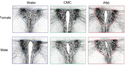
FIGURE 1. Effects of emulsifiers on vasopressin immunoreactivity in the PVN 8
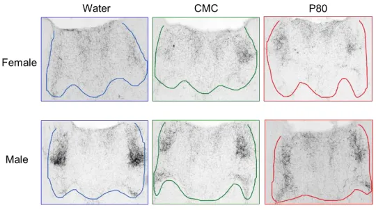
FIGURE 2. Effects of emulsifiers on vasopressin immunoreactivity in the PVT 9
INTRODUCTION
Microbiota are all the micro-organisms that colonize the internal and external surfaces of
the body (Montiel-Castro et al., 2013). The microbiota species that reside in the intestines or gut
play critical roles in digestion and absorption of food as well as in development of the immune
system. In addition, the gut microbiota has been shown to communicate bi-directionally with the
brain, via the microbiota-gut-brain axis. For example, there is a high comorbidity between
neurological and gastrointestinal disorders, which may be explained by this communication
pathway (Foster & Neufeld, 2013). Furthermore, patients with autism spectrum disorder (ASD)
show increased frequency of functional GI disorders, which are caused by altered
microbiome-gut-brain signals, specifically in the form of decreased tryptophan levels, increased serotonin
metabolite levels, and increased cytokine levels correlating with differences in microbiome
composition between patients with and without ASD (Luna et al., 2016).
Disruptions of the microbiota-gut-brain axis lead to significant changes in behavior. Mice
raised without microbiota, germ free (GF) mice, have an immature immune system, decreased
hypothalamic-pituitary axis stress response, and decreased anxiety-like behavior (De Palma et
al., 2014). GF mice also display reduced social behaviors (Desbonnet et al., 2014). In contrast,
mice with an increase in pro-inflammatory microbiota show increased anxiety-like behaviors
(Neufeld et al., 2011).
Dietary emulsifiers are detergent-like, ambiphillic molecules commonly added in
processed foods, such as ice cream, mayonnaise, margarine, and peanut butter, that prevent the
separation of the lipid and aqueous layers. It has been shown that dietary emulsifiers lead to the
proliferation of the pro-inflammatory microbiota, leading to symptoms of low-grade
(Chassaing et al., 2015). Two commonly found dietary emulsifiers are carboxymethylcellulose
(CMC) and polysorbate 80 (P80). These two emulsifiers also lead to robust effects in preliminary
behavior tests, such as in the open field test, elevated plus maze, and three-chambered sociability
test (Holder et al., 2016).
Further research in our lab has found that emulsifier administration increases anxiety-like
behaviors in both male and female mice and decreases social behaviors in female mice. These
sexually dimorphic behavior changes were seen with chronic low grade inflammation, increased
adiposity, and increased drive for feeding induced by dietary emulsifiers (Holder et al., 2016).
Although these physiological and behavior effects of dietary emulsifiers have been established, it
is unclear how administration of emulsifiers leads these effects. Perturbations in the gut
microbiome may induce these effects in several ways, from altered neurotransmitter or
neuropeptide levels, autonomic nervous system signaling, or changes in components of the
innate immune system (Li and Zhou, 2016).
A potential mechanism by which dietary emulsifiers may induce pathophysiological and
behavioral changes is via the vasopressin and oxytocin systems of the brain. Vasopressin (AVP),
or anti-diuretic hormone is expressed in a sexually dimorphic manner in the brain and has
numerous functions in the body including water and solute balance and stress response via the
hypothalamic-pituitary-adrenal (HPA) axis. Chronic stress can alter HPA axis activity, inducing
gut dysbiosis and changing the composition and function of the gut microbiota (Bienenstock et
al., 2015). Vasopressin is also released during chronic stress and may be a method by which
changes in the gut microbiome induce changes the brain. Oxytocin is a similar neuropeptide with
various functions, such as uterine contraction, milk letdown, energy homeostasis, and
bodies of neurons located in the paraventricular nucleus of the hypothalamus (PVN), a prominent
brain area involved in the stress response. Vasopressin neurons in the suprachiasmatic nucleus
(SCN) project to the paraventricular nucleus of the thalamus (PVT), an essential brain area in the
HPA axis that integrates multiple neural and humoral inputs to control food intake, energy
expenditure, and hormone levels (Fodor et al., 2013). Examining the AVP and OXT systems in
these specific brain areas would be beneficial to determine a possible mechanism of action of
dietary emulsifiers’ effects on behavior and the body.
Therefore, the overall research question is how does administration of two commonly
used dietary emulsifiers, CMC and P80, influence vasopressin and oxytocin, two neuropeptides
implemented in anxiety-like and social behavior? We predict that because vasopressin is a
neuropeptide released during stress and emulsifier-induced intestinal inflammation is a type of
stressor, vasopressin immunoreactivity in both the PVN and PVT will be increased in mice
treated with emulsifies. We also predicted that because oxytocin modulates social behaviors and
social behaviors are decreased during stress and following emulsifier treatment, oxytocin
METHODS
Animals
C57Bl/6 dams with litters (3 male and 3 female 14-day-old pups) were purchased from
Charles River Laboratories. Mice were housed in ventilated transparent OptiMouse plastic
cages with Bed-O-Cobs® and AlphaDri bedding (35.6 x 48.5 x 21.8cm; at Georgia State
University). Room lights were set to a 14h:10h light:dark cycle (lights off at 0900 ET), and
ambient temperature was maintained at 23°C. Food (Purina rodent chow no. 5001) and water
were available ad libitum. Mice were weaned postnatal day 21 (P21) and put into a new cage
(randomized to littermates) such that each experimental group contained mice from all litters and
that each litter was used for all experimental groups. All procedures were in accordance with the
Guide for Care and Use of Laboratory Animals and were approved by the Animal Care and Use
Committee at the Georgia State University.
Treatments
Upon weaning at age P21, male and female mice were transferred to new cages and
began treatment with water or a 1% concentrated solution of either sodium
carboxymethylcellulose (CMC) or Polysorbate 80 (P80) in the drinking water, forming six
different experimental groups of six animals each. Animals were monitored weekly, their body
weights were recorded, and they remained on treatment throughout the duration of the
experiment.
Behavioral Testing
After ten weeks on treatment starting at age P70, the animals underwent a standard
battery of behavior tests for anxiety-like, depression-like, and social behavior. One behavior test
open field test, elevated plus maze, light/dark box, marble burying test, three-chambered
sociability test, and forced swim test.
Tissue Collection
One week following the last behavior test, mice were deeply anesthetized under
isoflurane (5%v/v). Blood was collected by retrobulbar intraorbital capillary plexus.
Hemolysis-free serum was generated by centrifugation of blood using serum separator tubes (Becton
Dickinson, Franklin Lakes, NJ). Mice were euthanized by cervical dislocation, and the colons,
spleens, livers, and adipose were collected for subsequent analysis. Brains were removed and
submersed into a fixative solution of 5% acrolein in sodium phosphate buffer (0.1M, pH 7.4) at
4°C, followed by cryoprotection in 30% sucrose in phosphate buffered saline (PBS: 0.05M,
ph7.4). Brains were sectioned (30µm) in the coronal plane in a cryostat and stored in a
cryoprotectant solution (ethylene glycol/sucrose in sodium phosphate buffer) until
immunostained.
Immunohistochemistry
Immunohistochemistry (IHC) was conducted to visualize the immunoreactivity (IR) of
vasopressin (AVP) and oxytocin (OXT). The IHC process utilizes the natural and biological
function of antibodies binding to antigens to detect the presence of specific antigens, in this case
AVP and OXT, in brain areas of interest.
For the process of AVP immunohistochemistry, brain tissue underwent washes in 0.05 M
Tris-buffered saline (TBS) to remove all traces of the cryoprotectant, and then were incubated in
0.05 sodium citrate in TBS at 70°C, cooled, incubated in 0.1 M Glycine in TBS, and then in
blocking solution containing TBS, 10% normal goat serum (NGS), 0.4% Triton-X, and 1%
was incubated in rabbit anti-AVP primary antibody (Bachem, T-4563) overnight at a dilution of
1:46,000 in TTG, a solution containing 94% of 0.05 M TBS, 4% of 10% Triton-X, and 2% of
NGS. After this incubation, the series of tissue was rinsed in TTG and then incubated in
Biointylated goat-anti-rabbit IgG secondary antibody (Vector BA-7000) at a dilution of 1:800 in
TTG. Following rinses in TBS containing 0.4% Triton-X, the tissue was incubated in
avidin-biotin solution (Vector ABC-Elite standard kit) in TBS for 1 hour at room temperature. Next,
tissue underwent more TBS rinses and washes in sodium acetate solution, followed by a
30-minute incubation in DAB solution, containing sodium acetate buffer, nickel sulfate, 3, 3,
diaminobenzidine, and 5% hydrogen peroxide. After visualization, sections were rinsed in
sodium acetate buffer and TBS, the experimental sections were stored in TBS at 20°C until
mounting.
The process of OXT immunohistochemistry was a two-day protocol similar to that of
AVP IHC. Differences included no incubation in sodium citrate, and the tissue was incubated in
guinea pig anti-OXT primary antibody (Peninsula Labs) at a dilution of 1:12,000 in TTG, and
later in Biotinylated goat-anti guinea pig IgG secondary antibody (Vector BA-7000) at a dilution
of 1:800 in TTG. Upon completion of the DAB IHC protocol for OXT, sections were stored in
TBS at 20°C until mounting.
All experimental tissue was mounted rostro-caudally onto double-dipped, gelatin coated
slides. Slides were labeled to ensure unbiased analysis and allowed to dry overnight at room
temperature. The slides then underwent timed baths of increasing alcohol concentration to
dehydrate the tissue and then washes in xylene to clear the tissue of residual alcohol and fats.
They were coverslipped with glass coverslips using permount, allowed to dry overnight, then
Threshold Image Analysis
The processed sections were imaged with a photomicroscope at anatomically-matched
positions. Three to four images encompassing the PVN and PVT of the stained tissue were
captured for each animal. Microscope conditions were standardized to prevent variability
between images. Examples of these images for the various groups are included in the results
section. Next, photomicrographs were subjected to gray-level threshold analysis using a
previously-established algorithm in the Image J software (Murray et al., 2011; Rood et al., 2008).
The area of AVP or OXT-immunoreactivity (IR) in pixels was recorded for each image and the
average area was calculated for each subject.
Statistical Analysis
Data are represented as means + SEM or ± SEM where appropriate. The average IR of
each neuropeptide in each experimental group was determined and statistically analyzed using
repeated measures 2-way analysis of variance (ANOVA) where treatment and sex were the
RESULTS
Vasopressin
We hypothesized that administration of emulsifiers will increase AVP-IR in the PVN of
the brain. The average immunoreactivity area in pixels for each of the six groups was quantified.
There were no significant main effects of treatment (F(2, 29) = 1.802; P > 0.05) or sex (F(1, 29)
= 0.4303; P > 0.05). In addition, there was no interaction between sex and treatment (F(2,29) =
[image:18.612.182.435.264.395.2]0.06106; P > 0.05).
We hypothesized that administration of emulsifiers will increase AVP-IR in the PVT of
the brain. The average immunoreactivity area in pixels for each of the six groups was quantified.
There were no significant main effects of treatment (F(2, 29) = 0.6942; P > 0.05). There was a
significant main effect of sex observed (F (1, 29) = 5.429; p = 0.0270), in which females showed
significantly lower AVP-IR in the PVN compared to males. There was no interaction between
[image:19.612.170.440.245.396.2]sex and treatment (F(2,29) = 0.1601; p > 0.05).
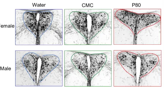
Oxytocin
We hypothesized that administration of emulsifiers will decrease OXT-IR in the PVN of
the brain. The average immunoreactivity area in pixels for each of the six groups was quantified.
There were no significant main effects of treatment (F(2, 29) = 0.8444; p > 0.05) or sex (F(1, 29)
= 0.08876; p > 0.05). In addition, there was no interaction between sex and treatment (F(2,29) =
[image:20.612.173.450.245.394.2]0.6430; p > 0.05).
DISCUSSION
Our results demonstrated that there were no significant effects of emulsifier treatment on
the AVP-IR and OXT-IR in the PVN and PVT areas of the mouse brain. Thus, our hypothesis
that administration of emulsifiers would increase AVP-IR in the PVN and PVT and decrease
OXT-IR in the PVN was refuted. This was a surprising finding as previously discovered effects
of emulsifier treatment on anxiety-like and social behavior indicated changes that may be linked
to alterations in the vasopressin and oxytocin systems of the brain. There are several
explanations and future directions for these results, including expanding on the various brain
areas analyzed and analysis using the age and developmental stage of the animals.
The PVN and PVT were two brain areas chosen for analysis due to their prominent
functions in the vasopressin and oxytocin systems and in stress-related and social behavior. The
lack of significant effects of emulsifier treatment in the PVN and PVT of the mice may indicate
the need to study other brain areas, such as the amygdala and bed nuclei of the stria terminalis
(BNST). The central amygdala has been shown to be critical in the processing of anxiety and
fear responses to stress. AVP is also synthesized in the medial amygdala (Smith et al., 2017) and
there are well-documented AVP projections to the amygdala from AVP-containing
magnocellular neurosecretory neurons (Hernández et al., 2016). Both AVP and OXT levels are
increased in the amygdala in response to stress (Dabrowska et al., 2011). Due to profound
behavior effects that AVP likely exerts in the amygdala, this area should be investigated as a
region where emulsifiers may induce changes in AVP. The BNST is another brain area involved
in the stress response, synthesizes AVP (Smith et al., 2017), and contains one of the highest
levels of OXT receptor mRNA. There is also anatomical support for the existence of direct
may be another region of action for stress-induced changes in OXT expression (Dabrowska et
al., 2011). OXT-IR in the PVT was not able to be analyzed due to time constraints, but should
also be performed to determine differences in this area. Although no significant differences were
found in the PVN and PVT from this study, other brain areas should be analyzed to determine
possible region-specific influences of microbiota on AVP and OXT immunoreactivity.
Furthermore, the lack of significant effects of emulsifier treatment on AVP-IR and
OXT-IR may be due to the developmental stage of the animals tested. In this experiment, there was
only one time schedule of treatment and all animals were administered a constant level of
emulsifiers throughout their entire development. In addition, the effects of dietary emulsifiers
were only determined at adulthood. It is possible that emulsifiers demonstrate their effects at
earlier time periods of development, such as neonatally or at juvenile age and that these effects
are abolished after the onset of puberty or during adulthood. Developmental differences in AVP
and OXT regulation of social behavior has been shown in that juvenile rats aged P35
demonstrate increased social behavior compared to their adult counterparts, and AVP and OXT
receptor binding density was significantly higher in juveniles than in adults in several brain areas
(Smith et al., 2017). Future studies can test the effects of these emulsifiers across various ages
through different scheduling of administration from birth through adulthood to pinpoint
developmental effects of emulsifier-induced intestinal inflammation.
Although there were no significant main effects of treatment on AVP-IR and OXT-IR in
the brain areas studied, there was a significant main effect of sex in the AVP-IR in the PVT, in
that males had increased AVP-IR than females. This result indicates a basal difference in AVP
expression in this area, unaffected by emulsifier treatment. Although this sex difference in this
the same manner in the BNST, and AVP-IR projections from the BNST to the lateral septum,
lateral habenular nucleus, and periaqueductal gray was significantly higher in males than in
females (De Vries and al-Shamma, 1990). In addition, conventionally colonized Swiss Webster
mice demonstrate a sex difference in AVP-IR in the PVN with males showing increased AVP-IR
compared to females (Mehta et al., 2015). It would be interesting to see whether OXT-IR
demonstrates a similar basal sex difference in the PVT.
In conclusion, this research did not reveal the vasopressin and oxytocin systems as
possible molecular mechanism for the effects of emulsifier treatment on physiological and
behavior changes in mice. Further findings in this field may uncover an underlying neural
mechanism for neurological and gastrointestinal diseases, leading to possible targets for
REFERENCES
Bienenstock, J., Kunze, W., & Forsythe, P. (2015). Microbiota and the gut–brain axis. Nutrition
reviews, 73(suppl 1), 28-31.
Borre, Y. E., O’Keeffe, G. W., Clarke, G., Stanton, C., Dinan, T. G., & Cryan, J. F. (2014).
Microbiota and neurodevelopmental windows: implications for brain disorders. Trends in
molecular medicine, 20(9), 509-518.
Chassaing, B., Aitken, J. D., Malleshappa, M., & Vijay-Kumar, M. (2014). Dextran Sulfate
Sodium (DSS)-Induced Colitis in Mice. Current Protocols in Immunology / Edited by
John E. Coligan ... [et Al.], 104, Unit–15.25.
http://doi.org/10.1002/0471142735.im1525s104
Chassaing, B., Koren, O., Goodrich, J., Poole, A., Srinivasan, S., Ley, R. E., & Gewirtz, A. T.
(2015). Dietary emulsifiers impact the mouse gut microbiota promoting colitis and
metabolic syndrome. Nature, 519(7541), 92–96.
http://doi.org.ezproxy.gsu.edu/10.1038/nature14232
Dabrowska, J., Hazra, R., Ahern, T. H., Guo, J.-D., McDonald, A. J., Mascagni, F., … Rainnie,
D. G. (2011). Neuroanatomical evidence for reciprocal regulation of the corticotrophin
releasing factor and oxytocin systems in the hypothalamus and the bed nucleus of the
stria terminalis of the rat: Implications for balancing stress and
affect. Psychoneuroendocrinology, 36(9), 1312–1326.
http://doi.org.ezproxy.gsu.edu/10.1016/j.psyneuen.2011.03.003
De Palma, G., Collins, S. M., & Bercik, P. (2014). The microbiota-gut-brain axis in functional
gastrointestinal disorders. Gut Microbes, 5(3), 419–429.
Desbonnet, L., Clarke, G., Shanahan, F., Dinan, T. G., & Cryan, J. F. (2014). Microbiota is
essential for social development in the mouse. Molecular Psychiatry, 19(2), 146–148.
http://doi.org.ezproxy.gsu.edu/10.1038/mp.2013.65
De Vries GJ, al-Shamma HA. Sex differences in hormonal responses of vasopressin pathways in
the rat brain. J Neurobiol. 1990;21:686–693. doi: 10.1002/neu.480210503.
Fodor, A., Pintér, O., Domokos, Á., Langnaese, K., Barna, I., Engelmann, M., & Zelena, D.
(2013). Blunted HPA axis response in lactating, vasopressin-deficient Brattleboro
rats. Journal of Endocrinology, 219(2), 89-100.
Foster, J. A., & Neufeld, K. A. M. (2013). Gut–brain axis: how the microbiome influences
anxiety and depression. Trends in neurosciences,36(5), 305-312.
Hernández, V. S., Hernández, O. R., Perez de la Mora, M., Gómora, M. J., Fuxe, K., Eiden, L.
E., & Zhang, L. (2016). Hypothalamic Vasopressinergic Projections Innervate Central
Amygdala GABAergic Neurons: Implications for Anxiety and Stress Coping. Frontiers
in Neural Circuits, 10, 92. http://doi.org.ezproxy.gsu.edu/10.3389/fncir.2016.00092
K.D. Mehta, M.K. Holder, N.V. Peters, G.J. de Vries. Effects of microbiota on vasopressin and
microglia in swiss webster mice. Poster presented in Atlanta, GA: Brain and Behavior
Summer Symposium, 2015.
Li, Q., & Zhou, J. M. (2016). The microbiota–gut–brain axis and its potential therapeutic role in
autism spectrum disorder. Neuroscience, 324, 131-139.
Luna, R. A., Oezguen, N., Balderas, M., Venkatachalam, A., Runge, J. K., Versalovic, J., …
Williams, K. C. (2017). Distinct Microbiome-Neuroimmune Signatures Correlate
and Molecular Gastroenterology and Hepatology, 3(2), 218–230.
http://doi.org.ezproxy.gsu.edu/10.1016/j.jcmgh.2016.11.008
M.K. Holder,B. Chassaing, N.V. Peters, J. Whylings, A. Gewirtz, G.J. deVries. Sex differences
in the effects of dietary emulsifiers on physiology and behavior in mice.Program No.
446.04 2016 Neuroscience Meeting Planner San Diego: Society for Neuroscience, 2016
Montiel-Castro, A. J., González-Cervantes, R. M., Bravo-Ruiseco, G., & Pacheco-López, G.
(2013). The microbiota–gut–brain axis: neurobehavioral correlates, health and
sociality. Beyond the borders: The gates and fences of Neuroimmune interaction, 7,
63-78.
Murray, E. K., Varnum, M. M., Fernandez, J. L., de Vries, G. J., & Forger, N. G. (2011). Effects
of neonatal treatment with valproic acid on vasopressin immunoreactivity and olfactory
behaviour in mice. Journal of neuroendocrinology, 23(10), 906-914.
Neufeld, K. M., Kang, N., Bienenstock, J., & Foster, J. A. (2011). Reduced anxiety‐like behavior
and central neurochemical change in germ‐free mice. Neurogastroenterology &
Motility, 23(3), 255-e119.
Rood, B. D., Murray, E. K., Laroche, J., Yang, M. K., Blaustein, J. D., & De Vries, G. J. (2008).
Absence of progestin receptors alters distribution of vasopressin fibers but not sexual
differentiation of vasopressin system in mice. Neuroscience, 154(3), 911-921.
Smith, C. J. W., Poehlmann, M. L., Li, S., Ratnaseelan, A. M., Bredewold, R., & Veenema, A.
H. (2017). Age and sex differences in oxytocin and vasopressin V1a receptor binding
densities in the rat brain: focus on the social decision-making network. Brain Structure &