Georgia State University
ScholarWorks @ Georgia State University
Nutrition Theses Department of Nutrition
Fall 11-16-2017
Synergistic Effects of d-Tocotrienol and
Xanthorrhizol in Prostate Cancer Cells
Chappell MadhaniFollow this and additional works at:https://scholarworks.gsu.edu/nutrition_theses
This Thesis is brought to you for free and open access by the Department of Nutrition at ScholarWorks @ Georgia State University. It has been accepted for inclusion in Nutrition Theses by an authorized administrator of ScholarWorks @ Georgia State University. For more information, please contact
Recommended Citation
Madhani, Chappell, "Synergistic Effects of d-Tocotrienol and Xanthorrhizol in Prostate Cancer Cells." Thesis, Georgia State University, 2017.
NOTICE TO BORROWERS
All theses deposited in the Georgia State University library must be used in accordance with the stipulations prescribed by the author in the preceding statement. The author of this thesis is:
Chappell Rebecca Madhani 3071 Lenox Road, Unit 13
Atlanta, GA 30324
The director of this thesis is:
Huanbiao Mo, PhD Chair and Professor Department of Nutrition
Byrdine F. Lewis College of Nursing and Health Professions Georgia State University
VITA
Chappell Rebecca Madhani
ADDRESS: 3071 Lenox Road, Unit 13 Atlanta, GA 30324
EDUCATION: M.S. 2017 Georgia State University Nutrition
B.S. 2012 Kennesaw State University Anthropology
PROFESSIONAL EXPERIENCE:
Senior Server, Trainer 2013-2014
Bistro Niko, Atlanta, GA
Laboratory and Field Technician - Intern 2012
Edwards Pitman Environmental, Inc., Smyrna, GA
Senior Server, Trainer 2010-2013
Outback Steakhouse, Roswell, GA
Office Assistant 2009-2010
River City Legal Group, Chattanooga, TN
PROFESSIONAL SOCIETIES AND ORGANIZATIONS:
Academy of Nutrition and Dietetics 2014-present
Georgia Dietetic Association 2014-present
Greater Atlanta Dietetic Association 2014-present
Nutrition Student Network, Georgia State University 2014-2015
AWARDS AND PUBLICATIONS:
Madhani, R., Todd, J., Mo, H. (2016) 2016 Dietary guidelines for Americans. 1:16 21. Publisher: China National Cereals, Oils & Foodstuffs Corporation Co. Ltd.
Dean’s List – Georgia State University 2014
ABSTRACT
SYNERGISTIC EFFECTS OF DELTA- δ-TOCOTRIENOL AND XANTHORRHIZOL IN PROSTATE CANCER CELLS
by
C. Rebecca Madhani
Background: Approximately one in seven American men will be diagnosed with
prostate cancer during their lifetime. The mevalonate pathway produces essential
intermediates for the post-translational prenylation and dolichylation of
growth-associated proteins including Ras, nuclear lamins and growth factor receptors.
Dysregulation of 3-hydroxy-3-methylglutaryl coenzyme A (HMG-CoA) reductase, the
rate-limiting enzyme of the mevalonate pathway, in prostate cancer cells supports tumor
growth and therefore can be targeted for prostate cancer prevention and therapy. Previous
studies have shown that isoprenoids including tocotrienols and xanthorrhizol suppress the
growth of prostate cancer cells with concomitant downregulation of HMG CoA
reductase.
Objective: To determine the synergistic effects of d-δ-tocotrienol and xanthorrhizol on
growth of human DU-145 prostate carcinoma cells.
Methods: DU-145 cells were incubated with d-δ-tocotrienol and xanthorrhizol,
individually and in blends, for 72 hours before viable cells were quantified by CellTiter
96® Aqueous One Solution. The impacts of these compounds, individually and in
expression of cell cycle related proteins in DU-145 cells were evaluated by flow
cytometry and Western-blot in follow-up studies.
Statistical analysis: All experiments were repeated 3 times. One-way analysis of
variance (ANOVA) was performed to assess the differences between groups using Prism
Ⓡ
4.0 software (GraphPad Software Inc., San Diego, CA). Differences in means was
assessed using Tukey’s test. Levels of significance are indicated as P < 0.05.
Results: Blends of d-δ-tocotrienol and xanthorrhizol showed greater inhibition of cell
growth than those of individual compounds. Current results did not show a significant
change in cell cycle distribution or down regulation of Cdk4 and cyclin D1 with the
combination of compounds. The level of procaspase-3 for apoptosis initiation was also
not altered.
Conclusion: Further studies are warranted to confirm these initial findings and
understand the mechanisms underlying the combined effect of d-δ-tocotrienol and
SYNERGISTIC EFFECTS OF DELTA- δ-TOCOTRIENOL AND XANTHORRHIZOL IN PROSTATE CANCER CELLS
by
C. Rebecca Madhani
A Thesis
Presented in Partial Fulfillment of Requirements for the Degree of
Master of Science in Health Sciences
The Byrdine F. Lewis College of Nursing and Health Professions
Department of Nutrition
Georgia State University
Atlanta, Georgia
ii
ACKNOWLEDGMENTS
I am deeply grateful to Huanbiao Mo, Ph.D. for sharing his lab and research, and
for allowing me to flourish as a student under his direction. I would also like to thank Dr.
Mo for introducing me to the fascinating world of tocotrienols, xanthorrhizol, and
nutritional cancer therapy in vitro. This opportunity has intensified my interest in
research and appreciation for time and work that is required for research. I would like to
thank Desiree Wanders, Ph.D. and Weiming Xia, Ph.D. for taking time to serve on my
committee and for offering their expertise.
Thank you to Rafaela Feresin, Ph.D. for sharing her western blot procedure and
best practices. I also appreciated her effort to organize and streamline our lab for a more
efficient work space. Thank you to Dr. Xiangming Ji, Ph.D. for sharing his expertise on
western blot and for suggesting the best companies from which to order antibodies. A big
thank you to Manal Elfakhani and Sophie Yount for guiding me through laboratory
methods and procedures. I would also like to thank both for helping me maintain the
DU-145 cells and procedures during my rotations outside of the lab. Their efforts were
integral to the completion of this project. Thank you to Darren Chan for sharing his
progress and troubleshooting with me. I would also like to thank him for discovering and
researching FCS Express for analysis of our cell cycle data. Finally, a big thank you to
Shaligram Sharma for always taking the time to answer my questions and for providing
iii
TABLE OF CONTENTS
List of Figures ... iv
Abbreviations ...v
Chapter I. INTRODUCTION ...1
Hypothesis and Specific Aims……….2
II. LITERATURE REVIEW ...3
Metabolic Regulation and Cancer Therapies ...3
Tocotrienol and Cancer Inhibition ...5
Xanthorrhizol and Cancer Inhibition ...7
III. METHODS AND PROCEDURES………... 10
IV. RESULTS……… ...15
V. DISCUSSION AND CONCLUSIONS ………. ...23
iv
LIST OF FIGURES
Figure Page
1. DU-145 Cell Viability ...16
2. DU-145 Cell Cycle Analysis ...18
a. G1 Phase ...18
b. S Phase ...18
c. G2 Phase ...19
3. DU-145 Western Blot Analysis ...20
a. Cdk4 ...20
b. Cyclin D1 ...21
v
ABBREVIATIONS
DU-145 human prostate cancer cells
d-δ delta
T tocotrienol
X xanthorrhizol
Cdk4 cyclin dependent kinase 4
E2F E2 transcription factor
RB retinoblastoma
P53 tumor protein p53
Ink4a A family of cyclin-dependent kinase inhibitors
FPP farnesyl pyrophosphate
GGPP geranylgeranyl pyrophosphate
GTPase hydrolase enzyme
HMG-CoA 3-hydroxy-3-methyl-glutaryl-CoA
TRF Tocotrienol rich fraction
DNA Deoxyribonucleic acid
mL milliliter
µL microliter
µM micromolar
vi x g times gravity
CO2 carbon dioxide
oC degrees Celsius
cm2 centimeters squared
RPMI Roswell Park Memorial Institute
FBS Fetal Bovine Serum
1x HBSS 1x Hank’s Balanced Salt Solution
1x PBS 1x Phosphate Buffer Solution
V volts
PVDF Immun-Blot Polyvinylidene difluoride
PBST Phosphate Buffered Saline containing 0.1% Tween-20 solution
ANOVA One-way analysis of variance
1 CHAPTER I
SYNERGISTIC EFFECTS OF DELTA- δ-TOCOTRIENOL AND XANTHORRHIZOL IN PROSTATE CANCER CELLS
Introduction
Approximately one in seven American men will be diagnosed with prostate
cancer during their lifetime. In 2017, the Centers for Disease Control and Prevention
reported 161,360North American men diagnosed with prostate cancer and 26,730 deaths
from prostate cancer. (1) Prostate cancer is currently treated with methods such as
chemotherapy, radiation, and surgery. Radiation and surgery may cause side effects such
as infections, impotence, or bladder and bowel problems.(2)Chemotherapy is
accompanied by disconcerting symptoms including loss of appetite, mouth sores, fatigue,
nausea, vomiting, diarrhea, and hair loss. In addition, common chemotherapy agents
including Docetaxel, Mitoxantrone, and Estramustine may put patients at risk for
peripheral neuropathy, leukemia, and blood clots, respectively. (3) Therefore, alternative
therapies are necessary for the treatment of prostate cancer.
Two dietary factors that could potentially serve as therapeutic agents for prostate
cancer are d-δ-tocotrienol and xanthorrhizol. d-δ-Tocotrienol, a minor form of vitamin E,
has been found to decrease cancer cell proliferation by inhibiting HMG-CoA reductase in
the mevalonate pathway, thereby preventing prenylation or dolichylation of
2
cell cycle arrest in the G1 phases with a concomitant inhibition of Cdk4 and caspase-3
activation and apoptosis in melanoma cells. (5) Xanthorrhizol is a bisabolane-type
sesquiterpene that has also exhibited anti-cancer effects by inducing cell cycle arrest
through decreased expression of Cyclin D1 and Cdk4, and apoptosis by activation of
procaspases 3 & 9 in colon cancer cells. (6)
Hypothesis and Specific Aims
Hypothesis: d-δ-Tocotrienol and xanthorrhizol synergistically suppress the
proliferation of DU-145 prostate carcinoma cells by inducing cell cycle arrest and
apoptosis.
Specific aim 1: To determine the effects of d-δ-tocotrienol and xanthorrhizol,
individually and in combination, on prostate cancer cell proliferation.
Specific aim 2: To determine the effects of d-δ-tocotrienol and xanthorrhizol,
individually and in combination, on cell cycle progression and cell cycle-related
proteins of prostate cancer cells.
Specific aim 3: To determine the effects of d-δ-tocotrienol and xanthorrhizol,
individually and in combination, on an apoptosis-related protein, procaspase-3, of
3 CHAPTER II
Literature Review
Metabolic Regulation and Cancer Therapies
The RB pathway plays a vital role in cell proliferation by activating and
suppressing growth related factors, mediating entry of cells into the cell cycle. The RB
pathway consists of five protein families including D-type cyclins, CDKN, (Ink4a),
E2F-transcription factors, cyclin-dependent protein kinases (e.g. Cdk4, Cdk6) and RB-family
pocket of proteins. (7)Cdk4 and Cdk6 are catalytic components that bind to D-cyclins to
form active kinase complexes and initiate cell progression through the cell cycle.(8)
Ink4a proteins inhibit the activity of Cdk4 and Cdk6 by competing with cyclin D1 to
suppress creation of the active cyclin-D1/Cdk4/6 kinase complex, which is activated
during normal cell cycle entry and proliferation. (9)Other cellular targets of the
D-cyclin/Cdk4/6 complexes are RB-proteins, which control chromatin structures and
E2F-transcription factor activities.(8)
The E2-factor is a family of transcription factors that are downstream effectors of
the RB pathway. E2F play important roles in entry of the S phase of the cell cycle(10)
and p53-dependent and p53-independent apoptosis.(11) RB-pocket proteins can be
directed to E2F-regulated promoters, thereby inhibiting transcription by suppressing E2F
transactivation. The D-cyclin/Cdk4/6 complex can phosphorylate RB-pocket proteins and
disrupt RB-E2F interaction, prompting activation of E2F-regulated gene expression.In
4
by failing to suppress E2F activity. Consequently, the D-cyclin/Cdk4/6 kinase
complex stimulates tumor cell proliferation, however Ink4a proteins operate as tumor
suppressors by inhibiting D-cyclins.(7)
Apoptosis is a regulated cellular suicide that is necessary to remove damaged
cells. An accumulation of evidence has shown mitochondria to be the major organelles
involved in apoptosis, through processes including DNA damage, growth factor down
regulation, and chemotherapeutic drugs. (12) Apoptosis is promoted through increased
E2F activity, which enhances the expression of pro-apoptotic genes such as caspases.
Caspases are cysteine proteases that are activated in apoptotic cells. Caspases 2, 8, and 9
are initiator caspases that cleave downstream effectors caspases 3, 6, and 7. Effector
caspases execute apoptosis by cleaving cellular proteins. (13)
The mevalonate pathway plays important roles in cell proliferation and cell death.
Intermediates of the mevalonate pathway, including farnesyl pyrophosphate (FPP) and
geranylgeranyl pyrophosphate (GGPP), are required for the prenylation, membrane
attachment, and biological activities of many growth-related proteins and dolichylation of
growth factor receptors. Among the growth-associated proteins that are prenylated are the
nuclear lamins and the small GTPases, including the Ras protein, that are involved in
signaling regulating cell division. (14)
The importance of mevalonate pathway intermediates in cell proliferation and cell
death renders the pathway a useful target for prostate cancer therapy. One approach is to
suppress the rate-limiting enzyme, 3-hydroxy-3-methylglutaryl coenzyme A (HMG-CoA)
reductase, of the pathway. (15) In eukaryotic cells, HMG CoA reductase is regulated at
non-sterol-5
mediated posttranscriptional downregulation; the latter includes ubiquitination and
proteasome-mediated degradation of HMG CoA reductase.(7) One class of HMG CoA
reductase inhibitors is statins. Statins are competitive inhibitors of HMG CoA reductase
and are widely prescribed for their hypocholesterolemic effect.(14)
Tocotrienol and Cancer Inhibition
Tumor cell proliferation is suppressed by down regulation of HMG-CoA
reductase, which is the committed step for the biosynthesis of sterols such as cholesterol
through the mevalonate pathway.In contrast, tocotrienol has been shown to down
regulate HMG CoA reductase, thereby inhibiting tumor cell proliferation. (14)
In addition to the statins mentioned above, tocotrienols are another class of HMG
CoA suppressors. Tocotrienols, a minor form of vitamin E, include four chemical forms
(ɑ-, β-, ɣ-, and δ-tocotrienol). Tocotrienols vary by the number and position of methyl
groups in the polar head of the molecules. Tocotrienol is derived from dietary sources
including palm oil, red annatto seeds, grape seed oil, flaxseed oil, buckthorn berry, rye,
oat, and barley.Tocotrienol studies began in the early 1990’s and focused on regulation
of cholesterol production via post-transcriptional suppression of HMG CoA reductase.
(16)Tocotrienols have been found to mimic sterols by promoting ubiquitination and
degradation of HMG-CoA reductase.(4) Over the past two decades, tocotrienol has been
found to suppress the proliferation of tumor cells originated from various tissues,
including colon, breast, prostate, skin, liver, lung, lymph gland, cervix, and nerve.(17)
Har et al. examined the effects of tocotrienol in BNL CL.2 normal liver cells and
6
liver cancer cells compared to normal liver cells in all experiments. Tocotrienol
significantly reduced cell viability of liver cancer cells in a dose-dependent manner.
Elevated caspase-3 activity was found in liver cancer cells in a time dependent manner
following 9, 12, and 24 hour treatments of tocotrienol. Finally, 24-hour tocotrienol
treatment induced DNA fragmentation in liver cancer cells compared to untreated cells.
These data suggest that tocotrienol decreases viability in 1ME A.7R.1 liver cancer cells
through apoptotic activity of caspase-3. (18)
Fernandes et al. looked at the effect of d-δ-tocotrienol in A2058 and A375 human
melanoma cells. d-δ-Tocotrienol caused a dose-dependent inhibition of cell proliferation
in both A2058 and A375 cell lines. Cell cycle analysis demonstrated significant increases
in the percentage of cells in the G1 phase for both cell lines after treatment with
d-δ-tocotrienol. d-δ-Tocotrienol (24 μM) raised the percentage of A2058 cells in the G1
phase from 46% to 63% (P < 0.05). Concomitantly, S% decreased from 35% to 20% (P <
0.05). G1 arrest was also seen in A375 cells treated with 33 μM d-δ-tocotrienol. G1 arrest
was congruent with a significant decrease in expression of Cdk4, a vital catalyst in the
transition from G1 to S phase, for both cell lines. (5)
A concentration-dependent cleavage of procaspase-3 was initiated by
d-δ-tocotrienol in A2058 cells using western blot analysis. Cleavage of procaspase-3 is shown
through lighter bands in western blot analysis. These data suggest that d-δ-tocotrienol
induces cell cycle arrest in A2058 and A375 cells and initiates caspase-3 activation and
apoptosis in A-2058 human melanoma cells.(5)
Srivastava and Gupta found significant growth inhibition of DU-145 human
7
14.77% tocotrienol, 33.97% γ-tocotrienol, 26.11% d-δ-tocotrienol, 18.10%
α-tocopherol, and 7.05% of other tocotrienol-like compounds. DU-145 cell growth
decreased by 17.9%, 20.1%, 46.5%, 78.3%, and 79.8%, after treatment with 5, 10, 20, 40,
and 80 µg/ml of TRF, respectively. In addition, TRF caused a dose-dependent increase of
the percentage of DU-145 cells in the G0/G1 phase, compared to the control. A
concomitant decrease in the percentage of cells in the S phase of cell cycle was shown,
compared to the control. These data suggest that TRF caused a dose-dependent decrease
in cell growth and increase in cell cycle arrest at the G0/G1 phase in DU-145 cells. (19)
Xanthorrhizol and Cancer Inhibition
Sesquiterpenes are a class of isoprenoids that have been shown to suppress the
mevalonate pathway and inhibit the biosynthesis of essential intermediates required for
the function of growth related proteins such as Cdk4. Consequently,
sesquiterpene-mediated downregulation of HMG CoA reductase induces cell cycle arrest and initiates
apoptosis.(14)
Xanthorrhizol is a sesquiterpene derived from Curcuma xanthorrhiza rhizome,
also known as Javanese turmeric. Xanthorrhizol is native to Indonesia and has exhibited a
variety of health benefits, including anti-inflammatory, antimicrobial, antioxidant,
hyperglycemic, antihypertensive, antiplatelet, nephroprotective, estrogenic, and
anticancer properties. It has been speculated that xanthorrhizol suppresses carcinogenesis
by delivering cytotoxic effects from its phenol group. Xanthorrhizol’s anti-cancer effects
8
Cheah et al. found a concentration-dependent growth inhibition in MDAMB-231
human breast cancer cells, caused by 48 hour treatments of xanthorrhizol. Apoptotic
levels of MDAMB-231 cells was measured using an apoptotic index after 24 hours of
treatment with xanthorrhizol. A significant increase (p<0.005) of apoptosis was found, in
a dose-dependent manner. Finally, caspase activity of MDAMB-231 cells after
xanthorrhizol treatment was also assessed, which found a significant increase (p<0.005)
in caspase-3 activity compared to control cells.These data suggest that xanthorrhizol
induced growth inhibition and apoptosis by activation of caspase-3 in MDAMB-231
human breast cancer cells.(20)
Kang et al. found that 24-hour treatments of xanthorrhizol inhibited growth and
increased percentage of HCT-116 human colon cancer cells at the G0/G1 phase of the
cell cycle, compared to control. Percentage of HCT-116 cells in the S phase also
decreased compared to the control. Concomitantly cyclin D1 and Cdk4 were
downregulated in a time- and dose-dependent manner. As previously mentioned, cyclin
D1 and Cdk4 form the active kinase complex for transition of cells from the G1 to S
phases of cell cycle. This suggests cell cycle arrest of the G0/G1 phase induced by
xanthorrhizol by down regulation of cyclin D1 and Cdk4. (21)
Kang et al. also found evidence of apoptosis through DNA fragmentation,
activation of procaspases 3 & 9, release of cytochrome c, and cleavage of
poly-(ADP-ribose) polymerase. DNA fragmentation was determined using an agarose gel
electrophoresis with 24 and 48 hour treatments of xanthorrhizol. Apoptosis was detected
in a time- and dose-dependent manner. Apoptosis was also detected using western blot
9
hour xanthorrhizol treatments. Additionally, expression of procaspase-3 was decreased in
a time- and dose-dependent manner. (21)
Handayani et al. examined the effects of xanthorrhizol in HepG2 human liver
cancer cells. IC50 values were used to conduct cytotoxicity assays; increasing
concentrations of xanthorrhizol was found to inhibit HepG2 cell growth in a dose
dependent manner. Apoptotic levels of HepG2 cells was measured using an apoptotic
index after 24, 48, and 72 hours of treatment with xanthorrhizol. The percentage of
apoptotic cells was increased to more than 70% in all treatment groups compared to the
less than 5% in the control. Western blot analysis of apoptosis-related proteins after 24,
48, and 72 hour treatments of xanthorrhizol showed a time-dependent decrease in
procaspase-3 and increased expression of caspase-3. (22)
In summary, tocotrienol has shown anti-proliferative effects in BNL 1ME A.7R.1
liver(18), A2058 and A375 melanoma(5), and DU-145 prostate(19) cancer cells.
Tocotrienol induced apoptotic events in BNL 1ME A.7R.1 liver cells(18) and A2058
melanoma cells(5). Tocotrienol also induced cell cycle arrest in DU-145 prostate cancer
cells(19) and A2058 and A375 melanoma cells(5). Xanthorrhizol has shown
anti-proliferative effects in MDAMB-231 breast(20), HCT-116 colon(21), and HepG2 liver
(18) cancer cells, but not prostate cancer. Xanthorrhizol has exhibited apoptotic events in
MDAMB-231 breast(20) and HepG2 liver(18) cancer cells, and both apoptosis and cell
cycle arrest in HCT-116 colon cancer cells(21). No studies to date have looked at the
10 CHAPTER III
Methods and Procedures
Materials
Human DU-145 prostate carcinoma cells were received as a gift from the Biology
Department at Georgia State University Biology and were used in all procedures. All
supplies needed for cell culturing were purchased from Fisher Scientific, Inc. (Pittsburgh,
PA) and Thermo Scientific (Waltham, MA). Xanthorrhizol and d-δ-tocotrienol were
received as a gift from American River Nutrition, Inc. (Hadley, MA).
Cell Culture
DU-145 cells were cultured in Roswell Park Memorial Institute (RPMI-1460)
media and supplemented with 10% Fetal Bovine Serum (FBS), 1%
penicillin-streptomycin, and 0.8% gentamicin in 25 cm2 flasks. Cells were then incubated for 48
hours at 37oC in a humidified atmosphere of 5% CO2. At 48 hours, cells were subcultured
into a 175 cm2 flask, grown to 80 - 90% confluence, and then prepared according to
experimental procedure. Nine treatments were used for each experiment, as follows:
control (C), d-δ-tocotrienol 5 µM (T5), d-δ-tocotrienol 10 µM (T10), xanthorrhizol 16.25
µM (X16.25), xanthorrhizol 32.5 µM (X32.5), d-δ-tocotrienol 5 µM & xanthorrhizol
16.25 µM (T5/X16.25), tocotrienol 5 µM & xanthorrhizol 32.5 µM (T5/X32.5),
d-δ-tocotrienol 10 µM & xanthorrhizol 16.25 µM (T10/X16.25), d-δ-d-δ-tocotrienol 10 µM &
xanthorrhizol 32.5 µM (T10/X32.5). Each treatment was dissolved in ethyl alcohol, with
11
Cell Proliferation
Cell titer was performed to quantify the effects of d-δ-tocotrienol, xanthorrhizol,
and a combination of both on DU-145 cell proliferation. Human prostate cancer
(DU-145) cells were cultured as previously described and seeded in 96-well plates at 1,500
cells per well. Cells were treated at 0.1% volume for 72 hours with respective treatments
of xanthorrhizol and d-δ-tocotrienol. At 72 hours, complete RPMI media was aspirated
from wells; cells were then washed with 1x Hank’s Balanced Salt Solution (1x HBSS),
and refreshed with 100 µL serum-free RPMI media and 20 µL Cell Titer 96® Aqueous
One Solution. Cells were then incubated for 2 hours at 37°C, 5% CO2. At 2 hours, the
plate was measured using a Biotek, Synergy HT plate reader; results were analyzed using
the Gen5TM software from Biotek® Instruments, Inc. (Winooski, VT, USA).
Cell Cycle Analysis
Xanthorrhizol and d-δ-tocotrienol were tested for their effects on cell cycle
distribution in DU-145 cells. Cells were seeded in 25 cm2 flasks (Becton Dickinson
Labware, Franklin Lakes, NJ) at 1.5x10⁶ cells per flask with 3 mL RPMI medium per
flask and incubated for 24 hours. At 24 hours, medium was aspirated and replaced with
fresh medium containing respective treatments of xanthorrhizol and d-δ-tocotrienol. After
24 hours of incubation, cells were harvested by trypsinization and pelleted by
centrifugation at 13,000 x g for 5 minutes. Cell pellets were then fixed in 1 mL of 70%
ethanol at -20 degrees Celsius overnight. In preparation for analysis, cells were washed
twice with a 1x Phosphate Buffer Solution (1x PBS) and centrifuged at 5 minutes after
each wash. Cells were re-suspended in 500 µL of 1x PBS containing RNase A
12
µL of a PBS-TritonX100-propidium iodide solution was added to each sample and held
in the dark, at room temperature for 15 minutes. Stained cell samples were divided into
200 µL aliquots and analyzed for DNA content using a BD LSR Fortessa flow cytometer
(BD Biosciences, San Jose, CA, USA). The distribution of cells in the G1, S, and G2
phases of the cell cycle was determined using FCS Express software (De Novo Software,
Glendale, CA).
Western Blot Analysis
Western-blot was performed to examine the proteins Cdk4, Cyclin D-1, and
Procaspase-3. Protein concentration of DU-145 cells treated with respective treatments of
xanthorrhizol and d-δ-tocotrienol was determined using a bicinchoninic acid protein
assay kit (Pierce, Rockford, IL, USA). Treatment samples containing 10 µg of protein
was mixed with 4x Laemmli Sample Buffer (Bio-Rad Laboratories, Hercules, CA, USA)
at a 1:1 ratio (v/v) and β-Mercaptoethanol before loading into a 12%
SDS-polyacrylamide gel. Gels were then loaded onto a Mini PROTEAN Tetra electrophoresis
unit (Bio-Rad Laboratories) and run at 100 volts (V) for 10 minutes, and 150 V for 1.5
hours. Proteins were transferred from the gel onto an Immun-Blot Polyvinylidene
difluoride (PVDF)membrane (Bio-Rad Laboratories) with a Trans Blot© Turbo™
Transfer System (Bio-Rad Laboratories) at 25 V and 1 amp (A) for 30 minutes.
Polyvinylidene difluoride membranes were then be incubated in blocking solution (5%
non-fat dry milk in 1x PBS) and shaken at room temperature for 20 minutes. After
blocking, membranes were rinsed with Phosphate Buffered Saline containing 0.1%
13
were then incubated with a 1:1000 dilution of PBST and monoclonal antibodies to
beta-actin at 4 degrees Celsius on a rocker overnight.
After overnight incubation, membranes were washed with PBST twice for five
minutes and once for ten minutes and incubated with a 1:1000 dilution of PBST and
secondary antibody (horseradish peroxidase linked; Cell Signaling Technology) for one
hour. After a third wash period, membranes were exposed to a Super Signal West Pico
Chemiluminescence Kit (Pierce) and then photographed with an Image Quant LAS 4000
system (GE Healthcare Life Sciences, Pittsburgh, PA).
Membranes were incubated again with blocking solution for twenty minutes,
washed three times, and incubated with primary antibodies of interest at 4 degrees
Celsius on a rocker overnight. Antibodies included cyclin-dependent kinase-4 (Cdk4)
(Santa Cruz Biotechnology, Santa Cruz, CA) and procaspase-3 (PC-3) (Santa Cruz
Biotechnology) with a 1:500 PBST solution, and Cyclin-D1 (Cell Signaling Technology,
Danvers, MA) with a 1:1000 PBST solution overnight. Membranes were washed again
three times and incubated with PBST and respective secondary antibodies in a 1:1000
dilution (Cdk4: mouse; Procaspase-3: mouse; Cyclin-D1: rabbit; horseradish peroxidase
linked) for one hour at room temperature. Finally, membranes were washed three times,
treated with the Super Signal Chemiluminescence solution, and imaged as described
14
Statistical Analysis
All experiments were repeated 3 times. One-way analysis of variance (ANOVA)
was performed to assess the differences between groups using PrismⓇ 4.0 software
(GraphPad Software Inc., San Diego, CA). Differences in means was assessed using
Tukey’s test. Values are mean ± standard error of the mean (SEM). Levels of significance
15 CHAPTER IV
Results
Cell proliferation of DU-145 cells was first analyzed using the predefined
treatment groups. Figure 1 shows the cytotoxic effect of d-δ-tocotrienol and xanthorrhizol
on DU-145 human prostate cancer cells. Proliferation of DU-145 cells treated with 5 µM
of d-δ-tocotrienol (T5) and 10 µM of d-δ-tocotrienol (T10) and 32.5 µM of xanthorrhizol
(X32.5) was 56 ± 37%, 67 ± 7%, and 43 ± 7%, respectively, of that of untreated cells.
Xanthorrhizol treatment reduced cell proliferation to a level significantly different from
that of untreated cells (P < 0.05). A combination of 5 μmol/L of d-δ-tocotrienol and 32.5
μmol/L of xanthorrhizol (T5/X32.5) decreased cell proliferation to 17 ± 7% of that of
untreated cells. The 83% inhibition of proliferation achieved by the combination is
greater than the 44% (d-δ-tocotrienol)and 57% (xanthorrhizol) inhibitions induced by the
two agents individually (P < 0.05). Similarly, the 81% inhibition of proliferation
achieved by the combination of 10 μmol/L of d-δ-tocotrienol and 32.5 μmol/L of
xanthorrhizol (T10/X32.5) is greater than the 33% (d-δ-tocotrienol)and 57%
16
Figure 1. Suppression of DU-145 prostate carcinoma cell proliferation induced by
d-δ-tocotrienol and xanthorrhizol. Cells were plated at a density of 1.5 x 10³ cells/well in
96-well plates with complete RPMI media. After 24 hours of incubation, wells were
divided into respective treatment groups (6 replicates/group) and incubated for an
additional 72 hours. Viable cell count was then determined using a CellTiter 96®
Aqueous One Solution. Data are based upon 3 assays. Vertical bars represent percentage
of viable cells according to respective treatments. Asterisks * and ** represent a
significant difference between T10 and groups T10/X16.25 (P < 0.01), and T10/X32.5 (P
< 0.001), respectively. Values are mean ± SEM, n = 18.
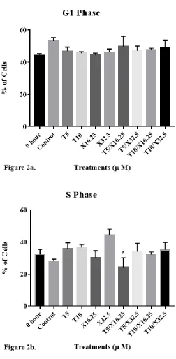
Cell cycle distribution of DU-145 cells was then evaluated (Fig. 2) to determine
whether inhibition of cell proliferation induced by d-δ-tocotrienol and xanthorrhizol was
17
hour (when the treatment started) and control groups at 24 h was 44 ± 2% and 53 ± 4%,
respectively. Cells treated with 5 μM of d-δ-tocotrienol (T5), 10 μM of d-δ-tocotrienol
(T10), 32.5 μM of xanthorrhizol (X32.5), 5 μM of d-δ-tocotrienol and 32.5 μM of
xanthorrhizol (T5/X32.5), and 10 μM of d-δ-tocotrienol and 32.5 μM of xanthorrhizol
(T10/X32.5) exhibited G1 percentages of 46 ± 6%, 46 ± 3%, 46 ± 5%, 47 ± 7%, and 49 ±
12%, respectively. The percentage of cells in the S phase for 0 hour and control groups
was 32 ± 6% and 28 ± 3%, respectively. Cells treated with T5, T10, X32.5, T5/X32.5,
and T10/X32.5 exhibited S phase percentages of 36 ± 9%, 37 ± 5%, 44 ± 10%, 34 ± 12%,
and 35 ± 11%, respectively. The percentage of cells in the G2 phase for 0 hour and
control groups was 24 ± 4% and 19 ± 6%, respectively. Cells treated with T5, T10,
X32.5, T5/X32.5, and T10/X32.5 exhibited G2 phase percentages of 19 ± 11%, 18 ± 6%,
9 ± 9%, 19 ± 11%, and 16 ± 6%, respectively. No significant differences in G1% were
observed with any of the treatments. X32.5 significantly reduced the G2% as compared
to untreated cells (P < 0.05), as noted by ***. Asterisks * and ** for G2 phase denote
18
19
Figure 2. The impact of individual and combined effects of d-δ-tocotrienol and
xanthorrhizol on the distribution of human DU-145 prostate cancer cells in the G1, S, and
G2 phases of the cell cycle following 24 h of incubation. DU-145 cells were plated at a
density of 1.5 x 10⁶ cells per 25 cm2 flask with 3 ml media and incubated for 24 hours.
Cells were then treated and incubated for 24 hours. After incubation, cells were
trypsinized, stained with propidium iodide, and analyzed for cell cycle distribution using
flow cytometry. Brackets in the S phase denotes a significant difference between T5 and
T5/X16.25; brackets in the G2 phase denotes a significant difference between X32.5 and
the control. Tukey’s multiple comparison test was used for repeated-measures analysis of
variance. Values are mean ± SEM, n = 6.
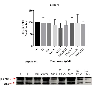
Figures 3a and 3b illustrate the impact of individual and combined treatments of
20
respectively, two regulatory proteins of the G1/S phase transition in the cell cycle of
DU-145 cells. Levels of Cdk4 were 97 ± 24%, 96 ± 39%, 76 ± 21%. 97 ± 40%, 91 ± 20% of
that of control for cells treated with T5, T10, X32.5, T5/X32.5 and T10/X32.5,
respectively. Levels for cyclin D1 were 128 ± 14%, 114 ± 16%, 112 ± 18%. 103 ± 32%,
155 ± 34% of that of control cells for cells treated with T5, T10, X32.5, T5/X32.5 and
T10/X32.5, respectively. No significant difference was observed between any groups.
Figure 3c. illustrates the impact of d-δ-tocotrienol (T) and xanthorrhizol (X),
individually and in combinations, on 24-h expression of procaspase-3 (PC-3), a
pro-apoptotic protein. Levels for PC-3 were 118 ± 15%, 91 ± 23%, 112 ± 25%, 77 ± 4%, 107
± 84% of that of control cells for cells treated with T5, T10, X32.5, T5/X32.5 and
[image:33.612.140.451.427.721.2]T10/X32.5, respectively. No significant difference was observed between any groups.
Figure 3. Western Blot Analysis
21
β-actin PC-3 Cyclin D1
22
Figure 3. Representative blots showing the impact of d-δ-tocotrienol (T) and
xanthorrhizol (X), individually and in combinations, on 24-h expression of cyclin D1 (a),
Cdk4 (b), and procaspase-3 (c), in human DU145 prostate cancer cells. Western blots
procedures were conducted on cell lysates and blots were detected by chemiluminescence
and quantified using ImageJ software. Tukey’s multiple comparisons test was used to
analyze significant variance between groups. Values are mean ± standard error of the
23 CHAPTER V
Discussion and Conclusions
Building on previous studies showing the anti-proliferative, cell cycle arresting,
and apoptotic effects of tocotrienols and xanthorrhizol in cancers of prostate, liver, breast,
colon and skin, this thesis studied the synergistic effects of tocotrienol and xanthorrhizol
in prostate cancer cells for the first time. Our cell proliferation data show a similar
pattern, with decreased cell proliferation shown in cells treated with d-δ-tocotrienol and
xanthorrhizol, individually and in combination. The combined treatments (T5/X32.5 and
T10/X32.5) showed a significant decrease in cell viability compared to the control (no
treatment). Proliferation of cells treated with d-δ-tocotrienol (T5, T10) was not
significantly lower than the control, whereas individual treatment with xanthorrhizol
(X32.5) led to cell proliferation significantly lower than that of the control. There was a
significant decrease in groups T5/X32.5 and T10/X32.5 (p < 0.001), compared to group
T10.
These data suggest that combined treatments of d-δ-tocotrienol and xanthorrhizol
may have a more potent impact on DU-145 cell viability compared to treatments of the
two compounds alone. The X16.25 group showed inconsistent results in cell
proliferation. Inadvertent variations in preparation of xanthorrhizol stock solution and
density of inoculated DU-145 cells may have contributed to the variations. Additional
studies are needed to more accurately determine the effect of xanthorrhizol on DU-145
24
the possibility of a CompuSyn analysis because a minimum of two concentrations of
either compounds in a combination is required for the isobologram analysis.
Cell cycle analysis showed no significant differences between any of the
treatment groups and the control in the G1, S, and G2 phases with one exception; X32.5
significantly reduced the percentage of cells in G2. Additionally, T5/X32.5 significantly
increased the percentage of cells in S phase compared to X32.5. It is perplexing that
X32.5 and T5/X16.25 caused opposite effect on the distribution of cells in the G2 phase.
Literature has shown that tocotrienols induce G1/S arrest in human prostate(19) and
melanoma(5) cancer cells and xanthorrhizol induces G1 and G2/M arrestsin human
colon cancer cells(21). The asynchronized cells that started in various phases of the cell
cycle before treatments and the timing and length of the treatments may have caused
some of the variations in these findings. Further studies are required to elucidate the
effect of these compounds on cell cycle distribution in DU-145 cells.
The lack of consistent cell cycle arrest induced by the compounds may be
congruent with western blot analysis finding no significant difference in Cdk4 and cyclin
D1 levels between treatment groups and the control. Previous studies have shown that
tocotrienols reduced Cdk4 expression in human melanoma cells.(5) Cell line-specific
responses, different sources of antibodies, and technique-related variations in blot
quantification may have led to the lack of detection of treatment effects. Additionally,
varying treatment times may have affected cell response. Cell viability was assessed after
72 hours of treatment, whereas cell cycle analysis and western blot lysate collection was
25
As one of the early events in initiating apoptosis, procaspase-3 is cleaved to a
shorter but active caspase-3 that drives apoptosis. Treatments with T5/X16.25, T5/X32.5,
and T10/X16.25 led to slight and non-significant decreases in the level of procaspase-3
(87%, 77%, and 88%, respectively) when compared to the control (100%). This finding
suggests that caspase-3 may be activated with combined treatments but that the doses of
treatments may need to be altered to show a significant effect for the initiation of
apoptosis.
Data from this study showed a significant decrease in cell viability from
individual and combined treatments of d-δ-tocotrienol and xanthorrhizol. However, we
did not show significant down regulation of Cdk4 or cyclin D1 for induction of cell cycle
arrest, or procaspase-3 activation for apoptosis initiation. Multiple explanations for these
observations are possible. Firstly, changes in doses of d-δ-tocotrienol and xanthorrhizol
in individual and combined treatments may increase their efficacies in downregulation of
the proteins of interest. Secondly, d-δ-tocotrienol is a fat-soluble vitamin and may have
contributed to the cellular uptake of xanthorrhizol.
Third, detached cells were not used in any of the experiments described. After
plating and treatment, some cells may detach before collection. Including detached cells
for analysis may contribute to a larger sample size with a higher proportion of apoptotic
cells with activation of pro-caspase-3.
Fourth, using a colorimetric assay to detect caspase-3 activity in DU-145 cells
may be assessed in future studies, providing a direct approach for detecting apoptosis.
Other caspases may also provide insight to apoptosis as a potential mechanism of action.
26
downstream effectors caspases 3, 6, and 7. Effector caspases execute apoptosis by
cleaving cellular proteins. Initiator and effector caspases may need to be studied
simultaneously to determine if initiation and/or execution of apoptosis has occurred in
DU-145 cells after individual and combined treatments of d-δ-tocotrienol and
xanthorrhizol.
Alternatively, suppression of cell proliferation may have been caused by other
signaling pathways that also regulate cell growth. For example, d-δ-tocotrienol has been
shown to decrease viability of prostate and esophageal cancer cells, respectively, by
inhibiting nuclear factor kappa B cells (NF-kB) of the Akt/NF-kB pathway. NF-kB, a
pro-inflammatory transcription factor, is an important regulator in cell survival.
Downregulation of NF-kB decreases inflammation, thereby decreasing the chance of
certain cancers.(16)
In conclusion, combination of d-δ-tocotrienol and xanthorrhizol inhibited the
growth of DU-145 cells with greater potencies than those of individual agents. Our
limited studies did not show a significant change in cell cycle arrest or down regulation
of Cdk4 and cyclin D1 in induction of cell cycle arrest, or activation of procaspase-3 for
27 REFERENCES
1. Key Statistics for Prostate Cancer. American Cancer Society.
https://www.cancer.org/cancer/prostate-cancer/about/key-statistics.html. Published 2017. Accessed October 27, 2017.
2. What Are the Symptoms of Prostate Cancer? Centers for Disease Control and Prevention. https://www.cdc.gov/cancer/prostate/basic_info/symptoms.htm. Published December 4, 2013. Accessed May 12, 2017.
3. Chemotherapy for Prostate Cancer. American Cancer Society.
https://www.cancer.org/cancer/prostate-cancer/treating/chemotherapy.html. Updated March 11, 2016. Accessed October 28, 2017.
4. Song BL, DeBose-Boyd RA. Insig-dependent ubiquitination and degradation of 3-hydroxy-3-methylglutaryl coenzyme a reductase stimulated by δ- and γ-tocotrienols. J Biol Chem. 2006; 281(35): 2554-61.
5. Fernandes NV, Guntipalli PK, Mo H. d-δ-Tocotrienol-mediated cell cycle arrest and apoptosis in human melanoma cells, Antican Res. 2010; 30: 4937-4944.
6. Seok Fang O, Nallappan M, Yew Hoong C, et al. Xanthorrhizol: a review of its pharmacological activities and anticancer properties. Can Cell Int. 2015; 15: 1-15.
7. Knudsen ES, Wang JY. Targeting the RB-pathway in cancer therapy, Clin Can Res. 2010; 16: 1094-1099.
8. Hydbring P, Malumbres M, Sicinski P. Non-canonical functions of cell cycle cyclins and cyclin-dependent kinases. Nat Rev Mol Cell Bio. 2016; 17(5): 280-292.
9. Gil J, Peter G. Regulation of the INK4b-ARF-INK4a tumour suppressor locus: all for one and one for all. Nat Rev Mol Cell Bio. 2016; 7(9): 667.
10. Dimova D, Dyson N. The E2F transcriptional network; old acquaintances with new faces. Onc. 2005; 24(17): 2810.
28
12. Pradelli L, Beneteau M, Ricci J. Mitochondrial control of caspase-dependent and independent cell death. Cell and Mol Life Sci. 2010; 67(10): 1589-1597.
13. Hengartner M. The biochemistry of apoptosis. Nat. 2000; 407(6805): 770.
14. Mo H, Elson CE. Studies of the isoprenoid-mediated inhibition of mevalonate synthesis applied to cancer chemotherapy and chemoprevention. Exp Biol Med. 2004; 229: 567-585.
15. Goldstein JL, DeBose-Boyd RA, Brown MS. Protein sensors for membrane sterols. Cell. 2006; 124: 35-46.
16. Tan, Barrie. Watson, Ronald. Preedy, Victor. Tocotrienols: Vitamin E Beyond Tocopherols. Boca Raton, FL: CRC Press; 2013.
17. Mo H, Elfakhani M, Shah A, et al. Mevalonate-suppressive tocotrienols for cancer chemoprevention and adjuvant therapy. Edtion ed. In: Watson RR, Preedy VR, Tan B, eds. Tocotrienols: vitamin E beyond tocopherols. Boca Raton: CRC Press, 2013: 135-49.
18. Har C, Keong C. Effects of tocotrienols on cell viability and apoptosis in normal murine liver cells (BNL CL.2) and liver cancer cells (BNL 1ME A.7R.1), in vitro. Asia Pac of Clin Nutr. 2005; 14(4): 374-380.
19. Srivastava JK, and Gupta S. Tocotrienol-rich fraction of palm oil induces cell cycle arrest and apoptosis selectively in human prostate cancer cells. Biochem and Biophy Res Comm. 2006; 346(2): 447-453.
20. Cheah YH, Nordin FJ, Sarip R, et al. Antiproliferative property and apoptotic effect of xanthorrhizol on MDAMB-231 breast cancer cells. Antican Res. 2008; 28: 3677–90.
21. Kang YJ, Park KK, Chung WY, Hwang JK, Lee SK. Xanthorrhizol, a natural sesquiterpenoid, induces apoptosis and growth arrest in HCT116 human colon cancer cells. J Pharm Sci. 2009; 111:276–84.