Biallelic mutations in IRF8 impair human NK
cell maturation and function
Emily M. Mace, … , James R. Lupski, Jordan S. Orange
J Clin Invest. 2017;
127(1)
:306-320.
https://doi.org/10.1172/JCI86276
.
Human NK cell deficiencies are rare yet result in severe and often fatal disease, particularly
as a result of viral susceptibility. NK cells develop from hematopoietic stem cells, and few
monogenic errors that specifically interrupt NK cell development have been reported. Here
we have described biallelic mutations in IRF8, which encodes an interferon regulatory
factor, as a cause of familial NK cell deficiency that results in fatal and severe viral disease.
Compound heterozygous or homozygous mutations in IRF8 in 3 unrelated families resulted
in a paucity of mature CD56
dimNK cells and an increase in the frequency of the immature
CD56
brightNK cells, and this impairment in terminal maturation was also observed in Irf8
–/–,
but not Irf8
+/–, mice. We then determined that impaired maturation was NK cell intrinsic, and
gene expression analysis of human NK cell developmental subsets showed that multiple
genes were dysregulated by IRF8 mutation. The phenotype was accompanied by deficient
NK cell function and was stable over time. Together, these data indicate that human NK
cells require IRF8 for development and functional maturation and that dysregulation of this
function results in severe human disease, thereby emphasizing a critical role for NK cells in
human antiviral defense.
Research Article
Immunology
Find the latest version:
Introduction
NK cell deficiency (NKD) is an inborn or primary immunodefi-ciency causing susceptibility to severe and often fatal viral infec-tion and malignancy (1–5). NKD can be classified as classical, aris-ing from profound or complete absence of the NK cell population or major subsets of peripheral NK cells, or functional, arising from impaired NK cell function with normal peripheral NK cell counts
(5, 6). There are presently 2 genetically defined causes of classical (MCM4, GATA2) and 1 of functional NKD (FCGR3A) (2–4, 7–9).
Human NK cell development results in generation of distinct phenotypic and functional subsets found in peripheral blood:
namely the CD56bright and CD56dim subsets. While identified
pri-marily by the density of CD56 on the cell surface, these subsets include the coordinated expression of cell surface and
intracellu-lar markers that reflect each subset’s unique function. CD56bright
NK cells are considered immunomodulatory and are described
phenotypically as CD16−/loCD57−perforinlo, CD117+, CD62Lhi,
and CD94/NKG2A+. While they constitute the minority of NK
cells in peripheral blood, they represent the dominant population
in secondary lymphoid tissue (10, 11). CD56dim NK cells, which
constitute more than 90% of peripheral blood NKs, are CD56dim
CD16hiperforinhiCD57+/− killer-cell immunoglobulin-like receptor+
(KIR+), and are the primary mediators of NK cell–mediated
cyto-toxicity. The relationship between the subsets is not complete-ly understood, but both in vivo and ex vivo evidence in humans
Human NK cell deficiencies are rare yet result in severe and often fatal disease, particularly as a result of viral susceptibility. NK cells develop from hematopoietic stem cells, and few monogenic errors that specifically interrupt NK cell development have been reported. Here we have described biallelic mutations in IRF8, which encodes an interferon regulatory factor, as a cause of familial NK cell deficiency that results in fatal and severe viral disease. Compound heterozygous or homozygous mutations in IRF8 in 3 unrelated families resulted in a paucity of mature CD56dim NK cells and an increase in the frequency
of the immature CD56bright NK cells, and this impairment in terminal maturation was also observed in Irf8–/–, but not Irf8+/–,
mice. We then determined that impaired maturation was NK cell intrinsic, and gene expression analysis of human NK cell developmental subsets showed that multiple genes were dysregulated by IRF8 mutation. The phenotype was accompanied by deficient NK cell function and was stable over time. Together, these data indicate that human NK cells require IRF8 for development and functional maturation and that dysregulation of this function results in severe human disease, thereby emphasizing a critical role for NK cells in human antiviral defense.
Biallelic mutations in
IRF8
impair human NK cell
maturation and function
Emily M. Mace,1,2,3 Venetia Bigley,4 Justin T. Gunesch,1,3 Ivan K. Chinn,1,2,5 Laura S. Angelo,1 Matthew A. Care,6 Sheetal Maisuria,7 Michael D. Keller,8 Sumihito Togi,9 Levi B. Watkin,1 David F. LaRosa,10 Shalini N. Jhangiani,5 Donna M. Muzny,5
Asbjørg Stray-Pedersen,5,11 Zeynep Coban Akdemir,5,11 Jansen B. Smith,1 Mayra Hernández-Sanabria,1 Duy T. Le,1
Graham D. Hogg,1 Tram N. Cao,1 Aharon G. Freud,12,13 Eva P. Szymanski,14 Sinisa Savic,7 Matthew Collin,4 Andrew J. Cant,15 Richard A. Gibbs,5 Steven M. Holland,14 Michael A. Caligiuri,13,16 Keiko Ozato,9 Silke Paust,1,2,17 Gina M. Doody,6
James R. Lupski2,5,11, and Jordan S. Orange1,2,3,17
1Center for Human Immunobiology, Texas Children’s Hospital, Houston, Texas, USA. 2Department of Pediatrics, and 3Department of Pathology and Immunology, Baylor College of Medicine, Houston,
Texas, USA. 4Institute of Cellular Medicine, University of Newcastle, Newcastle upon Tyne, England, United Kingdom. 5Human Genome Sequencing Center, Baylor College of Medicine, Houston, Texas, USA. 6Section of Experimental Haematology, Leeds Institute of Cancer and Pathology, University of Leeds, Leeds, United Kingdom. 7Department of Clinical Immunology and Allergy, St James’s University Hospital,
Leeds, United Kingdom. 8Division of Allergy and Immunology, Center for Cancer and Immunology Research, Children’s National Health System, Washington, DC, USA. 9Laboratory of Molecular Growth
Regulation, Program in Genomics of Differentiation, National Institute of Child Health and Human Development, NIH, Bethesda, Maryland, USA. 10Department of Pediatrics, University of Pennsylvania
School of Medicine, Children’s Hospital of Philadelphia Research Institute, Philadelphia, Pennsylvania, USA. 11Department of Molecular and Human Genetics, Baylor College of Medicine, Houston, Texas, USA. 12Division of Hematopathology, Department of Pathology, The Ohio State University, Columbus, Ohio, USA. 13The Ohio State University Comprehensive Cancer Center, The James Cancer Hospital and Solove
Research Center, Columbus, Ohio, USA. 14Laboratory of Clinical Infectious Diseases, NIAID, NIH, Bethesda, Maryland, USA. 15Department of Paediatric Immunology and Bone Marrow Transplantation,
Great North Children’s Hospital, Newcastle upon Tyne, United Kingdom. 16Division of Hematology, Department of Internal Medicine, The Ohio State University, Columbus, Ohio, USA. 17Dan L Duncan Cancer Center, Baylor College of Medicine, Houston, Texas, USA.
Conflict of interest: J.R.Lupski has stock ownership in 23andMe, is a paid consultant for Regeneron Pharmaceuticals, has stock options in Lasergen, Inc. is a member of the Scientific Advisory Board of Baylor Genetics, and is a co-inventor on multiple United States and European patents related to molecular diagnostics for inherited neuropathies, eye diseases, and bacterial genomic fingerprinting (5294533; 5306616; 5523217; 5599920; 5667968; 5780223; 6132954; 6713300; 7141420; 7189511; 7192579; 7273698; 7537899; 8129353; 9365899; 0424473; 0610396; 0989805). The Department of Molecular and Human Genetics at Baylor College of Medicine derives revenue from the chromosomal microarray analysis (CMA) and clinical exome sequencing offered in Baylor Genetics (BMGL: http://www.bmgl.com/BMGL/Default.aspx).
Submitted: December 30, 2015; Accepted: October 18, 2016.
ciency leading to specific loss of the CD11c+CD1c+ circulating DC
subset (19). K108E mutations of IRF8 have an even more profound impact, leading to the loss of DCs as well as monocytes (19).
Before recent advances in genomic sequencing technology, NKD was reported in a number of pediatric cases. Perhaps the most renowned was an isolated case of NKD and viral suscepti-bility ultimately proven as resulting from a GATA2 mutation (4, 22). The original NKD case, however, was reported in 1982 as a familial, non–X-linked susceptibility to EBV infection that led to the death of 2 affected siblings and ongoing illness in a third (1). Here we report that biallelic mutations in IRF8 cause intrinsic NKD with decreased NK cell number, decreased frequency of
the CD56dim subset, and accompanying susceptibility to viral and
mycobacterial infection. In doing so we define and characterize
IRF8 mutations in this original, unsolved family with NKD and
identify NKD in 2 unrelated patients with biallelic IRF8 mutations. These data identify IRF8 as a critical regulator of NK cell terminal maturation and define a novel cause of a rare human inborn NK cell developmental defect.
Results
Patients and clinical history. The index case proband (II.2)
present-ed with a history of sinopulmonary infections during childhood and severe infectious mononucleosis at age 22 years requiring pro-longed hospitalization (1). Born to non-consanguineous parents, his brother (II.6) died at age 16 years of multiple organ failure fol-lowing severe EBV mononucleosis, and his sister (II.5) died at age 38 years due to progressive pulmonary disease. She also had pro-longed hospitalization at age 17 years for severe EBV mononucleo-sis. Both siblings had lymphadenopathy and bronchiectasis begin-ning in childhood. Notably, the 3 siblings’ EBV-related illnesses occurred in separate calendar years. A fourth sibling (brother, II.3) had EBV mononucleosis at age 21 years at the same time as the proband, but had a typical course of illness and did not require hospitalization. The unusual nature of disease and clinical
pre-suggests that the CD56bright subset is a developmental precursor
of CD56dim, and it is hypothesized that secondary lymphoid tissue
serves as a site of NK cell terminal differentiation that is required for acquisition of cytotoxic function (12–15).
Specific loss of NK cells or NK cell subsets as occurs in classical NKD is rare but has been reported. Homozygous MCM4 mutation causes adrenal insufficiency, decreased numbers of circulating NK cells, and impaired terminal maturation, likely as a result of
chro-mosomal aberrations and impaired proliferation of the CD56bright
subset (2, 3). These patients show distinctive loss of the CD56dim
subset and correlative relative increase in the CD56bright subset (2).
In contrast, GATA2 mutations result in specific loss of CD56bright
NK cells and decreased frequency of NK cells overall in
circulat-ing blood. While GATA2 patients have CD56dim NK cells that vary
in number, they are largely nonfunctional, and affected patients develop severe herpesvirus and papillomavirus infections (4). NKD has also been reported in 1 isolated case of a child with a mutation in RTEL1, encoding regulator of telomere elongation helicase, although NK cell subsets were not examined in this case (16, 17).
Interferon regulatory factor 8 (IRF8; also known as interferon consensus binding protein, ICSBP) is a member of the IRF family of transcription factors that help shape the inflammatory response, particularly to viral infections. These transcription factors are also becoming increasingly recognized for their roles in human hema-topoiesis through rare, naturally occurring germline mutations.
Mutations of IRF8 in mice and humans cause cellular defi-ciency ranging from severe defects of monocyte and DC produc-tion with marked myeloproliferaproduc-tion to more subtle alteraproduc-tions of specific DC subsets with normal leukocyte counts (18–20). Gene dosage and mutation site–specific effects have been observed, sug-gesting that the level of IRF8 activity is critical to normal immune cell development and function. In addition to developmental aber-rations, mature cell function is affected as exemplified by defects in macrophage function (21). In humans, heterozygous IRF8 T80A mutations have been identified as a cause of primary
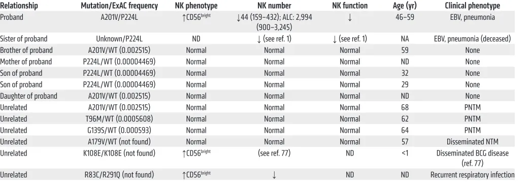
immunodefi-Table 1. NK characteristics and clinical phenotypes of subjects with IRF8 mutations
Relationship Mutation/ExAC frequency NK phenotype NK number NK function Age (yr) Clinical phenotype Proband A201V/P224L ↑CD56bright ↓44 (159–432); ALC: 2,994
(900–3,245) ↓ 46–59 EBV, pneumonia Sister of proband Unknown/P224L ND ↓ (see ref. 1) ↓ (see ref. 1) NA EBV, pneumonia (deceased) Brother of proband A201V/WT (0.002515) Normal Normal Normal 59 None Mother of proband P224L/WT (0.00004469) Normal Normal Normal ND None Son of proband P224L/WT (0.00004469) Normal Normal Normal 32 None Son of proband P224L/WT (0.00004469) Normal Normal Normal 29 None Daughter of proband A201V/WT (0.002515) Normal Normal Normal ND None
Unrelated A201V/WT (0.002515) Normal Normal Normal 68 PNTM
Unrelated T96M/WT (0.0005608) Normal Normal Normal 62 PNTM
Unrelated G139S/WT (0.000593) Normal Normal Normal 64 PNTM
Unrelated A179V/WT (not found) Normal Normal Normal 57 Disseminated NTM Unrelated K108E/K108E (not found) ↑CD56bright (see ref. 77) ND <1 Disseminated BCG disease
(ref. 77) Unrelated R83C/R291Q (not found) ↑CD56bright ↓ ND ND Recurrent respiratory infection
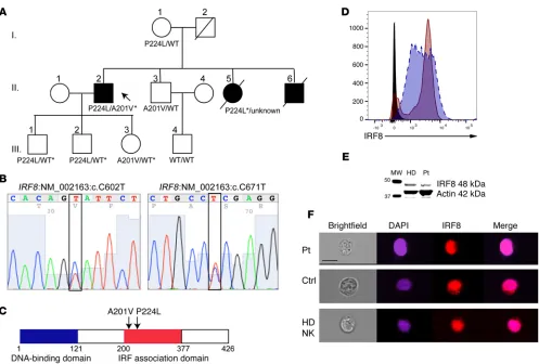
[image:3.585.42.564.83.262.2]samples (proband, 105X; mother, 124X; brother, 116X; son, 114X; fraternal nephew, 90X). The exome data suggested disease-relat-ed segregation of mutations in IRF8 (NM_002163:c.602C>T, 39 variant reads of 112 total in the proband; NM_002163:c.671C>T, 44 variant of 72 total proband reads) predicted by conceptual transla-tion to result in amino acid changes p.A201V and p.P224L, respec-tively. Of those mutations predicted to be damaging by SIFT, Poly-Phen, LRT, and MutationTaster algorithms, few aside from IRF8 were expressed highly in immune cells, particularly NK cells (see Supplemental Table 1 for list of variants identified by whole exome sequencing; supplemental material available online with this arti-cle; doi:10.1172/JCI86276DS1). Sanger sequencing of IRF8 in the proband’s sons and daughter confirmed the presence of both alleles in his children and the absence of either variant in his wife (Figure 1B and Supplemental Figure 1). The P224L variant was present in his mother, and the A201V variant was present in a surviving broth-er. Therefore, IRF8 mutations were confirmed with familial segre-gation in accordance with Mendelian expectations. In addition, the P224L mutation was confirmed from paraffin-embedded tissue sentation in the proband prompted immunological evaluation and
was described as a novel, non–X-linked immune deficiency lead-ing to susceptibility to EBV disease associated with a deficiency of NK cell cytotoxicity (1). Since the original description, the patient has continued to have fairly regular, recurrent episodes of sinusitis and has mucosal changes consistent with recurrent and chronic disease as well as presumably respiratory viral infections. He has no evidence of bronchiectasis or lymphadenopathy. T cell and B cell counts have been consistently normal, and immunoglobulins are present in normal quantities and quality. His surviving broth-er (II.3), mothbroth-er (I.1), children (III.1–3), and nephew (III.4) have unremarkable medical histories. Other unrelated patients are summarized in Table 1.
Genetics. The 1982 description of the proband’s family is
[image:4.585.43.540.51.387.2]with-out follow-up until now and did not include any genetic or genom-ic analysis. Thus, whole exome sequencing of genomgenom-ic DNA was performed using blood samples collected from the proband; his son, mother, and surviving brother; and the son of the brother (Fig-ure 1A). Excellent average per-base coverage was observed for all
Evaluation of the patient’s protein expression by flow cytom-etry in lymphoblastoid cell lines showed normal levels of expres-sion (Figure 1D), and Western blot analysis of IRF8 from patient EBV-transformed B cell lines confirmed protein expression at the expected molecular weight of full-length protein, as did healthy donor (Figure 1E; see complete unedited blots in the supplemental material). The only previously reported homozygous mutation in
IRF8, K108E, was shown to impair nuclear translocation of IRF8 in
transfected cells (21). To determine whether the novel compound heterozygous alleles found in our proband prevent the nuclear recruitment of IRF8, we evaluated the patient B cell line by imag-from the patient’s deceased sister (Supplemental Figure 1);
degra-dation of the DNA extracted from the same sample did not permit amplification and sequencing of the A201V mutation. The P224L mutation is reported 5 times in the Exome Aggregation Consortium
(ExAC) database (allelic frequency of 4.4 × 10⁻⁵) with no
[image:5.585.48.470.53.577.2]homozy-gous cases. The A201V mutation is present at an allelic frequency of 0.002515 with no homozygotes. Likewise, within the Baylor-Hop-kins Center for Mendelian Genomics database of over 6,000 exomes, no homozygous or compound heterozygous cases of IRF8 variants are found (23). Both mutations map to exon 7, in the region of IRF8 known as the IRF association domain (Figure 1C).
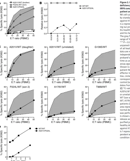
Figure 2. NK cell functional deficiency specifically occurs in IRF8 compound-heterozygous patient and is stable over time.
ing flow cytometry. In the absence of stimulation, IRF8 was detect-ed in both the cytoplasm and the nucleus of patient and control B cell lines. This was also observed for healthy donor NK cells, with colocalization between DAPI and IRF8 as calculated by similarity score (24) of 3.4 for patient cells, 2.2 for control B cells, and 2.2 for primary human NK cells (median values) (Figure 1F). Stimulation with IL-15 did not increase localization of IRF8 in primary human NK cells (similarity score 2.2, not shown). Together these data con-firm novel compound heterozygous mutations in IRF8 that do not abrogate protein expression or nuclear localization.
Biallelic IRF8 mutation leads to impaired NK cell cytotoxic func-tion. To specifically test the effect of biallelic mutations on NK
cell cytotoxicity, we performed standard 51Cr release assays. As
previously described, the proband has consistently low NK cell functional activity against the prototypical K562 target (ref. 1 and Figure 2A). The proband’s cytotoxic function was evaluated mul-tiple times during a 10-year time interval and consistently showed profoundly decreased activity relative to normal controls with specific lysis (at maximal effector-to-target ratio) that was 0% and 31% of normal control levels (Figure 2B). The patient’s children, carriers of the P224L or A201V mutations, had slightly decreased but normal cytotoxic function, as did his nephew and mother (Fig-ure 2, A and C, and not shown). Unrelated carriers of the A201V mutation from a separate family of a different nationality similarly had normal NK cell cytotoxic function (Figure 2C), despite having late onset of pulmonary nontuberculous mycobacterial disease
(Table 1). Other heterozygous patients (G139S/ WT, A179/WT, T96M/WT) also had normal NK cell function despite the presence of pulmo-nary nontuberculous mycobacterial infection (Figure 2C and Table 1). To determine wheth-er decreased NK cell lytic function in the pro-band was due to low NK cell frequency within the lymphocyte population, cytotoxicity assays using purified NK cells were performed, and showed significantly decreased lysis of targets (Figure 2D), suggesting that an intrinsic defect in NK cell cytotoxic function underlies the stably decreased impairment observed. Therefore, the compound heterozygous A201V/P224L muta-tion results in low/absent NK cell cytotoxic func-tion, an effect that is repeatedly observed over extended time and likely accounts for the pro-band’s profound susceptibility to viral disease.
Biallelic IRF8 mutation results in decreased NK cell number and impaired NK cell terminal dif-ferentiation. We next functionally examined NK
cells from probands with 2 different combinations of compound heterozygous and a homozygous (i.e., biallelic) variant allele: A201V/P224L, R83C/R291Q, and homozygous K108E. Human NK cells in peripheral blood fall largely within 2 dominant subsets with unique phenotypic and functional signatures. High density of CD56 and the presence of CD94/NKG2A, CD117, and CD62L on the cell surface classify the CD56bright subset phenotypically. These
cells are considered the most potent cytokine producers upon
stimulation, primarily of TNF-α and IFN-γ. The CD56dim subset
is characterized by markers of terminal differentiation such as CD57, activating and inhibitory KIRs, and CD16, and is thought to be derived from the CD56bright subset (13). In addition, CD56dim NK
cells express large quantities of perforin and granzymes; they are specialized for contact-mediated target cell lysis.
Consistently over the time of evaluation (>10 years) the patient had decreased frequency and absolute number of
CD56+CD3− NK cells within the lymphocyte population (1.7%–
6.0% vs. 4.6%–13.7% in healthy controls) (Figure 3, A–C, and Table 1). Evaluation of the phenotypic subsets within
peripher-al blood showed a relative paucity of the CD56dim subset in the
patient, accompanied by an overrepresentation of the CD56bright
subset. From our initial evaluation in 2004 and at all subsequent
times evaluated, patient 1’s CD56bright subset was between 30%
and 40% (27.9%–40.3%) of the NK cell population, in contrast to controls, which fell within the previously described range (4.1%–8.4%) (Figure 3, A–C, and ref. 25). Evaluation of
hetero-Figure 4. Irf8–/– mice have a block in terminal NK cell
maturation. Spleen and blood from Irf8–/–, Irf8+/–
lit-termates, and Irf8+/+ controls were analyzed by flow
[image:7.585.42.350.55.406.2]zygous A201V/WT subjects (both unrelated and related) showed normal frequencies of both NK cells within the peripheral blood
mononuclear cell (PBMC) population (13.9%) and CD56bright NK
cells (3.7%). Further, evaluation of other heterozygous individu-als (T96M/WT, G139S/WT, A179V/WT) showed frequencies of
NK cells and CD56bright NK cells consistent with normal controls
(Figure 3, B–D). In contrast, the R83C/R291Q and the homozy-gous K108E patient had low overall numbers of NK cells (3.9%
and 5.4%, respectively) and a significantly expanded CD56bright
population consistent with our proband (65.8% and 72.7%; Fig-ure 3, B–D). Therefore, compound heterozygous or homozygous mutations in IRF8 selectively result in aberrant generation of the terminally mature subsets of NK cells and a decreased frequency of NK cells within the lymphocyte population.
Because of the patient’s consistently low NK cell cytotoxic function and abnormal NK cell subset distribution, we performed multiparametric flow cytometric immune phenotyping of NK cell subsets, including intracellular cytokines IFN-γ and TNF-α as well as perforin and granzymes. We evaluated the patient, his clinical-ly unaffected single-heterozygous famiclinical-ly members, an unrelated heterozygous A201V/WT patient with pulmonary nontuberculous mycobacterial infection, a heterozygous T96M/WT patient, an
unrelated compound-heterozygous patient (R83C/R291Q), and a homozygous K108E patient (Table 1).
Multiparametric flow cytometric analysis of the proband’s lymphocytes showed that NK cell markers were consistent with
the expanded CD56bright subset but otherwise showed no
aber-rant expression of markers of activation, inhibition, or granzyme
B (Figure 3E). Despite decreased representation of the CD56dim
subset, markers of NK cell terminal differentiation and memo-ry (CD57, NKG2C) were expressed, as were activating receptors (NKG2D) and inhibitory receptors (NKG2A). Interestingly, how-ever, our proband had consistently low levels of perforin at multi-ple time points tested, as did the homozygous K108E patient (Fig-ure 3E). Upon stimulation, the proband, as well as those patients with compound heterozygous variants in IRF8, expressed normal
or higher than normal levels of IFN-γ and TNF-α measured both
as percentage of the population expressing cytokine and on a
per-cell basis (Figure 3E and not shown). The production of IFN-γ
was comparable between patient and control CD56bright NK cells,
which are generally the most potent producers of IFN; however,
the patient CD56dim NK cells had increased IFN-γ production
rela-tive to control. This accounted for the increase in IFN-γ production in his NK cells when considered as a total population (Figure 3F).
The patient with K108E/K108E mutations had impaired IFN-γ
production upon stimulation from both NK cell subsets, poten-tially underscoring the different effects of the different mutations on immune function even in the context of similar NK cell pheno-type. The observed decrease in perforin expression in our patient with A201V/P224L mutations and the patient with homozygous
K108E mutations was confined to the CD56dim subset, the
prima-ry cytolytic effectors. CD16 (FcγRIIIA) was expressed at normal
levels on CD56bright and CD56dim NK cells from the patients with
A201V/P224L and R83C/R291Q mutations (Figure 3, E and F). Low expression of CD16 on NK cells from the K108E patient was
restricted to the CD56dim NK cell subset (Figure 3F).
Given the important role that cytokine production plays in the control of the viral immune response and previously described DC deficiency in patients with IRF8 mutations, we sought to determine whether cytokine production in the context of costim-ulation by accessory cells was impaired in our patient. To do so, we incubated PBMCs from healthy donor or patient with a non-autologous EBV-transformed B cell line that had been induced
to lytic cycle. Secreted cytokines (IL-12p40, IFN-γ, TNF-α, and
IL-10) were measured by flow cytometric bead array. As suggest-ed by the intracellular flow cytometry data, the coculture-inducsuggest-ed
production of IFN-γ and TNF-α was not impaired in the patient,
[image:8.585.49.271.55.454.2]and, similarly to that observed in cytokine-stimulated NK cells,
numbers of these subsets (blood 0.45 × 10⁶ ± 0.4 × 10⁶, spleen 3.6
× 106 ± 1.9 × 10⁶) when compared with wild-type controls (blood
0.36 × 10⁶ ± 0.5 × 10⁶, spleen 0.31 × 10⁶ ± 0.3 × 10⁶). Conversely,
Irf8–/– mice had decreased frequency of mature Mac-1hiCD27lo NK
cells (48.3% ± 3.9%) compared with wild-type controls (64.3% ± 3.9%). This was despite increased absolute cell number (blood 1 × 10⁶ ± 0.6 × 10⁶, spleen 5.7 × 10⁶ ± 2.1 × 10⁶) compared with controls
(blood 0.7 × 10⁶ ± 0.5 × 10⁶, spleen 1.4 × 10⁶ ± 0.6 × 10⁶). Together,
these demonstrate impairment in terminal maturation in mice with homozygous, but not heterozygous, Irf8 loss-of-function muta-tions, suggesting an important parallel to our patients with biallelic
IRF8 mutations and increased frequency of immature NK cells. IRF8 is required for human NK cell development in a cell-intrinsic manner. Given the broad expression of IRF8 within immune cell
populations, we sought to determine whether the observed effect on NK cell function was due to NK cell–intrinsic mechanisms or representative of the need for other immune or nonimmune cell types in NK cell development. Thus, we evaluated the ability of
patient CD34+ hematopoietic stem cells (HSCs) to generate NK
cells in isolation. HSCs were sorted from our proband’s peripheral blood and incubated on OP9-DL1 stromal cells for 28 days in the presence of IL-7, FLT3L, c-Kit, and IL-15, as this in vitro experi-mental system has been previously demonstrated to promote NK cell development (14, 30, 31). As shown in Figure 5A, patient
NK cells derived from CD34+ HSCs were largely CD56bright (76%
vs. 19.7% from healthy donor control). The mean fluorescence intensity of CD56 was significantly higher on patient cells ver-sus the normal control (224 vs. 112 in healthy donor) (Figure 5B), consistent with immaturity and incomplete maturation. In addi-tion, while healthy donor–derived cells acquired CD16 (58%), the patient-derived cells had decreased CD16 expression yet retained CD94, a phenotype that is reflective of an immature state. These results show that the role for IRF8 is cell intrinsic and that IRF8 is functioning directly in the generation of appropriate and mature NK cell subsets during development.
IRF8 is expressed in human NK cell subsets and is upregulated upon IL-15 stimulation. Expression of IRF8 in human NK cell
devel-opmental subsets has not been previously determined. We per-formed real-time PCR and intracellular flow cytometry to evaluate the expression of IRF8 in human NK cell developmental subsets.
While human peripheral blood contains only the CD56bright and
CD56dim NK cell subsets, we used human tonsil as a source of
devel-oping NK cells and evaluated these cells based on known markers
the production of IFN-γ in these cocultures was increased (Figure
3G). Interestingly, elevated secretion of both IL-12 and IL-10 was also observed from patient cells.
As IRF8 serves a previously identified role in DC develop-ment and IRF8 deficiency has been shown to lead to a variable loss or absence of DC subsets, we performed phenotypic analy-sis of monocyte and DC populations. A subtle deficiency of
plas-macytoid dendritic cell (pDCs) and CD141+ DCs was observed in
the index patient relative to heterozygous family members and a wild-type control (Supplemental Figure 2). These differences were within the scope of those previously ascribed to human IRF8 mutations and were not as severe as those observed in the patient with homozygous K108E mutation (19).
Our findings, however, suggested a novel NK cell phenotype of arrested terminal maturation in the presence of biallelic IRF8 mutations. To further determine the role of IRF8 in NK cell
mat-uration, we performed phenotypic analysis of NK cells from Irf8–/–,
Irf8+/–, and Irf8+/+ mice by flow cytometry. While murine NK cells
do not express CD56, and not all transcription factor functions are evolutionarily conserved between human and murine NK cells, the developmental stages of murine NK cells can be stratified according to expression of CD27 and CD11b (Mac-1) (26). As
pre-viously reported (27–29), Irf8–/– mice had significant splenomegaly
and lymphocytosis, leading to increased absolute NK cell number in both spleen (14 × 106 ± 5.7 × 10⁶) and blood (1.2 × 10⁶ ± 0.7 ×
106) relative to that of Irf8+/+ controls (spleen 2.5 × 10⁶ ± 1.2 × 10⁶,
blood 0.89 × 10⁶ ± 0.6 × 10⁶). Despite this, as seen in Figure 4, NK
cells from blood and spleen of Irf8–/–, but not Irf8+/–, mice had an
increased frequency of immature Mac-1loCD27hi NK cells (22.5%
[image:9.585.44.287.56.347.2]± 2.7%) within the NK cell population when compared with wild-type controls (9.7% ± 1.8%). This was also reflected by absolute
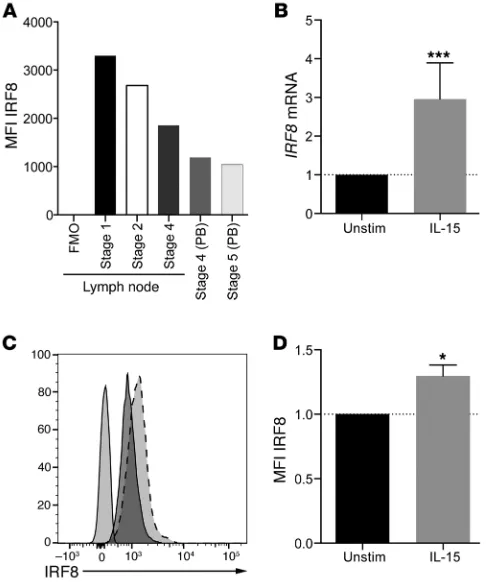
Figure 6. IRF8 is expressed in all NK cell developmental subsets and is upregulated upon IL-15 stimulation. (A) NK cell developmental subsets were isolated from tonsil as described in Methods, and IRF8 was detected by intracellular flow cytometry. Shown is 1 representative experiment from 3 independent experiments from 3 normal donors. FMO, fluorescence minus one; PB, peripheral blood. (B) NK cells were incubated for 6 hours with 10 ng/ml of IL-15 or vehicle control, and then RNA was extracted and qPCR performed as described in Methods. ***P < 0.001 by Student’s t test. Shown are the means of 3 independent experiments performed on 3 donors, each done in quadruplicate. (C) NK cells were stimulated as in
developmental subsets isolated from tonsil, as well as those from peripheral blood, expressed significant quantities of IRF8. Further, stimulation of primary human NK cells with IL-15 for as few as 6 hours resulted in an upregulation of IRF8 as measured by quanti-of differentiation (stages 1–5) (32). As shown in Figure 6A, healthy
human donors expressed IRF8 in all developmental subsets as measured by intracellular flow cytometry. Interestingly,
[image:10.585.91.491.56.562.2]expres-sion was highest in stage 1 (CD34+) NK cell precursors; however, all
Discussion
Genetic causes of aberrations in NK cell development are exceed-ingly rare but confer familial susceptibility to disease, particularly viral infection. Here we identify a novel human NK cell develop-mental defect and cause of NKD due to biallelic mutation in the
IRF8 gene. Interestingly, while homozygous and heterozygous IRF8 mutations lead to a spectrum of DC deficiency ranging from
mild to severe, biallelic variants in our patient and 2 additional unrelated patients result in NKD as a result of a block in terminal maturation. In our patient this phenotype occurs in the context of normal activation of PU.1 and IRF1, underscoring the varying impact of IRF8 mutations.
Our proband was originally diagnosed in 1980 and reported in 1982 with the first case of non–X-linked susceptibility to EBV as a result of NKD (1), and as such is within the first reported cohort of patients with NKD. Having the unique opportunity to study the 1 surviving affected patient from this family longitudinally has underscored the stability of the NK cell phenotype, namely
decreased NK cell number with a paucity of CD56dim NK cells and
interrupted maturation.
The IRF family plays a well-documented role in inflamma-tion, and these transcription factors are also becoming increasing-ly recognized for their importance in hematopoietic development (35, 36). In particular, a critical requirement for IRF8 in DC devel-opment is described in mice with both hypomorphic and knock-out mutations of IRF8, as well as previously described patients with heterozygous and homozygous mutations (19). Patients with homozygous K108E mutation are close in phenotype to the knockout mouse, with severe deficits of monocytes and DCs and a myeloproliferative disorder (29, 37–39). This homozygous mutation in the DNA-binding domain of IRF8 abrogates nucle-ar translocation but not protein expression, leading to impaired
function and defective IL-12, IFN-γ, and TNF-α production (21).
In contrast, the spontaneous heterozygous T80A mutation leads
to specific loss of the CD1c+ compartment of CD11c+ DCs but
retention of CD141+ DCs, monocytes, and CD123+
plasmacyt-oid DCs (19). Relative to disease caused by autosomal recessive K108E mutation, the T80A heterozygous mutation leads to a less severe clinical course. IL-12 production, but not type I IFN, is affected. In both cases, both B and T cell numbers are largely
normal; however, CD25hiFoxP3+ Treg numbers are decreased in
patients with K108E mutation (39).
The mechanism for the differential impact of IRF8 mutations on DC and NK cell homeostasis remains unclear. Gene dosage and mutation site–specific effects are commonly observed in transcriptions factors such as IRF8 that are regulated by superen-hancer structures (40). As opposed to A201V/P224L, the K108E mutation is unable to activate IRF1- and PU.1-dependent tran-scription and, in the biallelic state, is associated with monocyte and DC deficiency and a very marked distortion of NK cell
devel-opment in the ratio of CD56bright to CD56dim cells. The patient
described here with A201V/P224L mutation has an NK pheno-type with only subtle DC deficiency, in keeping with the ability of the 2 variants to activate transcription with PU.1 and IRF1. As these processes are required for DC development and function, these data suggest that additional IRF8-dependent mechanisms are required for normal NK cell homeostasis.
tative PCR (qPCR) (3-fold; Figure 6B) and flow cytometry (1.3-fold; Figure 6, C and D). Therefore, IRF8 is expressed in human NK cells and is responsive to IL-15 stimulation. This observation is consis-tent with a critical role for IRF8 during NK cell development.
IRF8 is required for NK cell expression of transcription factors and NK cell effector molecules. Previously reported homozygous
K108E mutations in IRF8 lead to impaired synthesis of IL-12 due to abrogated IRF1-dependent transcription of the IL12B gene (19). To determine whether the mutations in our proband affect-ed functional binding of IRF8 with cofactors IRF1 and PU.1, we performed luciferase assays with each allelic variant (WT, A201V, and P224L). As seen in Figure 7A, neither of our patient’s vari-ants affected transcriptional activity dependent on PU.1 or IRF1. This is consistent with the observation that DC development was only partially affected in the patient and contrasts with biallelic K108E mutation, in which transactivation with IRF1 was severely impaired and monocytes and DCs were absent (19). Collective-ly, this underscores the variability of mutational impact on the DC phenotype. To determine the effect that the IRF8 mutations have on human NK cell function, we performed NanoString gene expression analysis (Human Immunology v2 gene set) on isolat-ed NK cells from our proband, his family members, and 3 healthy donor controls. We identified 403 genes with a greater than 1.5-fold up- or downregulation in the patient relative to all 3 healthy donors (Supplemental Table 2). These included 18 of particular relevance to NK cell development, cytotoxicity, or homeostasis (Figure 7B), including transcription factors NFIL3, IKZF2, TBX21,
EOMES, and STAT5A/B. Differentially expressed effector
mol-ecules and cell surface receptors included PRF1, GZMA, GZMB,
CCR7, CD27, CD2, HAVCR2, and KLRG1. Ingenuity Pathway
and the significant downregulation of JAK3 and STAT5A and -B in our patient relative to 3 healthy donors supports this hypothesis.
While we show that the NK cell developmental defect is intrin-sic, an additional model for the requirement of IRF8 in NK cell development and function would be through the trans-presen-tation of IL-15 by DCs, which would potentially make the loss of DCs and functional defect of NK cells in IRF8 patients particularly damaging. Our data support a link between IL-15, IRF8, and NK cell development, as we show that IL-15 increases IRF8 protein expression in as few as 6 hours in ex vivo NK cells. This observation
suggests a need for the IL-15–induced proliferation of CD56bright
NK cells in the generation of the CD56dim subset, as proposed in
the report of MCM4 deficiency, the only other reported immune deficiency to result in this distinctive NK cell phenotype (2). This concept was originally proposed by Lutz et al. after they showed
that CD56bright NK cells undergo significantly greater proliferation
than the CD56dim subset (48). In addition, FLT3L-mediated
devel-opment of DCs is dependent on IRF8 signaling, and FLT3L-defi-cient mice have decreased NK cell number and impaired NK cell development (49). Therefore, our data could suggest a similar requirement for IRF8 in FLT3L-mediated signaling in NK cells.
While a direct role for IRF8 in NK cell development has not been described, IRF2-deficient mice have reduced NK cell num-ber and, as a result, impaired NK cell cytotoxic function (50, 51). Further, IRF2-deficient bone marrow is enriched for immature
NK cells (selective loss of the CD11bhiDX5hi subset) with higher
rates of apoptosis and decreased sensitivity to IL-15, a defect that
is cell intrinsic (51). Whether the loss of CD56dim NK cells in IRF8
deficiency is a result of accelerated apoptosis of this subset, as is the case for IRF2 mice, or a result of impairment in the transition
from CD56bright to CD56dim, remains to be seen. While
IRF1-defi-cient mice also have decreased NK cell number and function, IRF1 is required for IL-15 production by bone marrow stromal cells, and the NKD in these mice is due to the loss of IL-15 in the niche (52).
IRF8 plays a role in lineage commitment, particularly of DCs and B cells, and can substitute for the highly related factor IRF4 in some circumstances (18, 40, 53, 54). Both IRF4 and IRF8 have the capacity to act as either transcriptional activators or repressors but require other lineage-specific transcription factors as binding partners to achieve the appropriate outcome. For example, IRF4/ IRF8 can interact with PU.1 or SPI-B at cis-regulatory regions termed Ets-IRF composite elements (EICEs) in B cells, DCs, and macrophages. While PU.1 is not required for NK cell development, PU.1-deficient NK cells in mice showed reduced proliferation both at baseline and in response to IL-12 and IL-2 (55). However, PU.1 is difficult to detect in mature NK cells and is hypothesized to be required exclusively for lineage commitment (56). While EICEs are fairly well characterized, genetic approaches in T cells and DCs have recently revealed novel motifs recognized by IRF4/ IRF8, such as AP-1–IRF composite elements bound by BATF-IRF complexes (57, 58). Given the normal regulation of PU.1 and IRF1 elements by our patient’s IRF8 variants, work is currently under-way to identify novel binding partners of IRF8 in NK cells.
Recent high-resolution analysis of the ontology of both mouse and human ILC and NK cell lineages shows IRF8 highly expressed specifically in NK cells and, in the case of human cells, intraep-ithelial ILC1s (59, 60). Our gene expression analysis of purified While all of our patient’s surviving single-heterozygous
fam-ily members have unremarkable medical history, an unrelated patient with heterozygous A201V/WT mutation of IRF8 and late-onset pulmonary nontuberculous mycobacterial infection was identified, although a role for this IRF8 change in her disease state is unclear. The late onset of disease in an unrelated patient with A201V mutation suggests the relatively mild impact of this variant and that careful monitoring of heretofore clinically unaf-fected heterozygous family members may be prudent.
The apparent requirement for IRF8 in an NK cell–intrinsic manner, as we show using in vitro differentiation experiments, points to a direct role in the generation of appropriately
distrib-uted CD56bright and CD56dim NK cells and the subsequent
acquisi-tion of effector funcacquisi-tion. As with GATA2 mutaacquisi-tions, it is intriguing that perturbation of development that allows for the presence of
CD56dim NK cells in peripheral blood nevertheless significantly
affects the function of these cells. The profound disease in our patient and the consistent absence of NK cell cytotoxic function as a result of this developmental block speak to the strict
regula-tion of this process and support a model in which CD56bright cells
are the precursors of CD56dim. The impaired terminal maturation
of NK cells from Irf8–/–, but not Irf8+/–, mice suggests a conserved requirement for IRF8 in the final stages of NK cell development and supports a linear relationship between the subsets. As with
GATA2 mutations, CD56+CD3+ T cells are present in our patient,
often at higher frequencies than in normal controls.
IRF8 is known to play a role in both cell fate decision and func-tional control of viral infection. IRFs are well-described down-stream mediators of TLR signaling, and IRF8 mediates TLR9
sig-naling through TRAF6 and NF-κB (41, 42). Production of IFN-γ and
TNF-α was normal in our patient in NK cells stimulated by PMA/
ionomycin or EBV-transformed B cells, suggesting that disruption of cytokine production was not the primary mechanism of viral susceptibility in our patient. Elevated levels of IFN-γ (both intracel-lular and secreted in response to stimuli) were measured from our patient at multiple times of assessment. While the clinical signifi-cance of this is unclear, it may be related to PRDM1 downregulation (detected by gene expression array and qPCR of patient B cell lines), as PRDM1 has been previously shown to directly bind and
negative-ly regulate the IFN-γ locus in human NK cells (43). In contrast, as
previously reported in PBMCs (19), K108E/K108E NK cells did not
produce IFN-γ in response to stimulation, suggesting that the
ferential effect of the mutations on IRF1 activation may include
dif-fering effects on IFN-γ production in NK cells. In primary NK cells
or NK-92 cell lines, cytokine stimulation increases levels of
nucle-ar IRF8 and increased binding to DNA (43, 44). In CD8+ T cells,
common γ chain signaling in combination with CD3 cross-linking
results in increased IRF8 expression in concert with JAK/STAT
signaling and leads to differentiation of CD8+ effectors from naive
precursors. Deletion of IRF8 results in decreased, but not absent, T lymphocyte cytotoxicity, and it is hypothesized that presentation of
γc cytokine, namely IL-15, in combination with antigen by
neigh-boring DCs could drive CD8+ differentiation (45). IL-15 is unique
sity centrifugation. Human K562 target cells were used for 51Cr release
assays and maintained in supplemented RPMI media as previously described (72). K562 cells were acquired from American Type Culture Collection and routinely confirmed to be mycoplasma negative.
Murine NK cell phenotyping. Irf8–/– mice were generated as described
previously (29) and were a gift from Keiko Ozato (NIH, Bethesda, Mary-land, USA). Male and female mice between 9 and 12 weeks of age were euthanized upon arrival in accordance with Baylor College of Medi-cine’s animal care guidelines. Spleens were removed and single-cell suspensions prepared by mechanical separation and density centrifuga-tion as previously described (73). Blood was obtained by heart puncture immediately following euthanasia. All cells were maintained in PBS/2% FCS at 4°C throughout the staining procedure. Cells were stained for viability with LIVE/DEAD cell staining kit according to the manufac-turer’s instructions (Thermo Fisher). Rat anti-mouse Fc Block (Becton Dickinson) was added for 10 minutes on ice, and then fluorophore-con-jugated specific antibodies against extracellular markers were added for 30 minutes (antibodies listed in Supplemental Table 4). Cells were washed once in 2% FCS/PBS, then fixed in a final concentration of 1% paraformaldehyde. Cells were acquired and data analyzed as described for human flow cytometry and immune phenotyping.
Generation of lymphoblastoid cell lines. Lymphoblastoid cell lines
were generated from patient PBMCs by addition of EBV supernatant as previously described (74). Briefly, PBMCs were transduced with a B95-8 EBV supernatant and cultured in RPMI medium with 10% FBS, 2 mM glutamine, and 1 μg/ml cyclosporin A. Cell lines were confirmed mycoplasma negative before their use in experiments.
NK cell functional assays. Standard 51Cr release assays were
per-formed in round-bottomed plates against K562 target cells as pre-viously described (75).
Flow cytometry and immune phenotyping.
Fluorophore-conjugat-ed antibodies usFluorophore-conjugat-ed for multiparametric flow cytometry analysis of patient PBMCs are listed in Supplemental Table 4. Where indicated, stimulation for the detection of intracellular cytokines was by PMA/ ionomycin for 4 hours in the presence of brefeldin A followed by fixa-tion and permeabilizafixa-tion with BD Cytofix/Cytoperm. For intracellu-lar staining of IRF8 by flow cytometry, cells were permeabilized using the FoxP3 transcription buffer staining kit (eBioscience) followed by incubation with IRF8 eFluor 710 (clone V3GYWCH, eBioscience). Flow cytometry was acquired on a BD Fortessa configured for 14 col-ors. All analysis was done in FlowJo X (Tree Star). Positive populations were identified as those with fluorescence greater than that of isotype or fluorescence-minus-one controls. Cytokine bead arrays were per-formed with custom BD Biosciences CBA Flex Sets using IL-12, IFN-γ, TNF-α, and IL-10 probes. PBMCs (2 × 10⁶) were incubated for 4 hours
with 105 EBV-transformed B cells previously induced to lytic cycle
with 200 ng/ml phorbol 12-myristate 13-acetate. Data were analyzed with FlowJo X (Tree Star), and statistical analysis was performed and graphed using Prism 6.0 software (GraphPad Software).
Imaging flow cytometry. Cells were fixed and permeabilized using
FoxP3 transcription buffer (eBioscience) followed by incubation with IRF8 eFluor 710 (clone V3GYWCH, eBioscience) and DAPI. Samples were acquired on an ImageStream MkII (EMD Millipore) with a ×40 objective. Spectral compensation was applied using single-stained controls. Images were processed and exported using IDEAS software (EMD Millipore). Similarity score was calculated in IDEAS as a mea-sure of colocalization.
NK cells suggests that IRF8 plays a direct role in the regulation of NK cell genes required for maturation and function of human NK cells. These include NFIL3 (encoding E4BP4), PRDM1 (encod-ing BLIMP-1), TBX21 (encod(encod-ing T-bet), and IKZF2 (encod(encod-ing HELIOS), all previously shown to be required for mature NK cell
generation and/or homeostasis (43, 61–66). E4bp4–/– mice fail to
generate even immature NK cell precursors, indicating a very ear-ly blockade in development, and E4BP4 is considered a master regulator of NK cell fate (61, 64, 65). The previously demonstrat-ed gene dosage effect of E4BP4 suggests that repression but not absence of NFIL3, as seen in our patient, may lead to decreased mature NK cell numbers while not completely abrogating devel-opment (61), and placing NFIL3 downstream of IRF8 would be consistent with previous reports showing normal IRF8 expression in Nfil3–/– mice (67). Similarly, Tbx21–/– mice fail to generate mature NK cells and T-bet expression increases with NK cell maturation,
with T-bet highly expressed in the CD56dim NK cell subset (62, 68).
Both PRDM1 and HELIOS tune NK cell responsiveness; PRDM1 is a transcriptional repressor that binds directly to TNF and IFNG loci to regulate cytokine secretion in human NK cells (43) and is bound directly by IRF8 in B cells and macrophages (34), where-as HELIOS regulation is a mechanism of modulation of NK cell activity mediated through NKp46 ligation (69). Finally, the NK cell defect observed in our patient may be attributable to the sig-nificant downregulation of STAT5a and STAT5b, in concert with JAK3 and perforin. This signaling axis has been shown to be crit-ical for NK cell survival and function, and STAT5b regulates NK cell cytolytic activity by binding directly to the perforin promoter (70, 71). Collectively, our data suggest that IRF8 may be playing a role in both NK cell lineage commitment and effector responses, which is in keeping with the functional and developmental defects observed in NK cells from patients with biallelic IRF8 mutations.
In summary, we have defined a novel NKD and primary immunodeficiency that occurs specifically as a result of biallelic mutations in IRF8 encoding the transcription factor IRF8. These alterations result in impaired NK cell function, decreased NK
cell number, and loss of the CD56dim subset of human NK cells,
reflecting impaired NK cell terminal maturation also observed in
Irf8–/–, but not Irf8+/–, mice. Immunodeficiency occurring in asso-ciation with IRF8 mutation is likely to involve defects of NK cells in addition to previously described loss of monocytes, DCs, and macrophage function. These findings define a novel, critical role for IRF8 function in NK cell development, extending the previous-ly described range of phenotypes associated with IRF8 mutation in humans and identifying IRF8 mutation as the fourth genetic explanation for classical NKD leading to aberrant NK cell devel-opment and association with susceptibility to viral disease.
Methods
Human NK cell preparation and target cell lines. PBMCs were isolated by
den-was done using the mean of 8 negative control probes. Normalization was performed by calculation of the geometric mean of positive con-trols and content normalization parameters (15 housekeeping genes). Data processing was performed using nSolver software, and data were exported to Ingenuity Pathway Analysis for further analyses. Genes up- and downregulated greater than 1.5-fold in samples relative to the mean of all 3 healthy donor controls were considered significant.
Luciferase assays. Constructs encoding WT human IRF8 or allelic
variants A201V and P224L were generated in the pIRES2-EGFP expres-sion vector (Clontech). These were cotransfected with vectors encoding IRF1 or PU.1 and reporter luciferase vectors harboring interferon-stim-ulated response element (ISRE) or EICE motifs and quantified using the Promega dual luciferase assay system. Details of the PSMB8 and
TAPA-SIN promoter luciferase vectors have been described previously (76). Statistics. Statistical analysis was performed using Prism 6.0
(Graph-Pad Software). Unpaired 2-tailed Student’s t test was used to compare data sets. Ordinary 1-way ANOVA with Tukey’s multiple comparisons test was performed for multiple comparisons. For all data shown, error bars denote SD. A P value less than 0.05 was considered significant.
Study approval. All studies were performed in accordance with the
Declaration of Helsinki with the written and informed consent of all participants under the guidance of Children’s Hospital of Philadelphia, NIH, University of Newcastle, and Baylor College of Medicine Institu-tional Review Boards. Animal studies were performed in accordance with the Baylor College of Medicine and NIH Animal Care Committees.
Author contributions
The study was conceptualized by EMM, JRL, and JSO. Methodol-ogy was developed by EMM, VB, GMD, MC, SMH, M.A. Caligiu-ri, JRL, RAG, SP, and JSO. Investigation was performed by EMM, MDK, IKC, DFL, SNJ, DMM, ASP, ZCA, TNC, JTG, SP, LSA, GDH, JBS, MHS, LBW, DTL, ST, M.A. Care, SM, AGF, and EPS. EMM and JSO wrote the original draft of the manuscript. EMM, JRL, VB, MC, GMD, MDK, and JSO reviewed and edited the manuscript. Funding acquisition was by JSO and JRL. Clinical care was provid-ed by JSO, SMH, SS, MDK, AJC. Resources were providprovid-ed by KO.
Acknowledgments
The authors thank all the patients and their families, particularly the proband’s, for their engagement in this study. The authors also thank Linda Monaco-Shawver for technical assistance. This project was supported by the Genomic and RNA Profiling Core at Baylor College of Medicine and the expert assistance of the core director, Lisa D. White, and Mylinh Bernardi. This work was supported by NIH–National Institute of Allergy and Infectious Diseases grants R01AI067946 and R01 AI120989 to JSO, the Jeffrey Modell Foun-dation, and NIH (National Human Genome Research Institute/ National Heart, Lung, and Blood Institute) grant U54HG006542 to the Baylor-Hopkins Center for Mendelian Genomics.
Address correspondence to: Jordan S. Orange, Center for Human Immunobiology, Baylor College of Medicine and Texas Children’s Hospital, FC330 1102 Bates Avenue, Houston, Texas 77030, USA. Phone: 832.824.1319; E-mail: [email protected].
DFL’s present address is: Allergic Disease Associates, Philadel-phia, Pennsylvania, USA.
Western blotting. Lymphoblastoid cell lines (3 × 10⁵) from patient
or healthy donor were lysed, separated by 4%–10% SDS-PAGE gel, then transferred to nitrocellulose membrane, and blocked with skim milk. IRF8 was detected with anti-ICSBP (clone C-19, Santa Cruz Bio-technology) and actin with anti-actin (catalog A2066, Sigma-Aldrich) followed by goat anti-rabbit IRDye 700 secondary antibody. Proteins were detected on a LiCor Odyssey.
Quantitative reverse transcriptase PCR. RNA was extracted from
enriched NK cells or secondary lymphoid cells (SLC) using DirectZol (Genesee Scientific) or RNeasy (Qiagen), and cDNA was made using VILO (Invitrogen). IRF8, PRDM1, NFIL3, and IRF4 were amplified on a Roche Light Cycler by Taqman gene expression array. GAPDH was used to normalize expression for each sample. Each condition was performed in quadruplicate. Analysis was performed with Roche Light Cycler software and exported to Excel before graphing with Prism 6.0 (GraphPad software).
Genetic analyses. Genomic DNA was extracted from
EDTA-pre-served nonheparinized blood samples using the Gentra PureGene Blood Kit (Qiagen). Whole exome sequencing was conducted as pre-viously described (23). Briefly, exonic portions of genomic DNA frag-ments were captured in solution using custom VCrome probes (Nim-bleGen). Resulting DNA was sequenced using Illumina HiSeq 2500 equipment. Data processing and variant annotation were performed per standard pipeline analyses. Variants were evaluated using both recessive and dominant models. Bioinformatic filters were placed using a minor allelic frequency less than 0.005 in recessive mod-els within the Baylor-Hopkins Center for Mendelian Genomics (18), ESP5400, 1000 Genomes, and ExAC databases. Potential recessive variants were also excluded if present in the ExAC database with a homozygote or hemizygote count of 10 or greater. For compound het-erozygous analyses, variants were excluded if both variants were pres-ent in the maternal exome or in the exome of the unaffected brother. For autosomal dominant model analyses, variants were excluded with minor allelic frequencies greater than 0.001 in the same databases and if present in the ExAC database with an allele count greater than 5. Variants were confirmed by Sanger sequencing; primers were con-structed using Primer3 (http://biotools.umassmed.edu/bioapps/ primer3_www.cgi), and amplified DNA constructs were sequenced by the Baylor DNA Sequencing Core Facility. Sequences were visualized using 4Peaks software (Nucleobytes). For the deceased sister, forma-lin-fixed paraffin-embedded gallbladder tissue slices were retrieved from the Pathology Department at the University of Pennsylvania (Philadelphia, Pennsylvania, USA). DNA was extracted by the Baylor Human Genome Sequencing Center for Sanger sequencing.
In vitro NK cell differentiation. CD34+ precursors were isolated
from peripheral blood of the patient or healthy donor and highly puri-fied by fluorescence-based cell sorting. Following purification, cells were incubated on mitotically inactivated OP9-DL1 stromal cells (a gift from J.C. Zúñiga-Pflücker, University of Toronto, Toronto, Ontar-io, Canada) in media containing 5 ng/ml IL-3, 20 ng/ml IL-7, 10 ng/ml FLT3L, and 10 ng/ml IL-15 for 4 weeks. After culture, cells were phe-notyped by FACS in parallel with control cells from a healthy donor.
RNA isolation and gene expression analysis. NK cells were isolated
GDH’s present address is: Washington University School of Medi-cine, St. Louis, Missouri, USA.
EPS’s present address is: Johns Hopkins University School of Med-icine, Baltimore, Maryland, USA.
1. Fleisher G, Starr S, Koven N, Kamiya H, Douglas SD, Henle W. A non-x-linked syndrome with sus-ceptibility to severe Epstein-Barr virus infections.
J Pediatr. 1982;100(5):727–730.
2. Gineau L, et al. Partial MCM4 deficiency in patients with growth retardation, adrenal insuf-ficiency, and natural killer cell deficiency. J Clin
Invest. 2012;122(3):821–832.
3. Hughes CR, et al. MCM4 mutation causes adrenal failure, short stature, and natural killer cell deficiency in humans. J Clin Invest. 2012;122(3):814–820.
4. Mace EM, et al. Mutations in GATA2 cause human NK cell deficiency with specif-ic loss of the CD56(bright) subset. Blood. 2013;121(14):2669–2677.
5. Orange JS. Natural killer cell deficiency. J Allergy
Clin Immunol. 2013;132(3):515–525.
6. Orange JS. Unraveling human natural killer cell deficiency. J Clin Invest. 2012;122(3):798–801. 7. de Vries E, et al. Identification of an unusual
Fcγ receptor IIIa (CD16) on natural killer cells in a patient with recurrent infections. Blood. 1996;88(8):3022–3027.
8. Grier JT, et al. Human immunodeficiency-causing mutation defines CD16 in spontaneous NK cell cytotoxicity. J Clin Invest. 2012;122(10):3769–3780. 9. Jawahar S, Moody C, Chan M, Finberg R, Geha
R, Chatila T. Natural Killer (NK) cell deficiency associated with an epitope-deficient Fc recep-tor type IIIA (CD16-II). Clin Exp Immunol. 1996;103(3):408–413.
10. Cooper MA, Fehniger TA, Caligiuri MA. The biol-ogy of human natural killer-cell subsets. Trends
Immunol. 2001;22(11):633–640.
11. Fehniger TA, et al. CD56bright natural killer cells are present in human lymph nodes and are acti-vated by T cell-derived IL-2: a potential new link between adaptive and innate immunity. Blood. 2003;101(8):3052–3057.
12. Chan A, et al. CD56bright human NK cells differentiate into CD56dim cells: role of con-tact with peripheral fibroblasts. J Immunol. 2007;179(1):89–94.
13. Freud AG, Caligiuri MA. Human natural killer cell development. Immunol Rev. 2006;214:56–72. 14. Freud AG, et al. Evidence for discrete stages of
human natural killer cell differentiation in vivo.
J Exp Med. 2006;203(4):1033–1043.
15. Romagnani C, et al. CD56brightCD16– killer Ig-like
receptor– NK cells display longer telomeres and acquire features of CD56dim NK cells upon acti-vation. J Immunol. 2007;178(8):4947–4955. 16. Etzioni A, Eidenschenk C, Katz R, Beck R,
Casa-nova JL, Pollack S. Fatal varicella associated with selective natural killer cell deficiency. J Pediatr. 2005;146(3):423–425.
17. Hanna S, Béziat V, Jouanguy E, Casanova JL, Etzioni A. A homozygous mutation of RTEL1 in a child presenting with an apparently isolated nat-ural killer cell deficiency. J Allergy Clin Immunol. 2015;136(4):1113–1114.
18. Aliberti J, et al. Essential role for ICSBP in the in vivo development of murine CD8α+ dendritic
cells. Blood. 2003;101(1):305–310.
19. Hambleton S, et al. IRF8 mutations and human dendritic-cell immunodeficiency. N Engl J Med. 2011;365(2):127–138.
20. Tailor P, Tamura T, Morse HC, Ozato K. The BXH2 mutation in IRF8 differentially impairs dendritic cell subset development in the mouse.
Blood. 2008;111(4):1942–1945.
21. Salem S, et al. Functional characterization of the human dendritic cell immunodeficiency asso-ciated with the IRF8(K108E) mutation. Blood. 2014;124(12):1894–1904.
22. Biron CA, Byron KS, Sullivan JL. Severe herpes-virus infections in an adolescent without natural killer cells. N Engl J Med. 1989;320(26):1731–1735. 23. Lupski JR, et al. Exome sequencing resolves
apparent incidental findings and reveals further complexity of SH3TC2 variant alleles causing Charcot-Marie-Tooth neuropathy. Genome Med. 2013;5(6):57.
24. George TC, et al. Quantitative measurement of nuclear translocation events using similarity anal-ysis of multispectral cellular images obtained in flow. J Immunol Methods. 2006;311(1–2):117–129. 25. Angelo LS, et al. Practical NK cell phenotyping
and variability in healthy adults. Immunol Res. 2015;62(3):341–356.
26. Hayakawa Y, Smyth MJ. CD27 dissects mature NK cells into two subsets with distinct respon-siveness and migratory capacity. J Immunol. 2006;176(3):1517–1524.
27. Tamura T, Ozato K. ICSBP/IRF-8: its regulatory roles in the development of myeloid cells. J
Inter-feron Cytokine Res. 2002;22(1):145–152.
28. Masumi A, Tamaoki S, Wang IM, Ozato K, Komuro K. IRF-8/ICSBP and IRF-1 cooperatively stimulate mouse IL-12 promoter activity in mac-rophages. FEBS Lett. 2002;531(2):348–353. 29. Holtschke T, et al. Immunodeficiency and
chron-ic myelogenous leukemia-like syndrome in mchron-ice with a targeted mutation of the ICSBP gene. Cell. 1996;87(2):307–317.
30. De Smedt M, Hoebeke I, Reynvoet K, Leclercq G, Plum J. Different thresholds of Notch signaling bias human precursor cells toward B-, NK-, mono-cytic/dendritic-, or T-cell lineage in thymus micro-environment. Blood. 2005;106(10):3498–3506. 31. Jaleco AC, et al. Differential effects of Notch
ligands Delta-1 and Jagged-1 in human lymphoid differentiation. J Exp Med. 2001;194(7):991–1002. 32. Freud AG, et al. A human CD34(+) subset
resides in lymph nodes and differentiates into CD56bright natural killer cells. Immunity. 2005;22(3):295–304.
33. Langlais D, Barreiro LB, Gros P. The macrophage IRF8/IRF1 regulome is required for protection against infections and is associated with chronic inflammation. J Exp Med. 2016;213(4):585–603. 34. Xu H, et al. Regulation of bifurcating B cell
trajectories by mutual antagonism between tran-scription factors IRF4 and IRF8. Nat Immunol. 2015;16(12):1274–1281.
35. Collin M, Bigley V, Haniffa M, Hambleton S. Human dendritic cell deficiency: the missing ID?
Nat Rev Immunol. 2011;11(9):575–583.
36. Tamura T, Yanai H, Savitsky D, Taniguchi T. The IRF family transcription factors in immunity and oncogenesis. Annu Rev Immunol. 2008;26:535–584. 37. Giese NA, et al. Interferon (IFN) consensus
sequence-binding protein, a transcription factor of the IFN regulatory factor family, regulates immune responses in vivo through control of interleukin 12 expression. J Exp Med. 1997;186(9):1535–1546.
38. Turcotte K, Gauthier S, Tuite A, Mullick A, Malo D, Gros P. A mutation in the Icsbp1 gene causes susceptibility to infection and a chronic myeloid leukemia-like syndrome in BXH-2 mice. J Exp
Med. 2005;201(6):881–890.
39. Bigley V, et al. The human syndrome of dendritic cell, monocyte, B and NK lymphoid deficiency.
J Exp Med. 2011;208(2):227–234.
40. Grajales-Reyes GE, et al. Batf3 maintains auto-activation of Irf8 for commitment of a CD8α(+) conventional DC clonogenic progenitor. Nat
Immunol. 2015;16(7):708–717.
41. Tsujimura H, et al. Toll-like receptor 9 signaling activates NF-κB through IFN regulatory factor-8/ IFN consensus sequence binding protein in den-dritic cells. J Immunol. 2004;172(11):6820–6827. 42. Zhao J, et al. IRF-8/interferon (IFN) consensus
sequence-binding protein is involved in Toll-like receptor (TLR) signaling and contributes to the cross-talk between TLR and IFN-γ signaling path-ways. J Biol Chem. 2006;281(15):10073–10080. 43. Smith MA, et al. PRDM1/Blimp-1 controls
effec-tor cytokine production in human NK cells.
J Immunol. 2010;185(10):6058–6067.
44. Lehtonen A, Lund R, Lahesmaa R, Julkunen I, Sareneva T, Matikainen S. IFN-α and IL-12 acti-vate IFN regulatory factor 1 (IRF-1), IRF-4, and IRF-8 gene expression in human NK and T cells.
Cytokine. 2003;24(3):81–90.
45. Miyagawa F, Zhang H, Terunuma A, Ozato K, Tagaya Y, Katz SI. Interferon regulatory factor 8 integrates T-cell receptor and cytokine-sig-naling pathways and drives effector differen-tiation of CD8 T cells. Proc Natl Acad Sci U S A. 2012;109(30):12123–12128.
46. Kennedy MK, et al. Reversible defects in nat-ural killer and memory CD8 T cell lineages in interleukin 15-deficient mice. J Exp Med. 2000;191(5):771–780.
47. Ma A, Koka R, Burkett P. Diverse functions of IL-2, IL-15, and IL-7 in lymphoid homeostasis.
Annu Rev Immunol. 2006;24:657–679.
48. Lutz CT, et al. Human NK cells proliferate and die in vivo more rapidly than T cells in healthy young and elderly adults. J Immunol. 2011;186(8):4590–4598.
49. McKenna HJ, et al. Mice lacking flt3 ligand have deficient hematopoiesis affecting hematopoietic progenitor cells, dendritic cells, and natural killer cells. Blood. 2000;95(11):3489–3497.