Invasive Pulmonary Aspergillosis
Cathal E. O’Sullivan, Miki Kasai, Andrea Francesconi, Vidmantas Petraitis, Ruta Petraitiene,
Amy M. Kelaher, Alia A. Sarafandi, and Thomas J. Walsh*
Immunocompromised Host Section, Pediatric Oncology Branch, National Cancer Institute,
National Institutes of Health, Bethesda, Maryland
Received 17 March 2003/Returned for modification 11 July 2003/Accepted 20 September 2003
Invasive pulmonary aspergillosis (IPA) is a frequently fatal infection in immunocompromised patients that
is difficult to diagnose. Present methods for detection of
Aspergillus
spp. in bronchoalveolar lavage (BAL) fluid
and in tissue vary in sensitivity and specificity. We therefore developed an
A. fumigatus
-specific quantitative
real-time PCR-based assay utilizing fluorescent resonance energy transfer (FRET) technology. We compared
the assay to quantitative culture of BAL fluid and lung tissue in a rabbit model of experimental IPA. Using an
enzymatic and high-speed mechanical cell wall disruption protocol, DNA was extracted from samples of BAL
fluid and lung tissues from noninfected and
A. fumigatus
-infected rabbits. A unique primer set amplified
internal transcribed spacer regions (ITS) 1 and 2 of the rRNA operon. Amplicon was detected using FRET
probes targeting a unique region of ITS1. Quantitation of
A. fumigatus
DNA was achieved by use of external
standards. The presence of PCR inhibitors was determined by use of a unique control plasmid. The analytical
sensitivity of the assay was
<
10 copies of target DNA. No cross-reactivity occurred with other medically
important filamentous fungi. The assay results correlated with pulmonary fungal burden as determined by
quantitative culture (
r
ⴝ
0.72, Spearman rank correlation;
P
<
0.0001). The mean number of genome
equivalents detected in untreated animals was 3.86 log
10(range, 0.86 to 6.39 log
10) in tissue. There was a
3.53-log
10mean reduction of
A. fumigatus
genome equivalents in animals treated with amphotericin B (AMB)
(95% confidence interval, 3.38 to 3.69 log
10;
P
<
0.0001), which correlated with the reduction of residual fungal
burden in lung tissue measured in terms of log
10CFU/gram. The enhanced quantitative sensitivity of the
real-time PCR assay was evidenced by detection of
A. fumigatus
genome in infarcted culture-negative lobes, by
a greater number of mean genome equivalents compared to the number of CFU per gram in tissue and BAL
fluid, and by superior detection of therapeutic response to AMB in BAL fluid compared to culture. This
real-time PCR assay using FRET technology is highly sensitive and specific in detecting
A. fumigatus
DNA from
BAL fluid and lung tissue in experimental IPA.
Invasive pulmonary aspergillosis (IPA) is a major cause of
infectious pneumonic morbidity and mortality of patients
un-dergoing bone marrow transplantation, solid organ
transplan-tation, and treatment for hematological malignancies (12, 16).
Although there has been progress in attempts to expedite the
diagnosis of IPA (5, 21), prompt diagnosis of this infection
remains difficult. Unlike what occurs with many other serious
infectious processes,
A. fumigatus
is virtually never isolated
from blood cultures. Clinicians must therefore use other
mi-crobiological and radiological diagnostic approaches in cases
of IPA. The detection of galactomannan antigen in serum by
enzyme immunoassay is an important advance in the detection
of invasive aspergillosis (13, 21, 22). However, more recent
data demonstrate a lower sensitivity than has been previously
reported (9). Definitive diagnosis involves the combination of
positive cultures and the demonstration of histological tissue
invasion. Bronchoalveolar lavage (BAL) is usually performed
in lieu of biopsy in immunocompromised patients with
pulmo-nary infiltrates suspicious for IPA. However, the lack of
sensi-tivity of using BAL fluid to detect
A. fumigatus
in IPA cases has
been well documented (19). Consequently, there is a critical
need to improve the methodology of detection of
A. fumigatus
in BAL fluid.
We therefore developed a rapid, quantitative, sensitive, and
specific real-time PCR assay using fluorescence resonance
en-ergy transfer (FRET) technology to detect
A. fumigatus
. This
technology uses two oligonucleotide probes, a donor and an
acceptor, each of which is labeled with a different marker dye.
These probes are designed to hybridize to adjacent regions of
the target amplicon in a head-to-toe manner. When the labeled
probes align along the target in a sequence-specific manner,
fluorescent energy is transferred from one dye to another to
emit a signal in a different wavelength, hence the term FRET.
We further validated the assay in a well-established animal
model of IPA (6, 17, 24).
MATERIALS AND METHODS
Animal model. (i) Rabbits.Three groups of female New Zealand White rabbits (Hazleton Inc., Deutschland, Pa.) weighing 2.0 to 3.5 kg at the time of
* Corresponding author. Mailing address: Immunocompromised
Host Section, Pediatric Oncology Branch, National Cancer Institute,
Bldg. 10, Rm. 13N-240, Bethesda, MD 20892. Phone: (301) 402-0023.
Fax: (301) 402-0575. E-mail: [email protected].
5676
on May 15, 2020 by guest
http://jcm.asm.org/
inoculation were used in all experiments. Rabbits were individually housed and maintained according to National Institutes of Health guidelines for animal care and in fulfillment of American Association for Accreditation of Laboratory Animal Care criteria (15). Vascular access was established in each rabbit by the surgical placement of a silastic tunneled central venous catheter (23). The fol-lowing groups were studied: (i) untreated rabbits with experimental IPA (n⫽
10), (ii) rabbits treated with amphotericin B (AMB; 1 mg/kg of body weight/day) (n⫽9), and (iii) healthy controls (n⫽3).
(ii) Organism and inoculation.IPA was established as previously described (6). Briefly,A. fumigatus(NIH isolateA. fumigatus4215, ATCC no. MYA-1163) obtained from a fatal case of pulmonary aspergillosis was used in all experiments. The organism was subcultured from a frozen isolate (stored at⫺70°C) onto Sabouraud dextrose slants (BBL, Cockeysville, Md.) and incubated for 24 h at 37°C. The slants were then allowed to grow at room temperature for an addi-tional 5 days before harvesting of conidia. Conidia were harvested under a laminar airflow hood with a solution of 0.025% Tween 20 (Fisher Scientific, Fair Lawn, N.J.) in normal saline, transferred to a 50-ml conical tube, washed, and counted in a hemacytometer. The concentration was adjusted in order to give each rabbit a predetermined endotracheal inoculum of 5⫻107to 1.5⫻108 conidia ofA. fumigatusin a volume of 250 to 350l. The concentrations of the inocula were confirmed by serial dilution, and culturing was done on 5% Sab-ouraud glucose agar (SGA) plates. Inoculation was performed intravenously on day 2 of the experiments under general anesthesia with 0.8 to 1.0 ml of a 2:1 (vol/vol) mixture of ketamine (100 mg/ml; Fort Dodge Labs, Fort Dodge, Iowa) and xylazine (20 mg/ml; Mobay Corp., Shawnee, Ky.). Once satisfactory anes-thesia was obtained, a Flagg 0 straight-blade laryngoscope (Welch-Allyn, Ska-neateles Falls, N.Y.) was inserted until the vocal cords were clearly visualized and theA. fumigatusinocula was administered intratracheally with a tuberculin sy-ringe attached to a 16-gauge, 5[1/4]-inch Teflon catheter (Becton Dickinson, Sandy, Utah).
(iii) Induction, maintenance, and support of neutropenia. Immunosuppres-sion, neutropenia, and antimicrobial support were conducted as previously de-scribed (6, 17). Profound and persistent neutropenia (⬍100 cells/l) and throm-bocytopenia (30,000 to 50,000 platelets/l) were sustained throughout the course of infection by cytarabine (AraC) (Cytosar-U; Pharmacia-Upjohn, Kalamazoo, Mich.). Methylprednisolone (Abbott Laboratories, North Chicago, Ill.), at 5 mg/kg of body weight, was administered on days 1 and 2 of the experiments to inhibit macrophage activity against conidia and to facilitate establishment of infection. Ceftazidime (Glaxo, Inc., Research Triangle Park, N.C.), at a dose of 75 mg/kg given intravenously twice daily, gentamicin (Elkins-Sinn, Inc., Cherry Hill, N.J.), and vancomycin (Abbott Laboratories) were administered from day 4 of chemotherapy until study completion for prevention of opportunistic bacterial infections during neutropenia. To prevent antibiotic-associated diarrhea due to
Clostridium spiriforme, all rabbits continuously received 50 mg of vancomycin per liter of drinking water. White-blood-cell counts were monitored twice weekly using a Coulter counter (Coulter Corp., Miami, Fla.).
(iv) Treatment groups.Rabbits were grouped to receive either 1.0 mg of AMB/kg/day or no treatment. Antifungal therapy was initiated on the next day following endotracheal inoculation. AMB (Bristol Myers-Squibb, Princeton, N.J.) was administered slowly (0.1 ml every 10 s) intravenously at a dose of 1.0 mg/kg/day. Therapy was continued throughout the course of the experiments for a maximum of 14 days in surviving rabbits. Rabbits were euthanized by use of intravenous pentobarbital.
(v) Pulmonary lesion scores.The entire heart-lung block was carefully re-sected at autopsy. The heart was then disre-sected away from the lungs, leaving an intact tracheobronchial tree and lung preparation. The lungs were weighed and inspected by at least two observers who were blinded to the treatment group and who recorded hemorrhagic infarct lesions (if any) in each individual lobe.
(vi) Fungal cultures.Lung tissue in each individual rabbit was sampled and cultured by excision of a representative region of the lung. Each fragment was weighed individually, placed in sterile bags (Tekmar Corp., Cincinnati, Ohio), and homogenized with sterile saline for 15 s per tissue sample (Stomacher 80; Tekmar) (25). Lung homogenates were prepared in sterile saline in 10-fold dilutions of 1:10 and 1:100. Aliquots of 100l from homogenates and dilutions were plated onto SGA and incubated at 37°C for the first 24 h and then at room temperature for another 24 h. The CFU ofA. fumigatus were counted and recorded for each lobe and the CFU/gram were calculated. An aliquot (1 ml) of each homogenate lung lobe was stored at⫺20°C for PCR analysis.
(vii) Histopathology.Representative pulmonary lesions were excised and fixed in 10% neutral buffered formalin. Paraffin-embedded tissue sections were stained with periodic acid-Schiff with hematoxylin counterstain and Gomori methenamine silver stains. Tissues were microscopically examined for pulmonary injury and structural changes inAspergillushyphae.
(viii) BAL.BAL was performed on each postmortem resected lung prepara-tion by the instillaprepara-tion and subsequent withdrawal of 10 ml of sterile normal saline into the clamped trachea with a sterile 12-ml syringe. This process was repeated for a total infusion of 20 ml of normal saline. The lavage material was then centrifuged for 10 min at 500⫻g. The supernatant was discarded, leaving 2 ml of fluid in which the pellet was then resuspended. A 0.1-ml sample of this fluid and 0.1 ml of dilution (10⫺1) of this fluid were cultured on SGA plates.
DNA isolation. (i) Lung tissue.Frozen lung homogenates were thawed at room temperature before extraction was performed. Tissue specimens were initially processed by high-speed mechanical disruption (14). One gram of lysing matrix D (Q●BIOgene, Carlsbad, Calif.) was added to each (1-ml) sample before mechanical disruption in a FastPrep FP 120 instrument (Q●BIOgene). Samples were placed into a FastPrep instrument and processed at speed level 5 for 30 s and placed on ice for 5 min, a process that was repeated three times. Samples were centrifuged at 16,000⫻gfor 30 to 60 s and then gently vortexed. A 100-l aliquot was taken for further processing. Spheroplasts were formed by adding 100l of spheroplast buffer (1.0 M sorbitol, 50.0 mM sodium phosphate mono-basic, 0.1% 2-mercaptoethanol, 10 mg of lyticase [L-8137; Sigma]/ml) and 10l of lysing enzymes (Novozyme [20 mg/ml; L-1412; Sigma]). Samples were mixed well and incubated at 30°C for 1 h at 1,200 rpm in an Eppendorf thermomixer. Following spheroplasting, samples were centrifuged gently at 400⫻gfor 20 min. The supernatant was removed carefully and discarded without disturbing the pellet. The pellet was processed according to the protocol of the DNeasy Plant kit (Qiagen, Valencia, Calif.) with the following modification: after the 200-l preheated (65°C) AE buffer (Qiagen, Valencia, Calif.) was applied to the col-umn, the entire apparatus (column and collection tube) was heated at 65°C in the Eppendorf thermomixer for 5 min. A 100-l aliquot of sterile water was also processed as described above as a control for any contamination from the DNA extraction kit components (kit blank).
(ii) BAL.Frozen BAL fluid samples were thawed before extraction was per-formed. Samples were vortexed for 1 min before a 500-l aliquot was taken for processing. Following centrifugation for 10 min at 16,000⫻g, supernatant was discarded and the pellet was gently resuspended in 100l of spheroplast buffer and 10l of lysing enzymes and incubated at 37°C on a rocking platform for 1 h. After centrifugation for 20 min at 400⫻g, the spheroplast-BAL fluid pellet was resuspended in 400l of AP1 buffer (DNeasy Plant kit; Qiagen). The sample was added to FastRNA Green tubes (BioPulverizer System I; Q●BIOgene) and processed using the FastPrep instrument (see above). Specimens were then centrifuged at 16,000⫻g for 30 to 60 s and gently vortexed. The specimen (approximately 300l) was transferred to a new tube and adjusted to a volume of 400l with AP1 buffer. Four microliters of RNase A (100 mg/ml) was added, and the mixture was vortexed vigorously and incubated for 10 min at 65°C in the
FIG. 1. rDNA schematic showing the highly conserved 18S (partial), 5.8S, and 28S (partial) regions with the intervening less-conserved ITS1
and ITS2 regions of
A. fumigatus
.
on May 15, 2020 by guest
http://jcm.asm.org/
thermomixer at 1,200 rpm. The DNeasy Plant kit protocol was followed with the same modification stated above.
Real-time quantitative PCR assay. (i) Primer and probe design.A real-time PCR assay targeting internal transcribed spacer region 1 (ITS1), the 5.8S region, and ITS2 of the rRNA gene complex was designed (Fig. 1). The primers and probes were designed using Oligo software (Molecular Biology Insights, Cas-cade, Colo.) and purchased from Idaho Technologies (Salt Lake City, Utah). The primers and probes were designed based on a multiple-sequence alignment of rRNA sequences from GenBank by utilizing the Sequencher software package (Gene Codes Corp., Ann Arbor, Mich.) (Table 1). The National Center for Biotechnology Information BLAST database search program was used to deter-mine the uniqueness of the primers and probes forA. fumigatus. The amplicon generated was 253 bp in size.
(ii) PCR conditions.The PCR master mix consisted of 0.5M concentrations of each of the primers, 5 mM MgCl2, 0.025% bovine serum albumin (Sigma-Aldrich Corp., St. Louis, Mo.), 0.05 U of PlatinumTaqDNA polymerase
(In-denaturation, 95°C, 0 s; annealing, 58°C, 5 s; extension, 72°C, 15 s for 50 cycles. Quantitation standards were run in conjunction with each set of samples. For each rabbit screened (DNA extracted from six lobes/rabbit), the following addi-tional controls were included: (i) DNA extracted from normal lung, (ii) kit blank (water processed through extraction protocol), and (iii) a negative master mix control (water). BAL samples from each rabbit were screened with the following controls: (i) DNA extracted from normal BAL and (ii) a negative master mix control (water).
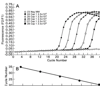
(iii) Validation of sensitivity and quantitation.The amplicon generated in the assay was cloned using a TA cloning kit and pCR 2.1 vector (Invitrogen Corp). The pCR 2.1 plasmid with cloned insert was sequenced (ABI 3100 capillary sequencer; Applied Biosystems, Inc., Foster City, Calif.) to confirm the insertion of a single copy of amplicon. The quantitation, accuracy, and precision of the real-time PCR assay were validated through serial dilutions of the pCR 2.1 plasmid with clonedA. fumigatusrRNA amplicon or gene (Fig. 2). The same plasmid was used as an external quantitation standard in all reaction runs. The conversion from copy number to genome equivalent was based on the estimated number of 100 ribosomal DNA (rDNA) gene complexes in theA. fumigatus
genome. For example, a reaction with 1,000 rDNA copies was equivalent to 10 genome equivalents (26). A positive quantitative-PCR signal for BAL was de-fined as detectable amplification at⬍36 cycles, particularly in recognition of the fact that a small quantity ofAspergillusconidia or hyphae may be present as background contamination or transient colonization. None of the healthy lungs sampled (n⫽19) had a detectable signal. Therefore, a positive quantitative-PCR
FIG. 2. (A) The wild-type amplicon generated from
A. fumigatus
genomic DNA was cloned into a pCR 2.1 vector (Invitrogen Corp). The
fluorescent signal is proportional to the number of plasmids, ranging from 2.5
⫻
10
4to 2.5
⫻
10
0plasmids. (B) The linear regression line
demonstrates both the linearity of the assay and also its efficiency (i.e., the slope) (
r
⫽ ⫺1.00).
ATT ATC G 3⬘
Cap RD 640
5⬘
ATC AGT TAA AAC TTT CAA
CAA CGG A 3⬘
62.0
Cap RD 705
5⬘
GAT CAT GAC AAG ATT CGC
TTC TAA T 3⬘
62.0
aTm, melting temperature.
on May 15, 2020 by guest
http://jcm.asm.org/
[image:3.603.42.283.81.202.2] [image:3.603.129.459.411.693.2]signal from lung samples was viewed as a true positive, reflecting the presence of
A. fumigatus.
(iv) Specificity: mutated-probe studies.The Cap RD 640 probe was mutated to create the Cap RD 705 probe, as delineated in Table 1. We demonstrated the specificity of the Cap RD 640 probe by the absence of signal when the Cap RD 705 probe was used instead.
(v) Specificity: cross-reactivity studies.Cross-reactivity of the assay was as-sessed by using DNA extracted fromAspergillus terreus,Aspergillus niger, Aspergil-lus flavus,Pseudallescheria boydii,Candida albicans,Candida glabrata, Cryptococ-cus neoformans, Penicillium chrysogenum, Penicillium citrinum, Penicillium purpurogenum, andTrichosporon beigeliias well as rabbit DNA and human DNA. (vi) Inhibition studies.To confirm the lack of PCR inhibitors from lung tissue and BAL samples, a separate set of PCR-FRET reactions was performed. A unique control plasmid and respective set of primers and probes were designed. A plasmid construct which incorporated a sequence-specific region of the 18S rRNA gene for Candida albicanswas designed. This was done to avoid any competition or cross-reaction with potentialA. fumigatusDNA in the experi-mental samples. Primers and probes which would hybridize only to this construct were designed. The presence of inhibitors was checked by comparing the am-plification efficiency of this reaction in the extracted samples against those from reactions run with water. Equivalent amplification efficiency compared to reac-tions run with water reflected the absence of inhibitors. Each lung and BAL sample was tested for inhibitors as described.
RESULTS
Analytical sensitivity and specificity.
The accuracy and
pre-cision of the real-time PCR assay were validated through serial
dilutions of the newly formed construct containing the
A.
fu-migatus
rRNA amplicon or gene. The assay reliably detected
ⱕ
10 copies of the pCR 2.1 plasmid per reaction with cloned
A.
fumigatus
rRNA amplicon or gene. The fluorescent signal was
proportional to the log concentration of the plasmid (Fig. 2).
The calculated PCR efficiency was 1.94. As a measure of
in-terday reproducibility, the coefficient of variation of the PCR
assay was 10.1%. The assay did not cross-react with rabbit
DNA, human DNA, or DNA from any of the following
clini-cally relevant organisms:
Aspergillus terreus
,
Aspergillus niger
,
Aspergillus flavus
,
Pseudallescheria boydii
,
Candida albicans
,
Candida glabrata
,
Cryptococcus neoformans
,
Penicillium
chryso-genum
,
Penicillium citrinum
,
Penicillium purpurogenum
, and
Trichosporon beigelii
.
Detection of
A. fumigatus
in lung tissue.
The quantitative
real-time PCR assay was more sensitive than culture in
detect-ing
A. fumigatus
in lung tissue. PCR detected
A. fumigatus
in
infarcted lung tissue in rabbits with experimental IPA in 60 of
60 specimens (100%), compared with 38 of 60 specimens
(63.3%) for culture (
P
⬍
0.0001). The log genome equivalent
of the real-time PCR assay correlated well (
r
⫽
0.72;
P
⬍
0.0001) with quantitative culture results of lung tissue (log
CFU/gram) (Fig. 3). However, the 22 tissues which were
cul-ture negative (0 CFU/g) also demonstrated a positive signal for
A. fumigatus
with log genome equivalents ranging from 0.86 to
5.11. Thus, the real-time PCR assay showed a sensitivity of
100% versus a sensitivity of 63.3% with the standard culture
method.
Consistent with this increased sensitivity of detection of
A.
fumigatus
in tissue, the mean log genome equivalent of 3.86
⫾
0.19/g of tissue was greater than that of the mean log CFU/
gram of culture of
A. fumigatus
(1.15
⫾
0.14) (Fig. 4).
Treat-ment with AMB significantly reduced the residual fungal
bur-den in lung tissue as measured by both quantitative PCR (
P
⬍
0.0001) and culture (
P
⬍
0.0001).
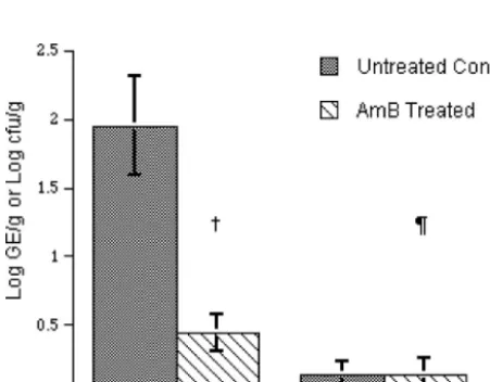
Detection of
A. fumigatus
in BAL fluid.
The relative
quanti-tative relationships between untreated controls and animals
treated with AMB were also consistent in BAL fluid when
analyzed by real-time PCR. There was a significant decline in
detectable signal in treated animals (0.44
⫾
0.14 log
10genome
equivalents) compared with that in untreated control animals
(1.95
⫾
0.36 log
10genome equivalents) (
P
⫽
0.0005) (Fig. 5).
By comparison, the level of detection of
A. fumigatus
in BAL
fluid was low (0.13
⫾
0.1 log
10CFU/ml) when measured by
culture in untreated controls and not significantly different in
comparison to that for treated animals (0.13
⫾
0.13 log
10 [image:4.603.119.471.70.289.2]CFU/ml) (
P
⫽
0.78). BAL samples from noninfected rabbits
FIG. 3. Correlation between real-time PCR with FRET technology (log genome equivalent [GE]) and quantitative culture (log
10CFU/gram)
in lobes from lungs of untreated rabbits with IPA (
r
⫽
0.72, Spearman rank correlation;
P
ⱕ
0.0001).
on May 15, 2020 by guest
http://jcm.asm.org/
were analyzed and found to be negative for
A. fumigatus
ge-nome at
ⱕ
36 cycles in 33 of 34 runs (specificity
⫽
97.1%).
DISCUSSION
We developed a sensitive, species-specific, real-time PCR
assay for
A. fumigatus
that was more sensitive than culture in
detecting
A. fumigatus
in BAL fluid from animals with
exper-imental IPA. Although the definitive diagnosis of IPA often
requires obtainment of tissue for histology and culture, such
procedures are often not feasible in immunocompromised
pa-tients. Instead, BAL is much more frequently performed for
the diagnosis of IPA. Unfortunately, BAL fluid samples are
often culture negative, necessitating lung biopsy or the use of
empirical therapy (11). The use of a sensitive and rapid
real-time assay, particularly in the case of BAL fluid samples, would
be valuable in establishing the diagnosis of IPA. Moreover, the
Beyond its potential clinical utility, this quantitative PCR assay
is a useful tool by which to study the pathogenesis and
treat-ment of experitreat-mental IPA.
Several key issues regarding the design of assay format,
primers, and probes warrant discussion. We purposely avoided
the use of a nested PCR approach as a method to increase the
sensitivity of the assay because of the major difficulties of
amplicon contamination inherent in such a method (10, 20). A
nested PCR format also undermines the benefits of the rapid
turnaround time associated with the real-time PCR approach.
The rRNA gene was chosen because (i) the multicopy
na-ture of the gene (at least 100 copies per genome) improves the
sensitivity of the assay, (ii)
A. fumigatus
cells separated by septa
form a multinucleate hyphal structure (4), thereby increasing
the number of copies of the target gene, and (iii) the large
amount of sequence data involving this gene complex across
many different strains of
A. fumigatus
and across many
differ-ent filamdiffer-entous fungi strengthen the design for a sensitive
assay. By utilizing the ITS regions of the complex for primer
design, it was possible to develop a species-specific assay while
preserving sensitivity. The high specificity of the primers in the
assay is critical in the case of nonsterile specimens such as BAL
fluid in order to avoid the occurrence of a competing PCR with
resultant reduction in sensitivity of
A. fumigatus
amplification.
In designing the fluorescent probes, a relatively short
dis-tance of approximately 1 to 5 bases is initially used in order to
allow for fluorescent energy transfer between the marker dyes.
By mutating the Cap RD 640 probe to Cap RD 705 (Table 1),
we demonstrated its specificity for the regional sequence of the
ITS1 gene of
A. fumigatus
.
The FRET assay detected
ⱕ
10 copies of
A. fumigatus
rRNA
gene per reaction. The assay was also highly specific, as shown
by the lack of cross-reactivity with other species of fungi and
bacteria. A wide linear range that extended out to 6 to 7 orders
of magnitude was demonstrated.
The quantitative PCR assay was more sensitive than culture,
detecting nearly 1,000-fold more genome equivalents in lung
tissue of nontreated animals. Culture detected only a 10-fold
difference. A similar pattern is seen in BAL. This increased
sensitivity presumably represented a combination of both free
A. fumigatus
DNA and nonviable fungal elements in untreated
lung tissues and BAL fluid. The assay detected 100 copies of
A.
fumigatus
rDNA (equivalent to one nucleus) per 10 mg of lung
tissue. Such a finding correlates with our measured extraction
efficiencies (see below).
Although not within the scope of the experiments reported
here, the enhanced sensitivity of the quantitative PCR assay
compared to that of culture may translate into earlier
detec-tion. To test this hypothesis, a separate series of experiments
could be designed to perform BAL on rabbits at designated
time points. The yield of quantitative PCR versus that of
cul-ture of BAL could then be compared.
[image:5.603.48.271.67.251.2]An internal inhibition control was incorporated into the
FIG. 4. Real-time PCR assay and standard culture assay reflecting
efficacy of AMB in lung samples from a rabbit model of IPA. Asterisks
indicate
P
value of
ⱕ
0.0001.
FIG. 5. Increased sensitivity of real-time PCR assay versus that of
standard culture in BAL fluid samples, as reflected by PCR assay
showing the efficacy of AMB in experimental IPA. †,
P
⫽
0.0005; ¶,
P
⫽
0.7805.
on May 15, 2020 by guest
http://jcm.asm.org/
[image:5.603.45.271.512.688.2]assay during its development. This permitted an assessment of
the presence or absence of inhibitors in the tissue and BAL
DNA extracts. Inhibitors were not demonstrated in lung tissue
or BAL fluid.
A number of different fungal DNA extraction methods were
initially assessed. These included the IsoQuick procedure
(Orca Research Inc., Bothwell, Wash.), Clontech’s (Palo Alto,
Calif.) DNA extraction kit, and Qiagen’s DNeasy Tissue and
DNeasy Plant kits. For the lung tissue samples, the method
that yielded the highest DNA extraction efficiency was
homog-enization with chaotropic high-speed disruption with lytic
ma-trices (HSCD/LM), followed by lyticase treatment (14) and
subsequent processing with the DNeasy Plant kit. For the BAL
fluid samples, the optimal approach involved using lyticase
followed by HSCD/LM, which was followed by the DNeasy
Plant kit. To highlight the difficulties encountered in fungal
DNA extraction, we calculated that for
A. fumigatus
conidia no
method assessed yielded more than 0.1% of fungal DNA, with
most yielding significantly less, consistent with previous
extrac-tion reports (7, 14).
All tissue samples from infected animals had hemorrhagic
infarcts or resolving infarct lesions, which correlate
histologi-cally with the presence of angioinvasive fungal infection (1).
Hence, the negative culture but positive PCR is not a
false-positive reaction but rather an indication of the greater
sensi-tivity of the PCR assay. These experimental observations also
correlate with the clinical observation that biopsies from
pa-tients that demonstrate histologically documented hyphal
in-vasion by fungi morphologically compatible with
Aspergillus
spp. may be negative by culture.
We demonstrated a very clear effect of treatment with AMB
in this animal model of IPA. For BAL fluid specimens, we
found a mean reduction of 1.51 log
10expressed as genome
equivalents. The reduction in tissue specimens was more
marked at 3.53 log
10, suggesting that the AMB is more effective
in tissue than in the bronchial mucosa, presumably secondary
to the respective drug levels. Another possibility is that the
assay may have detected some of the original conidia
inocu-lated but not cleared by AMB.
Although several studies have applied nonquantitative PCR
assays to human BAL specimens (2, 3, 8, 18), this is to our
knowledge the first report of the application of quantitative
PCR for the detection of experimental IPA. Moreover, we
have characterized and validated the assay in a well-defined
model of experimental IPA, where variables of
immunosup-pression, tissue burden, and therapy may be rigorously
con-trolled. This approach to characterization and validation of the
PCR assay provides a strong quantitative foundation for
un-derstanding data generated from its application to specimens
of BAL fluid from humans.
In summary, we have developed and validated a quantitative
real-time PCR assay to detect
A. fumigatus
. The assay is
sen-sitive and specific and easily demonstrates the effect of AMB in
the animal model of IPA. Assessment of the assay using human
specimens is presently ongoing.
REFERENCES
1. Berenguer, J., M. C. Allende, J. W. Lee, K. Garrett, C. Lyman, N. M. Ali, J. Bacher, P. A. Pizzo, and T. J. Walsh.1995. Pathogenesis of pulmonary aspergillosis. Granulocytopenia versus cyclosporine and methylprednisolone-induced immunosuppression. Am. J. Respir. Crit. Care Med.152:1079–1086.
2. Buchheidt, D., C. Baust, H. Skladny, M. Baldus, S. Brauninger, and R. Hehlmann.2002. Clinical evaluation of a polymerase chain reaction assay to detectAspergillusspecies in bronchoalveolar lavage samples of neutropenic patients. Br. J. Haematol.116:803–811.
3. Buchheidt, D., C. Baust, H. Skladny, J. Ritter, T. Suedhoff, M. Baldus, W. Seifarth, C. Leib-Moesch, and R. Hehlmann.2001. Detection ofAspergillus
species in blood and bronchoalveolar lavage samples from immunocompro-mised patients by means of 2-step polymerase chain reaction: clinical results. Clin. Infect. Dis.33:428–435.
4. Burnett, J. H.1975. Mycogenetics: an introduction to the general genetics of fungi. John Wiley & Sons, London, England.
5. Caillot, D., O. Casasnovas, A. Bernard, J. F. Couaillier, C. Durand, B. Cuisenier, E. Solary, F. Piard, T. Petrella, A. Bonnin, G. Couillault, M. Dumas, and H. Guy.1997. Improved management of invasive pulmonary aspergillosis in neutropenic patients using early thoracic computed tomo-graphic scan and surgery. J. Clin. Oncol.15:139–147.
6. Francis, P., J. W. Lee, A. Hoffman, J. Peter, A. Francesconi, J. Bacher, J. Shelhamer, P. A. Pizzo, and T. J. Walsh.1994. Efficacy of unilamellar liposomal amphotericin B in treatment of pulmonary aspergillosis in persis-tently granulocytopenic rabbits: the potential role of bronchoalveolarD -mannitol and serum galactomannan as markers of infection. J. Infect. Dis. 169:356–368.
7. Haugland, R. A., J. L. Heckman, and L. J. Wymer.1999. Evaluation of different methods for the extraction of DNA from fungal conidia by quan-titative competitive PCR analysis. J. Microbiol. Methods37:165–176. 8. Hayette, M. P., D. Vaira, F. Susin, P. Boland, G. Christiaens, P. Melin, and
P. De Mol.2001. Detection ofAspergillusspecies DNA by PCR in bron-choalveolar lavage fluid. J. Clin. Microbiol.39:2338–2340.
9. Herbrecht, R., V. Letscher-Bru, C. Oprea, B. Lioure, J. Waller, F. Cam-pos, O. Villard, K. L. Liu, S. Natarajan-Ame, P. Lutz, P. Dufour, J. P. Bergerat, and E. Candolfi.2002.Aspergillusgalactomannan detection in the diagnosis of invasive aspergillosis in cancer patients. J. Clin. Oncol. 20:1898–1906.
10. Kawamura, S., S. Maesaki, T. Noda, Y. Hirakata, K. Tomono, T. Tashiro, and S. Kohno.1999. Comparison between PCR and detection of antigen in sera for diagnosis of pulmonary aspergillosis. J. Clin. Microbiol.37:218–220. 11. Latge, J. P.1999.Aspergillus fumigatusand aspergillosis. Clin. Microbiol.
Rev.12:310–350.
12. Lortholary, O., S. Ascioglu, P. Moreau, R. Herbrecht, A. Marinus, P. Casas-sus, B. De Pauw, and D. W. Denning for the European Organization for Research and Treatment of Cancer/Invasive Fungal Infections Cooperative Group and the Intergroupe Francais du Myelome.2000. Invasive aspergil-losis as an opportunistic infection in nonallografted patients with multiple myeloma: a European Organization for Research and Treatment of Cancer. Clin. Infect. Dis.30:41–46.
13. Maertens, J., J. Verhaegen, H. Demuynck, P. Brock, G. Verhoef, P. Vandenberghe, J. Van Eldere, L. Verbist, and M. Boogaerts.1999. Au-topsy-controlled prospective evaluation of serial screening for circulating galactomannan by a sandwich enzyme-linked immunosorbent assay for hematological patients at risk for invasive aspergillosis. J. Clin. Microbiol. 37:3223–3228.
14. Muller, F. M., K. E. Werner, M. Kasai, A. Francesconi, S. J. Chanock, and T. J. Walsh.1998. Rapid extraction of genomic DNA from medically impor-tant yeasts and filamentous fungi by high-speed cell disruption. J. Clin. Microbiol.36:1625–1629.
15. National Research Council.1996. Guide for the care and use of laboratory animals. National Academy Press, Washington, D.C.
16. Paterson, D. L., and N. Singh.1999. Invasive aspergillosis in transplant recipients. Medicine (Baltimore)78:123–138.
17. Petraitiene, R., V. Petraitis, A. H. Groll, T. Sein, R. L. Schaufele, A. Francesconi, J. Bacher, N. A. Avila, and T. J. Walsh.2002. Antifungal efficacy of caspofungin (MK-0991) in experimental pulmonary aspergillosis in persistently neutropenic rabbits: pharmacokinetics, drug disposition, and relationship to galactomannan antigenemia. Antimicrob. Agents Che-mother.46:12–23.
18. Raad, I., H. Hanna, A. Huaringa, D. Sumoza, R. Hachem, and M. Albitar. 2002. Diagnosis of invasive pulmonary aspergillosis using polymerase chain reaction-based detection ofAspergillusin BAL. Chest121:1171–1176. 19. Saito, H., E. J. Anaissie, R. C. Morice, R. Dekmezian, and G. P. Bodey.1988.
Bronchoalveolar lavage in the diagnosis of pulmonary infiltrates in patients with acute leukemia. Chest94:745–749.
20. Skladny, H., D. Buchheidt, C. Baust, F. Krieg-Schneider, W. Seifarth, C. Leib-Mosch, and R. Hehlmann.1999. Specific detection ofAspergillus spe-cies in blood and bronchoalveolar lavage samples of immunocompromised patients by two-step PCR. J. Clin. Microbiol.37:3865–3871.
21. Stynen, D., A. Goris, J. Sarfati, and J. P. Latge.1995. A new sensitive sandwich enzyme-linked immunosorbent assay to detect galactofuran in pa-tients with invasive aspergillosis. J. Clin. Microbiol.33:497–500.
22. Verweij, P. E., Z. Erjavec, W. Sluiters, W. Goessens, M. Rozenberg-Arska, Y. J. Debets-Ossenkopp, H. F. Guiot, and J. F. Meis for the Dutch
![FIG. 3. Correlation between real-time PCR with FRET technology (log genome equivalent [GE]) and quantitative culture (login lobes from lungs of untreated rabbits with IPA (10 CFU/gram)r � 0.72, Spearman rank correlation; P � 0.0001).](https://thumb-us.123doks.com/thumbv2/123dok_us/8242529.831187/4.603.119.471.70.289/correlation-technology-equivalent-quantitative-culture-untreated-spearman-correlation.webp)