Isolation of
Salmonella
spp.
Sang-Hyun Park, Sangryeol Ryu, and Dong-Hyun Kang
Department of Food and Animal Biotechnology, Department of Agricultural Biotechnology, Center for Agricultural Biomaterials, and Research Institute for Agriculture and Life Sciences, Seoul National University, Seoul, Republic of Korea
We describe an improved selective, differential, and cost-effective medium, XA medium, which contains
D-arabinose, to
facili-tate the selective isolation of
Salmonella
spp. The sensitivity and the specificity of XA medium were compared to those of xylose
lysine desoxycholate agar (XLD) using stock cultures and naturally contaminated food samples. XA medium and XLD were
eval-uated with a total of 82
Salmonella
and 69 non-
Salmonella
stock cultures. Of 82 strains of
Salmonella
spp. tested, 76 produced a
characteristic black colony on XA medium and XLD. The remaining 6 strains belonged to
Salmonella enterica
serovars Berta
(
n
ⴝ
1), Paratyphi A (
n
ⴝ
1), Gallinarum (
n
ⴝ
2), and Pullorum (
n
ⴝ
2). The sensitivities of XA medium and XLD were identical
(92.7%).
Citrobacter freundii
(
n
ⴝ
21) and
Proteus mirabilis
(
n
ⴝ
21) stock cultures produced black colonies on XLD, whereas
only 4 strains of
P. mirabilis
appeared as black colonies on XA medium. In the second phase of the study, a total of 180 food
sam-ples were cultured onto XA medium and XLD after selective enrichment. The sensitivities of XA medium and XLD were equal
(100%), and a total of 6
Salmonella
strains were isolated from the 180 food samples. The specificity of XA medium (92.0%) was
superior to that of XLD (73.0%), with a total of 14 and 47 false-positive results found on XA medium and XLD, respectively. On
the basis of its good specificity, XA medium is useful for the isolation of
Salmonella
spp. from food samples.
S
almonella
has been associated with many food-borne diseases
across the world (
19
). It is the cause of an estimated 1.4 million
illnesses annually in the United States (
25
). Various foods, such as
chicken, beef, and pork, have been implicated in outbreaks caused
by
Salmonella
spp. (
8
,
27
,
33
). Thus, effective methods for the
isolation of
Salmonella
spp. from various foods are important to
ensure food quality and safety. The choice of a suitable sampling
procedure combined with a sensitive culture method is important
for the successful detection of
Salmonella
(
4
).
The use of selective and differential plating media is a simple
method for the isolation of
Salmonella
spp. A wide variety of
se-lective and differential media has been developed for this purpose,
including xylose lysine desoxycholate agar (XLD), Hektoen
en-teric (HE) agar, and bismuth sulfite (BS) agar (
6
). XLD and HE
agar are the most popular media for isolating
Salmonella
spp., and
their differentiation abilities rely on characteristics of
Salmonella
,
such as hydrogen sulfide production and the nonfermentation of
lactose (
28
). However, these characteristics are shared with other
microorganisms, such as
Proteus
and
Citrobacter
(
11
,
32
). Thus,
numerous false-positive results are observed on these media
which require further confirmation testing, a time-consuming
and labor-intensive activity (
13
). BS agar is the medium of choice
for the isolation of
Salmonella enterica
serovar Typhi, and it is used
for the isolation of atypical salmonellae, such as those which
fer-ment lactose (
7
). However, BS agar has several disadvantages, such
as low sensitivity and long incubation time for development of the
characteristic colony morphology (
18
).
Several chromogenic media have been developed to increase
the specificity of conventional selective and differential media for
the detection of
Salmonella
spp. (
14
,
21
,
26
,
23
,
31
). These media
incorporate chromogenic substrates which are metabolized by
Salmonella
spp. (
29
). Although chromogenic media have higher
specificities than conventional media, some of them have a low
sensitivity (
10
,
13
,
30
), which results in more false negatives
ob-served on these media. Also, chromogenic media are relatively
expensive, making them less appropriate for routine laboratory
use (
24
).
Recognizing the limits of currently used selective and
differen-tial media, it is desirable to improve the specificity and the
sensi-tivity of the medium while maintaining cost-effectiveness.
Partic-ularly, it is desirable to differentiate
Salmonella
spp. from
Proteus
spp., as well as from
Citrobacter
spp. Hydrogen sulfide production
depends on several factors, such as the sulfide production rate of
the microorganisms, the oxygen concentration in the colony, pH,
and the iron concentration in the medium (
28
). Acid production
by microorganisms in consequence of carbohydrate fermentation
could inhibit hydrogen sulfide production (
3
).
This study yielded XA medium, which contains
D-arabinose as
a differential agent and neutral red as a pH indicator to
differen-tiate
Salmonella
from
Citrobacter freundii
,
Proteus mirabilis
, and
other enteric bacteria. The effectiveness of XA medium was
com-pared to that of XLD using stock cultures and naturally
contami-nated food samples.
MATERIALS AND METHODS
Stock cultures.S. entericaserovar Typhimurium (ATCC 19585, ATCC 43971, ATCC 700408),S. entericaserovar Enteritidis ATCC 13078,C.
freundiiATCC 8090,Escherichia coli(ATCC 8739, ATCC 11775, ATCC
25922),E. coli O157:H7 (ATCC 35150, ATCC 43889, ATCC 43890),
Hafnia alvei(ATCC 13337, ATCC 29926, ATCC 29927),Yersinia
entero-colitica(ATCC 9610, ATCC 23715, ATCC 55075),Cronobacter sakazakii
(ATCC 12868, ATCC 29004),Klebsiella pneumoniaATCC 13883,Shigella
Received9 May 2012Returned for modification1 June 2012
Accepted16 July 2012
Published ahead of print18 July 2012
Address correspondence to Dong-Hyun Kang, [email protected].
Copyright © 2012, American Society for Microbiology. All Rights Reserved.
doi:10.1128/JCM.01228-12
on May 16, 2020 by guest
http://jcm.asm.org/
flexneri (ATCC 12022, ATCC 29903), and Pseudomonas aeruginosa
(ATCC 15692, ATCC 27853) were obtained from the American Type Culture Collection (ATCC; Manassas, VA).S. entericaserovar Agona NCCP 12231,S. entericaserovar Berta NCCP 10270,S. entericaserovar California NCCP 10319,S. entericaserovar Chester NCCP 10317,S.
en-terica serovar Derby NCCP 12238,S. enterica serovar Dublin NCCP
10860,S. entericaserovar Florida NCCP 10361,S. entericaserovar Galli-narum NCCP 10323,S. entericaserovar Heidelberg NCCP 10322,S.
en-tericaserovar Illinois NCCP 10437,S. entericaserovar Infantis NCCP
12233,S. entericaserovar London NCCP 10831,S. entericaserovar Maars-sen NCCP 10951,S. entericaserovar Montevideo NCCP 12211,S. enterica
serovar Newington NCCP 10440,S. entericaserovar Newport (NCCP 10325, NCCP 12235),S. entericaserovar Oranienburg NCCP 10441,S.
enterica serovar Panama NCCP 10333, S. enterica serovar Pullorum
NCCP 10335,S. entericaserovar SaintPaul NCCP 10329,S. enterica sero-var Senftenberg NCCP 12240,S. entericaserovar Schottmuelleri NCCP 10324,S. entericaserovar Schwarzergrund NCCP 12212,S. enterica sero-var Sloterdijk NCCP 10331,S. entericaserovar Stanley NCCP 10332,S.
entericaserovar Tennessee NCCP 10861,S. entericaserovar Thompson
NCCP 11011, andS. entericaserovar Weltevreden NCCP 12239 were obtained from the National Culture Collection for Pathogens (NCCP; Osong, South Korea).S. entericaserovar Choleraesuis (KCCM 13076, KCCM 14028),S. entericasubsp.arizonae(KCCM 41035, KCCM 41575),
S. entericasubsp. salamae (KCCM 41651, KCCM 41762),S. enterica
subsp.indicaKCCM 41759,S. entericasubsp.houtenaeKCCM 41760, and
S. entericasubsp.diarizonaeKCCM 41761 were obtained from the Korean
Culture Center of Microorganisms (KCCM; Seoul, South Korea).S. Gal-linarum KVCC 1457,S. Heidelberg KVCC 0506,S. Pullorum KVCC 2509,
S. Senftenberg KVCC 0590,S. Tennessee KVCC 0592, andS. enterica
serovar Virchow KVCC 0595 were obtained from the Korea Veterinary Culture Collection (KVCC; Anyang, South Korea). The remaining strains were isolates obtained from the bacterial culture collection of the Food Hygiene Laboratory at Seoul National University (SNCC; Seoul, South Korea). They were stored frozen at⫺80°C.
Preparation of the basal medium.Xylose lysine desoxycholate agar (XLD; Difco, Sparks, MD) was used as a base for the development of XA medium. The ingredients of the basal medium were as follows: 3.75 g
D-xylose (Fluka, Paris, France), 5.0 g ofL-lysine (Junsei Chemical Co. Ltd.,
Tokyo, Japan), 7.5 g of lactose (Difco), 7.5 g of sucrose (Difco), 5.0 g of sodium chloride (Samchun Chemical Co. Ltd., Pyeongtaeksi, South Ko-rea), 3.0 g of yeast extract (Difco), 2.5 g of sodium desoxycholate (Difco), 6.8 g of sodium thiosulfate (Junsei Chemical), 0.8 g of ferric ammonium citrate (Acros Organics, NJ), 15.0 g of Bacto agar (Difco), and 0.03 g of neutral red (Samchun Chemical) per liter. Various quantities ofD
-arabi-nose (7.5, 9.0, or 10.5 g/liter) (Fluka) were added to the basal medium to optimize the concentration of this component. The ingredients were added to distilled water, and the preparation was heated with agitation just until the medium boils. The medium was cooled to 50°C and poured into 9-cm-diameter petri dishes.
Evaluation of the effect ofD-arabinose concentration.Test bacteria (82 strains ofSalmonella, 21 strains ofC. freundii, and 21 strains ofP.
mirabilis) were incubated in 10 ml of tryptic soy broth (TSB; Difco) at
37°C for 18 h. After incubation, one loopful (ca. 10l) of each culture was streaked onto XLD and each of the basal media (containing 7.5, 9.0, or 10.5 g/literD-arabinose) to obtain single, isolated colonies and incubated at 37°C for 48 h. The colors of colonies were compared and recorded after 24, 36, and 48 h. The experiments were repeated twice with each tested strain. Colonies suspected of beingSalmonellaspp. were defined as black colonies on both XLD and the basal media.
The specificity testing of XA medium with other Gram-negative bac-teria.XA medium was prepared with the previously determined optimum
D-arabinose concentration. Twenty-seven strains of Gram-negative
bac-teria (seeTable 2) were incubated in 10 ml of TSB at 37°C for 18 h. After incubation, one loopful of each culture was streaked onto XLD and XA medium. The plates were incubated at 37°C for 48 h, and the colors of
colonies were compared and recorded. The experiments were repeated twice with each tested strain.
Assessment of the performance of XA medium using naturally con-taminated food samples.The conventional culture method described by the U.S. Food and Drug Administration (FDA) was used for the microbi-ological analysis of naturally contaminated foods (1). A total of 180 sam-ples consisting of chicken (n⫽80), ground beef (n⫽50), and ground pork (n⫽50) were purchased from local retail markets (Seoul, South Korea). Each sample (25 g) was homogenized with a stomacher (EASY MIX, AES Chemunex, Rennes, France) in sterile stomacher bags (Labplas Inc., Sainte-Julie, Quebec, Canada) containing 225 ml of lactose broth (Difco) for 2 min and incubated at 37°C for 22 h. One hundred microliters of preenriched culture was transferred to 10 ml of Rappaport-Vassiliadis (RV) medium (Difco) and incubated at 42°C for 24 h. After selective enrichment, one loopful of each RV culture was streaked onto XLD and XA medium and incubated at 37°C for 24 h. After incubation, a maximum of 5 colonies suspected of beingSalmonellaon XLD and XA medium were selected for identification. Bacterial colonies were identified using the API 20E system (bioMérieux SA) and theSalmonellalatex agglutination kit (Oxoid Ltd., Cambridge, United Kingdom). The specificity was evaluated by calculating the proportion ofSalmonella-negative samples correctly found to be negative (i.e., those that did not appear asSalmonella-like colonies).
RESULTS
A total of 124 strains (82 strains of
Salmonella
, 21 strains of
C.
freundii
, and 21 strains of
P. mirabilis
) were streaked onto each
basal medium (containing 7.5, 9.0, or 10.5 g/liter
D-arabinose)
(
Table 1
). At a
D-arabinose concentration of 7.5 to 10.5 g/liter,
most
Salmonella
strains produced typical black colonies after 24 h
of incubation, except
S
. Paratyphi A,
S
. Berta,
S
. Gallinarum, and
S
. Pullorum.
S
. Paratyphi A,
S
. Gallinarum, and
S
. Pullorum
pro-duced colorless colonies, and
S
. Berta appeared as pink colonies
on each basal medium.
Salmonella
strains belonging to
S. enterica
serovars California, Chester, Maarssen, and Montevideo
pro-duced pink colonies with black centers on each basal medium after
24 h of incubation. Also, 3 strains of
S.
Enteritidis,
S
. Florida, and
S
. London appeared as pink colonies with black centers at a
D-arabinose concentration of
ⱖ
9.0 g/liter. After 36 h of incubation,
these strains appeared as black colonies with no pink halo.
S
.
Typhi (
n
⫽
1) produced colonies with small black centers on both
XLD and XA medium.
C. freundii
produced no false-positive results on any of the
basal media within 48 h of incubation. Four strains of
P. mirabilis
produced pink colonies with small black centers on the basal
me-dium containing 7.5 g/liter of
D-arabinose after 24 h of incubation,
and among these, two strains produced pink colonies with small
black centers on the basal medium containing 9.0 g/liter of
D-arabinose. However, all
P. mirabilis
strains failed to produce black
colonies at a
D-arabinose concentration of 10.5 g/liter. The final
XA medium formulation contained 7.5 g/liter of
D-arabinose. The
sensitivities (the ratio of true positives over the number of true
positives and false negatives) of XA medium and XLD were
iden-tical (92.7%). Twenty-seven strains of other Gram-negative
bac-teria (
Table 2
) did not produce false-positive results on either XLD
or XA medium.
Figure 1
shows that colonies of
S.
Typhimurium,
C. freundii
,
and
P. mirabilis
formed on XLD and XA medium.
S.
Typhimu-rium (
Fig. 1A
) produced typical black colonies on XLD after 24 h
of incubation. However,
C. freundii
(
Fig. 1B
) produced black
col-onies or black colcol-onies surrounded by yellow halos, and
P.
mira-bilis
(
Fig. 1C
) appeared as black colonies on XLD, similar to those
on May 16, 2020 by guest
http://jcm.asm.org/
of
Salmonella
.
S.
Typhimurium (
Fig. 1D
) appeared as black
colo-nies on XA medium, whereas
C. freundii
and
P. mirabilis
produced
pink colonies (
Fig. 1E
and
F
).
Table 3
represents results of the bacteriological analysis of
nat-urally contaminated food samples. A total of 6
Salmonella
spp.
were isolated on both XLD and XA medium from the same food
samples (sensitivity, 100%) (data not shown). The specificity of
XA medium (92.0%) was superior to that of XLD (73.0%), with a
total of 14 and 47 false-positive results found on XA medium and
XLD, respectively. They consisted of
P. mirabilis
on XA medium
and
P. mirabilis
and
C. freundii
on XLD. No false-positive results
with
C. freundii
were observed on XA medium. All strains of
P.
mirabilis
that produced false-positive results on XA medium also
produced false-positive results on XLD.
DISCUSSION
Consumption of contaminated foods of animal origin is
respon-sible for the great majority of salmonellosis cases (
31
). Plating on
XLD, HE agar, and BS agar after preenrichment followed by
selec-tive enrichment in RV medium or tetrathionate (TT) broth has
been recommended for the isolation of
Salmonella
from foods by
the U.S. FDA (
1
). However, colonies of
C. freundii
and
Proteus
spp., such as
P. mirabilis
, may resemble these of
Salmonella
due to
hydrogen sulfide production on HE agar (
2
). Although XLD has a
high sensitivity and specificity,
Proteus
and
Citrobacter
produce
colonies indistinguishable from those of
Salmonella
on this
me-dium (
6
,
28
,
32
).
Acid formation in consequence of carbohydrate fermentation
may affect hydrogen sulfide production (
3
,
28
). Veron and Gasser
(
34
) reported that under the acid conditions of carbohydrate
me-tabolism, H
2S-positive
Enterobacteriaceae
were unable to produce
the black precipitate of iron sulfide. In the present study, the
car-bohydrate-fermenting abilities of 15 strains each of
C. freundii
and
P. mirabilis
were examined using the API CH50 system
(bio-Mérieux SA, Marcy 1
=
Etoile, France) for screening additional
car-bohydrate sources (data not shown).
C. freundii
and
P. mirabilis
strains commonly fermented
D-arabinose, ribose, galactose,
glu-cose,
N
-acetylglucosamine, and trehalose. Among these
carbohy-drates, we selected
D-arabinose to add to XA medium in addition
to
D-xylose, lactose, and sucrose, which are contained in XLD,
because most major
Salmonella
spp., including
S.
Typhimurium,
did not ferment
D-arabinose (data not shown).
The final XA medium formulation contained 7.5 g/liter of
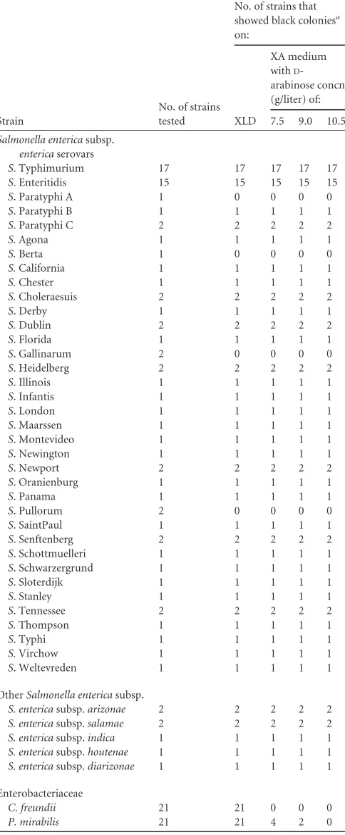
D-TABLE 1Colony color reactions ofSalmonellaspp.,C. freundii, andP.
mirabilison XLD and XA medium
Strain
No. of strains tested
No. of strains that showed black coloniesa
on:
XLD
XA medium withD
-arabinose concn (g/liter) of: 7.5 9.0 10.5
Salmonella entericasubsp.
entericaserovars
S. Typhimurium 17 17 17 17 17
S. Enteritidis 15 15 15 15 15
S. Paratyphi A 1 0 0 0 0
S. Paratyphi B 1 1 1 1 1
S. Paratyphi C 2 2 2 2 2
S.Agona 1 1 1 1 1
S.Berta 1 0 0 0 0
S.California 1 1 1 1 1
S.Chester 1 1 1 1 1
S.Choleraesuis 2 2 2 2 2
S. Derby 1 1 1 1 1
S.Dublin 2 2 2 2 2
S. Florida 1 1 1 1 1
S. Gallinarum 2 0 0 0 0
S. Heidelberg 2 2 2 2 2
S. Illinois 1 1 1 1 1
S. Infantis 1 1 1 1 1
S. London 1 1 1 1 1
S. Maarssen 1 1 1 1 1
S. Montevideo 1 1 1 1 1
S. Newington 1 1 1 1 1
S. Newport 2 2 2 2 2
S. Oranienburg 1 1 1 1 1
S. Panama 1 1 1 1 1
S. Pullorum 2 0 0 0 0
S. SaintPaul 1 1 1 1 1
S. Senftenberg 2 2 2 2 2
S. Schottmuelleri 1 1 1 1 1
S. Schwarzergrund 1 1 1 1 1
S. Sloterdijk 1 1 1 1 1
S. Stanley 1 1 1 1 1
S. Tennessee 2 2 2 2 2
S.Thompson 1 1 1 1 1
S. Typhi 1 1 1 1 1
S.Virchow 1 1 1 1 1
S. Weltevreden 1 1 1 1 1 OtherSalmonella entericasubsp.
S. entericasubsp.arizonae 2 2 2 2 2
S. entericasubsp.salamae 2 2 2 2 2
S. entericasubsp.indica 1 1 1 1 1
S. entericasubsp.houtenae 1 1 1 1 1
S. entericasubsp.diarizonae 1 1 1 1 1
Enterobacteriaceae
C. freundii 21 21 0 0 0
P. mirabilis 21 21 4 2 0
[image:3.585.299.545.87.220.2]aExpectedSalmonellaspp. color reactions: XLD, black; XA medium, black.
TABLE 2Colony colors of Gram-negative bacteria on XLD and XA medium
Strain
No. of strains tested
No. of strains that showed black coloniesa
on:
XLD XA medium
Escherichia coli 7 0 0
E. coliO157:H7 3 0 0
Hafnia alvei 5 0 0
Yersinia enterocolitica 3 0 0
Cronobacter sakazakii 3 0 0
Klebsiella pneumoniae 2 0 0
Shigella flexneri 2 0 0
Pseudomonas aeruginosa 2 0 0
aExpectedSalmonellaspp. color reactions: XLD, black; XA medium, black.
on May 16, 2020 by guest
http://jcm.asm.org/
[image:3.585.43.285.90.675.2]arabinose in spite of some false-positive results with
P. mirabilis
strains, because some
Salmonella
strains which fermented
D-arabinose produced pink colonies with black centers after 24 h of
incubation. It is assumed that these
Salmonella
strains produced
an initial acidification of the medium due to fermentation of
D-arabinose. Then, these strains apparently produced an alkaline
reversion in XA medium because of decarboxylation of lysine (
7
,
12
).
D-Arabinose may delay alkaline reversion by these strains, but
blackening of the colonies of these strains was obvious after 36 h.
This suggests that XA medium should be examined within 36 h
after inoculation.
All
C. freundii
stock cultures (
n
⫽
21) that produced black
colonies on XLD appeared as pink colonies on XA medium. Also,
no false-positive results due to
C. freundii
were observed on XA
medium from naturally contaminated food samples, whereas
false-positive results were observed on XLD. XA medium was
shown to be more specific than XLD for the differentiation of
Salmonella
from
P. mirabilis
, despite some false-positive results
with
P. mirabilis
which were still observed on XA medium.
Al-though
P. mirabilis
fermented
D-arabinose, it rarely fermented
lactose or sucrose contained in XA medium. It is quite likely that
the false-positive results with
P. mirabilis
strains were caused by
the fact that the amount of acid formation was not sufficient to
inhibit hydrogen sulfide production (
22
). Some
C. freundii
and
P.
mirabilis
strains showed variability in the size of black center of
colonies on XLD and XA medium. However, positive or negative
reactions were identical for each test.
Incorporating
D-arabinose does not affect the sensitivity of the
medium. The sensitivities were identical for both XA medium and
XLD.
S
. Paratyphi A (
n
⫽
1),
S
. Berta (
n
⫽
1),
S
. Gallinarum (
n
⫽
2), and
S
. Pullorum (
n
⫽
2) produced H
2S-negative colonies on
XA medium and XLD.
S
. Paratyphi A and
S
. Berta are known as
hydrogen sulfide negative; thus, their colonies do not appear as
black on media that detect hydrogen sulfide formation (
7
,
17
).
S
.
Gallinarum and
S
. Pullorum rarely produce hydrogen sulfide, and
the reaction occurred slowly (
5
). Also the hydrogen
sulfide-gen-erating ability of
S
. Typhi is weak or negative (
17
). On XA
me-dium,
S
. Typhi produced black colonies, but the black centers
were smaller than those of other
Salmonella
strains.
All strains of
S.
Typhimurium (
n
⫽
17) and
S.
Enteritidis (
n
⫽
15) were easily detected on XA medium. The detection of these
two
Salmonella
serotypes is important, as these serotypes are the
ones most frequently associated with food-borne outbreaks (
15
).
Also,
S.
Typhimurium and
S.
Enteritidis alone represent 73.9% of
all clinical isolates (
16
).
In the present study, XA medium contains neutral red as a dye
rather than phenol red contained in XLD. When phenol red was
used as a dye in XA medium, yellow-colored colonies due to low
pH were easily diffusible on agar (data not shown). Thus, neutral
red that produces less diffusible colored colonies on agar was used
in formulating XA medium.
Several rapid methods have been developed to detect
food-borne pathogens present in foods (
20
). However, conventional
selective and differential media are still important, due to several
advantages, including cost-effectiveness, ease of use, and
familiar-ity among users (
14
). A wide range of chromogenic media have
been developed and offered commercially. However, they are
more expensive than conventional media, largely due to the
addi-tional cost of enzyme substrates (
24
). High costs of chromogenic
media present a major obstacle for their use in routine practice.
D
-Arabinose incorporated in XA medium is cost-effective; thus, it
is adequate for routine laboratory use.
In conclusion, XA medium developed in this study has higher
specificity than XLD. Especially, XA medium easily differentiates
Salmonella
from
C. freundii
and
P. mirabilis
, which frequently
produce false-positive results on XLD. Due to the good specificity
of XA medium, labor and time can be saved due to fewer colonies
needing further confirmation tests. XA medium may provide a
valuable addition to the array of selective media available for the
FIG 1Colonies produced byS.Typhimurium,C. freundii, andP. mirabilison XLD (A, B, and C) and XA medium (D, E and F). Colonies ofS.Typhimurium
(A),C. freundii(B), andP. mirabilis(C) appeared as black colonies on XLD. On XA medium,S.Typhimurium (D) produced black colonies, whereasC. freundii
[image:4.585.125.461.67.237.2](E) andP. mirabilis(F) produced pink colonies.
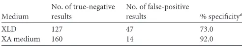
TABLE 3The specificity of XA medium compared with that of XLD on the microbiological analysis of naturally contaminated foods
Medium
No. of true-negative results
No. of false-positive
results % specificitya
XLD 127 47 73.0
XA medium 160 14 92.0
a(No. of true-negative results on the medium/no. of negative samples)⫻100.
on May 16, 2020 by guest
http://jcm.asm.org/
[image:4.585.39.285.668.715.2]detection of
Salmonella
spp. from foods. We recommend that XA
medium be used in parallel with HE agar and BS agar in the
ex-amination of food samples. Further studies are required to
evalu-ate the growth of more
Salmonella
serotypes on XA medium, and
it is necessary to assess the specificity of this new medium with
food samples other than meat products and stool samples.
ACKNOWLEDGMENTS
This work was supported by grant no. R32-2008-000-10183-0 from the World Class University (WCU) project of the Ministry of Education, Sci-ence & Technology (MEST) and the KOSEF through Seoul National Uni-versity.
REFERENCES
1.Andrews HW, Hammack TS.2002.Salmonella, p 10 –11.InFDA bacte-riological analytical manual online, 8th ed. U.S. Food and Drug Adminis-tration, Washington, DC.
2.Anomynous.2006. BBL™ Hektoen enteric agar quality control proce-dures. Becton, Dickinson and Company, Sparks, MD.http://www.bd.com /ds/technicalCenter/inserts/L007382(08)(0506).pdf.
3.Anomynous.2007. BBL™ XLD agar quality control procedures. Becton, Dickinson and Company, Sparks, MD.http://www.bd.com/ds/technical Center/inserts/L007426(09)(0907).pdf.
4.Carrique-Mas J, Davies R.2008. Sampling and bacteriological detection
ofSalmonellain poultry and poultry premises: a review. Rev. Sci. Tech.
27:665– 677.
5.Christensen JP, Olsen JE, Hansen HC, Bisgaard M.1992. Characteriza-tion ofSalmonella entericaserovargallinarumbiovars gallinarum and pul-lorum by plasmid profiling and biochemical analysis. Avian Pathol.21: 461– 470.
6.Cooke VM, Miles RJ, Price RG, Richardson AC.1999. A novel chromo-genic ester agar medium for detection of salmonellae. Appl. Environ. Mi-crobiol.65:807– 812.
7.Cox JM.1993. Lysine-mannitol-glycerol agar, a medium for the isolation
ofSalmonellaspp., includingS. typhiand atypical strains. Appl. Environ.
Microbiol.59:2602–2606.
8.de Jong B, Ekdahl K.2006. Human salmonellosis in travelers is highly correlated to the prevalence ofSalmonellain laying hen flocks. Euro Sur-veill.11:E060706.1.
9. Reference deleted.
10. Dusch H, Altwegg M.1993. Comparison of Rambach agar, SM ID me-dium, and Hektoen enteric agar for primary isolation of non-typhi salmo-nellae from stool samples. J. Clin. Microbiol.31:410 – 412.
11. Eigner U, Reissbrodt R, Hammann R, Fahr AM.2001. Evaluation of a new chromogenic medium for the isolation and presumptive identifica-tion ofSalmonellaspecies from stool specimens. Eur. J. Clin. Microbiol. Infect. Dis.20:558 –565.
12. Farmer JJ, et al.1985. Biochemical identification of new species and biogroups ofEnterobacteriaceaeisolated from clinical specimens. J. Clin. Microbiol.21:46 –76.
13. Gaillot O, Camillo D, Berche P, Courcol R, Savage C.1999. Comparison of CHROMagarSalmonellamedium and Hektoen enteric agar for isola-tion ofSalmonellafrom stool sample. J. Clin. Microbiol.37:762–765. 14. Gracias KS, Mckillip JL.2004. A review of conventional detection and
enumeration methods for pathogenic bacteria in food. Can. J. Microbiol.
50:883– 890.
15. Greig JD, Ravel A.2009. Analysis of foodborne outbreak data reported internationally for source attribution. Int. J. Food Microbiol.130:77– 87. 16. Herrera-León S, et al.2007. Blind comparison of traditional serotyping with three multiplex PCRs for the identification ofSalmonellaserotypes. Res. Microbiol.158:122–127.
17. Janda JM, Abbott SL.2008. The enterobacteria, 2nd ed. ASM Press, Washington, DC.
18. Jay LS, Davey GR.1989.Salmonella: characteristics, identification and enumeration, p 51– 82.InBuckle KA, Davey JA, Eyles MJ, Hocking AD, Newton KG, Stuttard EJ (ed), Foodborne microorganisms of public health significance. Australian Institute of Food Science and Technology New South Wales Food Microbiology Group, Sydney, Australia.
19. Jiménez SM, Tiburzi MC, Salsi MS, Moguilevsky MA, Pirovani ME.
2009. Survival ofSalmonellaon refrigerated chicken carcasses and subse-quent transfer to cutting board. Lett. Appl. Microbiol.48:687– 691. 20. Lund BM, Baird-Parker TC, Gould GW. 2000. The microbiological
safety and quality of foods. Aspen Publishers, Gaithersburg, MD. 21. Manafi M.2000. New developments in chromogenic and fluorogenic
culture media. Int. J. Food Microbiol.60:205–218.
22. O’Hara CM, Brenner FW, Miller JM.2000. Classification, identification, and clinical significance ofProteus,Providencia, andMorganella.Clin. Mi-crobiol. Rev.13:534 –546.
23. Perry JD, et al.1999. ABC medium, a new chromogenic agar for selective isolation ofSalmonellaspp. J. Clin. Microbiol.37:766 –768.
24. Perry JD, Freydière AM.2007. The application of chromogenic media in clinical microbiology. Lett. Appl. Microbiol.103:2046 –2055.
25. Perry JJ, Rodriguez-Romo LA, Yousef AE.2008. Inactivation of
Salmo-nella entericaserovar enteritidis in shell eggs by sequential application of
heat and ozone. Lett. Appl. Microbiol.46:620 – 625.
26. Poupart MC, et al.1991. A new chromogenic ready-to-use medium for
Salmonelladetection, abstr 1254. Abstr. Fifth Eur. Congr. Clin. Microbiol.
Infect. Dis.
27. Rabsch W, Tschape H, Baumler AJ.2001. Non-typhoidal salmonellosis: emerging problems. Microbes Infect.3:237–247.
28. Rambach A.1990. New plate medium for facilitated differentiation of
Salmonellaspp. fromProteusspp. and other enteric bacteria. Appl.
Envi-ron. Microbiol.56:301–303.
29. Ruiz J, et al.1996. Performance of six culture media for isolation of
Salmonellaspecies from stool samples. Eur. J. Clin. Microbiol. Infect. Dis.
15:922–926.
30. Ruiz J, et al.1996. Comparison of five plating media for isolation of
Salmonellaspecies from human stools. J. Clin. Microbiol.34:686 – 688.
31. Schönenbrücher V, Mallinson ET, Bülte M.2008. A comparison of standard cultural methods for the detection of foodborneSalmonella spe-cies including three new chromogenic plating media. Int. J. Food Micro-biol.123:61– 66.
32. Tate CR, Miller RG, Mallinson ET, Douglass LW, Johnston RW.1990. The isolation of salmonellae from poultry environmental samples by sev-eral enrichment procedures using plating media with and without novo-biocin. Poult. Sci.69:721–726.
33. Trevanich S, Tiyapongpattana S, Miyamoto T.2010. Application of an optimized 18-h method involving one step culturing and single primer-based PCR assay for detection ofSalmonellaspp. in foods. Food Control
21:593–598.
34. Veron M, Gasser F.1963. Sur la detection de l’hydrogene sulfure produit par certains enterobacteriacees dans les milieux dits de diagnostic rapide. Ann. Inst. Pasteur105:524 –534.