0095-1137/07/$08.00
⫹
0
doi:10.1128/JCM.00223-07
Copyright © 2007, American Society for Microbiology. All Rights Reserved.
Identification of Genotypically Diverse
Cryptococcus neoformans
and
Cryptococcus gattii
Isolates by Luminex xMAP Technology
䌤
M. Bovers,
1M. R. Diaz,
2F. Hagen,
1L. Spanjaard,
3B. Duim,
4C. E. Visser,
4H. L. Hoogveld,
5J. Scharringa,
6I. M. Hoepelman,
6J. W. Fell,
2and T. Boekhout
1,6*
CBS—Fungal Biodiversity Centre, Utrecht, The Netherlands
1; University of Miami, Rosenstiel School of Marine and Atmospheric Science
(RSMAS), Key Biscayne, Florida
2; The Netherlands Reference Laboratory for Bacterial Meningitis (AMC/RIVM), Department of
Medical Microbiology, Academic Medical Center, Amsterdam, The Netherlands
3; Department of Medical Microbiology,
Academic Medical Center, Amsterdam, The Netherlands
4; Netherlands Institute of Ecology (NIOO-KNAW), Centre for
Limnology, Nieuwersluis, The Netherlands
5; and Department of Internal Medicine and Infectious Diseases,
University Medical Centre Utrecht, Utrecht, The Netherlands
6Received 29 January 2007/Returned for modification 5 March 2007/Accepted 11 April 2007
A Luminex suspension array, which had been developed for identification of
Cryptococcus neoformans
and
Cryptococcus gattii
isolates, was tested by genotyping a set of 58 mostly clinical isolates. All genotypes of
C.
neoformans
and
C. gattii
were included. In addition, cerebrospinal fluid (CSF) obtained from patients with
cryptococcal meningitis was used to investigate the feasibility of the technique for identification of the infecting
strain. The suspension array correctly identified haploid isolates in all cases. Furthermore, hybrid isolates
possessing two alleles of the Luminex probe region could be identified as hybrids. In CSF specimens, the
genotype of the cryptococcal strains responsible for infection could be identified after optimization of the PCR
conditions. However, further optimization of the DNA extraction protocol is needed to enhance the usability of
the method in clinical practice.
Cryptococcus neoformans
and
Cryptococcus gattii
are
closely related pathogenic yeasts, as indicated by the
previ-ous description of
C. gattii
as a variety of
C. neoformans
(16).
Recently,
C. gattii
has been described as a separate species
because of differences in ecology, biochemical, and
molec-ular characteristics (17, 18).
C. neoformans
and
C. gattii
both
may cause meningoencephalitis, which is fatal unless
treated.
C. neoformans
occurs globally and is found
primar-ily in immunocompromised individuals, e.g., human
immu-nodeficiency virus (HIV)-infected patients. Although the
incidence of cryptococcosis in AIDS patients has decreased
because of the introduction of the highly active
antiretrovi-ral treatment, cryptococcosis remains a serious disease, with
a mortality rate of 10 to 30% in regions where access to
treatment is limited (3, 22), and it continues to be the most
important cause of fungal meningitis in
immunocompro-mised patients. In contrast to
C. neoformans
,
C. gattii
mainly
infects immunocompetent individuals and was thought to
occur only in (sub)tropical areas. However, one of the
ge-notypic groups of
C. gattii
is causing an ongoing outbreak on
Vancouver Island (14, 15, 27), which indicates that
C. gattii
may also occur in more temperate areas.
C. neoformans
and
C. gattii
differ not only in host range
and geographic distribution, but they also differ in clinical
manifestation. Although both species infect the central
ner-vous system,
C. gattii
appears to invade the brain
paren-chyma more commonly than
C. neoformans
. Furthermore, in
C. gattii
-infected patients, pulmonary infections are more
likely and pulmonary mass-like lesions occur more
com-monly than in
C. neoformans
-infected patients (23, 26).
Pa-tients infected with
C. gattii
seem to have had their
symp-toms longer before presentation and therapy is often needed
for a longer period of time (23, 26). Because of the
differ-ences in clinical manifestations and the outcomes of disease,
it is important to accurately identify the species responsible
for the infection.
Six haploid genotypic groups within
C. neoformans
and
C.
gattii
can be distinguished by several different molecular
meth-ods, e.g., by amplified fragment length polymorphism (AFLP)
analysis (4), PCR fingerprinting (21), and intergenic spacer
(IGS) genotyping (10). The haploid groups within
C.
neofor-mans
correspond to the two varieties
C. neoformans
var.
grubii
and
C. neoformans
var.
neoformans
, while
C. gattii
can be
di-vided into four genotypic groups. Besides these haploid
groups, hybrids have been described as well. Hybrids between
the two varieties of
C. neoformans
exist, these are the so-called
AD hybrids (4, 6, 20, 28), and hybrids between
C. neoformans
var.
neoformans
and
C. gattii
have recently been described (5).
The different genotypic groups and the relationship between
variety, serotype, and the different genotyping methods are
shown in Table 1.
Unfortunately, the diagnostic methods which are currently
used do not discriminate between all genotypic groups. As a
consequence, the differences in hosts and symptoms between
the genotypic groups are not known, which is especially true
for the genotypic groups within
C. gattii
. It is likely that more
differences in host range and symptoms will be found when the
exact genotype of the infecting cryptococcal strain is
deter-mined. Another disadvantage of the current diagnostic
meth-ods is that they take a considerable amount of time to complete
* Corresponding author. Mailing address: CBS—Fungal
Biodiver-sity Centre, Uppsalalaan 8, NL-3584CT Utrecht, The Netherlands.
Phone: 31(0)30-2122671. Fax: 31(0)30-2512097. E-mail: boekhout
@cbs.knaw.nl.
䌤
Published ahead of print on 18 April 2007.
1874
on May 16, 2020 by guest
http://jcm.asm.org/
(e.g., culturing) or they can only be used for a limited number
of species (e.g., antigen detection). Recently, Luminex xMAP
technology has been adapted for the detection of the
geno-types within
C. neoformans
and
C. gattii
(9). The xMAP
tech-nology is based on uniquely color-coded microspheres, which
allows as many as one hundred different species to be detected
in a single reaction. This technology has been used for the
detection of several species of bacteria and fungi (7, 8, 9, 11,
13, 24, 31). Recently, xMAP technology has been used in
sev-eral diagnostic kits for the detection of bacterial and viral
pathogens.
In our study, we used a set of 48 haploid and 10 hybrid
isolates to test a Luminex suspension array, which had been
developed for identification of
C. neoformans
and
C. gattii
strains (9). Our set contained isolates obtained from Dutch
cryptococcosis patients in the period between 1977 and
2001, as well as
C. gattii
isolates from our own collection. In
addition, cerebrospinal fluid (CSF) specimens obtained
from patients diagnosed with cryptococcal meningitis were
used to investigate the feasibility of this Luminex suspension
array for the identification of cryptococci in clinical
speci-mens.
MATERIALS AND METHODS
Genotyping of cryptococcal isolates. Thirty-four isolates obtained from Dutch patients and maintained in the cryptococcal collection of The Neth-erlands Reference Laboratory for Bacterial Meningitis (Academic Medical Center, Amsterdam, The Netherlands) were used for the genotyping assay. Because almost all of these isolates wereC. neoformans, we included twenty additionalC. gattiiisolates and four additional hybrid isolates from our own collection. The origins of the haploid and hybrid isolates are shown in Tables 2 and 3, respectively.
DNA was isolated from cultures as described by Bovers et al. (5). Isolates that had not been genotyped before were analyzed by AFLP (4) and C. neoformansmating- and serotype-specific PCRs. PCR amplifications were performed in 20-l volumes containing 1⫻PCR buffer (10 mM Tris-HCl, 50 mM KCl, 1.5 mM MgCl2, 0.01% gelatin, 0.1% Triton X-100, pH 8.3), 0.1 mM
deoxynucleoside triphosphates, 0.5 U ofTaq DNA polymerase (Gentaur, Bruxelles, Belgium), 2 to 3l of template DNA, and 0.1M of both primers. Amplification conditions were as follows: for serotype AD-MAT␣-specific primer pair JOHE1671/1672 (20), 96°C for 5 min, followed by 25 cycles of 96°C for 30 s, 66°C for 30 s, and 72°C for 30 s, and a final extension step of 72°C for 5 min; for serotype A-specific primer pair JOHE3241/JOHE2596 (20) and serotype D-specific primer pair JOHE3240/JOHE2596 (20), 96°C for
5 min, followed by 25 cycles of 96°C for 30 s, 55°C for 30 s, and 72°C for 45 s, and a final extension step of 72°C for 5 min. PCR conditions for the first serotype A-MATa-specific primer pair JOHE5169/JOHE5170 (20), the sec-ond serotype A-MATa-specific primer pair JOHE7270/JOHE7272 (1), the serotype A-MAT␣-specific primer pair JOHE7264/JOHE7265 (1), the sero-type D-MATa-specific primer pair JOHE7273/JOHE7275 (1), and the sero-type D-MAT␣-specific primer pair JOHE7267/JOHE7268 (1) were as follows: 96°C for 5 min, followed by 30 cycles of 96°C for 15 s, 66°C for 15 s, and 72°C for 1 min, and a final extension step of 72°C for 5 min.
Flow cytometry and sequencing of hybrid isolates.The diploid nature of all hybrid isolates was confirmed by flow cytometry according to the method of Bovers et al. (5). Furthermore, partial sequences of the IGS1 region of the ribosomal DNA and laccase (CNLAC1) gene were determined for all hybrid isolates. The primer sequences were those used by Diaz et al. (12) and Xu et al. (32). The amplicons were cloned intoEscherichia coliDH5␣cells with a TA cloning kit (Invitrogen, Carlsbad, CA) according to the manufacturer’s instructions. Clones were picked randomly, amplified, and purified with a GFX PCR DNA and gel band purification kit (Amersham Biosciences, Pis-cataway, NY). A BigDye v3.1 Chemistry kit (Applied Biosystems, Foster City, CA) was used for sequencing, and the amplicons were analyzed on an ABI 3700XL DNA analyzer (Applied Biosystems).
Clinical specimens.Clinical specimens were obtained from The Netherlands Reference Laboratory for Bacterial Meningitis in Amsterdam, the University Medical Centre in Utrecht, and the Erasmus Medisch Centrum in Rotterdam, all in The Netherlands, and the University Hospital Gasthuisberg in Leuven, Bel-gium. CSF from patients with culture-proven cryptococcal meningitis had been stored for up to five years at⫺80°C. The origins and volumes of the CSF specimens are described in Table 4. After thawing the CSF samples, they were centrifuged for 10 min at 16,000⫻gand the supernatant was removed. Five hundred microliters of distilled water was added, and the pellet was resuspended to remove human cells that might be present in the CSF. The samples were centrifuged for 10 min at 16,000⫻gand the supernatant was removed. One milliliter of Novozym 234 (1 mg/ml) (Novo Industri, Bagsvaerd, Denmark) sus-pended in sorbitol buffer (1 M sorbitol, 0.1 M sodiumcitrate; pH 5.5) was added to the samples. The samples were incubated for one hour at 37°C to generate protoplasts, after which the samples were centrifuged for 5 min at 4,600⫻gand the supernatant was removed. The tissue protocol of the QIAamp DNA Micro kit (QIAGEN, Venlo, The Netherlands) was used for DNA isolation. The DNA was eluted with 35l of AE buffer from the kit.
[image:2.585.43.542.81.213.2]Luminex suspension array.The Luminex suspension array, which detects 5⬘ biotin-labeled PCR amplicons hybridized to specific capture probes, was per-formed as described by Diaz and Fell (9). Specific oligonucleotide probes for each of the six haploid genotypic groups within theC. neoformansspecies com-plex as well as oligonucleotide probes targeting eitherC. neoformansorC. gattii were used. All probes had been designed based on the IGS1 region of the ribosomal DNA (9). An overview of the targets of each probe is given in Table 1. The probes were synthesized with a 5⬘-end Amino C-12 modification (Inte-grated DNA Technologies, Coralville, IA) and covalently coupled to different sets of 5.6-m polystyrene carboxylated microspheres using a slightly modified
TABLE 1. Overview of the varieties, serotypes, and genotypes within
C. neoformans
and
C. gattii
.
Species Serotypea,b AFLP genotypeb,c Molecular genotypea IGS genotyped Luminex probee
C. neoformans
CNNb
C. neoformans
var.
grubii
A
1
VNI/VNII
1A/1B/1C
CNN1b
C. neoformans
var.
grubii
⫻
C. neoformans
var.
neoformans
hybrid
AD
3
VNIII
C. neoformans
var.
neoformans
D
2
VNIV
2A/2B/2C
CNN2d
C. neoformans
var.
neoformans
⫻
C. gattii
AFLP4 hybrid
BD
8
C. gattii
CNG
C. gattii
B/C
4
VGI
4A/4B/4C
CNG4c
C. gattii
B/C
5
VGIII
5
CNG5b
C. gattii
B/C
6
VGII
3
CNG3
C. gattii
B/C
7
VGIV
6
CNG6
aMeyer et al. (21). bBovers et al. (5). cBoekhout et al. (4). dDiaz et al. (10). eDiaz and Fell (9).
on May 16, 2020 by guest
http://jcm.asm.org/
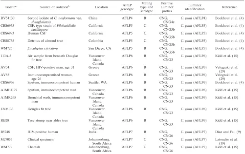
TABLE 2. Origins of haploid isolates and overview of results obtained by AFLP analysis, mating-serotype-specific PCRs, and Luminex
suspension array
Isolatea
Source of isolationb
Location AFLP genotype Mating type and serotype Positive Luminex probes Luminex identification Reference
AMC770704 CSF, HIV-negative woman, age 22 The Netherlands AFLP1 ␣A CNNb, CNN1b
C. neoformansvar. grubii(AFLP1)
This study
AMC830410 CSF, HIV-negative woman, age 58 The Netherlands AFLP1 ␣A CNNb, CNN1b
C. neoformansvar. grubii(AFLP1)
This study
AMC860743 CSF, HIV-positive man, age 47 The Netherlands AFLP1 ␣A CNNb, CNN1b
C. neoformansvar. grubii(AFLP1)
This study
AMC880696 Jugular gland, man, age 19 The Netherlands AFLP1 ␣A CNNb, CNN1b
C. neoformansvar. grubii(AFLP1)
This study
AMC900239 CSF, immunocompetent man, age 27
The Netherlands AFLP1 ␣A CNNb, CNN1b
C. neoformansvar. grubii(AFLP1)
This study
AMC900321 Lung, AIDS patient, man, age 43 The Netherlands AFLP1 ␣A CNNb, CNN1b
C. neoformansvar. grubii(AFLP1)
This study
AMC900906 CSF, immunocompetent woman, age 49
The Netherlands AFLP1 ␣A CNNb, CNN1b
C. neoformansvar. grubii(AFLP1)
This study
AMC901081 CSF, HIV-positive man, age 47 The Netherlands AFLP1 ␣A CNNb, CNN1b
C. neoformansvar. grubii(AFLP1)
This study
AMC922148 CSF, AIDS patient, man, age 29 The Netherlands AFLP1 ␣A CNNb, CNN1b
C. neoformansvar. grubii(AFLP1)
This study
AMC931394 CSF, HIV-negative, immunocompromised man, age 52
The Netherlands AFLP1 ␣A CNNb, CNN1b
C. neoformansvar. grubii(AFLP1)
This study
AMC940211 CSF, AIDS suspected, man, age 30
The Netherlands AFLP1 ␣A CNNb, CNN1b
C. neoformansvar. grubii(AFLP1)
This study
AMC940580 CSF, HIV-negative, immunocompromised man, age 73
The Netherlands AFLP1 ␣A CNNb, CNN1b
C. neoformansvar. grubii(AFLP1)
This study
AMC940751 CSF, HIV-positive man, age 44 The Netherlands AFLP1 ␣A CNNb, CNN1b
C. neoformansvar. grubii(AFLP1)
This study
AMC951535 CSF, prednisone usage, HIV-negative, immunocompromised man, age 58
The Netherlands AFLP1 ␣A CNNb, CNN1b
C. neoformansvar. grubii(AFLP1)
This study
AMC981683 CSF, AIDS patient, woman, age 23 The Netherlands AFLP1 ␣A CNNb, CNN1b
C. neoformansvar. grubii(AFLP1)
This study
AMC990558 CSF, encephalopathy, AIDS patient, man, age 37
The Netherlands AFLP1 ␣A CNNb, CNN1b
C. neoformansvar. grubii(AFLP1)
This study
AMC2040734 CSF, man, age 46 The Netherlands AFLP1 ␣A CNNb, CNN1b
C. neoformansvar. grubii(AFLP1)
This study
AMC9402891 CSF, AIDS patient, man, age 45 The Netherlands AFLP1 ␣A CNNb, CNN1b
C. neoformansvar. grubii(AFLP1)
This study
JS9901 CSF, sarcoidosis, man, age 43 The Netherlands AFLP1 ␣A CNNb, CNN1b
C. neoformansvar. grubii(AFLP1)
This study
MN HIV-positive man, age 30 The Netherlands AFLP1 ␣A CNNb, CNN1b
C. neoformansvar. grubii(AFLP1)
This study
P1953 HIV-negative, immunocompromised man, age 65
The Netherlands AFLP1 ␣A CNNb, CNN1b
C. neoformansvar. grubii(AFLP1)
This study
AMC890401 CSF, AIDS patient, man, age 35 The Netherlands AFLP2 aD CNNb, CNN2d
C. neoformansvar. neoformans (AFLP2)
This study
AMC940038 CSF, AIDS patient, man, age 29 The Netherlands AFLP2 aD CNNb, CNN2d
C. neoformansvar. neoformans (AFLP2)
This study
AMC941354 CSF, HIV-negative, immunocompromised man, age 69
The Netherlands AFLP2 ␣D CNNb, CNN2d
C. neoformansvar. neoformans (AFLP2)
This study
AMC2010488 CSF, AIDS patient, man, age 50 The Netherlands AFLP2 ␣D CNNb, CNN2d
C. neoformansvar. neoformans (AFLP2)
This study
AMC2031402 CSF, man, age 65 The Netherlands AFLP2 ␣D CNNb, CNN2d
C. neoformansvar. neoformans (AFLP2)
This study
AMC2020797A CSF, non-Hodgkin lymphoma, woman, age 41
The Netherlands AFLP2 ␣D CNNb, CNN2d
C. neoformansvar. neoformans (AFLP2)
This study
CBS1622 Tumor, man France AFLP4 B CNG,
CNG4c
C. gattii(AFLP4) Boekhout et al. (4)
CBS6289 Subculture of type strain of C. neoformansvar.gattii (RV20186: human CSF, Zaire)
AFLP4 B CNG,
CNG4c
C. gattii(AFLP4) Boekhout et al. (4)
CBS7229 Meningitis, type strain of C. neoformansvar.shanghaiensis
China AFLP4 B CNG,
CNG4c
C. gattii(AFLP4) Boekhout et al. (4)
CBS883 Infected skin, syntypeC. hondurianus
Honduras AFLP4 B CNG,
CNG4c
C. gattii(AFLP4) Boekhout et al. (4)
N114 HIV-positive man, age 47 The Netherlands AFLP4 NDb CNG, CNG4c
C. gattii(AFLP4) This study
Continued on following page
on May 16, 2020 by guest
http://jcm.asm.org/
carbodiimide method (8). Each microsphere set (MiraiBio, Alameda, CA) con-tains a unique spectral address by combining different ratios of red and infrared fluorochromes. In a typical reaction, 5⫻106
microspheres were resuspended in 25l 0.1 M MES (2[N-morpholino]ethanesulfonic acid), pH 4.5, with a deter-mined amount of probe (0.2 to 0.5 nmol). Probe coupling was performed as described by Diaz and Fell (8), and the microspheres were subsequently resus-pended in 100l of TE buffer (10 mM Tris-HCl, 1 mM EDTA; pH 8). A microsphere mixture was made by adding approximately 5,000 microspheres for each of the eight probes to a 1.5⫻TMAC (3 M tetramethyl ammonium chloride, 50 mM Tris [pH 8], 4 mM EDTA [pH 8], 0.1% Sarkosyl) solution.
To amplify the IGS1 region, forward primer IG1F (5⬘-CAG ACG ACT TGA ATG GGA ACG-3⬘) and reverse primer IG2R (5⬘-ATG CAT AGA AAG CTG TTG G-3⬘) were used (12). The reverse primer was biotinylated at the 5⬘end. The 1⫻HotStarTaq MasterMix (QIAGEN, Valencia, CA) containing 1.5 mM MgCl2, 0.2 mM deoxynucleoside triphosphates, and 2.5 units of HotStarTaq
polymerase was used for all PCRs. DNA had been extracted from the crypto-coccal isolates prior to amplification, although Diaz and Fell (9) directly used yeast cells for PCR amplification. PCRs were carried out in a total volume of 25
l. Primers IG1F and IG2R (0.6M) and 1.5l of template DNA were added to the MasterMix. Amplification conditions were as follows: 95°C for 15 min, followed by 35 cycles of 95°C for 30 s, 50°C for 30 s, and 72°C for 30 s, and a final extension step of 72°C for 7 min.
PCR amplification of the first two clinical samples was carried out using the HotStarTaq MasterMix in a 50-l total volume containing 3l of DNA and 0.4
M of both primer IG1F and primer IG2R. Amplification conditions were as described above.
Optimization of the PCR conditions resulted in the following reaction condi-tions, which were used for the remaining clinical samples. PCR amplification was carried out with the HotStarTaq MasterMix in a 50-l total volume containing 0.2% bovine serum albumin, 8l of DNA, and 0.6M of both primer IG1F and
primer IG2R. Amplification conditions were as follows: 95°C for 15 min, fol-lowed by 40 cycles of 95°C for 30 s, 50°C for 30 s, and 69°C for 30 s, and a final extension step of 69°C for 9 min. Amplicons were cleaned with a QIAGEN purification kit (QIAGEN, Valencia, CA) and eluted with elution buffer (10 mM Tris-HCl [pH 8.5]).
To genotype the cryptococcal isolates, 5 l of biotinylated amplicon was diluted with 12l of TE buffer (pH 8), and to genotype the clinical samples, 15
l of biotinylated amplicon was diluted with 2l of TE buffer (pH 8). Thirty-three microliters of the microsphere mixture was added. Each amplicon was tested in duplicate with the Luminex suspension array. The hybridization reac-tion was performed as described by Diaz and Fell (9).
The hybridized samples were analyzed on the Luminex 100 analyzer (Lu-minex Corporation, Austin, TX). One hundred microspheres of each set were analyzed, which represents a hundred replicate measurements. Median flu-orescence intensity (MFI) values were calculated with a digital signal pro-cessor and Luminex 1.7 proprietary software. A positive signal was defined as a signal that is at least twice the background level after subtraction of the background.
Nucleotide sequence accession numbers. All sequences were deposited at GenBank under the accession numbers DQ286656 to DQ286661, DQ286665 to DQ286670, and EF100569 to EF100594.
RESULTS AND DISCUSSION
AFLP analysis and mating-serotype-specific PCRs
per-formed on the isolates that had not been genotyped before
showed that twenty-one isolates belonged to
C. neoformans
[image:4.585.42.543.81.397.2]var.
grubii MAT
␣
serotype A (AFLP1), four isolates were
C.
TABLE 2—
Continued
Isolatea Source of isolationb Location AFLP genotype
Mating type and serotype
Positive Luminex probes
Luminex
identification Reference
RV54130 Second isolate ofC. neoformansvar. shanghaiensis
China AFLP4 B CNG,
CNG4c
C. gattii(AFLP4) Boekhout et al. (4)
CBS6955 CSF, type strain ofFilobasidiella bacillispora
California AFLP5 C CNG,
CNG5b
C. gattii(AFLP5) Boekhout et al. (4)
CBS6993 Human CSF California AFLP5 C CNG,
CNG5b
C. gattii(AFLP5) Boekhout et al. (4)
CBS8755 Detritus of almond tree Colombia AFLP5 C CNG,
CNG5b
C. gattii(AFLP5) Boekhout et al. (4)
WM726 Eucalyptus citriodora San Diego, CA AFLP5 B CNG,
CNG5b
C. gattii(AFLP5) Boekhout et al. (4)
113A-5 Air sample from beneath Douglas fir tree
Vancouver Island, Canada
AFLP6 B CNG,
CNG3
C. gattii(AFLP6) Kidd et al. (15)
AV54 CSF, HIV-positive man, age 31 Greece AFLP6 B CNG,
CNG3
C. gattii(AFLP6) Velegraki et al. (29) AV55 Immunocompromised woman,
age 26
Greece AFLP6 B CNG,
CNG3
C. gattii(AFLP6) Velegraki et al. (29) CBS6956 Sputum, immunocompetent human Seattle, WA AFLP6 B CNG,
CNG3
C. gattii(AFLP6) Boekhout et al. (4)
A1MF3179 Sputum, immunocompetent man Vancouver, Canada
AFLP6 B CNG,
CNG3
C. gattii(AFLP6) Kidd et al. (15)
A1MR265 Bronchial wash, immunocompetent man
Vancouver Island, Canada
AFLP6 B CNG,
CNG3
C. gattii(AFLP6) Kidd et al. (15)
ENV133 Douglas fir tree Vancouver
Island, Canada
AFLP6 B CNG,
CNG3
C. gattii(AFLP6) Kidd et al. (15)
RB28 Tree stump near alder tree Vancouver Island, Canada
AFLP6 B CNG,
CNG3
C. gattii(AFLP6) Kidd et al. (15)
B5748 HIV-positive human India AFLP7 B CNG,
CNG6
C. gattii(AFLP7) Diaz and Fell (9)
M27055 Clinical specimen Johannesburg, South Africa
AFLP7 C CNG,
CNG6
C. gattii(AFLP7) Latouche et al. (19)
WM779 Cheetah Johannesburg,
South Africa
AFLP7 C CNG,
CNG6
C. gattii(AFLP7) Kidd et al. (15)
aAMC, The Netherlands Reference Laboratory for Bacterial Meningitis, Academic Medical Center, Amsterdam, The Netherlands; CBS, CBS—Fungal Biodiversity Centre, Utrecht, The Netherlands; RV, BCCM/IHEM Biomedical Fungi and Yeast Collection, Brussels, Belgium; WM, Wieland Meyer, Molecular Mycology Research Laboratory, Westmead Hospital, Sydney, Australia.
bND, not determined.
on May 16, 2020 by guest
http://jcm.asm.org/
TABLE
3.
Origin
of
hybrid
isolates
and
overview
of
results
obtained
by
AFLP
analysis,
sequence
analysis,
mating-serotype-specific
PCRs,
and
Luminex
suspension
array
a Isolate b Source of isolation Location AFLP genotype Mating type and serotype Positive Luminex probes Luminex identification IGS1 sequences (no. of alleles)CNLAC1 sequences (no.
of alleles) Reference
AMC881205I
CSF,
HIV-positive
man
The
Netherlands
AFLP3
a
A-␣
D
CNNb,
CNN1b,
CNN2d
Hybrid
between
C.
neoformans
var.
grubii
and
C.
neoformans
var.
neoformans
(AFLP3)
2
2
This
study
CDC92-26
CDC
AFLP3
a
A-␣
D
CNNb,
CNN1b,
CNN2d
Hybrid
between
C.
neoformans
var.
grubii
and
C.
neoformans
var.
neoformans
(AFLP3)
2
2
This
study
Kl#1
Progeny
laboratory
crossing
H99
5-FOAr
⫻
JEC171
AFLP3
a
D-␣
A
CNNb,
CNN1b,
CNN2d
Hybrid
between
C.
neoformans
var.
grubii
and
C.
neoformans
var.
neoformans
(AFLP3)
2
2
Lengeler
et
al.
(20)
Kl#45
Progeny
laboratory
crossing
H99
5-FOAr
⫻
JEC171
AFLP3
a
D-␣
A
CNNb,
CNN1b,
CNN2d
Hybrid
between
C.
neoformans
var.
grubii
and
C.
neoformans
var.
neoformans
(AFLP3)
2
2
Lengeler
et
al.
(20)
ZG287
Duke
Medical
Center
permanent
strain
collection
AFLP3
a
D-␣
A
CNNb,
CNN1b,
CNN2d
Hybrid
between
C.
neoformans
var.
grubii
and
C.
neoformans
var.
neoformans
(AFLP3)
2
2
Lengeler
et
al.
(20)
AMC890351
CSF,
HIV-positive
man,
age
44
The
Netherlands
AFLP3
a
D-␣
A
CNNb,
CNN2d
C.
neoformans
var.
neoformans
(AFLP2)
1
2
This
study
AMC891529
CSF,
AIDS
patient,
man,
age
31
The
Netherlands
AFLP3
a
D-␣
A
CNNb,
CNN2d
C.
neoformans
var.
neoformans
(AFLP2)
1
2
This
study
AMC770616
(
⫽
CBS10488)
CSF,
brain
tumor
surgery,
man,
age
23
The
Netherlands
AFLP8
a
D-␣
B
CNNb,
CNN2d,
CNG,
CNG4c
Hybrid
between
C.
neoformans
var.
neoformans
and
AFLP4
C.
gattii
(AFLP8)
2
2
Bovers
et
al.
(5)
AMC2010404
(
⫽
CBS10489)
CSF,
idiopathic
intracranial
hypertension,
man,
age
35
The
Netherlands
AFLP8
a
D-␣
B
CNNb,
CNN2d,
CNG,
CNG4c
Hybrid
between
C.
neoformans
var.
neoformans
and
AFLP4
C.
gattii
(AFLP8)
2
2
Bovers
et
al.
(5)
AMC2011225
(
⫽
CBS10490)
CSF,
idiopathic
intracranial
hypertension,
man,
age
36
The
Netherlands
AFLP8
a
D-␣
B
CNNb,
CNN2d,
CNG,
CNG4c
Hybrid
between
C.
neoformans
var.
neoformans
and
AFLP4
C.
gattii
(AFLP8)
2
2
Bovers
et
al.
(5)
aAll isolates were diploid. bAMC, The Netherlands Reference Laboratory for Bacterial Meningitis, Academic Medical Center, Amsterdam, The Netherlands; CDC, Centers for Diseas e Control and Prevention, Atlanta, GA.on May 16, 2020 by guest
http://jcm.asm.org/
neoformans
var.
neoformans MAT
␣
serotype D (AFLP2), and
two isolates were
C. neoformans
var.
neoformans MAT
a
sero-type D (AFLP2). Finally, one isolate belonged to the
C. gattii
AFLP4 genotype. Some hybrid isolates between the two
vari-eties of
C. neoformans
were detected: two
MAT
a
serotype
A-
MAT
␣
serotype D (AFLP3) and two
MAT
a
serotype
D-MAT
␣
serotype A (AFLP3) isolates. Results of the AFLP
analysis and the mating-serotype-specific PCRs are presented
in Tables 2 and 3.
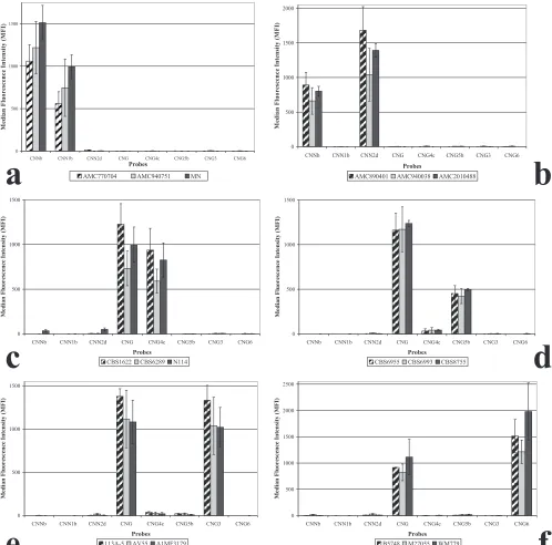
All haploid isolates were identified by two Luminex
suspen-sion array probes (Fig. 1). Probes CNNb and CNN1b identified
the AFLP1 isolates as
C. neoformans
var.
grubii
(AFLP1), and
probes CNNb and CNN2d identified the AFLP2 isolates as
C.
neoformans
var.
neoformans
(AFLP2). Probe CNG correctly
identified all
C. gattii
isolates as
C. gattii
. In addition, probe
CNG4c identified the AFLP4 isolates, probe CNG5b identified
the AFLP5 isolates, probe CNG3 identified the AFLP6
iso-lates, and probe CNG6 identified the AFLP7 isolates. These
results show that the Luminex suspension array correctly
geno-typed all haploid strains (Table 2).
The Luminex suspension array was also used to genotype ten
hybrid isolates. Five out of seven serotype AD (AFLP3) hybrid
isolates, namely AMC881205I, CDC92-26, Kl#1, Kl#45, and
ZG287, were identified as hybrids between the two varieties of
C. neoformans
by probes CNNb, CNN1b, and CNN2d (Fig.
2a). However, two serotype AD (AFLP3) hybrid isolates,
namely AMC890351 and AMC891529, were identified as
C.
neoformans
var.
neoformans
by probes CNNb and CNN2d (Fig.
2b). The three serotype BD (AFLP8) hybrid isolates were
identified as hybrids between
C. neoformans
var.
neoformans
and
C. gattii
AFLP4 by probes CNG, CNG4c, CNNb, and
CNN2d (Fig. 3). Interestingly, when the suspension array
iden-tified a hybrid isolate, low signal intensities, namely 16% to
53% of an average positive signal, were obtained for probes
which identified one of the parental genotypes. The results of
the suspension array correlated with the number of clones that
were found for each allele. For example, more AFLP4 than
AFLP2 IGS1 clones were obtained for the serotype BD hybrid
isolates. The Luminex probes gave a similar outcome: the
signals for probes CNG and CNG4c, which identify the
C. gattii
AFLP4 genotype, had normal intensities, i.e., 546 to 940 MFI.
The probes which identify
C. neoformans
var.
neoformans
(AFLP2), namely CNNb and CNN2d, were positive with MFI
values ranging from 119 to 311, but the MFI values that were
obtained were only 16% to 19% of an average positive signal
(Fig. 3).
Flow cytometry confirmed that all hybrid isolates were
dip-loid or close to dipdip-loid (data not shown). In addition, when a
part of the
CNLAC1
region was cloned and sequenced, two
alleles could be obtained for all hybrid isolates. However, when
the IGS1 region was used for cloning and sequencing, two
alleles were obtained for five serotype AD (AFLP3) hybrid
isolates, namely AMC881205I, CDC92-26, Kl#1, Kl#45, and
ZG287, but only one IGS1 allele was found in two serotype AD
(AFLP3)
hybrid
isolates,
namely
AMC890351
and
AMC891529, even though thirty clones were sequenced. All
three serotype BD (AFLP8) hybrid isolates possessed two
IGS1 alleles. Our results show that all hybrid isolates were
diploid or close to diploid, they possessed two
CNLAC1
alleles,
and most hybrid isolates possessed two IGS1 alleles. All hybrid
isolates that possessed two IGS1 alleles, the region on which
the Luminex probes are based, could be identified as hybrids.
In order to further improve the identification of cryptococcal
hybrids, a probe derived from another gene could be included.
In a multigene study of thirty-one serotype AD hybrid isolates
(AFLP3), five isolates possessed two IGS1 alleles, but
twenty-seven isolates had two
TEF1
␣
alleles, twenty-six isolates
pos-sessed two
RPB1
alleles, and twenty-three isolates had two
CNLAC1
alleles (M. Bovers, unpublished data). This indicates
that
CNLAC1
,
RPB1
, and
TEF1
␣
are potential regions that
could be used to improve the identification of cryptococcal
hybrids.
The detection limit of the suspension array was calculated
to vary from 4
⫻
10
1to 2
⫻
10
3cells for the different probes
[image:6.585.43.538.80.254.2](9). In clinical practice, the concentration of cryptococcal
cells ranges from 1,000 to 10,000,000 cells per ml of CSF
(25). Therefore, the Luminex technology could be a
power-ful tool for the detection and identification of cryptococcal
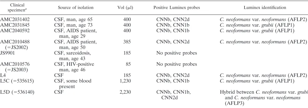
TABLE 4. Origin and volume of CSF for which amplicons could be obtained and the results of Luminex identification
Clinical
specimena Source of isolation Vol (l) Positive Luminex probes Luminex identification
AMC2031402
CSF, man, age 65
400
CNNb, CNN2d
C. neoformans
var.
neoformans
(AFLP2)
AMC2031845
CSF, man, age 73
400
CNNb, CNN1b
C. neoformans
var.
grubii
(AFLP1)
AMC2040592
CSF, AIDS patient,
man, age 29
400
CNNb, CNN1b
C. neoformans
var.
grubii
(AFLP1)
AMC2010488
(
⫽
JS2002)
CSF, AIDS patient,
man, age 50
385
CNNb, CNN2d
C. neoformans
var.
neoformans
(AFLP2)
JS9901
CSF, sarcoidosis,
man, age 43
185
No positive probes
AMC2010576
(
⫽
JS2003)
CSF, HIV-positive
man, age 46
85
No positive probes
L4
CSF
185
CNNb, CNN2d
C. neoformans
var.
neoformans
(AFLP2)
L5C (
⫽
535615)
CSF, some blood
present
1,230
CNNb, CNN1b
C. neoformans
var.
grubii
(AFLP1)
L5D (
⫽
536140)
CSF
2,230
CNNb, CNN1b,
CNN2d
Hybrid between
C. neoformans
var.
grubii
and
C. neoformans
var.
neoformans
(AFLP3)
aAMC, The Netherlands Reference Laboratory for Bacterial Meningitis, Academic Medical Center, Amsterdam, The Netherlands; JS and L, University Medical Centre Utrecht, Utrecht, The Netherlands.
on May 16, 2020 by guest
http://jcm.asm.org/
cells in CSF. CSF specimens from various Dutch and
Bel-gian hospitals that had been stored for up to five years at
⫺
80°C were used to test the Luminex suspension array on
clinical specimens. Only nine out of twenty CSF specimens
obtained from patients with culture-proven cryptococcal
meningitis gave an amplicon of the targeted IGS locus.
Unfortunately, we do not have additional information, i.e.,
about the number of (undamaged) cells or about the
pres-ence of interfering substances (30) in the samples, to explain
why an amplicon could not be obtained. Amplicons of the
[image:7.585.43.541.68.560.2]PCR-positive samples were subsequently used for genotypic
identification by the suspension array (Fig. 4), and the
re-sults are shown in Table 4. The first two samples that were
analyzed, namely JS9901 and AMC2010576, had probe
sig-nals that were too low to be considered positive.
Optimiza-tion of the PCR condiOptimiza-tions, i.e., adding 0.2% bovine serum
albumin and increasing the amount of DNA and primers in
the reaction mix as well as decreasing the temperature of the
elongation step to 69°C, improved detection. As a
conse-quence, the infecting agent of the remaining seven samples
FIG. 1. Results obtained with the Luminex suspension array for all six haploid groups within
C. neoformans
and
C. gattii
. Examples of the results
obtained with the AFLP1, -2, -4, -5, -6, and -7 genotypic groups are depicted in panels a, b, c, d, e, and f, respectively.
on May 16, 2020 by guest
http://jcm.asm.org/
could successfully be identified at species and genotypic
levels, but because no CSF of JS9901 and AMC2010576
remained, the genotype of the infecting agent could not be
determined for those two samples. Probes CNNb and
CNN1b, with MFI signals ranging from 260 to 1,402,
iden-tified the infecting agent in L5c, AMC2040592, and
AMC2031845 as
C. neoformans
var.
grubii
. Probes CNNb
and
CNN2d
identified
the
source
of
infection
of
AMC2010488, L4, and AMC2031402 as
C. neoformans
var.
neoformans
, with MFI signals ranging from 112 to 1,780.
Probes CNNb, CNN1b, and CNN2d, with MFI signals
rang-ing from 199 to 1,446, identified the cryptococcal strain
responsible for infection of the patient of specimen L5d as
a serotype AD (AFLP3) strain of
C. neoformans
.
Interest-ingly, CSF specimens L5c and L5d were obtained from the
same patient, thus suggesting a dual infection by a haploid
C. neoformans
var.
neoformans
and a serotype AD (AFLP3)
hybrid strain. Our results show that the suspension array is
highly specific, as both varieties of
C. neoformans
(
C.
neo-formans
var.
grubii
and
C. neoformans
var.
neoformans
) and
a hybrid could be identified in CSF specimens. The clinical
applicability of the Luminex suspension array might be
im-proved by optimization of the DNA isolation protocol, as
this is one of the critical steps in any molecular detection
system (2). In addition, to determine the robustness of the
method, fresh clinical specimens for which the amount of
cryptococcal cells and the antigen titers are known should be
tested and follow-up studies should be performed on
sam-ples with false-negative results.
[image:8.585.48.540.68.229.2]In summary, the Luminex suspension array has the
po-tential to become an efficient diagnostic method with high
specificity that not only identifies cryptococcal isolates at the
species and genotype levels but that also allows
identifica-tion of hybrid isolates that possess two IGS1 alleles.
Fur-thermore, our results show that the Luminex suspension
array is able to identify cryptococci in CSF specimens.
Iden-tification in CSF occurs at the species, genotype, and hybrid
levels, but optimization of DNA extraction methods is
FIG. 2. Results obtained with the Luminex suspension array for all of the serotype AD (AFLP3) hybrids of
C. neoformans
. Panel a
shows five serotype AD hybrids that were identified by three probes, and panel b shows two serotype AD hybrids that were identified by two
probes.
FIG. 3. Results obtained with the Luminex suspension array for the serotype BD (AFLP8) hybrid isolates.
on May 16, 2020 by guest
http://jcm.asm.org/
[image:8.585.134.450.517.712.2]needed before the method is suited for routine use in
clin-ical laboratories.
ACKNOWLEDGMENTS
Our special gratitude goes to A. van Belkum (Erasmus Medical
Center, Rotterdam, The Netherlands) and K. Lagrou (University
Hos-pital Leuven, Leuven, Belgium) for sending us clinical specimens.
This research was partially funded by National Institutes of Health
grant 1-UO1 AI53879-01, the ‘Odo van Vloten fonds’ and the
‘Neth-erlands-Florida Scholarship Foundation’.
REFERENCES
1.Barreto de Oliveira, M. T., T. Boekhout, B. Theelen, F. Hagen, F. A. Baroni, M. S. Lazera, K. B. Lengeler, J. Heitman, I. N. G. Rivera, and C. R. Paula.
2004.Cryptococcus neoformansshows a remarkable genotypic diversity in Brazil. J. Clin. Microbiol.42:1356–1359.
2.Baums, I. B., K. D. Goodwin, T. Kiesling, D. Wanless, and J. W. Fell.
Luminex detection of fecal indicators in river samples, marine recreational water, and beach sand. Mar. Pollut. Bull., in press.
3.Bicanic, T., and T. S. Harrison.2004. Cryptococcal meningitis. Br. Med. Bull.72:99–118.
4.Boekhout, T., B. Theelen, M. Diaz, J. W. Fell, W. C. Hop, E. C. Abeln, F. Dromer, and W. Meyer.2001. Hybrid genotypes in the pathogenic yeast Cryptococcus neoformans. Microbiology147:891–907.
5.Bovers, M., F. Hagen, E. E. Kuramae, M. R. Diaz, L. Spanjaard, F. Dromer, H. L. Hoogveld, and T. Boekhout.2006. Unique hybrids between fungal pathogensCryptococcus neoformansandCryptococcus gattii. FEMS Yeast Res.6:599–607.
6.Cogliati, M., M. Allaria, G. Liberi, A. M. Tortorano, and M. A. Viviani.2000. Sequence analysis and ploidy determination ofCryptococcus neoformansvar. neoformans. J. Mycol. Med.10:171–176.
7.Das, S., T. M. Brown, K. L. Kellar, B. P. Holloway, and C. J. Morrison.2006. DNA probes for the rapid identification of medically importantCandida species using a multianalyte profiling system. FEMS Immunol. Med. Micro-biol.46:244–250.
8.Diaz, M. R., and J. W. Fell.2004. High-throughput detection of pathogenic yeasts of the genusTrichosporon. J. Clin. Microbiol.42:3696–3706. 9.Diaz, M. R., and J. W. Fell. 2005. Use of a suspension array for rapid
identification of the varieties and genotypes of theCryptococcus neoformans species complex. J. Clin. Microbiol.43:3662–3672.
10.Diaz, M. R., T. Boekhout, T. Kiesling, and J. W. Fell.2005. Comparative analysis of the intergenic spacer regions and population structure of the species complex of the pathogenic yeastCryptococcus neoformans. FEMS Yeast Res.5:1129–1140.
11.Diaz, M. R., T. Boekhout, B. Theelen, M. Bovers, F. J. Caban˜es, and J. W. Fell.2006. Microcoding and flow cytometry as a high-throughput fungal identification system forMalasseziaspecies. J. Med. Microbiol.55:1197–1209. 12.Diaz, M. R., T. Boekhout, B. Theelen, and J. W. Fell. 2000. Molecular
sequence analyses of the intergenic spacer (IGS) associated with rDNA of the two varieties of the pathogenic yeast,Cryptococcus neoformans. Syst. Appl. Microbiol.23:535–545.
13.Dunbar, S. A., C. A. Vander Zee, K. G. Oliver, K. L. Karem, and J. W. Jacobson.2003. Quantitative, multiplexed detection of bacterial pathogens: DNA and protein applications of the Luminex LabMAP system. J. Micro-biol. Methods53:245–252.
14.Hoang, L. M. N., J. A. Maguire, P. Doyle, M. Fyfe, and D. L. Roscoe.2004. Cryptococcus neoformansinfections at Vancouver hospital and health sci-ences centre (1997–2002): epidemiology, microbiology and histopathology. J. Med. Microbiol.53:935–940.
15.Kidd, S. E., F. Hagen, R. L. Tscharke, M. Huynh, K. H. Bartlett, M. Fyfe, L. Macdougall, T. Boekhout, K. J. Kwon-Chung, and W. Meyer.2004. A rare genotype ofCryptococcus gattiicaused the cryptococcosis outbreak on Van-couver Island (British Columbia, Canada). Proc. Natl. Acad. Sci. USA101:
17258–17263.
16.Kwon-Chung, K. J., J. E. Bennett, and J. C. Rhodes. 1982. Taxonomic studies onFilobasidiellaspecies and their anamorphs. Antonie Leeuwenhoek
48:25–38.
17.Kwon-Chung, K. J., T. Boekhout, J. W. Fell, and M. Diaz.2002. Proposal to conserve the nameCryptococcus gattiiagainstC. hondurianusandC. bacil-lisporus(Basidiomycota,Hymenomycetes,Tremellomycetidae). Taxon51:804– 806.
18.Kwon-Chung, K. J., and A. Varma.2006. Do major species concepts support one, two or more species withinCryptococcus neoformans? FEMS Yeast Res.
6:574–587.
19.Latouche, G. N., M. Huynh, T. C. Sorrell, and W. Meyer.2003. PCR-restriction fragment length polymorphism analysis of the phospholipase B (PLB1) gene for subtyping ofCryptococcus neoformansisolates. Appl. En-viron. Microbiol.69:2080–2086.
20.Lengeler, K. B., G. M. Cox, and J. Heitman.2001. Serotype AD strains of Cryptococcus neoformansare diploid or aneuploid and are heterozygous at the mating-type locus. Infect. Immun.69:115–122.
21.Meyer, W., A. Castan˜eda, S. Jackson, M. Huynh, E. Castan˜eda, and the IberoAmerican Cryptococcal Study Group. 2003. Molecular typing of IberoAmericanCryptococcus neoformansisolates. Emerg. Infect. Dis.9:189– 195.
22.Mirza, S. A., M. Phelan, D. Rimland, E. Graviss, R. Hamill, M. E. Brandt, T. Gardner, M. Sattah, G. P. de Leon, W. Baughman, and R. A. Hajjeh.2003. The changing epidemiology of cryptococcosis: an update from population-based active surveillance in 2 large metropolitan areas, 1992–2000. Clin. Infect. Dis.36:789–794.
23.Mitchell, D. H., T. C. Sorrell, A. M. Allworth, C. H. Heath, A. R. McGregor, K. Papanaoum, M. J. Richards, and T. Gottlieb.1995. Cryptococcal disease of the CNS in immunocompetent hosts: influence of cryptococcal variety on clinical manifestations and outcome. Clin. Infect. Dis.20:611–616. 24.Page, B. T., and C. P. Kurtzman.2005. Rapid identification ofCandida
species and other clinically important yeast species by flow cytometry. J. Clin. Microbiol.43:4507–4514.
25.Perfect, J. R., D. T. Durack, and H. A. Gallis.1983. Cryptococcemia. Med-icine62:89–109.
26.Speed, B., and D. Dunt.1995. Clinical and host differences between infec-tions with the two varieties ofCryptococcus neoformans. Clin. Infect. Dis.
21:28–34.
[image:9.585.135.450.69.259.2]27.Stephen, C., S. Lester, W. Black, M. Fyfe, and S. Raverty.2002. Multispecies outbreak of cryptococcosis on southern Vancouver Island, British Columbia. Can. Vet. J.43:792–794.
FIG. 4. Luminex suspension array results for the amplicons obtained from CSF specimens.
on May 16, 2020 by guest
http://jcm.asm.org/
28.Tanaka, R., K. Nishimura, and M. Miyaji.1999. Ploidy of serotype AD strains ofCryptococcus neoformans. Jpn. J. Med. Mycol.40:31–34. 29.Velegraki, A., V. G. Kiosses, H. Pitsouni, D. Toukas, V. D. Daniilidis, and
N. J. Legakis.2001. First report ofCryptococcus neoformansvar.gattii sero-type B from Greece. Med. Mycol.39:419–422.
30.Wilson, I. G.1997. Inhibition and facilitation of nucleic acid amplification. Appl. Environ. Microbiol.63:3741–3751.
31.Wilson, W. J., A. M. Erler, S. L. Nasarabadi, E. W. Skowronski, and P. M. Imbro.2005. A multiplexed PCR-coupled liquid bead array for the si-multaneous detection of four biothreat agents. Mol. Cell. Probes19:137– 144.
32.Xu, J., R. Vilgalys, and T. G. Mitchell.2000. Multiple gene genealogies reveal recent dispersion and hybridization in the human pathogenic fungus Cryptococcus neoformans. Mol. Ecol.9:1471–1481.