0095-1137/10/$12.00
doi:10.1128/JCM.01265-10
Copyright © 2010, American Society for Microbiology. All Rights Reserved.
Hepatitis C Virus Genotyping Using an Oligonucleotide Microarray
Based on the NS5B Sequence
䌤
†
Dimitry Gryadunov,
1* Florence Nicot,
2Martine Dubois,
2Vladimir Mikhailovich,
1Alexander Zasedatelev,
1and Jacques Izopet
2Engelhardt Institute of Molecular Biology, Russian Academy of Sciences, Moscow, Russia,
1and CHU de Toulouse,
Ho
ˆpital Purpan, Laboratoire de Virologie, Toulouse F-31300, France
2Received 22 June 2010/Returned for modification 18 August 2010/Accepted 8 September 2010
The genotype of the hepatitis C virus (HCV) is essential for determining treatment duration in clinical
practice and for epidemiological and clinical studies. Currently, few genotyping assays that determine the HCV
subtype are available. This report describes a microarray-based molecular technique for identifying the HCV
genotype and subtype. It uses low-density hydrogel-based biochips containing genotype- and subtype-specific
oligonucleotides based on the sequences of the NS5B region of the HCV genome. The biochip contains 120
oligonucleotides that identify genotypes 1 to 6 and 36 (1a, 1b, 1c, 1d, 1e, 2a, 2b, 2c, 2d, 2i, 2j, 2k, 2l, 2m, 3a, 3b,
3k, 4a, 4c, 4d, 4f, 4h, 4i, 4k, 4n, 4o, 4p, 4r, 4t, 5a, 6a, 6b, 6d, 6g, 6h, and 6k) subtypes. The procedure included
amplification of a 380-nucleotide (nt) fragment of NS5B and its hybridization on the biochip. Tests on 345
HCV-positive samples showed that the assay agreed with NS5B sequencing 100% for the genotype and 99.7%
for the subtype. The hybridization on the microarray and the NS5B sequence were in 100% agreement for
identifying the most common subtypes, 1a, 1b, 4a, 4d, and 3a. This approach is a promising tool for HCV
genotyping, especially for implementing the new anti-HCV drugs that require accurate identification of
clinically relevant subtypes.
The hepatitis C virus (HCV) is a leading cause of chronic
liver disease and increased risk of cirrhosis and hepatocellular
carcinoma (51). More than 170 million people are infected
with HCV worldwide (42). This enveloped, single-stranded
positive-sense RNA virus is a member of the
Flaviviridae
fam-ily. The RNA genome contains a single large open reading
frame composed of over 9,000 nucleotides (nt) encoding
struc-tural and nonstrucstruc-tural proteins (5). One of these proteins is
an RNA-dependent RNA polymerase encoded by the
so-called NS5B region. This error-prone enzyme lacks
proofread-ing activity, which makes it responsible for the great genetic
variability of HCV. Sequencing studies of HCV strains have
identified 6 genotypes and more than 70 subtypes (43, 45).
The HCV genotype is considered to be the major baseline
predictor of a sustained virological response (SVR) to antiviral
therapy. Patients infected with HCV genotypes 2 and 3 are
more sensitive to combination therapy with interferon and
ribavirin than are those infected with genotype 1 (8, 11, 21).
The available data on HCV genotype 4 suggest that its
sensi-tivity to HCV treatment lies somewhere between those of
genotypes 1 and 2/3 (17). The sensitivity of genotypes 5 and 6
could be similar to that of genotype 2 or 3 (1, 9, 19). The HCV
subtype has recently been implicated as a potential predictor of
SVR. One study of 597 difficult-to-treat patients found that
subtypes 1b, 4a, and 4d were independently associated with
SVR (16). The virological response to new anti-HCV agents
could also be influenced by the HCV subtype (31, 42).
Several methods has been proposed for HCV genotyping
(50), including commercially available techniques based on
real-time PCR: the HCV genotyping analyte-specific reagent
(ASR) assay (Abbott Molecular Inc., Des Plaines, IL) (23),
semiautomated sequencing (the TruGene HCV 5
⬘
NC
geno-typing kit; Bayer HealthCare, Berkeley, CA) (10), and
auto-mated reverse hybridization (the Inno-LiPA HCV II assay;
Innogenetics, Ghent, Belgium) (46, 49). Most HCV genotyping
methods are based on analysis of the 5
⬘
noncoding (NC) region
of the HCV genome because the 5
⬘
NC region is regularly
amplified for HCV molecular diagnosis and quantification of
the viral load. However, this highly conserved region is not
suitable for accurately discriminating between subtypes and
can lead to genotyping or subtyping errors (2, 3, 15, 39, 43).
Hence, alternative genomic regions have been proposed for
genotyping HCV, including the core fragment (35, 49) and the
NS5B region (39). Sequencing and phylogenetic analysis of the
NS5B region are presently considered to be the gold standard
for HCV genotyping since they accurately identify the subtype
and can be used to establish an epidemiological picture of
circulating virus strains (27, 30, 39, 47). However, this method
includes steps of purification of the amplified product,
se-quencing, and phylogenetic analysis that require the skill of
laboratory personnel, a factor that can be a limitation to the
wide use of the technique in routine clinical laboratories.
Therefore, an assay was developed that involves
hybridiza-tion on an oligonucleotide microarray for identifying HCV
genotypes and subtypes. It uses a low-density hydrogel-based
microarray (biochip) that has been successfully used in many
fields of molecular diagnostics (26, 28, 37). The microarray
contains genotype- and subtype-specific oligonucleotides based
* Corresponding author. Mailing address: Engelhardt Institute of
Molecular Biology, Russian Academy of Sciences, 32 Vavilova St.,
Moscow 119991, GSP-1, Russia. Phone: 7 (499) 135 9846. Fax: 7 (499)
135 1405. E-mail: [email protected].
† Supplemental material for this article may be found at http://jcm
.asm.org/.
䌤
Published ahead of print on 15 September 2010.
3910
on May 16, 2020 by guest
http://jcm.asm.org/
on the corresponding sequences of the NS5B region. This
report compares this approach to accurately identifying HCV
genotype and subtype with direct NS5B sequencing.
MATERIALS AND METHODS
Collection of serum samples, HCV RNA isolation, and NS5B amplification.
All the HCV-positive patients attending Toulouse University Hospital between March 2007 and August 2008 for whom genotyping was requested were included in this study. A total of 345 samples from consecutive patients with HCV RNA
concentrations of 1,622 to⬎10,000,000 IU/ml were included. The viral load in
samples was quantified by the real-time RT-PCR Cobas AmpliPrep/Cobas TaqMan HCV test (CAP/CTM; Roche Diagnostic, Meylan, France) according to the manufacturer’s instructions.
HCV RNA for genotyping was extracted with the Cobas AmpliPrep total nucleic acid isolation kit (TNAI) (Roche Diagnostics, Basel, Switzerland) fol-lowing the manufacturer’s instructions. Briefly, reverse transcription-PCR
(RT-PCR) was performed using 10l of extracted RNA with primers Pr2r (5⬘-GG
CGGAATTCCTGGTCATAGCCTCCGTGAA-3⬘) and Pr1f (5⬘-TATGAYAC
CCGCTGYTTTGACTC-3⬘) as previously described (39). PCR products were
stored frozen for both NS5B sequencing and microarray genotyping.
Sequencing and phylogenetic analysis of the NS5B region.Performance of the developed microarray was tested by comparing the results of hybridization with phylogenetic analysis of NS5B sequences, which is a standard method to identify HCV genotypes and subtypes. In fact, it is representative of phylogenetic analysis of the complete HCV genome (12). Two microliters of RT-PCR amplification mix was used for sequencing the NS5B region as previously described (39). The NS5B nucleotide sequences were aligned with CLUSTAL_X 1.83 software (48), and phylogenetic trees were created by the neighbor-joining (NJ) method. The reproducibility of the branching pattern was tested by bootstrap analysis (100 replicates). Genotypes and subtypes were determined when the bootstrap value
was⬎70%. We used the TreeView 1.66 program to draw the phylogenetic trees
(32).
Phylogenetic analyses were performed with the NS5B sequences from patients and 191 reference sequences available from the Los Alamos HCV database (14).
These 191 reference sequences included genotype 1 (subtypes 1a, 1b, 1c, 1d, 1e, 1f, 1g, 1h, 1i, 1j, 1k, 1l, 1m, and 1 nontypeable), genotype 2 (subtypes 2a, 2b, 2c, 2d, 2e, 2f, 2g, 2h, 2i, 2j, 2k, 2l, 2m, 2o, 2p, 2q, 2r, and 2 nontypeable), genotype 3 (subtypes 3a, 3b, 3c, 3d, 3e, 3f, 3g, 3h, 3i, and 3k), genotype 4 (subtypes 4a, 4b, 4c, 4d, 4e, 4f, 4g, 4h, 4i, 4j, 4k, 4l, 4m, 4n, 4o, 4p, 4q, 4r, 4t, and 4 untypeable), genotype 5 (subtype 5a and 5 untypeable), and genotype 6 (subtypes 6a, 6b, 6c, 6d, 6f, 6g, 6h, 6i, 6j, 6k, 6l, 6o, 6p, 6q, 6t, and 6u). It allowed the determination of HCV genotype and subtypes of the samples subsequently tested by the mi-croarray.
Oligonucleotide design.NS5B region nucleotide sequences were aligned using Bioedit software (Ibis Therapeutics, Carlsbad, CA). A total of 1,232 NS5B region sequences from GenBank and the Los Alamos HCV sequence database were aligned (14) (nt 8256 to 8616; numbering according to reference 5). This multiple alignment was used to generate unique consensus sequences for each genotype. Genotype-specific probes were then selected from within the corresponding consensus sequences. As the HCV genome is highly variable, several probes were designed for each genotype wherever possible so as to increase the reliability of the method. The sequences of the genotype-specific probes selected by this procedure were located in different segments of the NS5B region.
Next, consensus sequences were deduced for each subtype, and segments of the NS5B region that discriminated between the maximum number of subtypes within each genotype were selected (Fig. 1). The number of such segments was optimized for reliable identification of each subtype. Finally, probes for identi-fying subtypes were designed based on the sequences of the selected segments. This procedure did not exclude the possibility that individual probes could detect simultaneously two or more subtypes in different subtype-specific segments of the analyzed NS5B region.
The melting temperatures were calculated, and the secondary structures of the designed oligonucleotides were estimated with an Oligo analyzer (Integrated DNA Technologies). The lengths of the oligonucleotides were adjusted to main-tain the range of melting temperatures within 2 to 3°C.
[image:2.585.43.545.68.331.2]The sequences of oligonucleotides are listed in Table S1 in the supplemental material, and they are also available in a published patent application (D. Grya-dunov, V. Mikhailovich, F. Nicot, M. Dubois, A. Zasedatelev, and J. Izopet, 2
FIG. 1. Alignment of the subtype-specific consensus sequences of the NS5B region. The subtypes are indicated in the left-hand column.
Residues identical to the consensus sequence of subtype 1a are indicated by dots. Numbering is from the first nucleotide of the H77 1a reference
sequence (GenBank accession no. NC_004102). The positions of segments of the NS5B region used for selecting subtype-specific probes are boxed.
The number of the segment corresponds to a group number and the designation of the subtype-specific oligonucleotide. Slashes indicate
discontinuous sequences within the NS5B region.
on May 16, 2020 by guest
http://jcm.asm.org/
February 2009, WO/2009/022939 2009, World Intellectual Property Organiza-tion).
Oligonucleotides for immobilization on the biochip and primers for amplifi-cation were synthesized and purified as described earlier (38). The molecular masses of oligonucleotides were measured with a matrix-assisted laser desorption ionization–time of flight (MALDI-TOF) mass spectrometer (Compact MALDI 4; Kratos Analytical, Chestnut Ridge, NY) using sinapinic acid or 2-amino-5-nitropyridine as a matrix.
Microarray design.The diagnostic biochip comprised 120 immobilized oligo-nucleotides, four marker cells (M) for accurate positioning (image acquisition) by the processing software, and four elements of empty gel (0) needed to
calculate the reference fluorescence intensityIref(background). The
arrange-ment of oligonucleotides immobilized on the biochip is shown in Fig. 2A. The oligonucleotides labeled “G” that identified the genotype of the HCV sample were immobilized in the two top rows (all six genotypes). The probes immobi-lized in the lower rows identified the HCV subtypes.
Four groups of oligonucleotides were designed to identify subtypes 1a, 1b, 1c, 1d, and 1e of genotype 1. Three groups of probes were designed to differentiate between subtypes 2a, 2b, 2c, 2d, 2i, 2j, 2k, 2l, and 2m of genotype 2. Three more groups of oligonucleotides identified the three subtypes of genotype 3—3a, 3b, and 3k. Finally, four groups of probes were included to identify subtypes 4a, 4c, 4d, 4f, 4h, 4i, 4k, 4n, 4o, 4p, 4r, and 4t of genotype 4. Genotype 5 has only one subtype, 5a; therefore, the three probes for identifying genotype 5 also identified subtype 5a. Subtypes 6a, 6b, 6d, 6g, 6h, and 6k of genotype 6 were identified using two groups of probes, each of which corresponded to a separate segment within the analyzed fragment of NS5B region (Fig. 1).
Biochip manufacture.The biochips were manufactured as described earlier
(38), with 35-l hybridization chambers (Biochip-IMB, Ltd., Moscow, Russia).
Each biochip contained semispherical gel elements 100m in diameter placed
300m apart. Quality control of large-scale microchip production was done by
measuring the quantity of immobilized oligonucleotides in each gel element using TestChip software provided by Biochip-IMB, Ltd.
Amplification of the NS5B fragment for genotyping on the microarray.The
PCR amplification step was performed with 1l RT-PCR mixture using the
primers Pr1f and Pr3r (5⬘-GCTAGTCATAGCCTCCGT-3⬘). The primer
con-centrations were 10 nM Pr1f and 100 nM Pr3r.
The reaction mixture (25l) contained 1.5 mM MgCl2; 10 mM KCl; 10 mM
Tris-HCl, pH 8.3; 0.2 mM (each) dATP, dCTP, dGTP, and dUTP (Sileks, Russia); 0.04 mM fluorescently labeled dUTP (IMD-515-dUTP; Biochip-IMB,
Ltd, Moscow, Russia); and 5 unitsTaqDNA polymerase (Sileks). Because of the
difference in the concentrations of forward and reverse primers within each pair, the reaction yielded predominantly single-stranded fluorescently labeled prod-uct. PCR was performed as follows: 4 min at 95°C; 36 cycles of 20 s at 95°C, 20 s at 60°C, and 30 s at 72°C; and 5 min at 72°C.
Hybridization on the biochip and registration of the results.Hybridization
mixtures were prepared by adding 12l of PCR mixtures to 23l of 1.5 M
guanidine thiocyanate (GuSCN), 0.075 M HEPES, pH 7.5, 7.5 mM EDTA. The biochip hybridization chamber was filled with the mixture, and the assembly was incubated for 14 to 16 h at 37°C. The chamber was then removed, and the
FIG. 2. (A) Diagram of the biochip for hybridization. Elements
with the letter G contain genotype-specific probes. Four probes (G1-1
to G1-4) are used to identify genotype 1, three (G2-1 to G2-3) are used
to identify genotype 2, two (G3-1 to G3-2) are used to identify
geno-type 3, three (G4-1 to G4-3) are used to identify genogeno-type 4, three
(G5-1 to G5-3) are used to identify genotype 5, and one (G6) is used
to identify genotype 6. The probes for identifying subtypes are named
ixN, where i is the genotype number, x indicates the subtype, and N is
the number of the group corresponding to a segment of the NS5B
region. (B) Fluorescence hybridization pattern of biochip elements
obtained by analyzing an HCV sample of genotype 1, subtype 1b. The
group of genotype-specific probes (G2-1 to G6) and groups 1 to 4 of
subtype-specific probes of genotype 1 are outlined with a broken line.
The histogram of normalized fluorescence signals of row 2 elements
containing genotype-specific oligonucleotides is shown above the
hy-bridization pattern. The histograms of normalized fluorescence signals
of elements comprising groups 1 to 4 of subtype-specific
oligonucleo-tides belonging to genotype 1 are outlined below the fluorescence
image. The calculated value of the mean signal of empty elements (
I
ref)
is shown by a solid bold line on all histograms.
on May 16, 2020 by guest
http://jcm.asm.org/
[image:3.585.42.286.76.713.2]microarray surface was washed three times (about 30 s each) with water at 37°C and air dried. The fluorescent pattern of biochips was registered using a fluo-rescence analyzer setup and specialized software (ImageWare; Biochip-IMB, Ltd.).
Interpretation of hybridization results. (i) Genotype identification (genotyp-ing).First, perfect hybridization duplexes were identified within the upper two rows containing oligonucleotides for identifying genotypes. Our statistics (33) indicated that the fluorescent intensity of perfect duplexes should be at least 2.0
times higher than the average background signal (Iref) with a standard deviation
of 0.2. Thus, a 2.0-fold intensity difference was taken as the threshold value for selecting positive signals.
The intensities of selected positive signals corresponding to perfect duplexes were compared within each genotype-specific group. If the maximum signal
Gimaxin one group exceeded the maximum signal in the other groups by more
than 1.5-fold, the analyzed specimen was considered to belong to the correspond-ing genotype.
If the ratio of the signals amongGimaxobserved within each individual group
did not exceed 1.5, the genotype of the analyzed specimen could not be accu-rately determined and identification of subtype was not performed. The program stopped further processing when the signals within each genotype-specific group were below the threshold value and could not pass the initial selection.
(ii) Subtype identification (subtyping).The subtype-specific oligonucleotides were combined in groups according to the selected segments of the NS5B region. Subtyping was performed strictly after the genotype had been successfully iden-tified. It was crucial for the identification strategy to consider only the subtype-specific oligonucleotides corresponding to the subtype-specific genotype while excluding all other signals as irrelevant.
The signals within each group of subtype-specific probes were considered positive if their intensities were at least 2.0 times higher than the average
background signalIref. Positive signals within each group of subtype-specific
probes that were at least 1.5 times stronger than other signals of the same group
were selected for further processing. These signals were designatedSixj(where i
is the genotype number, x is the symbol of a subtype according to the HCV subtype classification, and j is the number of the analyzed group of microarray elements). When two or more elements within the same group had signals
differing from one another by less than 1.5-fold, then all such signalsSixjwere
selected as positive. As a result, a set of elements from the various groups ix1, iy1, ix2, iz2, ix3, etc., were detected whose signals were at least 1.5 times the rest of the signals in their groups. If the number of elements homologous to one subtype in such a set, for example, ix1 and ix3, or ix1 and ixy2, exceeded the number of elements corresponding to other subtypes by at least 1, the conclusion was that the analyzed specimen belonged to subtype x of genotype i.
When the elements of different groups in the set so obtained corresponded to a different subtype, for example, ix1, iy2, and iz3, or ix1 and iyz3, the sets of signals corresponding to individual subtypes were compared to each other. If the signal of an element corresponding to subtype x in one group was 3 or more times stronger than the strongest signals from the groups corresponding to other subtypes, the conclusion was that the analyzed specimen belonged to subtype x.
If the ratio of signalsSix1/Siy2was 3 or less, we concluded that the subtype could
not be determined. Similarly, if the probe specific for two subtypes was the strongest signal in the group, for example, ixy1, and there were no valid signals in other groups of the elements, the conclusion was that the subtype could not be determined. Finally, if the signals of subtype-specific groups of elements did not
pass the primary signal filtration relative toIref, the conclusion was that the
subtype of the analyzed specimen could not be determined.
Statistical analysis.The kappa coefficient was measured using Stata SE 9.2 (StataCorp LP, College Station, TX) to evaluate the concordance between the HCV subtypes determined by NS5B sequencing and the NS5B biochip assays. The overall proportions of HCV subtypes determined by NS5B sequencing and
the NS5B biochip assays were checked using the chi-squared test.Pvalues of
⬍0.05 were considered significant.
RESULTS
Determining the genotype/subtype by biochip analysis of the
NS5B region.
HCV genotyping based on analysis of the NS5B
region was performed by hybridization on the biochip. The
procedure consisted of three steps: (i) RT-PCR to amplify the
NS5B region fragment, (ii) asymmetric PCR to obtain
fluores-cently labeled predominantly single-stranded DNA fragments,
and (iii) hybridization of the labeled product on the biochip
with gel elements carrying immobilized oligonucleotides.
Figure 2B shows an example of hybridization pattern and
distribution of normalized signals of biochip elements resulting
from analysis of a subtype 1b sample. As defined by the
algo-rithm described in Materials and Methods, only signals in the
G1 group containing genotype 1-specific probes were more
than 2 times the threshold, with the maximum signal being
produced by the G1-3 element (2.68). The signals in other
groups containing genotype-specific probes were close to
back-ground (the deviation from
I
refdid not exceed 0.12). The
conclusion was that this HCV sample belonged to genotype 1.
Further processing of the groups of elements containing
specific probes for genotype 1 subtypes produced the following
results. In group 1, the strongest signal was obtained from
element 1bd1 (4.98). In group 2 it was 1b2 (3.72), and in group
4 it was 1b4 (2.46). Group 3 contained no elements with
pos-itive signals (see corresponding histogram in Fig. 2B).
Conse-quently, this specimen was identified as subtype 1b.
Additional examples of fluorescence patterns by
hybridiza-tion with different HCV samples are shown in Fig. 3A to I. All
the genotype-specific probes hybridized with the
correspond-ing target genotypes without cross-reactcorrespond-ing with the other
ge-notypes. Analysis of some samples, for instance, 4d (Fig. 3G),
resulted in cross-hybridization with oligonucleotides specific
for subtypes of genotype 2. However, the data processing
algorithm uses only the elements with subtype-specific probes
of the genotype that was determined in the previous step
re-gardless of the signals in other biochip elements. As a result,
the subtypes for most samples were identified unambiguously.
The ability of biochips to identify mixed HCV infections was
examined. Specimens having 1a, 1b, 3a, and 4a subtypes were
each adjusted to an equal HCV RNA concentration and
sub-sequently mixed at different proportions as follows: 1a
⫹
4a,
1b
⫹
3a. The mixed infections were identified successfully as
long as the amount of the minor species was no smaller than
20% (data not shown). The lower concentration of the minor
subtype in a mixed sample led to decrease of signals in the
corresponding groups of elements, and such a sample was
identified as one that contained the dominant subtype only.
Analytical sensitivity and specificity.
The analytical
sensitiv-ity of this method was estimated by assaying 10-fold serial
dilutions (with seronegative plasma) of a plasma standard
con-taining 5.2
⫻
10
6IU/ml of HCV subtype 1b. Four replicates
were used for each dilution. The hybridization results obtained
with the 2.0
⫻
10
2-IU/ml concentration of viral RNA were
unambiguous.
The specificity of the procedure was tested using 24
sero-negative plasma samples. All were identified as sero-negative
sam-ples. With at least three replicates of each sample analyzed, the
deviations of signals of genotype- and subtype-specific
ele-ments for identical samples remained within 20% of the
aver-age background signal (
I
ref). Therefore, there were no
false-positive results.
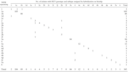
Comparison of biochip-based genotyping with NS5B
se-quencing.
Table 1 shows the results obtained by hybridization
on the biochip and direct sequencing of NS5B segments. All
(100%) of the 345 HCV RNA-positive sera analyzed were
successfully genotyped by biochip hybridization. They included
samples infected with all six HCV genotypes.
on May 16, 2020 by guest
http://jcm.asm.org/
The samples included subtypes 1a, 1b, 1d, 1e, 2a, 2b, 2c, 2i,
2j, 2k, 2l, 3a, 4a, 4c, 4d, 4f, 4h, 4k, 4p, 4r, and 5a and samples
of undetermined subtypes of genotypes 1, 2, 4, and 6, as
de-termined by sequencing. The two methods were concordant for
the subtypes of 329/330 samples (99.7%), with a kappa
coeffi-cient of 0.996 (
P
⬍
0.00001). One sample identified as 2c by
NS5B sequencing was identified as 2k by NS5B biochip
anal-ysis. The NS5B sequencing method failed to determine the
subtypes in 8 samples (2.3%), and the NS5B biochip methods
failed in 12 samples (3.5%) (
P
⫽
0.36). Samples with an
un-determined subtype by NS5B sequencing were identified as 1a,
1b, 2k, 4h, and 4r by NS5B biochip analysis. Samples with an
undetermined subtype by NS5B biochip analysis were assigned
to subtypes 1d, 2a, 2j, and 2l by NS5B sequencing. The
sub-types of 5 samples were not determined by either method.
DISCUSSION
The gold standard for HCV genotyping remains PCR
am-plification followed by sequencing of one of the
phylogeneti-cally informative coding regions of the HCV genome, such as
NS5B or core/E1, and comparison with the consensus
se-quences in GenBank or the Los Alamos hepatitis C virus
databases (14). We have developed a novel microarray-based
assay for identifying the HCV genotype and subtype and
eval-uated it in comparison with the phylogenetic analysis of the
NS5B region as a reference method. The new NS5B
microar-ray assay and NS5B sequencing were in almost complete
agree-ment.
The assay relies on hybridization of a 380-nt NS5B fragment
with oligonucleotides specific for HCV genotypes and subtypes
immobilized on a biochip. The reliable identification of each
individual genotype and subtype required the design of several
oligonucleotides for each of them, in consequence of the
vari-ability of the NS5B region. The results were interpreted using
an original algorithm that included preliminary processing of
the hybridization signal intensities from the biochip elements
and comparison of signals from elements within the sets of
genotype-specific probes and then from sets of subtype-specific
probes.
[image:5.585.92.494.67.440.2]The new method enabled us to determine all six HCV
ge-notypes with a sensitivity of approximately 2.0
⫻
10
2IU/ml of
FIG. 3. Hybridization patterns obtained using HCV samples belonging to subtype 1a (A), 1b (B), 2a (C), 2i (D), 3a (E), 4a (F), 4d (G), 5a (H),
and 6x (I). The groups of elements containing genotype- and subtype-specific oligonucleotides corresponding to the analyzed sample are
contoured.
on May 16, 2020 by guest
http://jcm.asm.org/
HCV RNA. This analytical performance using biochip-based
genotyping and subtyping is comparable to that of
commer-cially available assays (50), including the new generation of line
probe assays (49).
The new method was tested on 345 HCV-positive samples.
The results were 100% concordant for the genotype and 99.7%
concordant for the subtype with the results obtained by direct
sequencing of the NS5B segment. The accuracy and reliability
of the assay make it suitable for large-scale genotyping and
subtyping projects.
Hybridization on the biochip correctly identified HCV
iso-lates of subtypes 1a, 1b, 1e, 2a, 2b, 2c, 2i, 2k, 3a, 4a, 4c, 4d, 4f,
4k, 4p, 4r, and 5a. It failed to identify subtypes 1d, 2j, 2l, and
4h. This could be because there are fewer of these NS5B
sequences in GenBank and other databases, which resulted in
less accurate selection of subtype-specific probes. However,
these subtypes are very infrequent in Europe—2.9% for 2l,
0.9% for 2j, and 1% for 4h (30, 47). However, the hybridization
on the microarray and NS5B sequencing were in 100%
agree-ment for identifying the most widespread and clinically
rele-vant subtypes, such as 1a, 1b, 4a, 4d, and 3a. The only
limita-tion of the study is that not many samples of HCV genotype 6
were tested because this is very rare in France.
No mixed infections were encountered during the
evalua-tion. Testing the analytical mixed samples revealed that the
method is able to detect two different genotypes within the
sample if the concentration of the minor genotype constitutes
20% or more of the total HCV RNAs.
Some recent studies have shown that HCV subtypes can
predict the response to standard treatment regimens that
in-clude pegylated interferon and ribavirin. One French
[image:6.585.46.543.82.362.2]multi-center study of 597 treated patients showed that subtypes 1b,
4a, and 4d were independent predictors of SVR (16). A recent
study also demonstrated that patients infected with HCV
sub-type 1b had a higher antiviral response than did patients
in-fected with HCV subtype 1a (29). Another study of 1,532
patients infected with HCV genotype 4 showed that subtype 4a
was more sensitive to anti-HCV treatment than was subtype 4d
(36). Moreover, the development of new specific inhibitors of
HCV enzymes whose antiviral responses and resistance
pro-files may be determined by the HCV subtype may require
identification of the subtype prior to treatment (7, 20, 24).
Several HCV inhibitors appear to act selectively against certain
HCV genotype 1 subtypes, both
in vitro
and
in vivo
. Differences
in the activities of NS3/4A protease inhibitors (telaprevir and
boceprevir) against different subtypes have been reported.
There is evidence that the selection of resistant variants and
virus breakthrough is more frequent in patients infected with
subtype 1b than in those harboring subtype 1a (13, 25, 40). The
antiviral activities of nucleoside analogs of polymerase
inhibi-tors are similar regardless of the HCV subtype, while
non-nucleoside inhibitors are more active against subtype 1b than
against subtype 1a (18, 31, 41). These findings suggest that the
antiviral activity of new anti-HCV agents may also vary with
the subtypes of genotypes other than 1. It is therefore essential
to accurately discriminate between subtypes in order to tailor
anti-HCV treatment schedules with HCV protease and
poly-merase inhibitors. There are few methods presently available
other than direct sequencing of NS5B and core/E1 segments
for identifying numerous subtypes. One of the commercially
available methods, the INNO-LiPA v.2, discriminates better
between subtypes 1a and 1b than does the previous version,
TABLE 1. Comparison of HCV genotyping obtained by NS5B sequencing with that obtained by hybridization on the biochip
NS5B sequencing
No. of isolates with HCV genotype and subtype assigned by hybridization on biochip
1 1a 1b 1d 1e 2 2a 2b 2c 2i 2j 2k 2l 3a 4 4a 4c 4d 4f 4h 4k 4p 4r 5a 6 Total
1
1
1
1
3
1a
103
103
1b
88
88
1d
1
1
1e
1
1
2
3
1
4
2a
1
4
5
2b
5
5
2c
3
1
4
2i
7
7
2j
1
1
2k
5
5
2l
1
1
3a
84
84
4
0
4a
12
12
4c
1
1
4d
8
8
4f
1
1
4h
1
1
4k
1
1
4p
1
1
4r
2
2
4
5a
3
3
6
1
1
Total
2
104
89
0
1
6
4
5
3
7
0
7
0
84
3
12
1
8
1
0
1
1
2
3
1
345
on May 16, 2020 by guest
http://jcm.asm.org/
INNO-LiPA v.1, but does not discriminate between subtypes
4a, 4c, and 4d (4, 49). Our new method correctly identified
HCV subtypes 1a and 1b in more than 99% of samples; it also
identified subtypes 4a and 4d.
All the experimental microarray-based methods for HCV
genotyping use immobilized oligonucleotides from the 5
⬘
un-translated region of the HCV genome. They can therefore
identify only a small number of subtypes (1a, 1b, 2a/2b/2c, 3a,
3b, and 6a), although their determination of genotypes is
re-ported to be almost 100% (6, 22, 34). In this work, use of
probes complementary to subtype-specific sequences of the
NS5B region enabled us to identify more than 20 HCV
sub-types in the specimens tested. Other methods, such as
real-time PCR, can identify a limited number of subtypes and
genotypes (1a, 1b, 2a, 2b, 2c, 3, 4, 5, and 6) (23, 44). Only the
clip sequencing method can, in theory, discriminate as many
subtypes as can our procedure (35).
In conclusion, this new approach to analyzing the NS5B
region of HCV based on hybridization with a low-density
mi-croarray is a promising tool for rapidly, sensitively, and
accu-rately identifying viral genotype and subtype. It provides
clini-cians with the information needed for the choice of a correct
individual treatment of hepatitis C. In addition, the
perfor-mance of the new procedure and the range of identifiable
genotypes and subtypes make it suitable for epidemiological
surveys.
ACKNOWLEDGMENTS
This work was supported by contract 02.522.11.2019 with the
Fed-eral Agency of Sciences and Innovations of the Russian Federation.
We are grateful to E. Kreindlin for manufacturing of microarrays, to
S. Surzhikov and I. Grechishnikova for synthesis of oligonucleotides, to
A. Chudinov for synthesis and selection of the fluorescent dyes, and
to R. Urasov for help with the mathematical calculations and analysis
of experimental data. We are especially grateful to A. Kolchinsky
(Health Front Line, Ltd., Champaign, IL) for his assistance in the
preparation of this paper. The English text was edited by Owen Parkes.
REFERENCES
1.Bonny, C., H. Fontaine, T. Poynard, C. Hezode, D. Larrey, P. Marcellin, M. Bourliere, J. P. Bronowicki, P. Merle, J. P. Zarski, T. Sapey, C. Guillemard, S. Ughetto, C. Henquell, C. Nicolas, C. Roche, K. Randl, G. Bommelaer, and A. Abergel.2006. Effectiveness of interferon plus ribavirin combination in the treatment of naive patients with hepatitis C virus type 5. A French
multi-centre retrospective study. Aliment. Pharmacol. Ther.24:593–600.
2.Cantaloube, J. F., S. Laperche, P. Gallian, F. Bouchardeau, X. de Lambal-lerie, and P. de Micco.2006. Analysis of the 5⬘noncoding region versus the NS5b region in genotyping hepatitis C virus isolates from blood donors in
France. J. Clin. Microbiol.44:2051–2056.
3.Chen, Z., and K. E. Weck.2002. Hepatitis C virus genotyping: interrogation
of the 5⬘untranslated region cannot accurately distinguish genotypes 1a and
1b. J. Clin. Microbiol.40:3127–3134.
4.Chevaliez, S., M. Bouvier-Alias, R. Brillet, and J. M. Pawlotsky.2009. Hep-atitis C virus (HCV) genotype 1 subtype identification in new HCV drug
development and future clinical practice. PLoS One4:e8209.
5.Choo, Q. L., K. H. Richman, J. H. Han, K. Berger, C. Lee, C. Dong, C. Gallegos, D. Coit, R. Medina-Selby, P. J. Barr, et al.1991. Genetic organi-zation and diversity of the hepatitis C virus. Proc. Natl. Acad. Sci. U. S. A.
88:2451–2455.
6.Costi, C., C. M. da Silva, N. N. Da Fre, T. Grandi, F. I. Hamester, A. Zaha, C. Niel, and M. L. Rossetti.2009. Colorimetric microwell plate reverse-hybridization assay for detection and genotyping of hepatitis C virus. J. Virol.
Methods162:75–80.
7.Erhardt, A., K. Deterding, Y. Benhamou, M. Reiser, X. Forns, S. Pol, J. L. Calleja, S. Ross, H. C. Spangenberg, J. Garcia-Samaniego, M. Fuchs, J. Enriquez, J. Wiegand, J. Stern, K. Wu, G. Kukolj, M. Marquis, P. Beaulieu, G. Nehmiz, and J. Steffgen.2009. Safety, pharmacokinetics and antiviral effect of BILB 1941, a novel hepatitis C virus RNA polymerase inhibitor,
after 5 days oral treatment. Antivir. Ther.14:23–32.
8.Fried, M. W., M. L. Shiffman, K. R. Reddy, C. Smith, G. Marinos, F. L. Goncales, Jr., D. Haussinger, M. Diago, G. Carosi, D. Dhumeaux, A. Craxi, A. Lin, J. Hoffman, and J. Yu.2002. Peginterferon alfa-2a plus ribavirin for
chronic hepatitis C virus infection. N. Engl. J. Med.347:975–982.
9.Fung, J., C. L. Lai, I. Hung, J. Young, C. Cheng, D. Wong, and M. F. Yuen.
2008. Chronic hepatitis C virus genotype 6 infection: response to pegylated
interferon and ribavirin. J. Infect. Dis.198:808–812.
10.Germer, J. J., D. W. Majewski, M. Rosser, A. Thompson, P. S. Mitchell, T. F. Smith, S. Elagin, and J. D. Yao.2003. Evaluation of the TRUGENE HCV
5⬘NC genotyping kit with the new GeneLibrarian module 3.1.2 for
genotyp-ing of hepatitis C virus from clinical specimens. J. Clin. Microbiol.41:4855–
4857.
11.Hadziyannis, S. J., and J. S. Koskinas.2004. Differences in epidemiology, liver disease and treatment response among HCV genotypes. Hepatol. Res.
29:129–135.
12.Hraber, P. T., W. Fischer, W. J. Bruno, T. Leitner, and C. Kuiken.2006. Comparative analysis of hepatitis C virus phylogenies from coding and
non-coding regions: the 5⬘untranslated region (UTR) fails to classify subtypes.
Virol. J.3:103.
13.Kieffer, T. L., C. Sarrazin, J. S. Miller, M. W. Welker, N. Forestier, H. W. Reesink, A. D. Kwong, and S. Zeuzem. 2007. Telaprevir and pegylated interferon-alpha-2a inhibit wild-type and resistant genotype 1 hepatitis C
virus replication in patients. Hepatology46:631–639.
14.Kuiken, C., P. Hraber, J. Thurmond, and K. Yusim.2008. The hepatitis C
sequence database in Los Alamos. Nucleic Acids Res.36:D512–D516.
15.Laperche, S., F. Lunel, J. Izopet, S. Alain, P. Deny, G. Duverlie, C. Gaudy, J. M. Pawlotsky, J. C. Plantier, B. Pozzetto, V. Thibault, F. Tosetti, and J. J. Lefrere.2005. Comparison of hepatitis C virus NS5b and 5⬘noncoding gene
sequencing methods in a multicenter study. J. Clin. Microbiol.43:733–739.
16.Legrand-Abravanel, F., P. Colson, H. Leguillou-Guillemette, L. Alric, I. Ravaux, F. Lunel-Fabiani, M. Bouviers-Alias, P. Trimoulet, M. L. Chaix, C. Hezode, J. Foucher, H. Fontaine, A. M. Roque-Afonso, M. Gassin, E. Sch-voerer, C. Gaudy, B. Roche, M. Doffoel, L. D’Alteroche, S. Vallet, Y. Baazia, B. Pozzetto, V. Thibault, J. B. Nousbaum, D. Roulot, H. Coppere, T. Poinard, C. Payan, and J. Izopet.2009. Influence of the HCV subtype on the viro-logical response to pegylated interferon and ribavirin therapy. J. Med. Virol.
81:2029–2035.
17.Legrand-Abravanel, F., F. Nicot, A. Boulestin, K. Sandres-Saune, J. P. Vinel, L. Alric, and J. Izopet.2005. Pegylated interferon and ribavirin therapy for
chronic hepatitis C virus genotype 4 infection. J. Med. Virol.77:66–69.
18.Legrand-Abravanel, F., F. Nicot, and J. Izopet.2010. New NS5B polymerase
inhibitors for hepatitis C. Expert Opin. Invest. Drugs19:963–975.
19.Legrand-Abravanel, F., K. Sandres-Saune, K. Barange, L. Alric, J. Moreau, P. Desmorat, J. P. Vinel, and J. Izopet.2004. Hepatitis C virus genotype 5: epidemiological characteristics and sensitivity to combination therapy with
interferon-alpha plus ribavirin. J. Infect. Dis.189:1397–1400.
20.Liang, Y., H. Ishida, O. Lenz, T. I. Lin, O. Nyanguile, K. Simmen, R. B. Pyles, N. Bourne, M. Yi, K. Li, and S. M. Lemon.2008. Antiviral suppression vs restoration of RIG-I signaling by hepatitis C protease and polymerase
inhibitors. Gastroenterology135:1710–1718.
21.Manns, M. P., H. Wedemeyer, and M. Cornberg.2006. Treating viral
hep-atitis C: efficacy, side effects, and complications. Gut55:1350–1359.
22.Mao, H., H. Zhang, J. Zhao, Z. Lu, G. Jin, S. Gu, H. Wang, and Y. Wang.
2010. Clinical evaluation of a colorimetric oligonucleotide chip for
genotyp-ing hepatitis C virus. Clin. Biochem.43:214–219.
23.Martro, E., V. Gonzalez, A. J. Buckton, V. Saludes, G. Fernandez, L. Matas, R. Planas, and V. Ausina.2008. Evaluation of a new assay in comparison with reverse hybridization and sequencing methods for hepatitis C virus
genotyping targeting both 5⬘noncoding and nonstructural 5b genomic
re-gions. J. Clin. Microbiol.46:192–197.
24.McCown, M. F., S. Rajyaguru, S. Kular, N. Cammack, and I. Najera.2009. GT-1a or GT-1b subtype-specific resistance profiles for hepatitis C virus inhibitors telaprevir and HCV-796. Antimicrob. Agents Chemother.
53:2129–2132.
25.McHutchison, J. G., G. T. Everson, S. C. Gordon, I. M. Jacobson, M. Sulkowski, R. Kauffman, L. McNair, J. Alam, and A. J. Muir.2009. Tela-previr with peginterferon and ribavirin for chronic HCV genotype 1
infec-tion. N. Engl. J. Med.360:1827–1838.
26.Mikhailovich, V., D. Gryadunov, A. Kolchinsky, A. A. Makarov, and A. Zasedatelev.2008. DNA microarrays in the clinic: infectious diseases.
Bioes-says30:673–682.
27.Murphy, D. G., B. Willems, M. Deschenes, N. Hilzenrat, R. Mousseau, and S. Sabbah.2007. Use of sequence analysis of the NS5B region for routine
genotyping of hepatitis C virus with reference to C/E1 and 5⬘untranslated
region sequences. J. Clin. Microbiol.45:1102–1112.
28.Nasedkina, T. V., N. A. Guseva, O. A. Gra, O. N. Mityaeva, A. V. Chudinov, and A. S. Zasedatelev.2009. Diagnostic microarrays in hematologic
oncol-ogy: applications of high- and low-density arrays. Mol. Diagn. Ther.13:91–
102.
29.Nicot, F., L. Alric, K. Barange, S. Me´tivier, J. M. Dramard, J. M. Combis, B. Castan, J. J. Meurisse, J. L. Payen, D. Garipuy, H. Desmorat, J. M. Peron, S. Thebault, T. Morin, C. Renou, P. Barel, B. Guerin, Y. Imbert, S. Sire, K.
on May 16, 2020 by guest
http://jcm.asm.org/
Saune´, E. Chatelut, and J. Izopet.Influence of HCV genotype 1 subtypes on the virus response to peg interferon alpha-2a plus ribavirin therapy. J. Med. Virol., in press.
30.Nicot, F., F. Legrand-Abravanel, K. Sandres-Saune, A. Boulestin, M. Dubois, L. Alric, J. P. Vinel, C. Pasquier, and J. Izopet.2005. Heterogeneity of hepatitis C virus genotype 4 strains circulating in south-western France.
J. Gen. Virol.86:107–114.
31.Nyanguile, O., B. Devogelaere, L. Vijgen, W. Van den Broeck, F. Pauwels, M. D. Cummings, H. L. De Bondt, A. M. Vos, J. M. Berke, O. Lenz, G. Vandercruyssen, K. Vermeiren, W. Mostmans, P. Dehertogh, F. Delouvroy, S. Vendeville, K. VanDyck, K. Dockx, E. Cleiren, P. Raboisson, K. A. Sim-men, and G. C. Fanning.2010. 1a/1b subtype profiling of nonnucleoside
polymerase inhibitors of hepatitis C virus. J. Virol.84:2923–2934.
32.Page, R. D.1996. TreeView: an application to display phylogenetic trees on
personal computers. Comput. Appl. Biosci.12:357–358.
33.Pan’kov, S. V., V. R. Chechetkin, O. G. Somova, O. V. Antonova, O. V. Moiseeva, D. V. Prokopenko, R. A. Yurasov, D. A. Gryadunov, and A. V. Chudinov.2009. Kinetic effects on signal normalization in oligonucleotide
microchips with labeled immobilized probes. J. Biomol. Struct. Dyn.27:235–
244.
34.Park, J. C., J. M. Kim, O. J. Kwon, K. R. Lee, Y. G. Chai, and H. B. Oh.2010. Development and clinical evaluation of a microarray for hepatitis C virus
genotyping. J. Virol. Methods163:269–275.
35.Ross, R. S., S. Viazov, B. Wolters, and M. Roggendorf.2008. Towards a better resolution of hepatitis C virus variants: CLIP sequencing of an HCV core fragment and automated assignment of genotypes and subtypes. J.
Vi-rol. Methods148:25–33.
36.Roulot, D., V. Bourcier, V. Grando, P. Deny, Y. Baazia, H. Fontaine, F. Bailly, L. Castera, V. De Ledinghen, P. Marcellin, R. Poupon, M. Bourliere, J. P. Zarski, and F. Roudot-Thoraval.2007. Epidemiological characteristics and response to peginterferon plus ribavirin treatment of hepatitis C virus
genotype 4 infection. J. Viral Hepat.14:460–467.
37.Rubina, A. Y., A. Kolchinsky, A. A. Makarov, and A. S. Zasedatelev.2008.
Why 3-D? Gel-based microarrays in proteomics. Proteomics8:817–831.
38.Rubina, A. Y., S. V. Pan’kov, E. I. Dementieva, D. N. Pen’kov, A. V. Butygin, V. A. Vasiliskov, A. V. Chudinov, A. L. Mikheikin, V. M. Mikhailovich, and A. D. Mirzabekov.2004. Hydrogel drop microchips with immobilized DNA:
properties and methods for large-scale production. Anal. Biochem.325:92–
106.
39.Sandres-Saune, K., P. Deny, C. Pasquier, V. Thibaut, G. Duverlie, and J. Izopet.2003. Determining hepatitis C genotype by analyzing the sequence of
the NS5b region. J. Virol. Methods109:187–193.
40.Sarrazin, C., T. L. Kieffer, D. Bartels, B. Hanzelka, U. Muh, M. Welker, D. Wincheringer, Y. Zhou, H. M. Chu, C. Lin, C. Weegink, H. Reesink, S. Zeuzem, and A. D. Kwong.2007. Dynamic hepatitis C virus genotypic and phenotypic changes in patients treated with the protease inhibitor telaprevir.
Gastroenterology132:1767–1777.
41.Sarrazin, C., and S. Zeuzem.2010. Resistance to direct antiviral agents in
patients with hepatitis C virus infection. Gastroenterology138:447–462.
42.Simmonds, P.2004. Genetic diversity and evolution of hepatitis C virus—15
years on. J. Gen. Virol.85:3173–3188.
43.Simmonds, P., J. Bukh, C. Combet, G. Deleage, N. Enomoto, S. Feinstone, P. Halfon, G. Inchauspe, C. Kuiken, G. Maertens, M. Mizokami, D. G. Mur-phy, H. Okamoto, J. M. Pawlotsky, F. Penin, E. Sablon, I. T. Shin, L. J. Stuyver, H. J. Thiel, S. Viazov, A. J. Weiner, and A. Widell.2005. Consensus proposals for a unified system of nomenclature of hepatitis C virus
geno-types. Hepatology42:962–973.
44.Sohn, Y. H., S. Y. Ko, M. H. Kim, and H. B. Oh. 2010. Performance evaluation of the Abbott RealTime HCV Genotype II for hepatitis C virus
genotyping. Clin. Chem. Lab. Med.48:469–474.
45.Stuyver, L., W. van Arnhem, A. Wyseur, F. Hernandez, E. Delaporte, and G. Maertens.1994. Classification of hepatitis C viruses based on phylogenetic analysis of the envelope 1 and nonstructural 5B regions and identification of
five additional subtypes. Proc. Natl. Acad. Sci. U. S. A.91:10134–10138.
46.Stuyver, L., A. Wyseur, W. van Arnhem, F. Hernandez, and G. Maertens.
1996. Second-generation line probe assay for hepatitis C virus genotyping.
J. Clin. Microbiol.34:2259–2266.
47.Thomas, F., F. Nicot, K. Sandres-Saune, M. Dubois, F. Legrand-Abravanel, L. Alric, J. M. Peron, C. Pasquier, and J. Izopet.2007. Genetic diversity of
HCV genotype 2 strains in south western France. J. Med. Virol.79:26–34.
48.Thompson, J. D., T. J. Gibson, F. Plewniak, F. Jeanmougin, and D. G. Higgins.1997. The CLUSTAL_X windows interface: flexible strategies for multiple sequence alignment aided by quality analysis tools. Nucleic Acids
Res.25:4876–4882.
49.Verbeeck, J., M. J. Stanley, J. Shieh, L. Celis, E. Huyck, E. Wollants, J. Morimoto, A. Farrior, E. Sablon, M. Jankowski-Hennig, C. Schaper, P. Johnson, M. Van Ranst, and M. Van Brussel.2008. Evaluation of Versant
hepatitis C virus genotype assay (LiPA) 2.0. J. Clin. Microbiol.46:1901–1906.
50.Weck, K.2005. Molecular methods of hepatitis C genotyping. Expert Rev.
Mol. Diagn.5:507–520.
51.Younossi, Z., J. Kallman, and J. Kincaid.2007. The effects of HCV infection
and management on health-related quality of life. Hepatology45:806–816.