R E S E A R C H
Open Access
Wnt evolution and function shuffling in
liberal and conservative chordate genomes
Ildikó M. L. Somorjai
1,2*, Josep Martí-Solans
3, Miriam Diaz-Gracia
3, Hiroki Nishida
4, Kaoru S. Imai
4, Hector Escrivà
5,
Cristian Cañestro
3*and Ricard Albalat
3*Abstract
Background:What impact gene loss has on the evolution of developmental processes, and how function shuffling has affected retained genes driving essential biological processes, remain open questions in the fields of genome evolution and EvoDevo. To investigate these problems, we have analyzed the evolution of the Wnt ligand repertoire in the chordate phylum as a case study.
Results:We conduct an exhaustive survey of Wnt genes in genomic databases, identifying 156 Wnt genes in 13 non-vertebrate chordates. This represents the most complete Wnt gene catalog of the chordate subphyla and has allowed us to resolve previous ambiguities about the orthology of many Wnt genes, including the identification of WntA for the first time in chordates. Moreover, we create the first complete expression atlas for the Wnt family during amphioxus development, providing a useful resource to investigate the evolution of Wnt expression throughout the radiation of chordates.
Conclusions: Our data underscore extraordinary genomic stasis in cephalochordates, which contrasts with the liberal and dynamic evolutionary patterns of gene loss and duplication in urochordate genomes. Our analysis has allowed us to infer ancestral Wnt functions shared among all chordates, several cases of function shuffling amongWntparalogs, as well as unique expression domains forWntgenes that likely reflect functional innovations in each chordate lineage. Finally, we propose a potential relationship between the evolution ofWntAand the evolution of the mouth in chordates.
Keywords:Wnt evolution, Genome stasis, Gene loss, Function shuffling, Chordate WntA, Ascidians, Vertebrates, Amphioxus
Background
The era of comparative genomics is providing a new per-spective on the evolution of living diversity by revealing an unexpected and significant amount of genetic com-plexity that already existed in ancestral organisms. This new perspective implies that evolutionary
simplifica-tion—and not only complexification resulting from the
acquisition of gene novelties [1] or from the co-option of pre-existing or duplicated genes for novel functions [2–5]—has been a prominent trend across the Tree of Life (reviewed in [6]). At the genetic level, simplification has often been accompanied by pervasive gene loss, pro-viding an important source of genetic variation, in many
cases even eliciting major evolutionary adaptive re-sponses—“the less is more” principle [7]—(reviewed in
[8]). However, understanding the significance of gene
loss [8] and function shuffling among duplicated genes [9] on the generation of biodiversity, and especially their impact on the evolution of the genetic mechanisms of development of complex multicellular animals, is still a fundamental problem in evolutionary and developmental biology. To explore this problem, we focus here on the evolution of the Wingless/Wnt family in chor-dates as a paradigm; the Wnt family is among the best characterized of all metazoan gene families (reviewed in [8, 10, 11]) and plays conserved roles in fundamental developmental processes in all animals, including determination of the primary body axis, spatial cell patterning, cell fate specification, and cell proliferation and migration (reviewed in [11–13]).
The Wnt family encodes a set of secreted glycoprotein ligands that trigger a variety of signal transduction * Correspondence:[email protected];[email protected];[email protected]
1
Biomedical Sciences Research Complex, School of Biology, University of St Andrews, North Haugh, St Andrews KY16 9ST, Scotland, UK
3Departament de Genètica, , Microbiologia i Estadística, and Institut de
Recerca de la Biodiversitat (IRBio), Universitat de Barcelona, Barcelona, Spain Full list of author information is available at the end of the article
pathways to regulate gene transcription in target cells (e.g., [14]). The transduction of Wnt signaling can occur
via two main pathways, the “canonical” and the “
non-canonical”, although they are not mutually exclusive
[11]. The “canonical” Wnt/β-catenin pathway (a.k.a. cell-fate pathway) is mediated by the stabilization and transport ofβ-catenin into the nucleus, where it binds to transcription factors that regulate the expression of Wnt target genes, and thus specify cellular fates. The various Wnt signaling pathways that act independently of
β-catenin have been described as non-canonical and, des-pite their diverse functions, they can be broadly grouped into the so-called “Wnt cell polarity” pathway [11]. The ascription of each Wnt family member to a particular pathway is not straightforward and it largely depends on the ability of each ligand to modulateβ-catenin availability or, alternatively, to mediate cell behaviors. From cnidar-ians to vertebrates, for instance, Wnt1 and Wnt3 have been generally considered to signal through the canonical pathway, while Wnt5 and Wnt11 have been typically assigned to the Wnt cell polarity pathway [15–17]. The fact, however, that certain Wnt ligands can be promiscu-ous and activate more than one pathway (e.g., [18]) makes it difficult to assign them to any specific group.
Wnt genes are a metazoan novelty [1] found from
sponges to humans that duplicated and diversified into
13 subfamilies—Wnt1 to Wnt11, Wnt16, and WntA—
before the bilaterian and cnidarian split [19, 20].
Large-scale phylogenetic and genomic analyses have
re-vealed that several Wnt genes have been lost and
retained during animal evolution [8,10, 11,21–23]. For
instance, the gastropod Patella vulgata is the
proto-stome that has suffered the greatest number of losses (9 out of 13, only preserving Wnt1, Wnt2, Wnt10, and WntA subfamilies), which is in stark contrast with the only two losses (Wnt3 and Wnt8) seen in another gastropod, Lottia gigantea [24, 25]. Other species such as Drosophila melanogasterand Caenorhabditis elegans, which have lost six (Wnt2 to 4, 11, 16, and A) and eight (Wnt1 to 3, 6 to 8, 11, and 16) subfamilies, respectively, make it evident that each animal lineage has shaped its own repertoire ofWntgenes. They also reveal that while many gene losses are recurrent and occurred independ-ently in many lineages, others are ancestral and possibly important for the evolution of specific clades. For
in-stance, the patchy pattern of absence/presence of Wnt9
suggests that it has been lost at least eight times during the evolution of arthropods, annelids, platyhelminthes,
and cnidarians, while the absence of Wnt3in all
proto-stomes suggests an early loss ofWnt3in the stem proto-stome ancestor, likely affecting the evolution of the entire group (reviewed in [8,10,11]).
In contrast to protostomes, vertebrates appear refrac-tory to the loss of entire Wnt subfamilies. They have
preserved at least one member in 12 out of the 13 sub-families, with WntA the only subfamily that has not been found so far in vertebrates [24]. However, whether the tendency to retain Wnt subfamilies is specific to ver-tebrates or rather is a feature shared by all chordates (i.e., vertebrates + urochordates + cephalochordates) re-mains unkown. In urochordates (tunicates), the Wnt repertoire remains unresolved: phylogenetic classifica-tions ofWntgenes from partial studies in three ascidians species (i.e., Halocynthia roretzi, Ciona robusta, and
Botryllus schlosseri) have resulted in many unascribed
Wnt genes (referred to as “orphan” Wnt genes) and, in
some cases, conflicting orthologies due to the high sequence divergence typical of these species [26–33].
In cephalochordates, so far only eight Wnt genes
(Wnt1, 3, 4, 5, 6, 7, 8, 11) have been studied in one
amphioxus species, Branchiostoma floridae [34–39]
(reviewed in [40]), of which five (Wnt3, 5, 6, 7, and
8) have been partially characterized in another,B. lan-ceolatum [41, 42]. Consequently, the taxonomic diver-sity of the analyzed urochordate and cephalochordate species has been too narrow, and the phylogenetic
analysis of non-vertebrate chordate Wnt genes too
ambiguous, to draw general conclusions about the evolution and function of orthologous Wnt subfam-ilies in the chordate phylum.
In order to provide a comprehensive view of the evolu-tion of Wnt subfamilies in chordates, we have conducted an exhaustive survey ofWntgenes in genomic databases of ten ascidian (urochordate subphylum) and three amphioxus (cephalochordate subphylum) species and have generated the first complete atlas of developmental expression of the Wnt family in amphioxus. Our phylogenetic analysis repre-sents, to our knowledge, the first fully resolved reconstruc-tion of all Wnt subfamilies in the three chordate subphyla, resolving previous ambiguous or conflictive ascriptions of orthology. Our study reveals opposite trends in Wntgene losses and retentions in cephalochordates and urochor-dates: while amphioxus shows a conservative pattern of evolution, retaining the complete ancestral repertoire of chordate Wnt subfamilies, ascidians in contrast reveal a dy-namic pattern of evolution, with numerous gene losses and duplications. Our study also demonstrates for the first time the presence ofWntAgenes in chordates (both in cephalo-chordates and in urocephalo-chordates), which implies that the ab-sence of the WntA subfamily in vertebrates is not due to an ancestral loss in chordates as previously suggested, but to a specific gene loss occurring during the early evolution of vertebrates. Finally, our detailed atlas ofWntexpression
in amphioxus, including the newly identified WntAgenes
Results
The Wnt gene repertoire in non-vertebrate chordates
Our comprehensive survey ofWntgenes in genomic
data-bases of 13 non-vertebrate chordate species—three
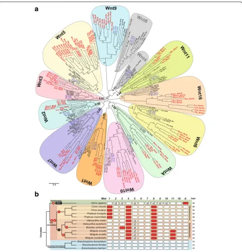
cephalochordate species and ten urochordate species representing five ascidian families from two different or-ders (two solitary Cionidae and two solitary Ascidiidae within Phlebobranchia; and two solitary Pyuridae, three solitary Molgulidae, and one colonial Styelidae within Sto-lidobranchia)—identified 156Wntgenes (Additional file1: Table S1), which constitutes the first comprehensive cata-log ofWntgenes in non-vertebrate chordates. Our phylo-genetic analyses, which included a total of 247 Wnt sequences from 19 species (sequence alignment in Additional file2) representing all major metazoan groups, from cnidarians to vertebrates, sorted the non-vertebrate chordate Wnt sequences into 13 monophyletic groups corresponding to the 13 known Wnt subfamilies (Fig.1).
Conservative Wnt evolution in cephalochordates
Our phylogenetic analyses revealed that the three
Bran-chiostomaspecies possess one ortholog for each of the 13 ancient Wnt subfamilies (Fig. 1a, b; see Additional file 2
for sequence alignment). Our results identified five Wnt
genes that had not been analyzed in cephalochordates be-fore and corroborated the orthology of eight previously described amphioxusWntgenes [34–39,41] (reviewed in [40]). We further extended our Wnt survey to the
tran-scriptome project ofAsymmetron lucayanum, a
cephalo-chordate distantly related to the other Branchiostoma
species [43]. We identified 13 Wnt sequences, mostly full length (Additional file1: Table S1), each one orthologous to one of the 13 Wnt subfamilies (Additional file1: Figure S4; see Additional file3for sequence alignment). The fact that all amphioxus Wnt orthologs form a single cluster nested within vertebrate and ambulacrarian sequences from each Wnt subfamily suggests that neither gene duplications nor gene losses affected the evolution of Wnt subfamilies in the cephalochordate subphylum. Our find-ings, therefore, reinforce the idea of genomic and morpho-logical evolutionary stasis attributed to cephalochordate species [41, 44–48] (reviewed in [49]), in spite of diver-gence times estimated at over tens of millions of years ago [43,50–53].
Liberal Wnt evolution with losses and duplications in ascidian urochordates
Our phylogenetic analyses provided the first fully
re-solved orthology of ascidian Wnt genes, allowing us to
rename previously described genes still classified as“ or-phan Wnts” with unclear orthology, as well as to settle conflicting orthology assignments, probably caused by limited species sampling [29–32] (Fig.1aand Additional
file 1: Table S1). The phylogenetic tree showed long
branches for ascidian Wntgenes, which rarely clustered
as the sister group of vertebrate Wntgenes within each
Wnt subfamily, as would be expected from their
taxo-nomic relationships, likely due to artifactual “
long--branch attraction” [54]. Our results showed that
ascidians, in contrast to amphioxus, do not conserve the entire Wnt repertoire and have suffered various events of gene loss, as well as gene duplications (Fig.1). While some of the losses appeared to be ancestral, resulting in the absence of Wnt subfamilies in all ascidian species, other losses and duplications affected different families and orders heterogeneously, suggesting a more dynamic
evolution ofWnt genes in ascidians than in the
conser-vative amphioxus.Wnt4and Wnt8, for instance, are
ab-sent in all analyzed species, plausibly due to two ancestral gene losses that occurred prior to the ascidian radiation, and therefore relevant to our understanding of the divergence in developmental mechanisms between
ascidians and other chordates (Fig. 1b). On the other
hand,Wnt1andWnt11appear to be absent in species of
the Phelobobranchia suborder and Molgula genus,
re-spectively, while Wnt3 is only absent in B. schlosseri. It
seems, therefore, that loss of Wnt genes might have
contributed to the genetic divergence between different groups or even single species of ascidians.
Our results, moreover, revealed that the Wnt5 subfamily in the Stolidobranchia order had experienced extensive amplification by gene duplication, affecting all six analyzed species (Fig.1b). The fact that many of theseWnt5 dupli-cates appeared to be located in the same genomic regions suggested that they originated by tandem gene duplica-tions (Additional file 1: Figure S1). The complex phylo-genetic reconstructions of the ascidian Wnt5 clade were difficult to interpret, suggesting either the occurrence of independent gene duplications in different lineages after events of speciation or, alternatively, ancestralWnt5 dupli-cations in stem Stolidobranchia, followed by multiple gene losses and events of gene conversion (Additional file 1:
Figure S1). In either case, the expansion of Wnt5 in
Stolidobranchia provides a singular case of Wnt subfamily amplification in non-vertebrate chordates, suggesting that the evolution of this order of ascidian species has been accompanied by a relaxation of the evolutionary con-straints that maintainWnt5genes as a single copy gene in other species. This may be linked to the recruitment of
new Wnt5 paralogs in biological innovations unique to
this group of ascidians.
Overall, the dynamic pattern of gene losses and
dupli-cations of Wnt genes observed among different orders,
WntA, lost and found in chordates
Our analysis also led to the identification ofWntAgenes in cephalochordates and urochordates (Fig.1and Additional file1: Table S1). This finding was of special interest since
WntAhad previously been identified in cnidarians, proto-stomes, and ambulacrarian deuterostomes (e.g., in the hemichordateSaccoglossus kowalevskiiand the echinoderm
Strongylocentrotus purpuratus), but not in any chordate, leading to the suggestion that the WntA subfamily had been lost in stem chordates [10,24]. The identification in
this study of WntA genes in both cephalochordate and
urochordate species implies, therefore, that WntA was
present in the last common chordate ancestor, preserved in cephalochodates and urochordates, but specifically lost during the early evolution of the vertebrate lineage.
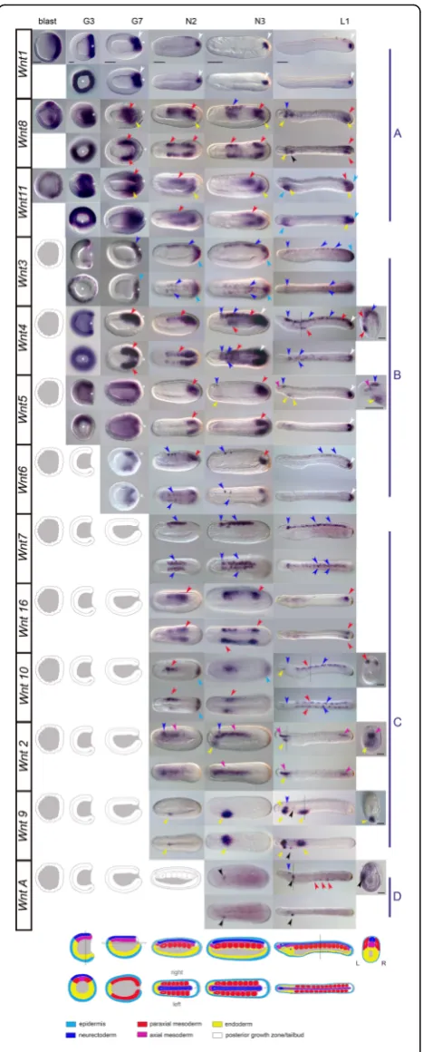
The complete expression atlas of cephalochordate Wnt genes
The apparent conservative evolutionary stasis shown in amphioxus and the finding that amphioxus possesses a full and non-redundant Wnt repertoire make this
organ-ism a very attractive model to infer the roles of Wnt
genes in the ancestral stem chordate, and from compara-tive studies, to analyze the impact of changes in the Wnt repertoire during the evolution of each chordate subphylum. In order to obtain the first comprehensive stage-matched developmental expression atlas for the ceph-alochordate Wnt repertoire, we performed whole-mount in
situ hybridization (WMISH) for all Wnt genes in the
European speciesB. lanceolatum (Figs.2 and 3). Our re-sults revealed that Wnt genes were expressed in a robust and precise tissue-specific fashion, with significant overlap
among several amphioxus Wntparalogs. Nevertheless, we
also saw a number of differences in the expression of differ-ent paralogs, suggesting a choreographed modulation of their expression domains throughout development to
gen-erate spatio-temporally complementary patterns (a
gene-by-gene detailed description of the expression
pat-terns of all Wnt genes shown in Fig. 2 is provided in
Additional file1: Text S1 and summarized in Fig.4). Over-all,Wntexpression appears to be highly dynamic, spanning a broad variety of tissues derived from all three germ layers, which precisely up- or down-regulate differentWnt para-logs throughout development.
Posterior dominance of Wnt expression
“Posteriority” is likely the most conspicuous hallmark observed in the expression domains of the majority of
amphioxus Wnt genes. At the blastula stage, Wnt1,
Wnt8, andWnt11, which were the firstWntgenes to be
expressed in amphioxus (class A genes in Fig.2), showed an evident restriction of their expression domains to the
posterior half of the embryo. Thus, while Wnt1
expres-sion clearly labeled the vegetal pole, Wnt8 and Wnt11
were expressed at the equator of the prospective poster-ior pole [55]. At the gastrula stage G3, along with the
early class A genes, Wnt3, Wnt4, and Wnt5 expression
domains appeared surrounding the blastopore (white as-terisks, class B genes in Fig. 2), as well as Wnt6 a little later (at stage G7). They were expressed in these poster-ior regions through neurulation (N2 and N3) until larval stages (L1). Close observation of the blastoporal view re-vealed some degree of overlap among gene expression
patterns, but also important differences among theWnt
expression domains of each paralog. For instance, while
Wnt1 signal labeled the entire circumference
surround-ing the blastopore (Fig. 2, G3 column), Wnt3
demar-cated a narrower outer ectodermal strip of cells into G7
(Fig. 2). Wnt8 and Wnt11 signals were excluded from
the edges of the blastopore and reached more central
endomesodermal regions (Fig. 2). At G3, Wnt4 signal
was also strongly visible in the endomesoderm near the blastopore, but rather than being restricted to the pos-terior region, it spanned the entire layer surrounding the gastrocoel and was excluded entirely from the ectoderm
(Fig. 2). Finally, Wnt5 most strongly marked the
endo-mesoderm of the dorsal blastopore lip (Fig. 2). Most of
these early posterior Wnt expression domains were
(See figure on previous page.)
Fig. 1Wnt family evolution in chordates.aThe ML phylogenetic tree reveals a conservative pattern of genomic evolution in cephalochordates (species names inblue), which preserve all 13 Wnt subfamilies, contrasting with the dynamic pattern of genomic evolution in urochordates (in red), which are characterized by several gene losses and duplications. Thescale barindicates amino acid substitutions. Values for the approximate likelihood ratio test (aLRT) are shown at nodes. Species are as follows. Chordate species: Urochordates:Botryllus schlosseri(Bsc),Ciona savignyi (Csa),Ciona robusta(Cro; formerlyCiona intestinallis),Halocynthia roretzi(Hro),Halocynthia aurantium(Hau),Mogula occulta(Moccu),Mogula oculata(Mocul),Mogula occidentalis(Mocci),Phallusia fumigata(Pfu), andPhallusia mammillata(Pma). Cephalochordates:Branchiostoma belcheri (Bbe),Branchiostoma floridae(Bfl),Branchiostoma lanceolatum(Bla). Vertebrates:Danio rerio(Dre),Homo sapiens(Hsa). Non-chordates species: hemichordateSaccoglossus kowalevskii(Sko), annelidCapitella teleta(Cte), molluskLottia gigantea(Lgi), and cnidarianNematostella vectensis(Nve).
steadily maintained throughout development. The Wnt1
expression domain, for instance, remained in the poster-ior wall of the neurenteric canal after elongation and closure of the blastopore until at least the early larval
stage (Fig. 2, L1 column). Wnt5 strongly marked the
posterior growth zone during neurulation (Fig. 2, N2
and N3 columns), culminating in strong tailbud expres-sion in larvae (Fig. 2, L1 column).Wnt3 signal was ob-served up to L1 stage in the posterior-most ectoderm
fated to become tailfin, abutting theWnt1domain.
Posteriority was also observed for some Wnt genes
with late expression onset (class C genes in Fig. 2). Of
these, Wnt10, for instance, showed a new ectodermal
expression domain in the most posterior part of the
embryo at N2 and N3 (Fig.2), which subsequently faded
concomitantly with the appearance ofWnt11expression
in this same region. This Wnt11 expression clearly
marked the entire fin in the one-gill-slit larval stage (Fig.2, L1 column, and Fig.3b). In summary, our data show that
nine out of the 13 Wnt amphioxusWntgenes showed
pos-terior expression domains (Fig.4), highlighting posteriority as one of the main hallmarks of this gene family.
Mesodermal Wnt expression and somite formation
In addition to the propensity for posterior dominance,
another important source ofWntsignaling was observed
in the presomitic and axial mesoderm, where seven out
of the 13 Wnt paralogs were expressed. Besides the
aforementioned early overlapping expression domains of
[image:6.595.58.292.86.667.2]Wnt8 and Wnt11 observed in the posterior endomeso-derm surrounding the blastopore at G3, new expression
domains of Wnt4, Wnt5, Wnt6, and Wnt16
consecu-tively appeared in the most posterior mesoderm by G7, N2, N3, and L1, respectively, in a temporally orches-trated manner. At the one-gill-slit larval stage,Wnt5and
Wnt16 expression was maintained in the posterior-most mesoderm (Fig. 2, L1 column, and Fig. 3b). In addition
to the Wnt-positive posterior mesodermal domain,
sev-eral other Wnt expression domains could be identified
in temporally dynamic complementary (as well as over-lapping) patterns, in some cases forming nested domains
along the anteroposterior axis (for instance, Wnt8/16–
Wnt11–Wnt8/16–Wnt4/5/6). In other casesWnt expres-sion domains differed in their dorso-ventral extent
within somites. For instance, Wnt10 appeared excluded
from dorsal domains compared with Wnt16 in the N2
stage. Wnt16 signal was observed in the last pair of
formed somites by L1, whileWnt10 appeared to be
dor-sally restricted in all mature somites (see cross-section in Fig. 2) and excluded from this last pair. No Wnt signal was observed in the anteriormost pair of somites at any stage of development.
In contrast to the paraxial mesoderm, surprisingly few
Wntgenes appeared to be expressed in chordamesoderm
or other mesoderm derivatives. For example, only Wnt2
andWnt5were expressed in the notochord.Wnt2signal
was localized to the anterior two-thirds of the
chorda-mesoderm during neurula stages (Fig. 2), becoming
restricted to the most caudal and rostral portions in one-gill-slit larvae (Fig. 3); in contrast, Wnt5 was con-spicuously expressed in the anterior notochord only in larval stages (Fig. 2, L1 column, and Fig.3). Finally, sev-eral non-myotomal structures of mesodermal origin
ap-peared to express Wnt genes in restricted domains.
Wnt4 signal was observed in mesothelial cells in late
a
b
[image:7.595.59.539.88.373.2]neurula and early larval stages on the left side adjacent
to the pharynx (Fig. 2). WntA was expressed in larval
stages in mesothelial cells between the ventral endoderm of the gut and the ectoderm in both early and late larvae (Figs.2,3b, and4).
Endodermal Wnt expression
In addition to mesodermal expression, Wnt signal was
also evident in endodermal derivatives. By the gastrula
stage, Wnt8 and Wnt11 expression domains were
already observed in the endodermal portion delimiting the blastopore, expression domains that persisted until N3 and L1, respectively (Fig.2). Many of theWntgenes expressed in the posterior growth zone or tailbud also labeled hindgut domains in that area (e.g., Wnt4; Fig.2).
During larval stages, new anterior Wnt expression
do-mains became evident in the anterior region and other parts of the digestive system (Fig.2, L1 column). Some,
such as Wnt5, Wnt2, and Wnt9, appeared to be already
expressed at late neurula stages in anterior endoderm
(Fig.2, N2 and N3 columns).Wntgenes labeling specific
derivatives by L1 included Wnt8, Wnt9, and WntA in
the mouth primordium, Wnt2,Wnt5, Wnt8, andWnt11
in Haetschek’s diverticula,Wnt8and Wnt9in the endo-style, andWnt9in the branchial primordium (Fig. 2, L1 column). Once the mouth was open and the first gill slit
was clearly formed,Wnt5labeled the club-shaped gland,
while Wnt8andWnt9labeled the preoral pit,WntA the
entire circumference of the mouth, andWnt9andWntA
the first gill slit (Figs. 2 and 3a). Wnt4and Wnt2 signal appeared in a few cells of the endostyle along withWnt8
and Wnt9.Wnt10signal was also clearly evident in cells lining the rostral coelom (Fig. 3a). Posteriorly,Wnt6 ap-peared to be expressed in a single line of cells demarcat-ing the posterior wall of the neurenteric canal, andWnt9
labeled cells within the posterior gut (Fig.3b).
Ectodermal Wnt expression
Ectodermal Wntexpression was detected in the
epider-mis as well as the nervous system. Besides the sequential
[image:8.595.58.538.87.394.2]coexpression of Wnt3, Wnt10, and Wnt11 in the tip of the tail, the ventral epidermis of the anteroventral region
at the level of the Haetschek’s diverticulum also
ap-peared to express Wnt11and Wnt5in L1 larvae (Fig. 2,
L1 column). At some stage, allWntgenes—with the
ex-ception of Wnt11—labeled the developing central
ner-vous system (CNS), consisting of cerebral vesicle and nerve cord (Figs.3and4). Thus, the invaginating neural
tube expressed Wnt7, Wnt3, Wnt6, Wnt2, Wnt4, and
Wnt8 starting in neurula stages, while Wnt5, Wnt10,
and WntA signal appeared later in early larvae (Fig. 2). The spatio-temporal expression profiles differed among
ligands, ranging from continuous Wnt7 expression
throughout the majority of the nervous system until lar-val stages, to the more restricted patterns shown by
Wnt2,Wnt4,Wnt6,WntA,Wnt10, orWnt5. Some, such as WntA or Wnt10, only labeled a few isolated cells within the cerebral vesicle in the early larval stage L1 (Fig. 2, L1 column). However, by the one-gill-slit larval stage, regionalisation of the cerebral vesicle became
ap-parent, with Wnt5 expression restricted to an anterior
domain encompassing the frontal eye (Fig. 3a), while
genes such as Wnt4, Wnt7, Wnt8, Wnt9, and Wnt10
(and possibly Wnt16) marked posterior regions of the
cerebral vesicle or the hindbrain (Fig.3b).
WntA expression in non-vertebrate chordates
We paid special attention to further investigating the function and evolution of our newly discovered chordate
WntA genes. We therefore analyzed the expression
pat-terns of WntA during embryonic development not only
in the amphioxus B. lanceolatum but also in two
add-itional chordate species, the ascidiansC. robusta (Phelo-bobranchia order) andH. roretzi(Stolidobranchia order).
WntA expression, however, was not detected during
embryonic development in either of these two ascidian species (Additional file1: Figure S2). This lack of
expres-sion of WntA was consistent with the absence of ESTs
from embryonic libraries in databases ofC. robusta and
H. roretzi; the only existing ESTs of the WntA gene
(FF969784 and FF969783 ofC. robusta) came from adult
animals. These results suggested, therefore, that WntA
might be exclusively expressed at postembryonic stages or during adulthood in ascidians.
In contrast to ascidians, we found that WntA was
expressed in a complex pattern during amphioxus embry-onic development. Our results revealed that amphioxus
WntAwas the lastWntgene to turn on in mid-late neuru-lae, with expression in only one or two cells located an-teriorly on the left side under the ectoderm (Fig.2), likely in the oral mesovesicle (OMV). The OMV is a coelomic vesicle that develops from the posterior ventral corner of the left first somite, and which has been associated with
amphioxus mouth opening [56]. This expression domain
persisted throughout development, accompanied by punc-tuated expression in the posterior cerebral vesicle from the late neurula until the pre-mouth larval stage (see
above). WntA signal was also observed in cells between
the ectoderm and endoderm of the forming gut of early and one-slit larvae (Fig. 2, cross-section, and Fig. 3),
possibly in the mesothelial precursors of the “
amoebo-cytes”, considered to be the homologs of the vertebrate blood cells [57]. Strong expression was further evident all around the mouth opening in these late larvae (Fig. 3a). This gene therefore represents a late-phaseWnt(class D) that plays a major post-neurulation role in differentiating structures such as the mouth and cerebral vesicle, and possibly in the circulatory system of amphioxus.
Discussion
Evolutionary patterns of Wnt subfamilies in non-vertebrate chordates
The identification and fully resolved phylogenetic re-construction of the entire Wnt repertoire in several
species of urochordate and cephalochordate (Fig. 1
and Additional file 1: Figure S4) permit the first
complete reconstruction of the evolution of the Wnt subfamilies in chordates, revealing that each subphy-lum follows different evolutionary trajectories. Cepha-lochordates show a conservative evolutionary pattern, without either apparent gene duplications or gene losses since the cephalochordate lineage diverged from other chordates more than half a billion years ago [43,50]. This finding suggests that the amphioxus genome has pre-served the complete and prototypical
chordate/deutero-stome/eumetazoan Wnt repertoire (Fig. 1b). Analyses of
many amphioxus gene families (e.g., Hox cluster [58],
homeobox gene families [59], gene families of the steroid and retinoic acid signaling pathway [60–62], the tyrosine kinase superfamily [63], DNA-methylation genes [64, 65], and the FGF gene family [66]) reinforces the idea of evolu-tionary stasis of cephalochordate genomes [43, 44, 48]. The expression patterns of allWntgenes analyzed here in
only some groups of ascidians (i.e.,Wnt1in the
Phelobo-branchia suborder andWnt11 in theMolgulagenus), or
limited to specific species (Wnt3 to B. schlosseri). Infer-ring the point at which Wnt losses occurred is not only important to better understand their possible impact on the divergence of developmental mechanisms between ascidians and other chordates, but will also help eluci-date how they may have contributed to genetic and morphological divergence during the ascidian radiation.
Moreover, the extensive gene duplications of the Wnt5
subfamily of the Stolidobranchia order may have also facilitated divergence within this order. Since this is the
only case of amplification of all Wnt subfamilies
ana-lyzed in non-vertebrate chordates, and recurrent tandem duplications have independently affected several species of the Stolidobranchia, the developmental constraints to
maintain Wnt5as single copy seem to have been
exclu-sively relaxed in this order of ascidians.
In contrast to cephalochordates and urochordates, many vertebrate Wnt subfamilies consist of two ohnolo-gues (e.g., human Wnt subfamilies 2, 3, 5, 7, 8, 9, and
10), which originated during the two rounds of
whole-genome duplications (2R-WGD) that occurred during early vertebrate evolution [4]. The differences in
the retention rate of Wnt paralogs between vertebrate
and non-vertebrate chordates are possibly due to the dif-ferent modes of duplication, i.e., genome duplication vs small-scale duplication (reviewed in [4, 8]), and suggest that duplication within Wnt subfamilies in non-vertebrate chordates might be impaired by dosage imbalance with the exception of the Wnt5 subfamily in the Stolidobranchia order. Remarkably, WntA is the only Wnt subfamily
ab-sent in vertebrates. To investigate when the WntA gene
was lost, we analyzed the Wnt catalog of two additional
vertebrate species: the lamprey Petromyzon marinus, an
agnathan representative, and the sharkCallorhinchus milii, a cartilaginous fish. We identified 24 Wnt sequences inP. marinus and 20 in C. milii databases (Additional file 1: Table S1). In our phylogenetic reconstruction, P.marinus
andC. miliisequences distributed among all Wnt subfam-ilies, with the exception of the WntA subfamily (Additional file 1: Figure S4; see Additional file 3for sequence align-ment), suggesting that WntA was lost early in vertebrate evolution, before the divergence of jawless and
gnathos-tome lineages. It can be argued, therefore, that WntA
plausibly became dispensable in the primitive vertebrate ei-ther because alternative pathways or genes compensated for its function, or because environmental or physiological changes made them dispensable [8]. Finally, taking advan-tage of genomic information on the newWntgenes identi-fied in this study, we have re-evaluated the conserved synteny of previously postulatedWntclusters [25]. Amphi-oxus has four clusters: theWnt9–Wnt1–Wnt6 cluster, the
Wnt2–Wnt16 cluster, the Wnt3–Wnt10 cluster, and the
Wnt5–Wnt7 cluster (Fig.5; Additional file1: Table S3). In ascidians, we have found evidence for a single cluster:
Wnt9–Wnt6–Wnt3in C. robusta and Wnt6–Wnt3 in C. savignyi(Fig.5; Additional file 1: Table S3). Lamprey con-serves at least five clusters: theWnt9–Wnt1–Wnt6cluster, the Wnt3–Wnt10and at least threeWnt5–Wnt7clusters (Fig. 5; Additional file 1: Table S3). Interestingly, cluster conservation is higher between lamprey and amphioxus
than between either of these and human (Wnt5–Wnt7,
andWnt5–Wnt7andWnt2–Wnt16, respectively), pointing to genomic rearrangements in the lineage leading to humans. In summary, our work highlights how distinct genomic rearrangements and patterns of gene
conserva-tion, loss, and duplication shaped differently the Wnt
repertoire in amphioxus, ascidians, and vertebrates, and provides an evolutionary scenario that will facilitate future investigations of how these changes relate to adaptations to the environmental and physiological requirements evolved by each subphylum.
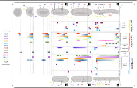
Comparative analysis of Wnt expression patterns during embryonic development in the three chordate subphyla
Our assignment of all non-vertebrate chordate Wnt
genes to the different Wnt subfamilies permits the first comparison of expression patterns of all orthologs among vertebrate and non-vertebrate chordates. With the complete cephalochordate dataset in hand, we were able to compare the expression patterns of allB. lanceo-latum Wnt genes with the reported patterns of
verte-brate Wnt genes and those available for ascidians
(Additional file 1: Figure S3 and Text S2). Overall, our analysis revealed three main situations: first, cases of
orthologousWntgenes that share expression domains in
homologous structures among chordate species, likely reflecting ancestral functional conservation; second,
homologous structures that share Wnt expression
do-mains, although the orthology of the ligands is not con-served among taxa, suggesting, therefore, gene function
shuffling; and third, Wnt expression domains absent in
amphioxus but present in other chordates, possibly
reflecting lineage-specific Wnt innovations during the
evolution of Olfactores (vertebrates + urochordates), or simplifications of the cephalochordate lineage.
Ancestral conserved Wnt functions in chordates
well as in the gill slits, the vertebrate homologs of amphi-oxus pharyngeal arches, and in the amphiamphi-oxus cerebral vesicle and the brain of vertebrates [70–73], while to our knowledge, only partial expression data have been docu-mented forWnt9orthologs in a colonial ascidian [33].
Fi-nally, shared expression domains are observed forWnt10
and Wnt16 in the neurectoderm, Wnt16 in somites, and
Wnt5, Wnt3, Wnt8, and possibly Wnt11 in the tailbud
(Additional file 1: Figure S3 and associated references). Globally, comparative analyses suggest a limited conserva-tion in the expression patterns of orthologous Wnt subfa-miles in the three chordate subphyla.
Function shuffling among Wnt paralogs
In vertebrates, expression ofWnt1is essential for proper anteriorposterior axial patterning of the brain and
specification of particular neuronal populations (the “mid-hindbrain organizer”), in some cases functioning
redundantly with other Wnt genes [74]. In amphioxus,
no Wnt1 expression has been observed in the anterior
neuroectoderm or the cerebral vesicle ([38] and this
study). However, we have identified a different ligand,
Wnt2, with expression in the developing cerebral vesicle at mid-neurula stages, which is compatible with a role of
amphioxus Wnt2 in cerebral vesicle/hindbrain
pattern-ing. Other Wnt genes, including Wnt3, Wnt5, Wnt7,
Wnt8, Wnt9, WntA, and Wnt16, are also turned on in
the amphioxus brain between neurulation and larval stages (reviewed in [40] and this study). These results are consistent with the unexpectedly complex genoarchi-tecture in the amphioxus neural tube that is conserved with vertebrates [42] and highlights events of “function
[image:11.595.57.538.87.453.2]shuffling”[9] among Wnt paralogs during chordate evo-lution. Other remarkable examples of function shuffling can be found in the notochord, one of the defining
syn-apomorphies of all chordates. BesidesWnt5and Wnt11,
which are near-ubiquitously expressed and may play more general functions (perhaps in cell movement or
polarity) across chordates, Wnt2 is the only paralog
expressed in the amphioxus notochord, while chick
Wnt16 (and maybe Xenopus Wnt4 andWnt8 according to Xenbase data) is expressed in the vertebrate structure (Fig. 2; reviewed in Additional file1: Figure S3). In
con-trast,Wnt5 inC. robustaand the Wnt-5α paralog in H.
roretzi are the onlyWntgenes so far identified with ex-pression in the asicidian notochord [26, 31, 75]. Similar examples of Wnt function shuffling are observed in early mesodermal derivatives (e.g., early paraxial mesoderm or
somites), which expressWnt10in amphioxus (this work)
but Wnt3 in both vertebrates and ascidians (Fig. 2;
reviewed in Additional file1: Figure S3).
Comparison of the development of the posterior pole of the embryo in all three subphyla reveals a complex scenario in which a variable number of Wnt subfamilies take part in different species. While in ascidians, Wnt5
seems to be the only ligand determining early posterior-ity in the primary axis, in amphioxus our study reveals a
highly redundant posterior Wnt expression system
in-volving at least eight out of the 13 subfamilies (i.e.,
Wnt1, 8, 11, and 3 surrounding the blastopore during
gastrulation, plus Wnt4,5,16, and6 in the most caudal part of the embryo later during neurulation and larval stages). In vertebrates, interestingly, while some species (similarly to ascidians) use a reduced number of Wnt
li-gands for determing early posteriority (e.g., Wnt8a in
zebrafish [76], Wnt11 (and Wnt5) in Xenopus [77, 78], andWnt3in mouse [79] (reviewed in [11]), other species such as chicken show a redundant posterior Wnt sys-tem, more similarly to amphioxus (e.g., [80, 81]; Add-itional file 1: Figure S3 and associated references). If the ancestral chordate relied on a simple Wnt system for determing posteriority, extensive Wnt function shuffling occurred during the evolution of the cephalochordate lineage as well as some vertebrate species such as chicken, recruiting other Wnt subfamilies for this posterior signal-ing role. Evidence from the direct-developsignal-ing hemichord-ate Saccoglossus kowalevskifurther corroborates the idea that posterior Wnt flexibility is a common occurrence during evolution, with a different but partially shared
combination ofWntgenes showing blastoporal expression
during gastrulation (Wnt1, Wnt3, Wnt4, Wnt6, and
Wnt16) [82]. It seems, therefore, that providingβ-catenin is asymmetrically localized (along the axis or in dividing daughter cells), then which particular Wnt ligands act up-stream, or how they are spatially organized relative to one another, may not be particularly important [13], making
the Wnt system one of the gene families most prone to function shuffling so far described. Importantly, the exten-sive events of function shuffling that occurred during Wnt evolution challenge the notion of establishing homologies simply based on the expression of orthologous genes and highlight the need to consider these events when analyz-ing cases of deep homology.
Wnt expression domains absent in amphioxus but present in other chordates
Different Wnt expression domains have been observed at
different stages of germline formation and gonadogenesis
in Olfactores, such as Wnt5 in two ascidian species and
mouse,Wnt4andWnt8in zebrafish and mouse,Wnt1and
Wnt3 in chick, and Wnt11 in Xenopus(Additional file 1: Figure S3 and associated references), while no evidence has been found suggesting any specificWntexpression in ceph-alochordate primordial germ cells (PGCs) or the germline (this work and [46,67,83,84]; reviewed in [49]). Similarly, the mesodermal cardiac-related expression domain of
Wnt9 in Olfactores—i.e., the heart endocardium of verte-brates [85] and the epithelial walls of the vasculature of the colonial ascidian B.schlosseri[33]—is not paralleled by any
Wntparalog in amphioxus. Moreover, in vertebrates, many
Wntgenes are expressed in placodal derivatives and neural crest (Additional file 1: Figure S3 and associated refer-ences), and complex modulation of Wnt signals both within ectoderm and from other tissues is required for their specification and later differentiation [86–88]. In
amphi-oxus, none of the ectodermal Wnt domains seem to be
compatible with the presence of placode-like structures, which supports the notion that placodes may have been an evolutionary innovation of Olfactores [89–91], similar to migratory cells with neural crest-like properties [92]. It seems, therefore, that the appearance of important func-tional novelties during chordate diversification—e.g., germ-line, heart or placode development—were accompanied by new expression domains and functions ofWntgenes.
Evolution of WntA: another new mouth?
Our work demonstrates the presence ofWntA orthologs
in both cephalochordates and urochordates, suggesting that its absence in vetebrates is likely due to a specific gene loss in this lineage. Our expression analyses reveal WntA function may be linked to mouth development in amphioxus, since it is clearly expressed in the region where the mouth will open at neurula stages, and around the periphery of the opening mouth in late larvae. A role for Wnt signaling in cephalochordate mouth formation may be further supported by the expression of Wnt antag-onistDkk1/2/4 in the region in which the dissolution of basal laminae and mouth perforation occur [56].
the chordate mouth shifted from its now dorsal position
[93], implying that chordates evolved a new means of
mouth formation. Two possible evolutionary scenarios have been proposed for the origin of the amphioxus mouth, which would have had different consequences on the evolu-tion ofWntA. In the first scenario, the amphioxus mouth would share its evolutionary origins with the ambulacrarian coelomic pore-canal [56]. In this case, the absence ofWntA
expression associated with pore-canal formation in
Parentrotus lividus [94] may suggest that WntA function was co-opted in the cephalochordate lineage (or secondarily lost in sea urchin). In the second scenario, the amphioxus mouth would represent a specialized gill slit [95] supported by the fact that both structures utilize Wnt (now including WntA), Fgf, and Hh signaling pathways for their formation [41,96,97], and that uncoupling of the gene regulatory net-work for mouth formation from the blastopore could be
key deuterostome innovations. The expression ofWntAin
hemichordates is of particular relevance for discriminating between these hypotheses. Although evidence points to a conserved pharyngeal transcriptional network in deutero-stomes [98], the expression patterns reported forWntAinS. kowalveskiiare for stages too early to properly evaluate a pu-tative conserved role in gill slit (or mouth) formation [82].
Regarding a role for WntA in ascidian mouth
develop-ment, it should be noted that Olfactores and amphioxus pri-mary mouths may not be homologous structures [56, 99]. To open a new mouth, Olfactores developed an anterior placode or stomodaeum, whereas amphioxus utilized an al-ternative system probably owing to its inability to form pla-codes [100]. With an alternative mode to open a mouth, Olfactores no longer neededWntAfor this purpose, which might favor its loss in vertebrates and redeployment in uro-chordates. The dorsal-ventral inversion hypothesis, besides postulating changes in mouth formation mechanisms, pro-poses associated changes in brain architecture, which may have further relaxed constraints onWntA(and otherWnt) gene expression in this structure in different chordate
lineages. Losses or redeployments of WntA have indeed
been frequent during evolution. WntA has been lost in
different species of arthropods, annelids, platyhelminthes and cnidarians [8], or used in a variety of conserved or novel structures and processes in different prototostome species [21,101–104].WntAevolution illustrates, therefore, the ver-satility of Wnt signaling in controlling diverse biological processes in metazoans.
Functional diversification and loss of Wnt signaling during animal evolution
Function shuffling has significant consequences because it makes it difficult to predict biological function from orthology, challenging the so-called“orthology–function
conjecture” (reviewed in [105]). This consequence is
supported by the divergent expression patterns we
observe here for orthologous chordate Wnt genes. Far
from being a specific property of the chordate Wnt fam-ily, however, substantial differences have also been
re-ported forWntexpression in many other animal species,
mainly arthopods and annelids [21,25,101,106–110]. A
detailed analysis of many Wnt genes in different
proto-stome species, for instance, leads to the conclusion that the repertoire of Wnt ligands used during segment addition has evolved differentially among arthropod line-ages, that the general role of Wnt8in regulating
poster-ior development has been altered in annelids [21] and
onychophorans [104], and thatWnt5 and Wnt16
ortho-logs are differentially expressed in annelids [25]. Outside bilaterians, substantial differences in the expression
pat-terns of the ctenophore Mnemiopsis leidyi Wnt genes
render difficult comparisons with those of other meta-zoans, including clades such as cnidarians, placometa-zoans, and poriferans [111].
Finally, the loss ofWntAin vertebrates, as well as of a
number of Wnt genes in ascidians, illustrate another
general feature of Wnt evolution: the “pervasiveness” of
the loss of Wntgenes during animal evolution. The loss
of Wnt subfamilies in ascidians might have contributed to the morphological diversification of urochordates.
Likewise, the loss of Wnt6 in hemipterans has been
linked to the loss of maxillary palps in this group of in-sects [112], while the loss ofWnt2 and Wnt4 in insects [107] might be related with arthropod diversification. Thus, pervasive gene loss accompanied by numerous events of function shuffling appear to be two of the main features that characterize the evolution of the Wnt fam-ily not only in chordates but also in all branches of metazoan evolution.
Conclusions
Up until now, Wnt research has mainly focused on iden-tifying “conserved” biological functions. Here, we argue that essential information can also be gleaned from the
analysis of Wnt “differences”, many of them derived
from events of function shuffling and gene loss. Under-standing the biological basis of such differences will help uncover how highly conserved developmental pro-cesses—such as axial patterning, germlayer specification,
or body segmentation—might be controlled by
molecu-lar mechanisms (e.g., Wnt signaling) with remarkable genetic and functional diversity.
Methods
Genome database searches and phylogenetic analyses Protein sequences of the Wnt repertoire from vertebrate
Homo sapiens, urochordate C. robusta, and cephalochord-ate B. floridae were used as queries in BLASTp and tBLASTn searches in genome databases of selected species:
genome.bucm.edu.cn/lancelet/ for B. belcheri; https://blas t.ncbi.nlm.nih.gov/Blast.cgiforB. floridae; NCBI Sequence
Read Archive accession SRX437623 forAsymmetron
lucaya-num; http://www.aniseed.cnrs.fr/, https://blast.ncbi.nlm.nih. gov/Blast.cgi and http://octopus.obs-vlfr.fr/public/botryllus/ blastbotryllus.php for ascidian species (C. robusta, C. savignyi,P. fumigata, P. mammillata,H. roretzi, H. auran-tium,B. schlosseri,M. occulta,M. oculata, andM. occiden-talis); https://genomes.stowers.org/organism/Petromyzon/ marinus and https://www.ensembl.org/Petromyzon_mari nus/Info/Index for P. marinus; and NCBI database for C. milii. Orthologies of the non-vertebrate chordateWntwere initially assessed by the reciprocal best blast hit (RBBH) approach and corroborated by phylogenetic analyses. Phylo-genetic reconstructions were based on ML inferences calcu-lated with PhyML v3.0 and automatic mode of selection of substitution model [113] using protein alignments generated
with MUSCLE [114] and CLUSTALX [115] programs and
reviewed manually. Accession numbers and protein align-ment for phylogenetic tree reconstruction are provided in Additional file1: Table S1 and Additional file2, respectively.
Animal collection and gene expression analysis by WMISH
C. robusta type A adults were obtained from the National Bio-Resource Project for Ciona (AMED, Japan).
H. roretzi adults were purchased from fishermen in Ao-mori and Iwate prefectures, Japan.B. lanceolatumadults were collected in Argelès-sur-Mer, France, and induced to spawn as in [116].
Fragments of Wnt genes were PCR amplified and
cloned to synthesize gene-specific riboprobes forH. ror-etzi and B. lanceolatum Wnt genes (Additional file 1:
Table S2). For C. robusta, cDNA clones were obtained
from the cDNA clone collections [117, 118]. WMISH
experiments were performed as previously described in [119] forC. robusta; as in [120] forH. roretziwith minor modifications (the acetylation step was not carried out before prehybridization, and after the antibody incuba-tion the specimens were washed with PBST 12 times, 20 min each, and stored overnight at 4 °C); and as in
[41] forB. lanceolatum. NBT/BCIP (Roche) or
BMPur-ple (Roche) were used for the chromogenic reaction.
Comparative studies of expression patterns
Vertebrate and ascidian Wnt gene expression patterns
were identified and cross- and back- referenced using published literature as well as public database searches. These included ANISEED for ascidians species [121,122] (http://www.aniseed.cnrs.fr/), ZFIN for zebrafish [123] (www.zfin.org), Xenbase for Xenopus [124–126] (http:// www.xenbase.org/, RRID:SCR_003280), Geisha for chick [80] (www.geisha.arizona.edu/geisha/), and the EMAGE gene expression database for mouse [127] (http://www.e mouseatlas.org/emage/). As no such database is currently
available for cephalochordates, published literature images were examined by eye only. In all cases, special care was taken to ensure gene name nomenclature in the literature
matched our results for Wnt gene assignation. In
verte-brates, the expression of paralogs was grouped for ease of comparison across subphyla, under the assumption that both neo- and subfuctionalization events would be ad-equately represented. Please see Additional file1: Figure S2 for additional details.
Additional files
Additional file 1:Figure S1.Evolution ofWnt5in ascidians.Figure S2.
Expression ofWntAin two ascidian species.Figure S3.ChordateWnt expression.Figure S4.Wnt subfamilies inA. lucayanum,P. marinus, andC. milii.Table S1.ChordateWntgenes analyzed in this study.
Table S2.Branchiostoma lanceolatumandHalocynthia roretziprimer and probe sequences.Table S3.Wntsynteny in lancelets (B. lanceolatum,B. belcheriandB. floridae) and vertebrates (H. sapiens andP. marinus). Text S1.Branchiostoma lanceolatum Wntexpression as shown in Fig.2.Text S2.References for Figure S3. (PDF 11566 kb)
Additional file 2:Wnt sequence alignment for Fig.1. (FASTA 307 kb)
Additional file 3:Wnt sequence alignment for Additional file1: Figure S4. (FASTA 99 kb)
Acknowledgements
The authors thank all team members of the CC and RA laboratories for fruitful discussions on Wnt signaling, gene loss, and evolution. The authors thank the PhallusiaandHalocynthiagenome sequencing consortia, as well as all other ascidian laboratories for sharing results ahead of their publication. This work was supported by theBranchiostoma lanceolatumgenome consortium, which provided access to theBranchiostoma lanceolatumgenome sequence. The non-human silhouettes used in Fig.5are courtesy of Tom Barton-Owen, with modifications by IS.
Funding
IS was supported by the European Union Horizon 2020 research and innovation programme under grant agreement numbers 654428 (“CORBEL”). HN was supported by Grants in Aid from The Japan Society for The Promotion of Science (22370078, 15H04377) and The Ministry of Education, Culture, Sports, Science and Thechnology, Japan (23112714, 25113518). KI was supported by a Grant in Aid from the Japan Society for the Promotion of Science (26711014). HE was supported by ANR-16-CE12-0008-01 grant from Agence Nationale de la Recherche. CC was supported by BFU2016-80601-P. RA was supported by BIO2015-67358-C2-1-P grant from Ministerio de Economía y Competitividad (Spain). CC and RA were also supported by grants BFU2010-14875 from Ministerio de Ciencia e Innovación (Spain), and SGR2014-290 and SGR2017-1665 from Generalitat de Catalunya.
Availability of data and materials
The data that support the findings of this study are available from public databases:
http://amphiencode.github.io/forB. lanceolatum;http://genome.bucm.edu.cn/ lancelet/forB. belcheri;https://blast.ncbi.nlm.nih.gov/Blast.cgiforB. floridae; NCBI Sequence Read Archive accession SRX437623 forAsymmetron lucayanum;http:// www.aniseed.cnrs.fr/,https://blast.ncbi.nlm.nih.gov/Blast.cgi, and http://octopus.obs-vlfr.fr/public/botryllus/blastbotryllus.phpfor ascidian species (C. robusta,C. savignyi,P. fumigata,P. mammillata,H. roretzi,H. aurantium,B. schlosseri,M. occulta,M. oculata, andM. occidentalis);https://genomes.stowers.org/organism/Petromyzon/marinus
andhttps://www.ensembl.org/Petromyzon_marinus/Info/IndexforP. marinus; and NCBI database forC. milii. Additional files1,2, and3, including the alignments used for phylogenetic analyses and the accessions numbers of the genes, are available athttps://doi.org/10.6084/m9.figshare.6477494[128].
Authors’contributions
hybridization experiments for expression analysis. IS collated all expression data and did the comparative analyses of chordateWntgenes. JM-S, MD-G, RA, and CC collected all ascidian Wnt sequences from databases. CC and RA performed phylogenetic calculations, with support from JM-S and MD-G, and elaborated the evolutionary inferences. JM-S, KI, and HN analyzed the ex-pression of ascidianWntA.IS, CC, and RA designed the study and ana-lyzed data. IS, CC, and RA wrote the manuscript with imput from JM-S, HE, and HN. IS, JM-S, CC, and RA finalized figures, tables, and text. All authors commented on the manuscript and agreed to its final version.
Ethics approval and consent to participate
Not applicable.
Competing interests
The authors declare that they have no competing interests.
Publisher’s Note
Springer Nature remains neutral with regard to jurisdictional claims in published maps and institutional affiliations.
Author details 1
Biomedical Sciences Research Complex, School of Biology, University of St Andrews, North Haugh, St Andrews KY16 9ST, Scotland, UK.2Scottish Oceans Institute, School of Biology, University of St Andrews, East Sands, St Andrews KY16 8LB, Scotland, UK.3Departament de Genètica, , Microbiologia i Estadística, and Institut de Recerca de la Biodiversitat (IRBio), Universitat de Barcelona, Barcelona, Spain.4Department of Biological Sciences, Graduate School of Science, Osaka University, Toyonaka, Osaka 560-0043, Japan. 5Sorbonne Universités, UPMC Univ Paris 06, CNRS, Biologie Intégrative des
Organismes Marins (BIOM), Observatoire Océanologique, F-66650 Banyuls/ Mer, France.
Received: 11 January 2018 Accepted: 22 June 2018
References
1. Paps J, Holland PWH. Reconstruction of the ancestral metazoan genome reveals an increase in genomic novelty. Nat Commun. 2018;9:1730. 2. Suga H, Chen Z, de Mendoza A, Sebe-Pedros A, Brown MW, Kramer E, Carr M,
Kerner P, Vervoort M, Sanchez-Pons N, et al. The Capsaspora genome reveals a complex unicellular prehistory of animals. Nat Commun. 2013;4:2325. 3. Taylor JS, Raes J. Duplication and divergence: the evolution of new genes
and old ideas. Annu Rev Genet. 2004;38:615–43.
4. Cañestro C, Albalat R, Irimia M, Garcia-Fernandez J. Impact of gene gains, losses and duplication modes on the origin and diversification of vertebrates. Semin Cell Dev Biol. 2013;24:83–94.
5. Cañestro C, Catchen JM, Rodríguez-Marí A, Yokoi H, Postlethwait JH. Consequences of lineage-specific gene loss on functional evolution of surviving paralogs: ALDH1A and retinoic acid signaling in vertebrate genomes. PLoS Genet. 2009;5:e1000496.
6. O'Malley MA, Wideman JG, Ruiz-Trillo I. Losing complexity: the role of simplification in macroevolution. Trends Ecol Evol. 2016;31:608–21. 7. Olson MV. When less is more: gene loss as an engine of evolutionary
change. Am J Hum Genet. 1999;64:18–23.
8. Albalat R, Cañestro C. Evolution by gene loss. Nat Rev Genet. 2016;17:379–91.
9. McClintock JM, Carlson R, Mann DM, Prince VE. Consequences ofHoxgene duplication in the vertebrates: an investigation of the zebrafishHox paralogue group 1 genes. Development. 2001;128:2471–84.
10. Holstein TW. The evolution of the Wnt pathway. Cold Spring Harb Perspect Biol. 2012;4:a007922.
11. Loh KM, van Amerongen R, Nusse R. Generating cellular diversity and spatial form: Wnt signaling and the evolution of multicellular animals. Dev Cell. 2016;38:643–55.
12. Guder C, Philipp I, Lengfeld T, Watanabe H, Hobmayer B, Holstein TW. The Wnt code: cnidarians signal the way. Oncogene. 2006;25:7450–60. 13. Petersen CP, Reddien PW. Wnt signaling and the polarity of the primary
body axis. Cell. 2009;139:1056–68.
14. Niehrs C. The complex world of WNT receptor signalling. Nat Rev Mol Cell Biol. 2012;13:767–79.
15. Rigo-Watermeier T, Kraft B, Ritthaler M, Wallkamm V, Holstein T, Wedlich D. Functional conservation of Nematostella Wnts in canonical and noncanonical Wnt-signaling. Biol Open. 2012;1:43–51.
16. Andre P, Song H, Kim W, Kispert A, Yang Y. Wnt5a and Wnt11 regulate mammalian anterior-posterior axis elongation. Development. 2015;142:1516–27. 17. Kraus Y, Aman A, Technau U, Genikhovich G. Pre-bilaterian origin of the
blastoporal axial organizer. Nat Commun. 2016;7:11694.
18. van Amerongen R, Fuerer C, Mizutani M, Nusse R. Wnt5a can both activate and repress Wnt/beta-catenin signaling during mouse embryonic development. Dev Biol. 2012;369:101–14.
19. Kusserow A, Pang K, Sturm C, Hrouda M, Lentfer J, Schmidt HA, Technau U, von Haeseler A, Hobmayer B, Martindale MQ, Holstein TW. Unexpected complexity of the Wnt gene family in a sea anemone. Nature. 2005;433:156–60.
20. Borisenko I, Adamski M, Ereskovsky A, Adamska M. Surprisingly rich repertoire of Wnt genes in the demosponge Halisarca dujardini. BMC Evol Biol. 2016;16:123.
21. Janssen R, Le Gouar M, Pechmann M, Poulin F, Bolognesi R, Schwager EE, Hopfen C, Colbourne JK, Budd GE, Brown SJ, et al. Conservation, loss, and redeployment of Wnt ligands in protostomes: implications for understanding the evolution of segment formation. BMC Evol Biol. 2010;10:374.
22. Murat S, Hopfen C, McGregor AP. The function and evolution of Wnt genes in arthropods. Arthropod Struct Dev. 2010;39:446–52.
23. Rentzsch F, Technau U. Genomics and development of Nematostella vectensis and other anthozoans. Curr Opin Genet Dev. 2016;39:63–70. 24. Prud'homme B, Lartillot N, Balavoine G, Adoutte A, Vervoort M. Phylogenetic
analysis of the Wnt gene family. Insights from lophotrochozoan members. Curr Biol. 2002;12:1395.
25. Cho SJ, Valles Y, Giani VC Jr, Seaver EC, Weisblat DA. Evolutionary dynamics of the wnt gene family: a lophotrochozoan perspective. Mol Biol Evol. 2010; 27:1645–58.
26. Sasakura Y, Ogasawara M, Makabe KW. HrWnt-5: a maternally expressed ascidian Wnt gene with posterior localization in early embryos. Int J Dev Biol. 1998;42:573–9.
27. Sasakura Y, Makabe KW. Ascidian Wnt-7 gene is expressed exclusively in the tail neural tube of tailbud embryos. Dev Genes Evol. 2000;210:641–3. 28. Miya T, Nishida H. Isolation of cDNA clones for mRNAs transcribed
zygotically during cleavage in the ascidian, Halocynthia roretzi. Dev Genes Evol. 2002;212:30–7.
29. Hino K, Satou Y, Yagi K, Satoh N. A genomewide survey of developmentally relevant genes in Ciona intestinalis. VI. Genes for Wnt, TGFbeta, hedgehog and JAK/STAT signaling pathways. Dev Genes Evol. 2003;213:264–72. 30. Hotta K, Takahashi H, Ueno N, Gojobori T. A genome-wide survey of the
genes for planar polarity signaling or convergent extension-related genes in Ciona intestinalis and phylogenetic comparisons of evolutionary conserved signaling components. Gene. 2003;317:165–85.
31. Imai KS, Hino K, Yagi K, Satoh N, Satou Y. Gene expression profiles of transcription factors and signaling molecules in the ascidian embryo: towards a comprehensive understanding of gene networks. Development. 2004;131:4047–58.
32. Rosner A, Alfassi G, Moiseeva E, Paz G, Rabinowitz C, Lapidot Z, Douek J, Haim A, Rinkevich B. The involvement of three signal transduction pathways in botryllid ascidian astogeny, as revealed by expression patterns of representative genes. Int J Dev Biol. 2014;58:677–92.
33. Di Maio A, Setar L, Tiozzo S, De Tomaso AW. Wnt affects symmetry and morphogenesis during post-embryonic development in colonial chordates. Evodevo. 2015;6:17.
34. Schubert M, Holland LZ, Holland ND. Characterization of two amphioxus Wnt genes (AmphiWnt4 and AmphiWnt7b) with early expression in the developing central nervous system. Dev Dyn. 2000;217:205–15.
35. Schubert M, Holland LZ, Holland ND. Characterization of an amphioxus wnt gene, AmphiWnt11, with possible roles in myogenesis and tail outgrowth. Genesis. 2000;27:1–5.
36. Schubert M, Holland LZ, Holland ND, Jacobs DK. A phylogenetic tree of the Wnt genes based on all available full-length sequences, including five from the cephalochordate amphioxus. Mol Biol Evol. 2000;17:1896–903. 37. Schubert M, Holland LZ, Panopoulou GD, Lehrach H, Holland ND.
Characterization of amphioxus AmphiWnt8: insights into the evolution of patterning of the embryonic dorsoventral axis. Evol Dev. 2000;2:85–92. 38. Holland LZ, Holland ND, Schubert M. Developmental expression of
39. Schubert M, Holland LZ, Stokes MD, Holland ND. Three amphioxus Wnt genes (AmphiWnt3, AmphiWnt5, and AmphiWnt6) associated with the tail bud: the evolution of somitogenesis in chordates. Dev Biol. 2001; 240:262–73.
40. Bertrand S, Le Petillon Y, Somorjai I, Escriva H. Developmental cell-cell communication pathways in the cephalochordate amphioxus: actors and functions. Int J Dev Biol. 2017;61:697–722.
41. Somorjai I, Bertrand S, Camasses A, Haguenauer A, Escriva H. Evidence for stasis and not genetic piracy in developmental expression patterns of Branchiostoma lanceolatum and Branchiostoma floridae, two amphioxus species that have evolved independently over the course of 200 Myr. Dev Genes Evol. 2008;218:703–13.
42. Albuixech-Crespo B, Lopez-Blanch L, Burguera D, Maeso I, Sanchez-Arrones L, Moreno-Bravo JA, Somorjai I, Pascual-Anaya J, Puelles E, Bovolenta P, et al. Molecular regionalization of the developing amphioxus neural tube challenges major partitions of the vertebrate brain. PLoS Biol. 2017;15:e2001573.
43. Yue JX, Yu JK, Putnam NH, Holland LZ. The transcriptome of an amphioxus, Asymmetron lucayanum, from the Bahamas: a window into chordate evolution. Genome Biol Evol. 2014;6:2681–96.
44. Cañestro C, Albalat R. Transposon diversity is higher in amphioxus than in vertebrates: functional and evolutionary inferences. Brief Funct Genomics. 2012;11:131–41.
45. Louis A, Roest Crollius H, Robinson-Rechavi M. How much does the amphioxus genome represent the ancestor of chordates? Brief Funct Genomics. 2012;11:89–95.
46. Dailey SC, Febrero Planas R, Rossell Espier A, Garcia-Fernàndez J, Somorjai IML. Asymmetric distribution of pl10 and bruno2, new members of a conserved Core of early germline determinants in cephalochordates. Front Ecol Evol. 2016;3:156.
47. Yong LW, Bertrand S, Yu JK, Escriva H, Holland ND. Conservation of BMP2/4 expression patterns within the clade Branchiostoma (amphioxus): resolving interspecific discrepancies. Gene Expr Patterns. 2017;25–26:71–5. 48. Paps J, Holland PW, Shimeld SM. A genome-wide view of transcription
factor gene diversity in chordate evolution: less gene loss in amphioxus? Brief Funct Genomics. 2012;11:177–86.
49. Somorjai I, et al. Amphioxus regeneration: evolutionary and biomedical implications. Int J Dev Biol. 2017;61:689–96.
50. Cañestro C, Albalat R, Hjelmqvist L, Godoy L, Jornvall H, Gonzalez-Duarte R. Ascidian and amphioxus Adh genes correlate functional and molecular features of the ADH family expansion during vertebrate evolution. J Mol Evol. 2002;54:81–9.
51. Nohara M, Nishida M, Nishikawa T. New complete mitochondrial DNA sequence of the lancelet Branchiostoma lanceolatum (Cephalochordata) and the identity of this species’sequences. Zool Sci. 2005;22:671–4. 52. Kon T, Nohara M, Yamanoue Y, Fujiwara Y, Nishida M, Nishikawa T.
Phylogenetic position of a whale-fall lancelet (Cephalochordata) inferred from whole mitochondrial genome sequences. BMC Evol Biol. 2007;7:127.
53. Igawa T, Nozawa M, Suzuki DG, Reimer JD, Morov AR, Wang Y, Henmi Y, Yasui K. Evolutionary history of the extant amphioxus lineage with shallow-branching diversification. Sci Rep. 2017;7:1157.
54. Felsenstein J. Cases in which parsimony or compatibility methods will be positively misleading. Syst Zool. 1978;27:401–10.
55. Holland LZ, Holland ND. A revised fate map for amphioxus and the evolution of axial patterning in chordates. Integr Comp Biol. 2007;47:360–72. 56. Kaji T, Reimer JD, Morov AR, Kuratani S, Yasui K. Amphioxus mouth after
dorso-ventral inversion. Zoological Lett. 2016;2:2.
57. Pascual-Anaya J, Albuixech-Crespo B, Somorjai IM, Carmona R, Oisi Y, Alvarez S, Kuratani S, Munoz-Chapuli R, Garcia-Fernandez J. The evolutionary origins of chordate hematopoiesis and vertebrate endothelia. Dev Biol. 2013;375:182–92. 58. Garcia-Fernàndez J, Holland PW. Archetypal organization of the amphioxus
Hox gene cluster. Nature. 1994;370:563–6.
59. Holland LZ, Albalat R, Azumi K, Benito-Gutierrez E, Blow MJ, Bronner-Fraser M, Brunet F, Butts T, Candiani S, Dishaw LJ, et al. The amphioxus genome illuminates vertebrate origins and cephalochordate biology. Genome Res. 2008;18:1100–11.
60. Cañestro C, Postlethwait JH, Gonzàlez-Duarte R, Albalat R. Is retinoic acid genetic machinery a chordate innovation? Evol Dev. 2006;8:394–406. 61. Albalat R. The retinoic acid machinery in invertebrates: ancestral elements
and vertebrate innovations. Mol Cell Endocrinol. 2009;313:23–35.
62. Albalat R, Brunet F, Laudet V, Schubert M. Evolution of retinoid and steroid signaling: vertebrate diversification from an amphioxus perspective. Genome Biol Evol. 2011;3:985–1005.
63. D'Aniello S, Irimia M, Maeso I, Pascual-Anaya J, Jimenez-Delgado S, Bertrand S, Garcia-Fernandez J. Gene expansion and retention leads to a diverse tyrosine kinase superfamily in amphioxus. Mol Biol Evol. 2008;25:1841–54. 64. Albalat R. Evolution of DNA-methylation machinery: DNA methyltransferases
and methyl-DNA binding proteins in the amphioxus Branchiostoma floridae. Dev Genes Evol. 2008;218:691–701.
65. Albalat R, Marti-Solans J, Cañestro C. DNA methylation in amphioxus: from ancestral functions to new roles in vertebrates. Brief Funct Genomics. 2012; 11:142–55.
66. Oulion S, Bertrand S, Escriva H. Evolution of the FGF gene family. Int J Evol Biol. 2012;2012:298147.
67. Zhang QJ, Luo YJ, Wu HR, Chen YT, Yu JK. Expression of germline markers in three species of amphioxus supports a preformation mechanism of germ cell development in cephalochordates. Evodevo. 2013;4:17.
68. Blader P, Strahle U, Ingham PW. Three Wnt genes expressed in a wide variety of tissues during development of the zebrafish, Danio rerio: developmental and evolutionary perspectives. Dev Genes Evol. 1996;206:3–13.
69. Yagi K, Satoh N, Satou Y. Identification of downstream genes of the ascidian muscle determinant gene ci-macho1. Dev Biol. 2004;274:478–89.
70. Curtin E, Hickey G, Kamel G, Davidson AJ, Liao EC. Zebrafish wnt9a is expressed in pharyngeal ectoderm and is required for palate and lower jaw development. Mech Dev. 2011;128:104–15.
71. Quinlan R, Graf M, Mason I, Lumsden A, Kiecker C. Complex and dynamic patterns of Wnt pathway gene expression in the developing chick forebrain. Neural Dev. 2009;4:35.
72. Cox AA, Jezewski PA, Fang PK, Payne-Ferreira TL. Zebrafish Wnt9a,9b paralog comparisons suggest ancestral roles for Wnt9 in neural, oral-pharyngeal ectoderm and mesendoderm. Gene Expr Patterns. 2010;10:251–8. 73. Zhang B, Tran U, Wessely O. Expression of Wnt signaling components
during Xenopus pronephros development. PLoS One. 2011;6:e26533. 74. Lekven AC, Buckles GR, Kostakis N, Moon RT. Wnt1 and wnt10b function
redundantly at the zebrafish midbrain-hindbrain boundary. Dev Biol. 2003; 254:172–87.
75. Niwano T, Takatori N, Kumano G, Nishida H. Wnt5 is required for notochord cell intercalation in the ascidian Halocynthia roretzi. Biol Cell. 2009;101:645–59. 76. Lu FI, Thisse C, Thisse B. Identification and mechanism of regulation of the
zebrafish dorsal determinant. Proc Natl Acad Sci U S A. 2011;108:15876–80. 77. Tao Q, Yokota C, Puck H, Kofron M, Birsoy B, Yan D, Asashima M, Wylie CC, Lin X, Heasman J. Maternal wnt11 activates the canonical wnt signaling pathway required for axis formation in Xenopus embryos. Cell. 2005;120: 857–71.
78. Cha SW, Tadjuidje E, Tao Q, Wylie C, Heasman J. Wnt5a and Wnt11 interact in a maternal Dkk1-regulated fashion to activate both canonical and non-canonical signaling in Xenopus axis formation. Development. 2008;135: 3719–29.
79. Liu P, Wakamiya M, Shea MJ, Albrecht U, Behringer RR, Bradley A. Requirement for Wnt3 in vertebrate axis formation. Nat Genet. 1999;22:361–5.
80. Antin PB, Yatskievych TA, Davey S, Darnell DK. GEISHA: an evolving gene expression resource for the chicken embryo. Nucleic Acids Res. 2014;42: D933–7.
81. Martin A, Maher S, Summerhurst K, Davidson D, Murphy P. Differential deployment of paralogous Wnt genes in the mouse and chick embryo during development. Evol Dev. 2012;14:178–95.
82. Darras S, Fritzenwanker JH, Uhlinger KR, Farrelly E, Pani AM, Hurley IA, Norris RP, Osovitz M, Terasaki M, Wu M, et al. Anteroposterior axis patterning by early canonical Wnt signaling during hemichordate development. PLoS Biol. 2018;16:e2003698.
83. Wu HR, Chen YT, Su YH, Luo YJ, Holland LZ, Yu JK. Asymmetric localization of germline markers vasa and Nanos during early development in the amphioxus Branchiostoma floridae. Dev Biol. 2011;353:147–59. 84. Yue JX, Li KL, Yu JK. Discovery of germline-related genes in
cephalochordate amphioxus: a genome wide survey using genome annotation and transcriptome data. Mar Genomics. 2015;24(Pt 2):147–57. 85. Person AD, Garriock RJ, Krieg PA, Runyan RB, Klewer SE. Frzb modulates
Wnt-9a-mediated beta-catenin signaling during avian atrioventricular cardiac cushion development. Dev Biol. 2005;278:35–48.