www.impactjournals.com/oncotarget/ Oncotarget, Vol. 5, No. 20
TM4SF4 overexpression in radiation-resistant lung carcinoma
cells activates IGF1R via elevation of IGF1
Soo-Im Choi1,2, Seo-Yeon Kim1, Jaeha Lee1,2, Eun-Wie Cho3 and In-Gyu Kim1,2
1 Department of Radiation Biology, Environmental Radiation Research Group, Korea Atomic Energy Research Institute
(KAERI), Daedeok-daero, Yuseong-gu, Daejeon, South Korea
2 Department of Radiation Biotechnology and Applied Radioisotope, Korea University of Science and Technology,
Daedeok-daero, Yuseong-gu, Daejeon, South Korea
3 Epigenomics Research Center, Korea Research Institute of Bioscience and Biotechnology (KRIBB), Gwahak-ro, Yuseong-gu,
Daejeon, South Korea
Correspondence to: Eun-Wie Cho , email: [email protected]
Correspondence to: In-Gyu Kim , email: [email protected]
Keywords: TM4SF4, lung adenocarcinoma, IGF1R activation, IGF1
Received: April 25, 2014 Accepted: September 07, 2014 Published: September 08, 2014
This is an open-access article distributed under the terms of the Creative Commons Attribution License, which permits unrestricted use, distribution, and reproduction in any medium, provided the original author and source are credited.
ABSTRACT
Transmembrane 4 L six family member 4 (TM4SF4) is a member of the tetraspanin L6 domain family. Other members of this family, TM4SF1 (also known as L6-Ag) and TM4SF5, have been shown to be upregulated in multiple tumors and involved in epithelial-to-mesenchymal transition and cell migration. However, unlike its homologs, little is known about TM4SF4. Here, we show that TM4SF4 was highly expressed in radiation-resistant lung adenocarcinoma cells, such as A549 and Calu-3 cells, and its expression activated cell growth, migration, and invasion. Overexpression of TM4SF4 in A549 cells increased the activation of PI3K, AKT, and NF-kappaB and the expression of PTEN. IGF1R was clearly activated by overexpression of TM4SF4, although EGFR was also slightly activated. TM4SF4 expression was correlated with the increased expression of IGF1, consequently resulting in IGF1R activation. Tumorigenic activity of TM4SF4 in lung adenocarcinoma cells was also demonstrated by xenograft assay; however, this activity was almost completely suppressed by treatment with anti-TM4SF4 antibody. Our results suggest that anti-TM4SF4 overexpression in lung carcinoma cells results in resistance to radiotherapy via IGF1-induced IGF1R activation and
blocking the activity of TM4SF4 using specific antibody can be a promising therapeutics
against TM4SF4-overexpressing lung adenocarcinoma.
INTRODUCTION
Transmembrane 4 L six family member 4 (TM4SF4) is a member of the tetraspanin L6 domain family [1], which includes TM4SF1/L6, TM4SF4/IL-TMP, and TM4SF5/L6H. TM4SF1 and TM4SF5 were
originally identified as tumor-associated antigens [2,
3], and their overexpression was reported in multiple tumors, including lung, breast, colon, prostate cancer, and
hepatocellular carcinoma [2, 4-8]. TM4SF1 and TM4SF5
affect migratory mechanisms crucial to cancer invasion
and metastasis [2, 4, 9-11], making them as crucial targets for cancer therapy [10, 12]. Also, TM4SF1 is defined as a cancer stem cell marker [13], and TM4SF1 and TM4SF5
are reported to be involved in epithelial-to-mesenchymal transition [7, 14], which is also associated with stemness properties [15].
TM4SF4 was originally cloned from intestinal epithelium and liver; for this reason, it was named intestine and liver tetraspan membrane protein (IL-TMP) [16, 17]. TM4SF4 is a 202-amino acid membrane
protein that contains four hydrophobic transmembrane
domains and two hydrophilic regions [1]. It is classified
as a more divergent member of the tetraspanin L6 domain
family, owing to a lack of characteristic cysteine residue motifs in the EC2 extracellular domain, and has 50%
cells differentiate and migrate out of intestinal crypts
[16]. Likewise, in the liver, TM4SF4 is expressed in
non-dividing hepatocytes that retain high proliferative potential in the presence of the appropriate stimulus, and
is upregulated during liver injury [17, 18], which implies
that its functions are related to cellular differentiation or
proliferation. However, little is known about functions of
TM4SF4 in cancer cells. Only recently, it was reported that TM4SF4/IL-TMP mRNA and protein levels were upregulated in 80% of hepatocellular carcinoma tissues [19].
Lung cancer is a lethal cancer in both men and women. Non-small cell lung cancer (NSCLC) comprises
the majority (greater than 75%) of lung cancers and,
when clinically extensive, it is typically characterized by inexorable disease progression despite treatment
with chemotherapy and/or irradiation [20]. Because
chemotherapy and irradiation induce programmed cell death, or apoptosis, recent efforts have been made to understand molecular events that confer therapeutic
resistance. Based on these efforts, the phosphatidylinositol-3-kinase (PI3K)/ protein kinase B (AKT) pathway [21] and the IGF1/IGF1R signaling pathway [22] have emerged
as potential determinants of radiation resistance in human lung cancer cells.
Here, we show that TM4SF4 is highly expressed in radiation-resistant lung adenocarcinoma cells, such as
A549 and Calu-3 cells, and its expression activates cell
growth, migration, and invasion via IGF1R activation. Overexpression of TM4SF4 elevated the level of IGF1 induction, which resulted in IGF1R activation and radiation resistance. Treatment of TM4SF4-overexpressing lung carcinoma cells with anti-TM4SF4 antibody suppressed cell growth, which was mediated by
suppression of IGF1 expression. Based on these results,
we discuss the use of anti-TM4SF4 antibody against TM4SF4-overexpressing and radiation-resistant lung cancer therapy.
RESULTS
TM4SF4 is overexpressed in radiation-resistant lung adenocarcinoma A549 cells.
A549 NSCLC adenocarcinoma cancer cells are
more invasive and resistant to radiation than the H460
NSCLC cell line [23, 24]. To identify novel genes involved
in radiation resistance of NSCLC cells, expression levels
of 30,000 human genes in A549 and H460 cells were compared using DNA microarray analysis. Among
hundreds of differentially regulated genes, a dramatic difference in the expression level of TM4SF4 was
observed between these cells; A549 cells expressed
TM4SF4 at a level approximately 30-fold greater than
that observed in H460 cells (data not shown). Based on
these results, expression of TM4SF4 in various NSCLC
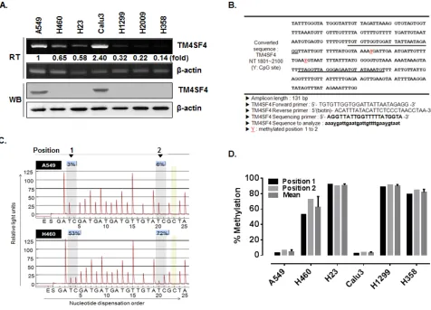
cells, including A549, H460, H23, Calu-3, H1299, H2009 and H358 cells, were analyzed by RT-PCR and Western blotting (Figure 1A). Most of lung cancer cells examined expressed low levels of TM4SF4; however, A549 and
Calu-3 cells showed exceptionally high levels of TM4SF4 expression.
A significant difference in gene expression is usually regulated by DNA methylation, a common epigenetic
signaling tool that cells use to repress transcription. To examine the regulation of TM4SF4 expression by methylation in the NSCLC cells tested above, puta tive CpG islands within the promoter and 5’-untranslated region (5’-UTR) of the TM4SF4 gene were analyzed using the Methprimer program (http://www.urogene.org//
methprimer) [25], and two CpG islands were suggested as methylation sites (Figure 1B). In A549 cells, the two positions were methylated less than 10%. In contrast, the
TM4SF4 gene in H460 cells showed a level of methylation
greater than 50% (Figure 1C). The methylation percentage
of the TM4SF4 gene was also analyzed in other lung
cancer cells. As shown in Figure 1D, lung cancer cells including H23, H1299 and H358, showed high methylation levels, of greater than 80%. However, Calu-3 cells as well as A549 cells showed very low levels, of less than 10% DNA methylation.
In normal lung cells, TM4SF4 is expressed at a very
low level [17]. Also, previous studies of TM4SF4 were
focused on its functions in intestinal epithelium and liver and showed that TM4SF4 is a negative regulator of cell
proliferation [16, 18]. Therefore, TM4SF4 functions in
cancer, especially in lung cancer, have not been studied, although other members of the tetraspanin L6 family, such as TM4SF1 and TM4SF5, have been investigated as
inducers of tumorigenesis. Based on our findings about
the expression and methylation patterns of TM4SF4 in NSCLC cells, we hypothesized that overexpression of TM4SF4 in some aggressive NSCLC adenocarcinoma cells is critical for tumorigenesis and radiation resistance, and we examined this hypothesis using
TM4SF4-overexpressing or knockdown A549 NSCLC cells.
Overexpression of TM4SF4 in A549 cells enhanced cell growth, migration, and invasion.
First, we analyzed the effect of downregulation of
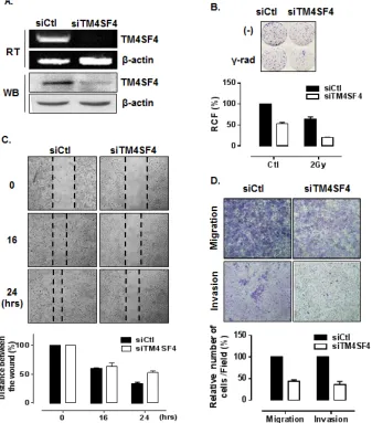
TM4SF4 in A549 cells to examine whether the suppression of TM4SF4 inhibit the tumorigenicity of A549 cells. Short interfering RNA (siRNA)-mediated suppression of TM4SF4 in A549 cells (Figure 2A) retarded cell growth, which was reduced to 50% of control value (Figure 2B).
Resistance to gamma irradiation was also decreased severely. Gamma irradiation reduced relative colony
TM4SF4-knockdown cells to 38%, of the level seen in control cells (Figure 2B). To monitor cell migration, a wound healing assay was performed. As shown in Figure 2C, the rate of wound closure was slower in TM4SF4-knockdown cells.
Results of transwell migration assay showed the same
tendencies (Figure 2D, upper panel). It was also confirmed
that TM4SF4 is involved in invasiveness of tumor cells.
With suppression of TM4SF4, invasion of A549 cells through Matrigel was significantly decreased (Figure 2D,
lower panel).
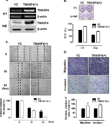
In contrast, A549 cells transfected with
TM4SF4-overexpressing vector, which express TM4SF4 at a
higher level than untransfected A549 cells (Figure 3A)
showed enhanced cell growth and radiation resistance
when compared by colony-forming assay (Figure 3B).
The rates of wound closure (Figure 3C) and transwell migration (Figure 3D, upper panel) were also increased by overexpression of TM4SF4. Finally, Matrigel assay with TM4SF4-overexpressing cells showed again that TM4SF4 expression is closely involved in cell invasive
ability (Figure 3D, lower panel).
TM4SF4 is involved in activation of PI3K, AKT, NF-kappaB, and IGF1R.
Overexpression of TM4SF4 in the invasive A549
NSCLC adenocarcinoma cell line, and its effects on the cellular characteristics of lung carcinoma cells, revealed that TM4SF4 is critical for tumorigenesis and radiation
resistance in A549 NSCLC cells. Calu-3, which is
another NSCLC cell possessing the hypomethylated CpG islands of TM4SF4 gene (Figure 1D), also overexpressed
TM4SF4 (Figure 1A) and showed similar patterns of cell growth and radiation resistance as A549 cells when
expression of TM4SF4 was suppressed or overexpressed
(Supplementary Figure 1A).
To investigate the mechanism of TM4SF4-mediated cell signaling related to tumorigenicity of
[image:3.612.72.548.315.658.2]A549 cells, activations of the PI3K/AKT, NF-kappaB,
Figure 1: TM4SF4 expression in lung cancer cell lines is regulated by methylation. (A) RT-PCR and Western blot analysis of TM4SF4 expression in the indicated lung cancer cell types. Band intensities were measured using Image J software (National Institute of Health, Bethesda, MD, USA), normalized to β-actin and fold increase were indicated. (B) Bisulfite-converted sequence of TM4SF4. Each Y of underlined sequences indicates a methylated position. (C) Pyrosequencing diagram of A549 and H460 cells and comparison of methylation percentages at each CpG position. Each gray box in the diagram indicates the position of the two Y’s shown in panel B. (D)
and ERK signaling pathways, which are involved in cell
growth, radiation resistance, migration, and invasion
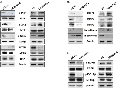
were analyzed. As shown in Figure 4A, suppression of TM4SF4 inhibited activity of the PI3K/AKT pathway. Phosphorylation of AKT and PI3K was severely inhibited, although total levels of AKT and PI3K were not affected.
In these cells, PTEN, a phosphatase involved in inhibition
of AKT signaling [26], was elevated in correlation with TM4SF4 expression level. Phosphorylation of NF-kappaB
was also decreased without an increase in its expression
level. However, ERK phosphorylation was not influenced
by TM4SF4 expression level. In contrast, overexpression
of TM4SF4 elevated phosphorylation of AKT, PI3K, and NF-kappaB. PTEN expression was also completely
suppressed by TM4SF4 overexpression. Matrix metalloproteases (MMPs), which are involved in cell
migration and invasion, were also analyzed (Figure 4B). MMP-2, 7, and 9 were reduced in TM4SF4-knockdown
cells and increased in TM4SF4-overexpressing cells, which is consistent with the effect of TM4SF4 on cellular migration and invasion.
There are several upstream factors which control
[image:4.612.137.474.219.602.2]activation of the PI3K/AKT pathway. In lung cancer cells, IGF1R and EGFR signaling play key roles and
have been suggested as major factors upstream of the
PI3K/AKT pathway. Phosphorylation of EGFR in A549 cells was inhibited by TM4SF4 knockdown and slightly
increased by TM4SF4 overexpression; however, TM4SF4
knockdown severely inhibited phosphorylation of IGF1R (Figure 4C). Overexpression of TM4SF4 also significantly
increased the level of phosphorylated IGF1R. These results suggest that TM4SF4 reinforces tumorigenicity of lung cancer cells primarily through the IGF1R signaling pathway.
TM4SF4 is involved in the activation of IGF1R by up-regulation of IGF1 via NF-κB activation.
Most members of TM4SF family mediate signal transduction events related to the regulation of cell development, activation, growth, and motility and these effects have been investigated primarily in the context of their cooperation with integrin signaling within the plasma
membrane [27]. TM4SF family proteins also mediate
[image:5.612.133.520.199.634.2]signal transduction events by regulating the expression
Figure 3: Overexpression of TM4SF4 in A549 cells enhanced cell growth, migration, and invasiveness. (A) RT-PCR and Western blot analysis of TM4SF4 expression in pcDNA-TM4SF4-transfected A549 cells: RNA and proteins were extracted from cells 72 h post-transfection. (B) Colony-forming assay of TM4SF4-overexpressing A549 cells: 24 h after transfection, cells were irradiated with a single dose of 2 Gy and, 24 h later, plated for colony-forming assay. (C) Wound-healing assay of TM4SF4-overexpressing A549 cells: A549 cells transfected with pcDNA-TM4SF4 or control vector were used for assay 72 h after transfection. At each period of assay, cells were photographed and wound size was determined. (D) Assay of migration (upper panel) and invasion (lower panel)of
of growth factors and their receptors [28]. TM4SF4
expression is also related to the expression of several growth factors and receptors, such as TNF-alpha, TNFR1,
and c-Met in liver cells [18]. Activation of IGF1R which is
essential for the initiation and progression of many cancers
[29, 30] starts by ligand-initiated kinase activation with IGF1 or IGF2. Both types of IGF are synthesized in many
fetal and adult tissues and secreted IGFs act in autocrine or paracrine manner. However, most of extracellular IGFs are
complexed with IGFBP-3, the most prevalent extracellular
IGF binding protein, which indicate that free IGFs in serum is important for the activation of IGF1R [31-36].
To examine the mechanism of IGF1R activation in
TM4SF4-overexpressing A549 cells, we first analyzed the
expression level of each component in the IGF1R pathway.
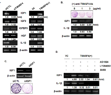
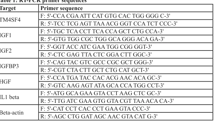
As shown in Figure 4C, levels of IGF1R and EGFR were not influenced by knockdown or overexpression of TM4SF4. The transcripts of their specific ligands (IGF1 and IGF2) and their regulatory component (IGFBP-3) were also analyzed by RT-PCR. As shown in Figure 5A, expression of IGF2 and IGFBP3 was not altered by
knockdown or overexpression of TM4SF4. The expression
of hepatocyte growth factor (HGF), which was analyzed as a growth factor unrelated to the IGF1R signaling pathway, was also unaltered. In contrast, IGF1 expression
was decreased or increased about 20-fold, depending on
the expression of TM4SF4. These results suggest that IGF1R activation mediated by TM4SF4 overexpression
in A549 cells may be the result of regulation of IGF1 expression, specifically, the concentration of free
extracellular IGF1. Interaction between TM4SF4 and IGF1R on the plasma membrane was also investigated by co-immunoprecipitation using anti-IGF1R antibody, as in our previous study of IGF1R activation by transgelin [37]; however, direct association of TM4SF4 and IGF1R was not observed (data not shown). In addition, IL1beta
expression, which is known to facilitate metastasis of lung cancer[38], was also altered depending on the level of TM4SF4.
TM4SF4 is a membrane protein which can be easily
targeted by specific antibody. We examined next whether
[image:6.612.87.546.324.664.2]antibody-mediated inactivation of TM4SF4 suppresses
tumor cell growth or the expression of growth factors.
Cells were treated with anti-TM4SF4 antibody (1~3μg/ mL) and colony-forming units were assessed. As shown in Figure 5B, antibody treatment greatly suppressed colony formation by A549 cells. In these cells, levels of
IGF1 and IL1beta transcripts were also decreased, which might cause the decrease in cell growth. Correlation between the decrease of IGF1 and cell growth was also
confirmed by colony formation assay of IGF1-knockdown A549 cells (Figure 5C). When TM4SF4-overexpressing
Calu-3 cells were treated with anti-TM4SF4 antibody or anti-IGF1 antibody colony formation was reduced also
(Supplementary Data 1B).
As shown in Figure 4, overexpression of TM4SF4 activated PI3K, AKT, NF-kappaB, and IGF1R in A549
cells. To confirm signaling molecules related to
mediated induction of IGF1 and IL1beta,
TM4SF4-overexpressing A549 cells were treated with the specific inhibitors AG1024 (inhibits IGF1R), LY294002 (inhibits PI3K), SN50 (inhibits NF-kappaB nuclear translocation),
and the expression patterns of IGF1 and IL1beta were
examined by RT-PCR. As shown in Figure 5D, AG1024
inhibited the expression of IGF1 and IL1beta. Inhibition
of PI3K or NF-kappaB also almost completely suppressed
the expression of IGF1 and IL1beta, which indicates that TM4SF4-mediated induction of IGF1 requires IGF1R
[image:7.612.83.550.242.642.2]mediated PI3K activation and nuclear translocation of NF-kappaB.
Antibody-mediated inactivation of TM4SF4 inhibited the growth of xenograft tumors.
We analyzed TM4SF4 levels in A549 NSCLC cells and identified TM4SF4 as an inducer of lung cancer cell
tumorigenicity. Moreover, treatment with anti-TM4SF4 antibody was an effective method for suppressing the
growth of TM4SF4-overexpressing A549 cells. To
evaluate the importance of TM4SF4 in human lung cancer and its potential as a therapeutic target, TM4SF4
expression in lung cancer tissue was confirmed by immunohistochemical staining. As shown in Figure 6, lung adenocarcinoma tissues (A1~A6) were stained dominantly
compared to non-neoplastic tissues (N1~N3); however, large cell carcinoma tissues (L1~L4) were not stained. These results suggest that TM4SF4 expression is related
to lung adenocarcinoma and it can be used as a biomarker
as well as a therapeutic target of lung adenocarcinoma.
The therapeutic efficacy of anti-TM4SF4 antibody
was evaluated in mouse xenograft models of
TM4SF4-overexpressing human lung adenocarcinomas. When athymic BALB/c nude mice were subcutaneously inoculated with TM4SF4-overexpressing A549 human
lung cancer cells, visible tumors were usually detectable
18 days later (Figure 7A). Inoculated mice were divided
into two groups, one group in which anti-TM4SF4
antibody was injected peritumorally at six time points after tumor injection, and the control group, in which
vehicle (phosphate-buffered saline [PBS]) was injected. As shown in Figure 7A, treatment with anti-TM4SF4
antibody almost completely eliminated the growth of the xenograft tumors, compared with the control group.
Without antibody treatment, tumor burdens increased up to 2,500 mm3 49 days after injection of tumor cells; however tumor volume in the anti-TM4SF4 antibody-treated group was less than 100 mm3. In addition, anti-TM4SF4 antibody treatment enhanced radiation sensitivity of lung
cancer cells. As shown in Figure 7B, gamma irradiation suppressed survival of A549 cells to about 50% that of
untreated cells, and treatment with anti-TM4SF4 antibody
also effectively suppressed the survival of A549 cells.
However, treatment of gamma-irradiated cells with anti-TM4SF4 antibody more severely decreased the tumor cell
survival. More importantly, we confirmed that repetitive radiation on A549 lung cancer cells increased the
expression of TM4SF4 (Figure 7C). Overall, these results
suggest that TM4SF4 is a key protein conferring radiation
[image:8.612.112.535.390.683.2]resistance in lung adenocarcinoma and a combination therapy employing radiation and targeting TM4SF4 might be effective therapy against radiation-resistant lung adenocarcinoma.
Figure 6: TM4SF4 expression in lung adenocarcinoma but not in large cell carcinoma. Non-neoplastic tissue (n=3, N1~N3),
DISCUSSION
In this study, we demonstrated that TM4SF4 is overexpressed in lung adenocarcinoma cells as a result of hypomethylation in the promoter region of the
TM4SF4 gene, and it enhances activation of the IGF1R pathway and, consequently, enhances tumorigenicity. Immunohistochemical staining of human lung cancer
tissue also confirmed that TM4SF4 is overexpressed
in lung adenocarcinoma and that it can be used as an indicator of lung adenocarcinoma. Overexpression of
TM4SF4 in lung cancer cells increased phosphorylation
of IGF1R and activated signaling components of the PI3K
pathway, the most important signaling pathway in lung
cancer cells, including PI3K, AKT, and NK-kappaB. In
these lung cancer cells, TM4SF4 overexpression increased the expression of IGF1, a ligand that activates IGF1R,
which appears to be a key event in activation of the IGF1R
pathway. Suppression of PTEN, an inhibitory phosphatase
of the AKT signaling pathway, also enhances activation of the PI3K/AKT signaling pathway in
[image:9.612.71.548.209.643.2]TM4SF4-overexpressing lung cells.
Figure 7: Anti-TM4SF4 antibody treatment inhibited the growth of xenograft tumor established by injection of TM4SF4-overexpressing A549 cells. (A) Anti-TM4SF4 antibody was injected into the implantation site of TM4SF4-overexpressing A549 cells at six time points (19, 21, 23, 26 and 28 d) after tumor cell implantation in mice and tumor volume was measured. (B) Colony-forming assay of TM4SF4-overexpressing A549 cells treated with anti-TM4SF4 antibody. Gamma-irradiated cells were also treated with
anti-TM4SF4 antibody. Ten days after plating, colonies were counted and relative cell viability was plotted. (C) TM4SF4 expression in
However, at present it is unclear how overexpression of TM4SF4 initiates these events. Previous studies on other tetraspan family proteins have suggested that tetraspanins form complexes with integrins and growth factor receptors to create massive tetraspanin-enriched domains that consequently function in cell adhesion,
proliferation, and motility [39, 40]. Further studies on the
function of TM4SF4 in lung cancer cells can be started based on the interaction of TM4SF4 with other molecules, such as integrins, growth factor receptors, and cytoplasmic components, which might reveal the mechanism of IGF1
induction. IL1beta expression, which is known to facilitate
metastasis of lung cancer [38], is also altered depending
on the expression of TM4SF4. Although we did not
investigated further about the induction mechanism or action of IL1beta related to tumorigenicity of TM4SF4-overexpressed lung adenocarcinoma cells in this study,
NF-kappaB and PI3K signaling, key components regulating the expression of IGF1 in A549 cells, also
regulate the expression of IL1beta as shown in results and investigation about its action in lung cancer cells might show another way of cancer treatment.
Tumorigenic activity of TM4SF4 in lung adenocarcinoma cells was dramatically shown by xenograft assay; moreover, the growth of xenograft tumors was effectively suppressed by treatment with TM4SF4 antibody, which suggests that anti-TM4SF4 antibody can be used for anti-cancer therapy of TM4SF4-overexpressing lung adenocarcinoma. In addition, combination treatment of anti-TM4SF4 antibody and radiation was shown to be more effective in tumor removal.
Approximately 50% of all cancer patients receive
some type of radiation therapy during the course of their treatment. Patients with lung cancer are also frequently treated with radiation therapy, and more than half of those diagnosed with NSCLC receive radiation therapy. Radiation is used to destroy cancer cells by damaging
DNA; however, the DNA can often be repaired, resulting in only temporary responses to treatment. What is worse,
in some cases, is that radiation actually increases the expression of cancer-promoting genes, such as EGFR
[41], resulting in radiation resistance. Therefore, to overcome these situations, many efforts have been made
to increase sensitivity or efficiency of radiation therapy.
Recently, studies have shown that adding molecularly targeted agents to radiation therapy can prevent repair of radiation-induced damage and thereby improve the treatment response of patients. Drugs that inhibit proteins
central to cancer growth or DNA repair, such as the EGFR inhibitor cetuximab, can impede DNA repair and make
cancer more susceptible to radiation [41]. In this study, we showed that TM4SF4 can be a therapeutic target in lung cancer. In addition, we demonstrated the potential of combination therapy of targeting TM4SF4 and radiation against lung cancer, although at present this has been
confirmed in in vitro cell-based assay. Further studies on this combinational therapy in vivo and development of novel targeted cancer therapy based on these results may be valuable for treatment of patients with radiation-resistant lung cancer.
METHODS
Cell culture
The human lung cancer cell lines A549, H460, H23, Calu-3, H129, H2009, and H358 were obtained from the Korea Cell Line Bank (Seoul, Korea) and grown in RPMI 1640 medium supplemented with 10% (v/v) fetal bovine serum (FBS; Invitrogen, Carlsbad, CA, USA) and 1%
penicillin/streptomycin. Cells were incubated at 37°C in
a humidified atmosphere of 5% CO2.
DNA methylation analysis
Genomic DNA was extracted from target cells using the DNeasy® Blood & Tissue Kit (Qiagen, Valencia, CA, USA) according to the manufacturer’s protocol and modified by sodium bisulfite using the EZ DNA Methylation-Gold Kit (Zymo Research, Orange, CA, USA) according to the manufacturer’s instructions, as previously described [23, 42].
Pyrosequencing of bisulfite-modified DNA was
performed to validate methylation status of candidate
loci. Primers for amplification and sequencing were 5´- TGT GTT GGT GGA TTATTAATAGAG G-3´ (forward), 5´-biotin- ACA TTT ATA CAT TCT CCC TAA CCT AA-3´ (reverse) and 5´- AGG TTA TTG GTT TTT ATG GTA-3´ (sequencing). Primers were designed using the Pyrosequencing (PSQ) Assay Design Program (Biotage AB, Uppsala, Sweden) and PCR was carried out as described previously [23]. All primers were obtained from Bioneer (Daejeon, Korea). Pyrosequencing of PCR products was performed using the PyroGold Reagent Kit (Biotage AB) according to the manufacturer’s instruction.
Two CpG sites were analyzed and the expected sequence
was AAA YGA TTG AAT GAT TGT TTT GAA YGT AAT
(Y = T or C, i.e., methylation positions). The methylation percentage at individual CpG sites was then analyzed
using the Pyro Q-CpG software (Biotage AB) and the
methylation percentage of TM4SF4 was calculated by averaging the methylation percentages at two CpG sites.
siRNA transfection
using Lipofectamine® RNAiMAX reagent (Invitrogen). Cells were incubated for 72 h after transfection and then
harvested for RT-PCR or western blot analyses. The
sequences of Stealth RNAiTM for targeting the TM4SF4 gene were as follows: sense, 5´-gcc ucu caa ugu ggu ucc cug gaa u-3´ and antisense, 5´-auu cca ggg aac cac auu gag agg c-3´. The sequences of Stealth RNAiTM for targeting the IGF1 gene were as follows: sense, 5´-CUG UUC ACC AAA UUG UGA A-3´ and antisense, 5´-UUC ACA AUU UGG UGA ACA G-3´.
Construction and transfection of TM4SF4 overexpression vector
Poly(A) mRNA was isolated from A549 cells, and a 626-bp insert of human TM4SF4 mRNA was amplified
by reverse transcription (RT)-PCR using the following primers: EcoRI/forward, 5´- CCA CGA ATT CAT GTG CAC TGG GGG C -3´ and XhoI/reverse, 5´- TCC TCG AGT TAA ACG GGT CCA TCT CCC -3´. TM4SF4 cDNA
inserts were cloned into the mammalian expression vector
pcDNA3.1 (Invitrogen), and the resultant expression vector (pcTM4SF4) was transfected into A549 cells using
Lipofectamine® 2000 (Invitrogen).
Colony-forming assay
Cells were plated in 35-mm culture dishes at a
density of 2 × 10³ cells per plate, incubated for 10 days, and stained with 0.5% crystal violet. Colonies, defined as groups of ≥50 cells, were then counted and relative
colony-forming percentage was plotted. For
gamma-irradiation, cells were cultured in T25 flasks at a density of 1 × 10⁶ cells/flask and, after 24 h, irradiated by exposure to a single dose of 20 Gy (60Co gamma ray source; dose rate, 2 Gy/min). Then the cells were plated for colony-forming assay. To inactivate TM4SF4 on the surface of A549 cells,
anti-TM4SF4 antibody was added to medium used for the
colony-forming assay at concentration of 1 or 3 µg/ml.
Wound-healing assay
A549 cells were transfected with TM4SF4-specific siRNA or TM4SF4 overexpression vector. After 72 h,
cells were re-plated in 35-mm culture dishes and cultured
until the monolayer was 100% confluent. Cells were then
serum-starved overnight by incubation in RPMI 1640
medium containing 0.5% FBS, followed by introduction
of a scratch wound with a plastic pipette tip. Medium was replaced with fresh growth medium, and images
were taken at regular intervals over the course of 12–36 h. Quantification of cellmigration was carried out by measuring the distances between 10 randomly selected points within the wound edge and the mean values with
standard deviation were plotted.
Migration assay
Untransfected control A549 cells or A549 cells 48 h after transfection with pcTM4SF4 or TM4SF4-specific siRNA were analyzed in a transwell migration assay. In brief, the lower culture chamber of a 24-transwell plate (Cell Biolabs, San Diego, CA, USA) was filled with 500
μL migration medium, consisting of RPMI 1640 and 10% FBS. Cells were seeded in the upper chamber, at a density of 2 × 10⁵ cells in 300 µl serum-free medium/well, and incubated for 24 h at 37°C in a humidified atmosphere of 5% CO2. Non-migratory cells in the upper chamber were removed by wiping with a cotton swab. Migratory cells on the bottom of chambers were stained with crystal violet, and cells were counted under a light microscope.
Invasion assay
Cell invasion was determined using Matrigel-coated
invasion chambers (8-µm pores; BD Biosciences, Bedford, MA, USA) according to the manufacturer´s instructions. Untransfected control A549 cells or A549 cells 48 h after transfection with pcTM4SF4 or TM4SF4-specific siRNA were incubated for 24 h in serum-free RPMI 1640 and
then detached from the cell culture plates using a
non-enzymatic cell dissociation solution (Sigma-Aldrich, St Louis, MO, USA). Cells were resuspended in serum-free
RPMI 1640, placed in the upper invasion chamber at a
density of 5 × 10⁴ cells/well, and RPMI 1640 containing 10% (v/v) FBS was added to the lower chamber. The plates were incubated in a at 37°C in a humidified atmosphere of 5% CO2 for 24 h, and noninvasive cells in the upper chamber were removed by wiping with a cotton swab. The invasive cells in the underside of inserts were
fixed with 4% (w/v) formaldehyde in phosphate-buffered saline and stained with 2% (w/v) crystal violet in 2% (v/v)
ethanol. The stained cells that had penetrated the Matrigel were counted under a light microscope.
cDNA synthesis and PCR amplification
Total RNA was isolated from cells with TRIzol
reagent (Invitrogen) following the manufacturer’s
CAT G-3’. PCR was carried out under conditions of initial denaturation at 94°C for 5 min; followed by 30 cycles of 94°C for 1 min, 59°Cfor 30 sec, and 72°C for 1 min; and a final extension at 72°C for 5 min. The amplified PCR products were separated on 1% agarose gels (Intron Biotechnology) and stained with ethidium bromide.
Western blot analysis
Anti-TM4SF4 antibody for western blot analysis was purchased from Sigma-Aldrich. Antibodies against phospho-Ser473 AKT, AKT, phospho-ERK, ERK, phospho-NF-kappaB (p65), NF-kappaB, phospho-PI3K, PI3K, PTEN, Tyr1068 EGFR, EGFR,
phospho-IGF1R beta (Tyr1131)/insulin receptor beta (Tyr1146),
IGF-1R beta (111A9), and anti-beta-actin antibodies were
purchased from Cell Signaling Technology (Danvers,
MA, USA). Anti-MMP-2, anti-MMP-7, and anti-MMP-9 antibodies were purchased from Santa Cruz Biotechnology (Santa Cruz, CA, USA). Western blot analysis was performed as described previously [37]. Antibodies
against N-cadherin and E-cadherin were purchased from
BD Biosciences.
Xenograft tumor growth assay
Tumorigenicity of TM4SF4-overexpressing
cells was estimated by xenograft assay. A549 cells (5 × 10⁵ cells per injection) overexpressing TM4SF4 were resuspended in 100 µL PBS and subcutaneously inoculated into the flanks of 5-week old athymic BALB/c female nude mice (n=5 per group). At 3-day intervals
after tumor cell injection, tumor size was measured using calipers (calculated volume=shortest diameter2×longest
diameter/2). Seven weeks after cell inoculation, the
grafts were removed and photographed. To evaluate the
antitumor efficacy of anti-TM4SF4 antibody treatment
on lung cancer, two groups of xenograft tumor mice
inoculated with TM4SF4-overexpressing A549 cells were prepared and anti-TM4SF4 antibody (7 µg/18 g body
weight) was injected into the tumor inoculation site at six
time points (19, 21, 23, 26, 28, and 30 d after tumor cell
injection) after the tumor burdens became measurable.
The control group was injected with PBS. Tumor size was measured at 3-day intervals; 6 weeks after inoculation, the grafts were removed and photographed. All animal
studies were approved by the institutional review board
(KRIBB Institutional Animal Care and Use Committee/ KRIBB-IACUC, approval number: KRIBB-AEC-4253),
and all procedures were performed in accordance with institutional guidelines for animal care.
Immunohistochemistry
Lung cancer tissue sections (AccuMax Array; ISU ABXIS Co, Seongnam, Gyeonggi, Korea) were heated at 60°C for 30 min. Sections were deparaffinized and
rehydrated by incubation in three changes of xylene (3 min
each), three changes of 100% ethanol (2 min each), 95% ethanol for 2 min, 85% ethanol for 2 min, 75 % ethanol for 2 min, and two changes of distilled water. For antigen retrieval, sections were incubated in 0.3 % hydrogen
peroxide at room temperature for 30 min to inactivate endogenous peroxidase, followed by three washes in
PBS. Tissue sections were incubated with anti-TM4SF4 polyclonal antibody (1:100; Sigma-Aldrich) and incubated
for 1 h at room temperature. For a negative control, the
[image:12.612.134.493.55.259.2]primary antibody was replaced with PBS. After three washes with PBS, sections were incubated with secondary reagent (Vectastain ABC Immunohistochemistry Kit for rabbit; Vector Laboratories, Burlingame, CA, USA).
Table 1: RT-PCR primer sequences
Target Primer sequence
TM4SF4 F: 5'-CCA CGA ATT CAT GTG CAC TGG GGG C-3'R: 5'-TCC TCG AGT TAA ACG GGT CCA TCT CCC-3'
IGF1 F: 5'-TGC TCA CCT TCA CCA GCT CTG CCA-3'R: 5'-GTG TGG CGC TGG GCA GGG ACA GA-3'
IGF2 F: 5'-GGT ACC ATC GAA TGG CGG GGT-3'R: 5'-CTC GAG TTA CTC GGA CTT GGC-3'
IGFBP3 F: 5'-CAG TAC GTC GCC CGC GCT GGG-3'R: 5'-CGT CTA CTT GCT CTG CAT GCT-3'
HGF F: 5'-CCA TGA TAC CAC ACG AAC ACA GC-3'R: 5'-GTC AAG AGT ATA GCA CCA TGG CCT-3'
IL1 beta F: 5'-ATG GCA GAA GTA CCT AAG CTC GC-3'R: 5'-TTG ATC GAA GTG GTA CGT TAA ACA CA-3'
Beta-actin F: 5'-CAT CCT CAC CCT GAA GTA CCC-3'R: 5'-AGC CTG GAT AGC AAC GTA CAT G-3'
Sections were washed with water to stop the reaction. Sections were counterstained with hematoxylin, rehydrated by passage through a graded series of alcohols
(100%, 95%, 85%, and 75%), and mounted with xylene.
Statistical analysis
Each experiment was repeated three times, and a representative result was shown. Statistical analysis was performed using PRISM version 5.0 (GraphPad,
San Diego, CA, USA). Data are presented as the
mean±standard deviation (SD) and error bar represent standard deviation.
ACKNOWLEDGEMENTS
This research was mainly supported by grants from the Ministry of Science, ICT, and Future Planning (Nuclear Research and Development Program) of the
Republic of Korea and was also partially supported by a creative program of the Korea Atomic Energy Research Institute (cooperation with Korea Research Institute of Bioscience and Biotechnology).
REFERENCES
1. Wright MD, Ni J and Rudy GB. The L6 membrane
proteins--a new four-transmembrane superfamily. Protein science : a
publication of the Protein Society. 2000; 9(8):1594-1600. 2. Marken JS, Schieven GL, Hellstrom I, Hellstrom KE and
Aruffo A. Cloning and expression of the tumor-associated antigen L6. Proceedings of the National Academy of Sciences of the United States of America. 1992;
89(8):3503-3507.
3. Hellstrom I, Beaumier PL and Hellstrom KE. Antitumor effects of L6, an IgG2a antibody that reacts with most human carcinomas. Proceedings of the National Academy of Sciences of the United States of America. 1986; 83(18):7059-7063.
4. Muller-Pillasch F, Wallrapp C, Lacher U, Friess H, Buchler M, Adler G and Gress TM. Identification of a new
tumour-associated antigen TM4SF5 and its expression in human
cancer. Gene. 1998; 208(1):25-30.
5. Kaneko R, Tsuji N, Kamagata C, Endoh T, Nakamura M, Kobayashi D, Yagihashi A and Watanabe N. Amount of
expression of the tumor-associated antigen L6 gene and transmembrane 4 superfamily member 5 gene in gastric
cancers and gastric mucosa. The American journal of gastroenterology. 2001; 96(12):3457-3458.
6. Chen Q, Watson JT, Marengo SR, Decker KS, Coleman I, Nelson PS and Sikes RA. Gene expression in the LNCaP
human prostate cancer progression model: progression associated expression in vitro corresponds to expression changes associated with prostate cancer progression in vivo.
Cancer letters. 2006; 244(2):274-288.
7. Lee SA, Lee SY, Cho IH, Oh MA, Kang ES, Kim YB, Seo WD, Choi S, Nam JO, Tamamori-Adachi M, Kitajima S, Ye SK, Kim S, Hwang YJ, Kim IS, Park KH, et al.
Tetraspanin TM4SF5 mediates loss of contact inhibition through epithelial-mesenchymal transition in human
hepatocarcinoma. The Journal of clinical investigation. 2008; 118(4):1354-1366.
8. Simpson NE, Lambert WM, Watkins R, Giashuddin S, Huang SJ, Oxelmark E, Arju R, Hochman T, Goldberg JD, Schneider RJ, Reiz LF, Soares FA, Logan SK and Garabedian MJ. High levels of Hsp90 cochaperone p23
promote tumor progression and poor prognosis in breast cancer by increasing lymph node metastases and drug
resistance. Cancer research. 2010; 70(21):8446-8456. 9. Kao YR, Shih JY, Wen WC, Ko YP, Chen BM, Chan
YL, Chu YW, Yang PC, Wu CW and Roffler SR.
Tumor-associated antigen L6 and the invasion of human lung
cancer cells. Clinical cancer research : an official journal of the American Association for Cancer Research. 2003; 9(7):2807-2816.
10. Lee SA, Kim TY, Kwak TK, Kim H, Kim S, Lee HJ, Kim SH, Park KH, Kim HJ, Cho M and Lee JW. Transmembrane
4 L six family member 5 (TM4SF5) enhances migration and
invasion of hepatocytes for effective metastasis. Journal of cellular biochemistry. 2010; 111(1):59-66.
11. Allioli N, Vincent S, Vlaeminck-Guillem V, Decaussin-Petrucci M, Ragage F, Ruffion A and Samarut J. TM4SF1, a
novel primary androgen receptor target gene over-expressed in human prostate cancer and involved in cell migration.
The Prostate. 2011; 71(11):1239-1250.
12. Kwon S, Kim D, Park BK, Cho S, Kim KD, Kim YE, Park CS, Ahn HJ, Seo JN, Choi KC, Kim DS, Lee Y and Kwon HJ. Prevention and therapy of hepatocellular carcinoma by vaccination with TM4SF5 epitope-CpG-DNA-liposome complex without carriers. PloS one. 2012; 7(3):e33121. 13. Seo DC, Sung JM, Cho HJ, Yi H, Seo KH, Choi IS, Kim
DK, Kim JS, El-Aty AA and Shin HC. Gene expression profiling of cancer stem cell in human lung adenocarcinoma A549 cells. Molecular cancer. 2007; 6:75.
14. Muschel RJ and Gal A. Tetraspanin in oncogenic epithelial-mesenchymal transition. The Journal of clinical investigation. 2008; 118(4):1347-1350.
15. Brabletz T. EMT and MET in metastasis: where are the cancer stem cells? Cancer cell. 2012; 22(6):699-701. 16. Wice BM and Gordon JI. A tetraspan membrane
glycoprotein produced in the human intestinal epithelium and liver that can regulate cell density-dependent
proliferation. The Journal of biological chemistry. 1995; 270(37):21907-21918.
17. Liu Z, Zhao M, Yokoyama KK and Li T. Molecular cloning of a cDNA for rat TM4SF4, a homolog of human il-TMP
(TM4SF4), and enhanced expression of the corresponding
acta. 2001; 1518(1-2):183-189.
18. Qiu J, Liu Z, Da L, Li Y, Xuan H, Lin Q, Li F, Wang Y, Li Z and Zhao M. Overexpression of the gene for
transmembrane 4 superfamily member 4 accelerates liver
damage in rats treated with CCl4. Journal of hepatology. 2007; 46(2):266-275.
19. Li Y, Wang L, Qiu J, Da L, Tiollais P, Li Z and Zhao M.
Human tetraspanin transmembrane 4 superfamily member 4 or intestinal and liver tetraspan membrane protein is overexpressed in hepatocellular carcinoma and accelerates
tumor cell growth. Acta biochimica et biophysica Sinica. 2012; 44(3):224-232.
20. Brognard J, Clark AS, Ni Y and Dennis PA. Akt/protein kinase B is constitutively active in non-small cell lung
cancer cells and promotes cellular survival and resistance
to chemotherapy and radiation. Cancer research. 2001; 61(10):3986-3997.
21. Schuurbiers OC, Kaanders JH, van der Heijden HF, Dekhuijzen RP, Oyen WJ and Bussink J. The
PI3-K/AKT-pathway and radiation resistance mechanisms in non-small
cell lung cancer. Journal of thoracic oncology : official publication of the International Association for the Study of Lung Cancer. 2009; 4(6):761-767.
22. Riesterer O, Yang Q, Raju U, Torres M, Molkentine D, Patel N, Valdecanas D, Milas L and Ang KK. Combination of anti-IGF-1R antibody A12 and ionizing radiation in
upper respiratory tract cancers. International journal of
radiation oncology, biology, physics. 2011; 79(4):1179-1187.
23. Kim EJ, Lee SY, Kim TR, Choi SI, Cho EW, Kim KC and Kim IG. TSPYL5 is involved in cell growth and the resistance to radiation in A549 cells via the regulation of p21(WAF1/Cip1) and PTEN/AKT pathway. Biochemical and biophysical research communications. 2010; 392(3):448-453.
24. Kim EJ, Lee SY, Woo MK, Choi SI, Kim TR, Kim MJ, Kim KC, Cho EW and Kim IG. Fibulin-3 promoter methylation
alters the invasive behavior of non-small cell lung cancer
cell lines via MMP-7 and MMP-2 regulation. International journal of oncology. 2012; 40(2):402-408.
25. Li LC and Dahiya R. MethPrimer: designing primers for methylation PCRs. Bioinformatics. 2002;
18(11):1427-1431.
26. Chalhoub N and Baker SJ. PTEN and the PI3-kinase pathway in cancer. Annual review of pathology. 2009; 4:127-150.
27. Yunta M and Lazo PA. Tetraspanin proteins as organisers
of membrane microdomains and signalling complexes. Cell
Signal. 2003; 15(6):559-564.
28. Choi S, Lee SA, Kwak TK, Kim HJ, Lee MJ, Ye SK, Kim SH, Kim S and Lee JW. Cooperation between integrin alpha5 and tetraspan TM4SF5 regulates VEGF-mediated angiogenic activity. Blood. 2009; 113(8):845-855. 29. Worrall C, Nedelcu D, Serly J, Suleymanova N, Oprea I,
Girnita A and Girnita L. Novel mechanisms of regulation
of IGF-1R action: functional and therapeutic implications.
Pediatric endocrinology reviews : PER. 2013; 10(4):473-484.
30. Werner H. Tumor suppressors govern insulin-like growth
factor signaling pathways: implications in metabolism and
cancer. Oncogene. 2012; 31(22):2703-2714.
31. Douglas JB, Silverman DT, Pollak MN, Tao Y, Soliman AS and Stolzenberg-Solomon RZ. Serum I, IGF-II, IGFBP-3, and IGF-I/IGFBP-3 molar ratio and risk of
pancreatic cancer in the prostate, lung, colorectal, and ovarian cancer screening trial. Cancer epidemiology,
biomarkers & prevention : a publication of the American Association for Cancer Research, cosponsored by the American Society of Preventive Oncology. 2010; 19(9):2298-2306.
32. Lann D and LeRoith D. The role of endocrine insulin-like growth factor-I and insulin in breast cancer. Journal of mammary gland biology and neoplasia. 2008; 13(4):371-379.
33. Parkinson C, Renehan AG, Ryder WD, O’Dwyer ST, Shalet SM and Trainer PJ. Gender and age influence the
relationship between serum GH and IGF-I in patients with
acromegaly. Clinical endocrinology. 2002; 57(1):59-64. 34. Schairer C, Hill D, Sturgeon SR, Fears T, Pollak M, Mies
C, Ziegler RG, Hoover RN and Sherman ME. Serum concentrations of IGF-I, IGFBP-3 and c-peptide and risk
of hyperplasia and cancer of the breast in postmenopausal
women. International journal of cancer Journal international du cancer. 2004; 108(5):773-779.
35. Lee HY, Chun KH, Liu B, Wiehle SA, Cristiano RJ, Hong WK, Cohen P and Kurie JM. Insulin-like growth factor
binding protein-3 inhibits the growth of non-small cell lung
cancer. Cancer research. 2002; 62(12):3530-3537.
36. Fürstenberger G, Senn HJ. Insulin-like growth factors and cancer. Lancet Oncol. 2002; 3(5):298-302.
37. Kim TR, Cho EW, Paik SG and Kim IG. Hypoxia-induced SM22alpha in A549 cells activates the IGF1R/PI3K/Akt
pathway, conferring cellular resistance against chemo- and
radiation therapy. FEBS letters. 2012; 586(4):303-309. 38. Yano S, Nokihara H, Yamamoto A, Goto H, Ogawa
H, Kanematsu T, Miki T, Uehara H, Saijo Y, Nukiwa T and Sone S. Multifunctional interleukin-1beta promotes
metastasis of human lung cancer cells in SCID mice via enhanced expression of adhesion-, invasion- and
angiogenesis-related molecules. Cancer science. 2003; 94(3):244-252.
39. Stipp CS, Kolesnikova TV and Hemler ME. Functional
domains in tetraspanin proteins. Trends in biochemical
sciences. 2003; 28(2):106-112.
40. Yang X, Kovalenko OV, Tang W, Claas C, Stipp CS and
Hemler ME. Palmitoylation supports assembly and function
41. Selzer E and Kornek G. Targeted drugs in combination
with radiotherapy for the treatment of solid tumors: current state and future developments. Expert review of clinical
pharmacology. 2013; 6(6):663-676.