comment
reviews
reports
deposited research
interactions
information
refereed research
Research
Identification of
Schistosoma mansoni
gender-associated gene
transcripts by cDNA microarray profiling
Karl F Hoffmann*, David A Johnston
and David W Dunne*
Addresses: *Department of Pathology, University of Cambridge, Tennis Court Road, Cambridge CB2 1QP, UK. Biomedical Parasitology Division, Department of Zoology, The Natural History Museum, Cromwell Road, London SW7 5BD, UK.
Correspondence: Karl F Hoffmann. E-mail: [email protected]
Abstract
Background: Parasitic helminths of the genus Schistosoma mate, achieve sexual maturity and produce eggs in the bloodstream of their definitive hosts, and the most important pathological consequences of the infection are associated with this process. We have used cDNA microarray technology to initiate genome-wide gene-expression studies of sex and sexual development in mature Schistosoma mansoniparasites.
Results: An S. mansoni-specific cDNA microarray was fabricated using 576 expressed sequence tags selected from three cDNA libraries and originating from two different parasite developmental stages. Five independent cDNA microarray hybridizations were analyzed using stringent filtering criteria and careful quality control, leading to the identification of 12 new female-associated and 4 new male-associated gene transcripts in the mature adult schistosome. Statistical analysis of variation demonstrated high levels of agreement within a cDNA microarray (correlation coefficient 0.91; median coefficient of variation 11.1%) and between cDNA microarrays (correlation coefficient 0.90; median coefficient of variation 14.4%). RT-PCR analysis confirmed the cDNA microarray results, thereby supporting the reliability of the system.
Conclusions: Our study expands the list of S. mansoni gender-associated gene transcripts from all previous studies by a factor of two. Among the new associations identified, a tyrosinase ortholog was preferentially expressed in the adult female, and a dynein light-chain ortholog was highly induced in the adult male. cDNA microarrays offer the potential for exponential leaps in the understanding of parasite biology and this study shows how molecules involved in sexual biology can be rapidly identified.
Published: 25 July 2002
GenomeBiology2002, 3(8):research0041.1–0041.12
The electronic version of this article is the complete one and can be found online at http://genomebiology.com/2002/3/8/research/0041 © 2002 Hoffmann et al., licensee BioMed Central Ltd
(Print ISSN 1465-6906; Online ISSN 1465-6914)
Received: 23 April 2002 Revised: 24 May 2002 Accepted: 11 June 2002
Background
Schistosomes are members of a medically important group of parasitic helminths that contribute to severe morbidity and mortality in people in 74 tropical and subtropical devel-oping nations [1]. A virtually unique trait of all species of
Schistosoma (phylum Platyhelminthes) is their evolution from hermaphrodite ancestors into sexually dimorphic
within the infected host. Thus, in developing approaches to prevent egg-induced pathology (as well as blocking trans-mission), knowledge about the molecules associated with sexual maturation of the female parasite and development of the viable egg will be critical. To this end, past studies have provided insight into eggshell organization [2-4], reproduc-tive duct morphology [5] and vitellarium biology [6,7] as well as gastrodermis composition [8], and cysteine-protease enzymatic activity [9]. However, only a few other detailed studies into female gene expression have added to our knowledge of this sexs genetic complexities [10,11].
Even less is known about the expression of male-associated transcripts, although these molecules clearly exist [12]. As female worms of some Schistosoma species (including
S. mansoni) are incapable of reaching sexual maturity in the absence of sexually mature males [13], identifying the gene products expressed by adult male schistosomes may uncover molecules integral to female sexual maturation. Therefore, identifying gene transcripts associated with sexually mature male and female adult schistosomes represents an important first step in developing strategies for blocking the worms reproductive life cycle, blocking their potential to induce host morbidity and ultimately preventing successful transmission.
The databases of available schistosome expressed sequence tag (EST) sequence information [14-16] offer an excellent source for genomic investigations and have led to the char-acterization of numerous individual gene products [17-21]. However, the information that can be obtained by this mole-cule by molemole-cule approach is limited in respect of subse-quent interpretation of gene-expression studies in the modern genomic age. Gene discovery and expression studies can now be carried out on a much larger scale through the use of cDNA microarrays [22]. These tools (as well as genomic DNA arrays) have proved a major contributor in various disciplines ranging from cancer phenotyping [23] to host-pathogen interactions [24] and have slowly begun to have an impact on parasite genomic investigations [25,26]. Using DNA sequences deposited in the schistosome EST databases we have fabricated a schistosome-specific cDNA microarray useful for large-scale gender-specific gene dis-covery studies.
This first-generation schistosome cDNA microarray consisted of 576 putatively nonredundant elements from three different cDNA libraries (mixed adult (male/female); female-enriched; cercarial libraries) and two different stages of the parasites life cycle (adult; cercarial stages). Of the 576 arrayed cDNA elements (representative examples of the S. mansoni EST database), 36% shared sequence similarity to known mole-cules in the GenBank databases, whereas the remaining 64% had no significant homology matches. The studies described here show that cDNA microarrays are a reproducible, rapid and highly efficient method for profiling schistosome gender-associated gene expression. Furthermore, the genes identified
in this study will contribute to our understanding of schisto-some sexual biology and will lead to the identification of asso-ciations, processes and pathways previously unappreciated during the development of schistosome parasites.
Results
Male and female gender-specific gene transcripts are reproducibly detected by S. mansonicDNA
microarrays
As our report represents the first use of cDNA microarrays to study gene expression in schistosomes, we investigated the reproducible nature of this functional genomics tool. Our first objective, however, was to ensure that similar levels of high-quality total RNA were isolated from each sexually mature parasite population. As differences in the starting quality and/or quantity of input RNA from each sample being compared can have dramatic effects on the measure-ment of gene expression, we took extreme care in isolating total parasite RNA from 7-week, sexually mature schisto-somes. Denaturing gel electrophoresis of equivalent amounts of total male and female RNA showed that each sample pool was harvested intact, was of high quality, and displayed minimal degradation (Figure 1a), as judged by the characteristic staining pattern of schistosome 18S rRNA [27]. Additional evidence that equivalent quantities of male and female total RNA were used for each cDNA microarray hybridization experiment was obtained by examining the mean fluorescent intensities generated from hybridization to the schistosome genomic DNA spots contained on the cDNA microarray in three representative independent experiments (Figure 1b). No significant difference in mean fluorescent intensities was observed between male and female cDNA samples hybridizing to genomic DNA elements, suggesting that equivalent amounts of total RNA were used to prime cDNA synthesis. Therefore, the variation in gene expression between genders observed in this study is due to true biolog-ical differences and not to disparity between starting input RNA quantities.
comment
reviews
reports
deposited research
interactions
information
refereed research
intra-array and inter-array replication analyses (Figure 2c and d). Intra-array replication analysis compared the cali-brated ratio (male/female gene-expression measurement) of each spotted DNA element to its printed duplicate within a single, representative cDNA microarray hybridization exper-iment (Figure 2c). As can be seen, the regression line gener-ated from this comparison (natural log calibrgener-ated ratio spot 1 versus natural log calibrated ratio spot 2) was almost identi-cal to the line of symmetry. Furthermore, almost every duplicated measurement (median coefficient of variation = 11.1%) was found within the 99% confidence interval (gener-ated from the predicted values and measured away from the slope of the regression line). The coefficient of variation measure generated in this comparison is consistent with those values previously reported for replicate sample analy-ses [29,30]. On the basis of this comparison, the correlation coefficient of 0.91 represents a good measure of agreement between observations and, therefore, the intra-array cali-brated ratios obtained from duplicated spots on our cDNA microarrays were highly reproducible.
Inter-array replication analysis compared the average cali-brated ratio (average of duplicate measurements on each microarray) obtained from one representative cDNA microarray hybridization (hybridization 1) to those average calibrated ratios obtained from an independent hybridization
(hybridization 2) (Figure 2d). Again, the regression line gen-erated from this comparison (natural log average calibrated ratios from microarray experiment 1 versus natural log average calibrated ratios from microarray experiment 2) was almost identical to the line of symmetry. Furthermore, the correlation coefficient was approaching 1 (r = 0.90), the median coefficient of variation was 14.4%, and the vast majority of all measurements fell within the 99% confidence interval. Therefore, very little variability was observed in the measurement of gene expression between two independent cDNA microarray hybridizations (that is, the inter-array reproducibility was high).
Gene-expression profiling reveals several novel gender-specific associations in sexually mature adult
S. mansoni
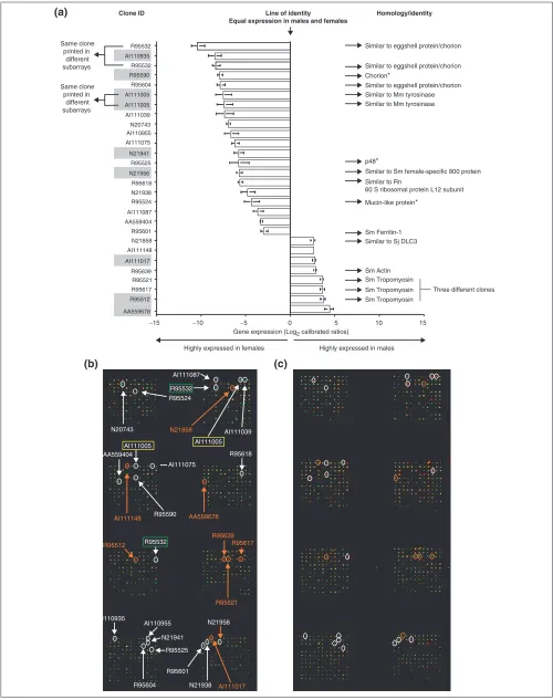
[image:3.609.58.554.88.318.2]After carrying out five independent cDNA microarray hybridizations and subjecting the accumulated data to strin-gent quality-control measures (summarized in Materials and methods), we assigned novel, sex-associated transcription to 12 female-associated and 4 male-associated transcripts in the 7-week adult schistosome (Figure 3a). In addition to these specific gender associations, all the positive control DNA elements (chorion, p48, mucin-like protein) printed on the fabricated schistosome cDNA microarray showed expression profiles similar to those of previous observations
Figure 1
High-quality total RNA was isolated from adult male and female schistosomes and reverse transcribed into cDNA which hybridized with equal affinity to control S. mansonigenomic elements.(a)Total RNA was isolated from 7-week-old male (M) and female (F) parasites by iterative phase separation and affinity chromatography techniques. A 10 g sample of each RNA preparation was electrophoresed under denaturing conditions on a 0.1% agarose gel [58]. Intense 18S rRNA bands are visible with minimal apparent sample degradation. (b) Analysis of mean fluorescent signal intensities (MFI) detected from all arrayed S. mansonigenomic elements after hybridization with cDNA generated from 10 g male and female RNA samples. Three representative hybridization experiments (out of five) are shown, which show no statistically significant difference in the mean fluorescent intensity (MFI) between male and female cDNA samples, and support the contention that equivalent quantities of both sample materials were used in each experimental hybridization. Standard errors of the MFI are indicated.
600
500
400
18S rRNA
300
200
Male cDNA
Female cDNA
p = 0.98
p = 0.10
p = 0.10
100
1
2
Experiments
MFI
(arbitr
ar
y units)
3
M
F
0
Figure 2
Measurement of adult S. mansonimale and female gene expression using cDNA microarrays was highly reproducible.(a)S. mansonicDNA microarrays were constructed on glass slides in a format consisting of eight individual subarrays (arranged in four rows by two columns). Each subarray consisted of 144 DNA elements (arranged in 12 rows by 12 columns) with each DNA element being spotted twice (6 rows by 12 columns) within each subarray (indicated by brackets). Images were obtained by hybridization of Cy5-conjugated male cDNA and Cy3-conjugated female cDNA. (b)Analysis of subarrays 3 and 4 from five independent hybridization experiments (Hyb 1-Hyb 5) demonstrated consistent patterns of gene expression in adult male and female parasites. In experiments 1-4, male parasite cDNA was labeled with Cy5 (red) and female parasite cDNA was labeled with Cy3 (green), whereas in experiment 5, the fluorescent dyes were switched. (c)Statistical analysis of intra-array variation (comparing the natural log converted calibrated ratio from one DNA element to the natural log converted calibrated ratio of its corresponding duplicate DNA element for one representative experiment) demonstrated high reproducibility and tight associations between duplicates. The median coefficient of variation (CV = 11.1%) and the correlation coefficient (r= 0.91) indicated a high degree of agreement. (d)Statistical analysis of inter-array variation (comparing the mean natural log converted calibrated ratio of each DNA element in one hybridization experiment to the mean natural log converted calibrated ratio of the same DNA element in a second hybridization experiment) indicated a high degree of association between measurements and high reproducibility. The median coefficient of variation (CV = 14.4%) and the correlation coefficient (r= 0.90) supported this contention. Dashed lines in (c) and (d) represent the line of symmetry. The remaining solid lines in both (c) and (d) represent the regression line and the predicted 99% confidence intervals of the supplied data.
Subarrrays 3 and 4
Spot duplication
Spot duplication
Spot duplication
Spot duplication
Hyb1
Hyb2
Hyb3
Hyb5 Dyes switched
Hyb4
(a)
(c)
(d)
(b)
Natural log converted average calibrated ratio measurement from hybridization 2
Natural log converted average calibrated ratio
measurement from hybridization 1
−8 −6 −4 −2 0 2 4 −8 −6 −4 −2 0 2 4
2
0
−2
−4
−6
−8 4
2
0
−2
−4
−6
−8 4 r = 0.91
CV = 11.1%
r = 0.90 CV = 14.4%
Intra-array reproducibility Inter-array reproducibility
Natural log converted calibrated ratio measurement from spot 2
Natural log converted calibrated ratio
comment
reviews
reports
deposited research
interactions
information
[image:5.609.55.555.86.719.2]refereed research
Figure 3(see legend on the next page)
R95617 R95521 AI111148 N21858 R95601 AA559404 AI111087 N21938 R95618 AI111075 AI110955 AI111039 R95604 R95532 R95532
0 5 10
Highly expressed in females Highly expressed in males
Gene expression (Log2 calibrated ratios)
15
−5
−10
−15
Similar to eggshell protein/chorion Similar to eggshell protein/chorion
Chorion*
Similar to eggshell protein/chorion
p48*
Similar to Rn
60 S ribosomal protein L12 subunit
Mucin-like protein*
Sm Tropomyosin Sm Tropomyosin Sm Tropomyosin Similar to Sj DLC3 Sm Ferritin-1
Sm Actin
Three different clones Same clone
printed in different subarrays Same clone printed in
different subarrays
Similar to Sm female-specific 800 protein Similar to Mm tyrosinase
Similar to Mm tyrosinase
R95639 N20743 AI110935
AA559678 AI111005 R95590
AI111005
AI111017 N21956 N21941
R95512 R95524 R95525
Clone ID Line of identity Homology/identity Equal expression in males and females
R95532
AI110935
R95532 R95590
R95604
AI111005 AI111005
AI111039 N20743
AI110955 AI111075
N21941 R95525
N21956 R95618
N21938 R95524
AI111087
AA559404
R95601
N21858
AI111148
AI111017 R95639
R95521 R95617 R95512
AA559678 (a)
[3,5,31]. This can also be extended to cytochrome oxidase subunit I, where no difference in gene expression was observed between sexes (data not shown) as previously shown [32]. Every subarray contained at least one of the dif-ferentially expressed gene products, showing that no local-ized hybridization bias occurred during our experiments (Figure 3b). In addition, as all 23 differentially expressed gene products (16 novel, sex-associated transcripts in this study and control elements) had similar expression profiles when the dyes used to label the starting cDNA pools were switched (Figure 3c), fluorescent dye differences did not con-tribute to the observations. Note that multiple elements cor-responding to tropomyosin and eggshell-protein genes were arrayed and detected in this study. This resulted from the fact that ESTs with these homologies formed multiple contigs under the assembly parameters used for EST clustering.
Independent confirmation of gender associations predicted by the schistosome cDNA microarrays by reverse transcription PCR
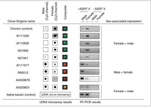
To verify the cDNA microarray predictions for a subset of gene products (gray-shaded clone IDs in Figure 3a), we examined gene expression in the sexually mature male and female schistosome RNA samples by RT-PCR analysis (Figure 4). All PCR reactions (except for alpha-tubulin and clone AA559631) were carried out for 35 cycles (well within the plateau of cycling parameters) to determine whether any product could be obtained in the sex where the cDNA microarray hybridizations detected minimal or no tran-script. In every case, differentially transcribed gene products examined by RT-PCR analysis displayed the gender-specific associations predicted by the cDNA microarray hybridiza-tions. Even subjecting the parasite cDNA to 35 rounds of amplification failed to yield significant product in the male sample for clones AI111005, AI110935, N21956, and N21941 or in the female sample for clones AI111017, AA59678 and R95512. This suggested that these clones (and probably all the others identified by the cDNA microarray hybridiza-tions) were highly expressed in a sex-dependent manner. Control RT-PCR reactions also showed predicted patterns of
gene expression (chorion and alpha-tubulin). Clone AA559631, which was shown to be expressed at similar levels in 7-week male and female schistosomes by cDNA microar-ray hybridization experiments, was also found to be expressed at similar levels by RT-PCR analysis.
Discussion
The modern tools of this post-genomic age are slowly devel-oping alongside the well-established parasite sequencing efforts and will undoubtedly change the path and scope of all future, genome-wide, functional investigations. We describe the first use of one specific post-genomic tool (cDNA microarrays) in the study of schistosome biology and illus-trate several key points. First, our fabricated schistosome cDNA microarrays provided an efficient means to examine gender-associated gene expression en masse. Second, the results obtained with these cDNA microarrays were highly reproducible and capable of independent confirmation, and finally, microarrays provided information previously unap-preciated in schistosome sexual biology.
Schistosomes are complex parasitic metazoans that have co-evolved with humans since before historical records were kept. They have excelled at parasitism by adopting develop-mental strategies virtually unique amongst the Platy-helminthes. The separation of sexes is one of these traits and so is perfectly suited for the initiation of schistosome cDNA microarray investigations. In this study, we developed a schistosome cDNA microarray from a small subset of EST elements deposited in one of the current schistosome EST databases [14]. The resultant 576-element schistosome cDNA microarray contained only about 4% of the predicted open reading frames (ORFs) encoded by the schistosome genome [33], but was sufficient to identify 23 differentially expressed genes between the sexes. Some of these genes were positive control elements intentionally printed on the cDNA microarray (chorion, p48, mucin-like protein), but 16 genes demonstrated novel sex-specific associations in the adult schistosome (12 female- and 4 male-associated
Figure 3 (see figure on the previous page)
transcripts) (Figure 3a). In comparison to all previous gender-associated genomic investigations undertaken in
Schistosoma, this one study, which exploited a very limited subset of the available EST clone archive, was able to double the number of genes identified as possessing sex-specific transcription.
Of the 18 cDNA clones (Figure 3a) that showed female-asso-ciated gene expression in our study, most of the annotated DNAs (those that had a positive match in the GenBank data-bases) that passed the applied filters were (unsurprisingly) either selected control elements or components involved in schistosome egg production (Figure 3a). Mature females devote a tremendous amount of metabolic effort to produc-ing eggs and thus a large proportion of mRNA generated by this sex is for this purpose. It has been estimated that the eggshell-protein genes of S. mansoni, encoding p14 and p48,
contribute 5-10% and 0.3-0.5%, respectively, of the total mRNA pool in a mature female [3]. These genes only repre-sent two of the many that contribute to the production, assembly and laying of the approximately 100-300 eggs per day often observed in S. mansoniinfections. Thus, it is not surprising that molecules associated with this fundamental biological process would be a large component of the mature female RNA population and subsequently be identified in our cDNA microarray study (especially when compared to mature male parasite RNA pools). Ferritin-1 (clone R95601), a clone related to S. mansoni female-specific 800 protein (clone N21956), and a tyrosinase ortholog (clone AI111005) are three such molecules. Ferritins are involved in hemoglo-bin metabolism where they serve as anchoring proteins for iron [34]. Schussler and colleagues demonstrated the expression of ferritin-1 predominantly in the female schisto-some (possibly in yolk platelets in the vitellarium) [35] and
comment
reviews
reports
deposited research
interactions
information
[image:7.609.57.555.86.442.2]refereed research
Figure 4
RT-PCR analysis confirmed gene expression results obtained by cDNA microarray hybridizations. A 1 g sample of total RNA was used to prime cDNA synthesis for both adult male and female samples (in the presence or absence of reverse transcriptase; +SSRT or -SSRT). PCR primers (Table 1) were designed for cDNA clone IDs AI111005, AI110935, N21956, N21941, AI111017, R95512, AA59678, AA559631 as well as chorion and alpha-tubulin. PCR amplification was performed for 35 cycles (except for alpha-tubulin and clone AA559631: 23 cycles). Spot morphology and color intensity for each RT-PCR-verified clone, obtained from one representative cDNA microarray hybridization, are included for comparison (except for alpha-tubulin which is not present on the cDNA microarray). The sex-associated expression of each transcript, as classified by both techniques, is also indicated.
Composite
Male
(Cy5 channel)
Female
(Cy3 channel)
Clone ID/gene name
Alpha-tubulin (control) cDNA not on microarray
cDNA microarray results AI110935
N21956
N21941
R95512 AI111017
AA559631
Female > male
Male > female
Female = male
RT-PCR results
MaleFemaleMale Female
+SSRT II −SSRT II
Sex-associated expression
Chorion (control)
our results support this finding. The exact function of fer-ritin-1 in vitellarium biology remains unclear. Clone N21956 has a moderate degree (BLAST Evalue 6 x 10⁻⁴) of sequence similarity to a female-specific cDNA previously character-ized [7]. Reis et al.reported that this cDNA (female-specific 800 protein) contained two large ORFs and the correspond-ing protein product(s) localized to vitelline cells [7]. Clone N21956 may represent a novel S. mansoni female-specific 800 protein family member (divergent ORF), but the func-tion of any of these cDNA products in vitelline cell biology is still unknown. The finding here that a tyrosinase ortholog (clone AI111005) is differentially transcribed in the adult female is intriguing. Vitelline cells avidly take up tyrosine, where it is stored in vitelline droplets and endoplasmic retic-ulum [36]. In addition, the tyrosine residues on eggshell pre-cursor proteins have been hypothesized to be substrates for enzymes and enzyme activities during the tanning process of eggshell formation (reviewed in [37]). One of these enzy-matic activities has been characterized by Eshete and LoVerde in extracts of S. mansoniand is related to those described for tyrosinases [38]. We now show here that adult female parasites transcribe the mRNA for a tyrosinase ortholog at a much higher rate than adult males, and so this enzyme may be responsible for the phenol oxidase/tyrosi-nase activities described previously in S. mansoni[38-40]. It is also possible that this enzyme ortholog (or family member) participates in additional schistosome-related metabolic reactions (alkaloid biosynthesis I, riboflavin metabolism or melanin synthesis) described for other organ-isms [41]. This molecule is being characterized further.
In addition to these cDNAs, several other clones had novel female-specific associations. One clone had sequence simi-larity to a 60S ribosomal protein L12 subunit (R95618) and clearly has a role in protein production, but why it is prefer-entially transcribed in adult female schistosomes is, as yet, unclear. The remaining nine clones possessed no significant database match. The functional roles of these nine novel female-associated cDNA clones in schistosome biology are currently under investigation. They could be newly identified components of the females egg-producing machinery or be involved in other sex-specific biological processes. These molecules represent a new collection of female-associated transcripts that may be useful in understanding the processes that lead to host immunopathology.
Three annotated proteins whose genes show male-associated expression are tropomyosin (clones R95521, R95617, and R95512), actin (clone R95639) and a dynein light-chain ortholog (Sj DLC3 - clone N21858) (Figure 3a). Although actin has previously been shown to be differentially expressed in adult males [12], our study provides information on the sex-associated expression of two other tegument-associated molecules, tropomyosin and the dynein light chain [42,43]. Tropomyosin and actin are also located in other schistosome tissues such as parenchyma, muscle and spines [44].
The coordinate expression of tropomyosin and actin in the adult male schistosome is probably due to the fact that these two molecules interact intimately. Actin polymers (microfil-aments) provide mechanical stability for the cytoskeleton of eukaryotes and serve as tracks for motor proteins such as tropomyosin [45]. As male schistosomes are significantly larger than females, contain a greater volume of tegument and muscle, and are more physically active, our expression results may reflect this bias. It is also possible, however, that the preferential transcription of tropomyosin, actin and dynein light chains in the male has an important develop-mental role for copulating worm pairs. Evidence for this comes from studies where unpaired female parasites are developmentally and sexually stunted in comparison to paired female parasites [46-48]. In the paired state, male schistosomes are thought to provide mechanical (among other types of) support to the maturing female [49]. This mechanical support enables the female to acquire proper nutritional supplements from the hepatic portal system, which allows the development of full sexual maturity. As a function of tropomyosin (binding to actin) is in contractile muscle regulation and a hypothesized function of dynein light chains is in tegument biology [43], our results suggest that these molecules, preferentially transcribed in the male, may participate in this mechanical and support mainte-nance. With the male firmly securing the female in its gynecophoral canal, she can devote more of her metabolic and transcriptional machinery to egg production (as our results indicate). Several other schistosome tegument-associated transcripts (EST clones corresponding to Sm DLC, Sm 20.8, Sm 22.6, Sm 21.7 and Sm 22) were also differen-tially expressed in the adult male in our study, further sup-porting this hypothesis. These molecules narrowly missed inclusion in our final dataset because they failed in one of the applied filtering criteria. Taken together, these data show that tegument-associated gene products are transcribed in a gender-related manner, which may reflect differences in size and/or physical activity, or a differential functional require-ment for each of these transcripts. The identity and func-tional role of the three unknown male-associated transcripts identified in this study await further investigation.
(Figure 4). Second, the importance of fabricating cDNA microarrays from EST sequences derived from different developmental stages and libraries was evident. Orphan ESTs AA559404 and AA559678 were both derived from cer-carial cDNA libraries, yet showed adult fe and male-associated gene expression, respectively (Figure 3a). Whether both male and female cercariae express these two molecules, and then alter their expression as the parasites mature, or whether a gender-specific association is already present at this immature developmental stage remains to be determined. This point could be addressed by examination of gene transcription in sexed cercariae [50]. Nevertheless, the gender-specific association of these two transcripts in adults would not have been identified if cercarial elements had not been included in our schistosome cDNA microar-rays. Third, it was observed that two male-associated tran-scripts hybridized to cDNA clones that originated from an adult female-enriched cDNA library (clones AI111148 and AI111017, Figure 3a). This showed that associations not often looked for (differentially expressed male transcripts hybridizing to female-enriched cDNA clones) could be iden-tified using cDNA microarrays. Together, these points show that cDNA microarrays constructed in a highly reproducible manner from diverse ESTs and different cDNA libraries are extremely useful for gene identification/expression pur-poses. Attention to these details will maximize the informa-tion obtained from future experimental hybridizainforma-tions.
Conclusions
This report describes the first use of cDNA microarrays in functional analyses of schistosome biology. We have shown that these tools were of high quality, capable of reproducibly detecting gene-expression differences between genders, and useful for identifying new biological associations. The 16 novel, gender-associated transcripts identified here serve as starting points for functional investigations. Continued refinement and expansion of schistosome cDNA microarrays (fabrication of a 4,000-element array is underway) and development of new post-genomic techniques will provide the means to initiate numerous other investigations aimed at unraveling the biological complexities of this parasitic helminth and the pathology it induces.
Materials and methods
ParasitesA Puerto Rican strain of S. mansoniwas used in this study. Adult male and female schistosomes were perfused from per-cutaneously infected TO outbred mice (Harlan) challenged 7 weeks earlier with 125 cercaria [51]. The parasite was passaged through Biomphalaria glabrataintermediate snail hosts.
Schistosome cDNA microarray fabrication
Parasite ESTs deposited on cDNA microarrays were selected from a putatively nonredundant sequence set contained in
the March 2000 cluster-analysis database [14]. This particu-lar arrayed clone set was selected for two main reasons: we could successfully grow this subset of bacterial clones at the time of array fabrication; and the set contained similar rep-resentative percentages of known and unknown annotated sequences deposited in the schistosome EST database. Clus-ters were assembled using an exhaustive search-and-compare algorithm in Sequencher v3.1.1 (Gene Codes Corp., Ann Arbor MI), set to assemble contigs from sequences dis-playing ⱖ90% homology over ⱖ60 bases, with a maximum of two allowable consecutive mismatched bases. EST prod-ucts were generated via standard PCR techniques from cDNA clones archived by the Schistosome Genome Network and maintained at The Natural History Museum (London). The source libraries were constructed in LambdaZap vectors (Stratagene, La Jolla, CA) and inserts were amplified from phage suspension or from lysed bacterial colonies containing excised pBluescript plasmid using M13 forward and reverse primer pairs. EST PCR products were 1,000 bp on average as assessed by DNA gel electrophoresis (over 98% of the PCR reactions generated a single product). PCR products were purified, using Multiscreen-FB 96-well filtering units (Milli-pore, Bedford, MA), away from primers, salt and other potential contaminants. Purified EST products were diluted (1:4) in 4x spotting buffer (600 mM sodium phosphate, 0.04% sodium dodecyl sulfate) and printed in ordered arrays (8 subarrays composed of 12 columns x 12 rows) on gamma-aminopropyl silane-coated glass slides (Corning) using a Microgrid II robotic arrayer (BioRobotics Ltd, Cam-bridge, UK). Slides were processed after printing by baking for 2 h at 80°C and UV cross-linking at 450 mJ to perma-nently fix DNA on the glass slides, followed by boiling for 2 min to denature fixed DNA. The 1,152 elements (576 ele-ments printed in duplicate) arrayed on each glass slide were composed of the PCR-amplified parasite ESTs, positive con-trols (S. mansoni genomic DNA and known female-specific cDNAs: chorion [2], mucin-like protein [5] and p48 [3]), and negative controls (yeast tRNA, pBluescript DNA, lambda DNA, and spotting buffer only).
Total RNA isolation, cDNA synthesis and hybridization
After perfusion of experimentally infected mice (7 weeks post-infection), male and female schistosomes were sepa-rated manually. Approximately equal numbers of adult male or female worms were pooled and used as the starting mate-rial for total RNA isolation. A procedure to isolate total RNA from the parasite was adapted from previous studies [52] and involved both phase extraction (TRIZOL reagent, Invit-rogen, Life Technologies, Paisley, UK) and column chro-matography (Qiagen RNeasy maxi affinity columns, Qiagen, Crawley, UK). Denaturing gel electrophoresis assessed RNA quality from each sample. Amino-allyl dUTP (Sigma-Aldrich) labeled cDNA targets (nomenclature recommendation described in [53]) were generated from a 10g male RNA sample and a 10g female RNA sample using Superscript
comment
reviews
reports
deposited research
interactions
information
Reverse Transcriptase II (Invitrogen). Cy dye (Amersham Pharmacia Biotech, Little Chalfont, UK) conjugation to each cDNA target was carried out according to standard protocols [54]. Fluorescent cDNA targets were hybridized to the cDNA microarray probes in a modified hybridization buffer (40% deionized formamide, 5x Denhardts reagent, 5x SSC, 1 mM sodium pyrophosphate, 50 mM Tris pH 7.4, 0.1% SDS, 0.25g/l mouse Cot1 DNA (Life Technologies), 0.44g/l poly(dA) (Amersham Pharmacia Biotech), and 0.22g/l yeast tRNA (Sigma-Aldrich) at 48°C using hybridization chambers (TeleChem, Sunnyvale, CA) for a minimum of 16 h. Post-hybridization processing involved three succes-sive 5-min washes in 0.5x SSC/0.1% SDS, 0.5x SSC/0.01% SDS and 0.06x SSC. Slides were spun dried (500gfor 5 min) to remove all washing buffer, stored at room temperature in the dark, and scanned at 10 m resolution using a Packard ScanArray Express microarray scanner (Packard BioScience, Pangbourne, UK).
RT-PCR analysis
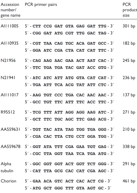
For RT-PCR analysis, 1 g male or female RNA was used for cDNA synthesis to confirm cDNA microarray results. RT-PCR was carried out as described [55] to confirm the cDNA microarray gender-specific gene-expression results for clones AI110935, N21956, N21941, AI11017, R95512, AA559678, AI111005 and AA559631. Additional cDNAs examined by this method included two well characterized controls - alpha-tubulin (housekeeping transcript [56]) and chorion (female-associated transcript [31]). The primers for all transcripts are included in Table 1. Thirty-five cycles of PCR were used to amplify all transcripts except for alpha-tubulin and clone AA559631 (to ensure that amplification was within the linear range of PCR in both male and female cDNA, 23 cycles were used for these clones). All amplicons were electrophoresed on a 1% agarose gel and stained with ethidium bromide. Images were captured by a digital camera and analyzed by gel-documentation software (Kodak 1D 2.0 electrophoresis documentation and analysis system 120, Eastman Kodak, New Haven, CT). Semi-quantitative analy-sis of the differentially expressed transcripts was not carried out owing to the cycling conditions used in this study (35 cycles). RT-PCR was used, therefore, to verify the cDNA microarray results and not used to obtain exact or relative amounts of gene transcript present in each sex.
Statistical analysis
Microarray Suite (Scanalytics, Fairfax, VA) software was used to process signal-intensity information from all 16-bit TIFF files generated in the cDNA microarray experiments. Hybridization signals representing gene expression (Cy5/Cy3 ratios) from cDNA microarray experiments were processed by several filtering criteria after global mode nor-malization (each experiment normalized to itself) and local background subtraction. Filters one and two evaluated target/probe spot morphology and were used to eliminate any questionable data (weak signals, poorly defined spots,
and background noise). The first criterion required each spot to be greater in size than the lowest tenth percentile of all spots within an experiment (removal of small or irregularly shaped spots). The second criterion required one fluor (either Cy5 or Cy3) from each spot to be greater in intensity than one standard deviation above the mean intensity from all negative control array elements (removal of spots that were not significantly above background intensities).
[image:10.609.312.553.140.480.2]Intra-array and inter-array reproducibility were next exam-ined as filtering criteria. As each EST was printed twice on every array (intra-array reproducibility), only those ESTs that showed highly reproducible gene-expression ratios (within the 99% confidence interval of each experimental hybridization as predicted by the derived regression equa-tion) were included. Each of the duplicates subsequently had to show expression levels significantly greater than the median gene-expression ratio for each cDNA microarray
Table 1
PCR primer pairs used for RT-PCR confirmatory assays on S.
mansonigender-associated transcripts identified by cDNA microarray analysis
Accession PCR primer pairs PCR
number/ product
gene name size
AI111005 5´ - CTT CCG GAT GTA GAG GAT TTG- 3´ 301 bp 5´ - CGG GAT ATG CGT TTG GAC TAG- 3´
AI110935 5´ - CGT TAA CAG TGC ACA GAT GCC- 3´ 182 bp 5´ - GGA ATC CGA CTA CAT CAT TTC- 3´
N21956 5´ - CAG AAG AAC GAA ACT AAT CAC- 3´ 245 bp 5´ - TTC TGA TGA TAC GAT ACC GTG- 3´
N21941 5´ - ATC ATC ATT ATG GTA CAT CAT- 3´ 236 bp 5´ - TGA ATT TCA ACG TAT ATT CTC- 3´
AI111017 5´ - AAG TGT CCC TGA CAC AAC AAC- 3´ 137 bp 5´ - GCC TGT TTC ATT TTC ACC TTC- 3´
R95512 5´ - TCG TTT ATT AGG AGG AAG ATC- 3´ 271 bp 5´ - GCT TTC TGC AGC TTC GAG ACG- 3´
AA559631 5´ - TGT TAC ATA TAG TGG TGA GGG- 3´ 210 bp 5´ - CGA CAC TTA CTG CCT GGA TGG- 3´
AA559678 5´ - GGT ATA TTT CGA GAA TGT GAG- 3´ 338 bp 5´ - CGC TTA GGT TAA TCA TGA ATG- 3´
Alpha 5´ - GGC GGT GGT ACT GGT TCT GGG- 3´ 291 bp tubulin 5´ - CAT TTA GCG CAC CAT CGA AGC- 3´
Chorion 5´ - GAA ACA GTC ACT CAC ACT CG- 3´ 461 bp 5´ - ATG GCT GGG TTT GTA AGT GC- 3´
hybridization (outside the 99% confidence interval derived from all gene-expression ratios).
Finally, gene-expression measurements for each cDNA (including duplicates) had to have passed the previous filters in four out of five independent hybridizations to be included in the final dataset. Statistical analysis of gene-expression ratios between two of these five independent hybridizations showed low variation (that is, inter-array reproducibility). The 28 calibrated ratio values surviving these stringent filters were log2transformed and stored in a table (rows, individual cDNA clones; columns, gene-expression ratios for each inde-pendent hybridization). All clones showing differential gene expression were resequenced to confirm their annotated identity, as human error can sometimes lead to well-to-well cross-contamination of bacterially derived clone sets [57].
Additional data files
Additional files containing the primary data for the individ-ual microarray experiments are available with the online version of this article.
Acknowledgements
We thank Francis Jones, Maureen Laidlaw, Karen Plant, Mike Anderson, Viv Tuffney, and Susan Arnold for excellent technical assistance in maintain-ing the schistosome life cycle and separation of adult parasites. We also thank Rhian Hayward, Tom Wynn, and Thomas McCarty for critically reviewing this manuscript. Finally, we thank David Latto for many helpful discussions and in the fabrication of the cDNA microarrays. K.F.H. is sup-ported by a 2-year, long-term research fellowship awarded by the Euro-pean Molecular Biology Organization. This work was also supported by means of Wellcome Trust and Medical Research Council programme grants to the Cambridge laboratory and a UNDP/WORLD BANK/WHO Special Programme for Research and Training in Tropical Diseases (T.D.R.) grant (ID no 980502) awarded to D.A.J. at The Natural History Museum.
References
1. Bergquist NR, Colley DG: Schistosomiasis vaccines: research
to development.Parasitol Today 1998, 14:99-104.
2. Bobek LA, Rekosh DM, LoVerde PT: Small gene family encoding
an eggshell (chorion) protein of the human parasite
Schisto-soma mansoni.Mol Cell Biol 1988, 8:3008-3016.
3. Chen LL, Rekosh DM, LoVerde PT: Schistosoma mansoni p48 eggshell protein gene: characterization, developmentally regulated expression and comparison to the p14 eggshell
protein gene.Mol Biochem Parasitol 1992, 52:39-52.
4. Johnson KS, Taylor DW, Cordingley JS: Possible eggshell protein
gene from Schistosoma mansoni. Mol Biochem Parasitol 1987,
22:89-100.
5. Menrath M, Michel A, Kunz W: A female-specific cDNA
sequence of Schistosoma mansoni encoding a mucin-like
protein that is expressed in the epithelial cells of the
repro-ductive duct.Parasitology 1995, 111:477-483.
6. Koster B, Dargatz H, Schroder J, Hirzmann J, Haarmann C, Symmons P, Kunz W: Identification and localisation of the products of a
putative eggshell precursor gene in the vitellarium of
Schis-tosoma mansoni.Mol Biochem Parasitol 1988, 31:183-198. 7. Reis MG, Kuhns J, Blanton R, Davis AH: Localization and pattern
of expression of a female specific mRNA in Schistosoma
mansoni.Mol Biochem Parasitol 1989, 32:113-119.
8. Schussler P, Kohrer K, Finken-Eigen M, Michel A, Grevelding CG, Kunz W: A female-specific cDNA sequence of Schistosoma mansoniencoding an amidase that is expressed in the
gas-trodermis.Parasitology 1998, 116:131-137.
9. Dalton JP, Clough KA, Jones MK, Brindley PJ: Characterization of
the cathepsin-like cysteine proteinases of Schistosoma
mansoni.Infect Immun 1996, 64:1328-1334.
10. Drew AC, Brindley PJ: Female-specific sequences isolated from Schistosoma mansoniby representational difference analysis.
Mol Biochem Parasitol 1995, 71:173-181.
11. Fantappie MR, Correa-Oliveira R, Caride EC, Geraldo EA, Agnew A, Rumjanek FD: Comparison between site-specific DNA
binding proteins of male and female Schistosoma mansoni.
Comp Biochem Physiol B Biochem Mol Biol 1999, 124:33-40.
12. Davis AH, Blanton R, Klich P: Stage and sex specific differences
in actin gene expression in Schistosoma mansoni.Mol Biochem
Parasitol 1985, 17:289-298.
13. Moore DV, Yolles TK, Meleney HE: The relationship of mature
worms to the sexual development of female Schistosoma
mansoni.J Parasitol. 1954, 40:166-185.
14. The WHO/UNDP/World Bank Schistosoma Genome
Network[http://www.nhm.ac.uk/hosted_sites/schisto]
15. Schistosoma mansoniEST Genome Project [http://verjo18.iq.usp.br/schisto/]
16. The Institute for Genomic Research Schistosoma mansoni
genome project[http://www.tigr.org/tdb/e2k1/sma1/index.shtml]
17. Hoffmann KF, Davis EM, Fischer ER, Wynn TA: The guanine protein coupled receptor rhodopsin is developmentally
reg-ulated in the free-living stages of Schistosoma mansoni. Mol
Biochem Parasitol 2001, 112:113-123.
18. Kampkotter A, Ridgers I, Johnston DA, Rollinson D, Kunz W, Grev-elding CG: Schistosoma mansoni: cloning and characterization
of the Ras homologue.Exp Parasitol 1999, 91:280-283.
19. Beall MJ, McGonigle S, Pearce EJ: Functional conservation of Schistosoma mansoni Smads in TGF-beta signaling. Mol Biochem Parasitol 2000, 111:131-142.
20. Osman A, Niles EG, LoVerde PT: Characterization of the Ras
homologue of Schistosoma mansoni.Mol Biochem Parasitol 1999,
100:27-41.
21. Williams SA, Johnston DA: Helminth genome analysis: the current status of the filarial and schistosome genome pro-jects. Filarial Genome Project. Schistosome Genome
Project.Parasitology 1999, 118:S19-S38.
22. Shalon D, Smith SJ, Brown PO: A DNA microarray system for analyzing complex DNA samples using two-color
fluores-cent probe hybridization.Genome Res 1996, 6:639-645.
23. DeRisi J, Penland L, Brown PO, Bittner ML, Meltzer PS, Ray M, Chen Y, Su YA, Trent JM: Use of a cDNA microarray to analyse
gene expression patterns in human cancer.Nat Genet 1996,
14:457-460.
24. Hoffmann KF, McCarty TC, Segal DH, Chiaramonte M, Hesse M, Davis EM, Cheever AW, Meltzer PS, Morse HC 3rd, Wynn TA: Disease fingerprinting with cDNA microarrays reveals dis-tinct gene expression profiles in lethal type 1 and type 2
cytokine-mediated inflammatory reactions. FASEB J 2001,
15:2545-2547.
25. Hayward RE, Derisi JL, Alfadhli S, Kaslow DC, Brown PO, Rathod
PK: Shotgun DNA microarrays and stage-specific gene
expression in Plasmodium falciparum malaria. Mol Microbiol
2000, 35:6-14.
26. Ben Mamoun C, Gluzman IY, Hott C, MacMillan SK, Amarakone AS, Anderson DL, Carlton JM, Dame JB, Chakrabarti D, Martin RK, et al.: Co-ordinated programme of gene expression during asexual intraerythrocytic development of the human
malaria parasite Plasmodium falciparum revealed by
microarray analysis. Mol Microbiol 2001, 39:26-36.
27. Tenniswood MP, Simpson AJ: The extraction, characterization and in vitro translation of RNA from adult Schistosoma mansoni.Parasitology 1982, 84:253-261.
28. Lee ML, Kuo FC, Whitmore GA, Sklar J: Importance of replica-tion in microarray gene expression studies: statistical methods and evidence from repetitive cDNA
hybridiza-tions.Proc Natl Acad Sci USA 2000, 97:9834-9839.
29. Bartosiewicz M, Trounstine M, Barker D, Johnston R, Buckpitt A: Development of a toxicological gene array and quantitative
assessment of this technology. Arch Biochem Biophys 2000,
376:66-73.
30. Carlisle AJ, Prabhu VV, Elkahloun A, Hudson J, Trent JM, Linehan WM, Williams ED, Emmert-Buck MR, Liotta LA, Munson PJ, Krizman, DB: Development of a prostate cDNA microarray
and statistical gene expression analysis package.Mol Carcino-genesis 2000, 28:12-22.
31. Bobek L, Rekosh DM, van Keulen H, LoVerde PT: Characteriza-tion of a female-specific cDNA derived from a
developmen-tally regulated mRNA in the human blood fluke Schistosoma
mansoni.Proc Natl Acad Sci USA 1986, 83:5544-5548.
32. Brito CF, Oliveira GC, Oliveira SC, Street M, Riengrojpitak S, Wilson RA, Simpson AJ, Correa-Oliveira R: Sm14 gene
expres-sion in different stages of the Schistosoma mansonilife cycle
and immunolocalization of the Sm14 protein within the
adult worm.Braz J Med Biol Res 2002, 35:377-381.
33. Franco GR, Valadao AF, Azevedo V, Rabelo EM: The Schistosoma
gene discovery program: state of the art.Int J Parasitol 2000,
30:453-463.
34. Ford GC, Harrison PM, Rice DW, Smith JM, Treffry A, White JL, Yariv J: Ferritin: design and formation of an iron-storage
molecule.Phil Trans R Soc Lond B Biol Sci 1984, 304:551-565.
35. Schussler P, Potters E, Winnen R, Bottke W, Kunz W: An isoform
of ferritin as a component of protein yolk platelets in
Schis-tosoma mansoni.Mol Reprod Dev 1995, 41:325-330.
36. Erasmus DA: The subcellular localization of labelled tyrosine
in the vitelline cells of Schistosoma mansoni.Z Parasitenkd 1975,
46:75-81.
37. Coles GC: Recent advances in schistosome biochemistry. Par-asitology 1984, 89:603-637.
38. Eshete F, LoVerde PT: Characteristics of phenol oxidase of Schistosoma mansoni and its functional implications in
eggshell synthesis.J Parasitol 1993, 79:309-317.
39. Bennett JL, Seed JL, Boff M: Fluorescent histochemical
localiza-tion of phenol oxidase in female Schistosoma mansoni.J
Para-sitol 1978, 64:941-944.
40. Seed JL, Boff M, Bennett JL: Phenol oxidase activity: induction in
female schistosomes by in vitro incubation. J Parasitol 1978,
64:283-289.
41. Kyoto Encyclopedia of Genes and Genomes
[http://www.genome.ad.jp/kegg/]
42. Xu H, Miller S, van Keulen H, Wawrzynski MR, Rekosh DM, LoVerde PT: Schistosoma mansonitropomyosin: cDNA char-acterization, sequence, expression, and gene product
local-ization.Exp Parasitol 1989, 69:373-392.
43. Hoffmann KF, Strand M: Molecular identification of a Schisto-soma mansoni tegumental protein with similarity to
cyto-plasmic dynein light chains.J Biol Chem 1996, 271:26117-26123.
44. MacGregor AN, Shore SJ: Immunocytochemistry of
cytoskele-tal proteins in adult Schistosoma mansoni.Int J Parasitol 1990,
20:279-284.
45. Vilfan A: The binding dynamics of tropomyosin on actin.
Biophys J 2001, 81:3146-3155.
46. Clough ER: Morphology and reproductive organs and
oogene-sis in bisexual and unisexual transplants of mature
Schisto-soma mansonifemales.J Parasitol 1981, 67:535-539.
47. Popiel I, Cioli D, Erasmus DA: The morphology and
reproduc-tive status of female Schistosoma mansonifollowing
separa-tion from male worms.Int J Parasitol 1984, 14:183-190.
48. Popiel I, Basch PF: Reproductive development of female Schis-tosoma mansoni (Digenea: Schistosomatidae) following
bisexual pairing of worms and worm segments. J Exp Zool
1984, 232:141-150.
49. Gupta BC, Basch PF: The role of Schistosoma mansonimales in
feeding and development of female worms. J Parasitol 1987,
73:481-486.
50. Liberatos JD, Short RB: Identification of sex of schistosome
larval stages. J Parasitol 1983, 69:1084-1089.
51. Smithers SR, Terry RJ: The infection of laboratory hosts with
cercariae of Schistosoma mansoni and the recovery of the
adult worms.Parasitology 1965, 55:695-700.
52. National Human Genome Research Institute Microarray
Project[http://www.nhgri.nih.gov/DIR/Microarray/main.html]
53. Phimister B: Going global.Nat Genet 1999, 21:1.
54. Qiagen/Operon homepage[http://www.operon.com/]
55. Wynn TA, Eltoum I, Cheever AW, Lewis FA, Gause WC, Sher A: Analysis of cytokine mRNA expression during primary
gran-uloma formation induced by eggs of Schistosoma mansoni.
J Immunol 1993, 151:1430-1440.
56. Webster PJ, Seta KA, Chung SC, Mansour TE: A cDNA encoding
an alpha-tubulin from Schistosoma mansoni.Mol Biochem
Para-sitol 1992, 51:169-170.
57. Halgren RG, Fielden MR, Fong CJ, Zacharewski TR: Assessment of clone identity and sequence fidelity for 1189 IMAGE cDNA
clones.Nucleic Acids Res 2001, 29:582-588.
![Figure 1High-quality total RNA was isolated from adult male and female schistosomes and reverse transcribed into cDNA which hybridized with equal affinity to[58]](https://thumb-us.123doks.com/thumbv2/123dok_us/8649920.867380/3.609.58.554.88.318/figure-quality-isolated-schistosomes-reverse-transcribed-hybridized-affinity.webp)