Naturally processed T cell epitopes from human
glutamic acid decarboxylase identified using
mice transgenic for the type 1
diabetes-associated human MHC class II allele,
DRB1*0401.
L S Wicker, … , R Cummings, P J Whiteley
J Clin Invest.
1996;
98(11)
:2597-2603.
https://doi.org/10.1172/JCI119079
.
The identification of class II binding peptide epitopes from autoimmune disease-related
antigens is an essential step in the development of antigen-specific immune modulation
therapy. In the case of type 1 diabetes, T cell and B cell reactivity to the autoantigen
glutamic acid decarboxylase 65 (GAD65) is associated with disease development in
humans and in nonobese diabetic (NOD) mice. In this study, we identify two
DRB1*0401-restricted T cell epitopes from human GAD65, 274-286, and 115-127. Both peptides are
immunogenic in transgenic mice expressing functional DRB1*0401 MHC class II molecules
but not in nontransgenic littermates. Processing of GAD65 by antigen presenting cells
(APC) resulted in the formation of DRB1*0401 complexes loaded with either the 274-286 or
115-127 epitopes, suggesting that these naturally derived epitopes may be displayed on
APC recruited into pancreatic islets. The presentation of these two T cell epitopes in the
islets of DRB1*0401 individuals who are at risk for type 1 diabetes may allow for
antigen-specific recruitment of regulatory cells to the islets following peptide immunization.
Research Article
Find the latest version:
J. Clin. Invest.
© The American Society for Clinical Investigation, Inc. 0021-9738/96/12/2597/07 $2.00
Volume 98, Number 11, December 1996, 2597–2603
Naturally Processed T Cell Epitopes from Human Glutamic Acid Decarboxylase
Identified Using Mice Transgenic for the Type 1 Diabetes-associated Human MHC
Class II Allele, DRB1*0401
Linda S. Wicker,* Shiow-Ling Chen,* Gerald T. Nepom,¶**John F. Elliott,** Daniel C. Freed,* Alka Bansal,* Song Zheng,i
Andrew Herman,¶** Åke Lernmark,**‡‡ Dennis M. Zaller,‡ Laurence B. Peterson,§ Jonathan B. Rothbard,‡‡ Richard Cummings,i
and Phyllis J. Whiteley*
*Department of Autoimmune Diseases Research, ‡Department of Molecular Immunology, §Department of Immunopharmacology, and
iDepartment of Molecular Design and Diversity, Merck Research Laboratories, Rahway, New Jersey 07065-0900; ¶Virginia Mason
Research Center, **Department of Immunology, and ‡‡Department of Medicine, University of Washington School of Medicine, Seattle,
Washington 98195; §§Department of Medical Microbiology and Immunology, University of Alberta, Edmonton, Alberta, Canada; and
iiDepartment of Medicine, Division of Immunology, Stanford University Medical School, Stanford, California 94305-5303
Abstract
The identification of class II binding peptide epitopes from
autoimmune disease-related antigens is an essential step in
the development of antigen-specific immune modulation
therapy. In the case of type 1 diabetes, T cell and B cell
re-activity to the autoantigen glutamic acid decarboxylase 65
(GAD65) is associated with disease development in humans
and in nonobese diabetic (NOD) mice. In this study, we
identify two DRB1*0401-restricted T cell epitopes from
hu-man GAD65, 274–286, and 115–127. Both peptides are
immunogenic in transgenic mice expressing functional
DRB1*0401 MHC class II molecules but not in
nontrans-genic littermates. Processing of GAD65 by antigen
present-ing cells (APC) resulted in the formation of DRB1*0401
complexes loaded with either the 274–286 or 115–127
epitopes, suggesting that these naturally derived epitopes
may be displayed on APC recruited into pancreatic islets.
The presentation of these two T cell epitopes in the islets of
DRB1*0401 individuals who are at risk for type 1 diabetes
may allow for antigen-specific recruitment of regulatory
cells to the islets following peptide immunization. (
J. Clin.
Invest.
1996. 98:2597–2603.) Key words: autoimmunity
•di-abetes mellitus, insulin-dependent
•peptide autoantigens
•MHC class II
•mice, inbred NOD
Introduction
A number of recent studies on type 1 diabetes in humans and nonobese diabetic (NOD)1 mice have underscored the
impor-tance of the immune response to glutamic acid decarboxylase (GAD). The two isoforms of GAD, glutamic acid decarboxy-lase 65 (GAD65; 65 kD) and glutamic acid decarboxydecarboxy-lase 67 (GAD67; 67 kD), are encoded by separate genes. Although both forms are expressed in the b cells of the islets of Langer-hans and in the brain, in humans, GAD65 is the predominant form expressed in b cells and appears to be the primary GAD autoantigen in type 1 diabetes. Both antibodies and T cell re-activity specific for GAD65 and GAD67 have been detected in prediabetic and diabetic individuals (1, 2). Interestingly, sev-eral groups have demonstrated that administration of GAD can prevent the development of diabetes in NOD mice (3–5). It has been hypothesized that the induction of Th2 T cells spe-cific for GAD are able to downregulate pathogenic T cells within the islets.
Modulation of the immune response with GAD or peptides derived from GAD is a potential method to prevent diabetes in humans. The identification of dominant, GAD-derived T cell epitopes presented by human class II molecules is an im-portant step in the development of a vaccine that could be used to downregulate a b cell-specific, pathogenic autoim-mune response in genetically susceptible humans. In the cur-rent study, we used a panel of overlapping 20mer peptides de-rived from human GAD65 to define peptides that bind to the human class II molecules DRB1*0401 and DQ3.2, which are encoded by the DRA1*0101, DRB1*0401 and DQA1*0301, DQB1*0302 genes, respectively. Both the DRB1*0401 and DQ3.2 molecules are found in a majority of Caucasian type 1 diabetics and are thought to contribute to the disease by pre-senting b cell-associated self-proteins to the immune system in a manner that stimulates a pathogenic T cell response (6, 7). In addition, a panel of 13mer peptides derived from human GAD65, predicted to be DRB1*0401-binding epitopes (8), were assessed for binding to DRB1*0401. Several DRB1*0401-binding peptides were studied further by testing for immuno-genicity in B10.M transgenic mice that express a MHC class II molecule with the DRA1*0101/DRB1*0401 peptide binding domain (9). 2 of the 10 peptides examined were found to be immunogenic in DRB1*0401-transgenic but not in nontrans-genic mice. Responses to human GAD65-derived peptides oc-curred even though the sequences tested were identical to those within murine GAD65. Finally, T cell hybridomas de-rived from mice immunized with GAD peptides were shown to recognize naturally processed GAD65. This study demon-strates a strategy that can be used to define T cell epitopes rel-evant for human autoimmune disease.
Address correspondence to Dr. Linda Wicker, Merck Research Lab-oratories, Mail Code R80W-107, P.O. Box 2,000, 126 East Lincoln Avenue, Rahway, NJ 07065-0900. Phone: 594-7511; FAX: 908-594-7299; E-mail: [email protected]
Received for publication 19 March 1996 and accepted in revised form 3 October 1996.
Methods
Cell lines and antibodies. The human EBV-transformed B-LCL, Preiss (homozygous HLA-DRB1*0401, DQ3.2) was maintained in RPMI supplemented with 10% fetal calf serum, 2 mM L-glutamine, and anti-biotics. GAD6 is a murine monoclonal antibody that is specific for the GAD65 protein (10).
Construction of the human GAD65 cDNA expression vector. The 1.7-kb fragment encompassing the cDNA for human GAD65 (11), was released from the pEX9 plasmid (Invitrogen Corp., San Diego, CA) by digestion with BamH1 and Kpn1. The cDNA insert was blunt-end li-gated into the EcoR1 site in the pMCFR-pac vector after both DNA species had been complemented using T4 DNA polymerase. The pM-CFR-pac expression plasmid, (a gift from L.K. Denzin and P. Cress-well, Yale University School of Medicine, Howard Hughes Medical In-stitute, New Haven, CT; 12), directs expression of puromycin N-acetyl transferase, thus allowing for selection with puromycin in mammalian cells (13), and contains promoter elements derived from the LTR of a murine thymotropic retrovirus and the enhancer from the R-U5 region of HTLV-1 (14, 15). The resultant plasmid is named pMCFR-pac/GAD.
Generation of human GAD65 expressing cell lines. Stable trans-fectants of the human B-LCL, Preiss, expressing human GAD65, were isolated after electroporation. Briefly, pMCFR-pac/GAD, at a concentration of 10 mg/ml, was incubated with 1 3 107 cells in 0.8 ml
of serum free RPMI medium supplemented with DEAE-dextran (10
mg/ml). The cells were electroporated using a Gene Pulser apparatus (Biorad Laboratories, Richmond, CA) set at 250 V and 500 mF. The electroporated cells were allowed to recover on ice for 10 min, trans-ferred to growth medium, and incubated for approximately 30 h be-fore selection using 0.75 mg/ml puromycin (Sigma Chemical Corp., St. Louis, MO).
Drug resistant cell lines were screened for expression of GAD65 by immunoblotting with the mAb GAD6 on lysates electrophoresed on 8% SDS-PAGE minigels (Hoefer Sci. Instr., San Francisco, CA). After transfer onto nitrocellulose, blots were incubated with GAD6 hybridoma supernatant and developed with peroxidase-conjugated AffiniPure F(ab)2 goat anti–mouse IgG 1 IgM (Jackson ImmunoRe-search Labs., Inc., West Grove, PA) diluted 1:4000. Bound mAb was detected by chemiluminescence (ECL; Amersham Corp., Arlington Heights, IL).
Among the Preiss transfectants screened by Western blotting, PRGD-1 was shown to be the highest GAD65-expressing cell line and was cloned by limiting dilution. In contrast, the PRGD-8 cell line was negative for GAD65 expression by immunoblotting. GAD65 ex-pression was also verified by immunoprecipitating GAD65 with GAD6-coupled Sepharose from cell lysates, and subsequently apply-ing the eluted material to SDS-PAGE, followed by transfer and im-munoblotting as described above.
Class II binding assays. The DRB1*0401 class II binding assay was performed as described previously (16). Briefly, 10 nM detergent solubilized, affinity purified DRB1*0401 was incubated with 1 nM bi-otinylated rat myelin basic protein 90–102 (B-RMBP 90–102, ED50
for binding DRB1*0401 5 10 nM) in the presence of various concen-trations of GAD65 peptides. Following binding at 378C for 4 h, the DRB1*0401 was immobilized on 96-well plates coated with the DR-specific monoclonal antibody, LB3.1. B-RMBP 90–102/DRB1*0401 complexes were detected using a europium chelate of streptavidin. The concentration of GAD65 peptide required to inhibit 50% of the binding of B-RMBP 90–102 to DRB1*0401 (IC50) was calculated
us-ing a four-parameter logistical curve fit (KaleidaGraph, Synergy Soft-ware, Reading, PA). In the DQ3.2 binding assay, 20 nM detergent solubilized DQ3.2, which was affinity purified from the Preiss cell line using a DQ-specific monoclonal antibody (IVD12) coupled to sepharose, was incubated with 800 nM biotinylated lambda repressor 12–24 (B-l12–24) (LEDARRLKAIYEK, ED50 for binding DQ3.2 5
1.5 mM) in the presence of various concentrations of GAD65 pep-tides. Binding was performed in calcium- and magnesium-free PBS containing 0.1 M KH2PO4 (pH 6.5) and 0.02% digitonin for 48 h at
378C. B-l12–24/DQ3.2 complexes were immobilized on 96-well plates coated with a DQ-specific monoclonal antibody, SPVL3, and analyzed as described above.
Animals. NOD/MrkTacfBR (NOD) mice were obtained from Taconic Farms Inc. (Germantown, NY). C57BL/10SnJ (B10) and B10.M/Sn (H2f) mice were purchased from the Jackson Laboratory
(Bar Harbor, ME). The B10.H2g7 and B10.M/DR4 strains were
de-rived as previously described (9, 17). 2- to 4-mo-old mice of either sex were used for immunization.
Human GAD65, murine GAD67, and GAD65-derived peptides.
The production and purification of recombinant murine GAD67 has been described previously (5). Recombinant human GAD65 (resi-dues 90–585; NH2 terminus MHHHHHHLVPRGSGIRARGS/
AFLH) was produced and purified using identical methods. Based on the sequence for human islet GAD65 (11), a total of 57 peptides of 20 residues (overlapping each other by 10 residues) were synthesized us-ing a peptide synthesizer (model 430A; Applied Biosystems, Inc., Foster City, CA) and were kindly provided by Dr. Howard Grey (Cy-tel Corp., La Jolla, CA). Peptides were purified by HPLC, lyo-philized, and dissolved for use in DMSO/water. 13mer peptides for which the human and murine sequences are identical (18), were syn-thesized and purified as described (16). Peptide purity was confirmed by amino acid analysis. Additional batches of GAD65 peptides 274– 286 and 115–127 were synthesized and their compositions were con-firmed by fast atom bombardment mass spectrometry.
T cell proliferation. Mice were injected in the hind foot pad with GAD peptides emulsified in complete Freund’s adjuvant (CFA, 25
mg/foot pad). 9 to 11 d later, popliteal and inguinal lymph node cells were cultured (4 3 105/well) in 96-well cell culture plates (Costar
Corp., Cambridge, MA) in RPMI 1640 medium (Mediatec, Washing-ton, DC) supplemented with 1% Nutridoma-SP (Serum Free Media Supplement, Boehringer Mannheim Biochemicals, Indianapolis, IN), 2 mM L-glutamine, 0.1 mM nonessential amino acids, 10 mM Hepes, 1 mM sodium pyruvate, 50 mM 2-mercaptoethanol, and 50 mg/ml gen-tamicin. Cultures were incubated for 3 d in a humidified atmosphere of 5% CO2 in air and pulsed for 8–16 h with 1 mCi [3H]thymidine
be-fore being harvested. Incorporation of [3H]thymidine was measured
by a 1205 Betaplate Liquid Scintillation counter (LKB Wallac, Gaith-ersburg, MD). The data are shown as the mean of triplicate cultures and the standard errors were less than 20% in all cases.
T cell hybridomas. T cell hybridomas specific for GAD65 peptides and HA307-319 were established by polyethylene glycol-induced fu-sion of lymph node cells with BW5147 thymoma cells. Lymph node cells from B10.M/DR4 mice injected with peptides in CFA were stim-ulated with 10 mM antigen in vitro for 3 d before cell fusion. Cells were cultured in selective media and emerging hybridoma cells were screened for reactivity to peptide. The screening cultures contained approximatlely 1 3 105 T hybridoma cells and 5 3 105 antigen
pre-senting cells (APC; either B10.M or B10.M/DR spleen cells) with or without peptide. Culture medium was RPMI 1640 supplemented with 10% heat-inactivated FCS, 2 mM glutamine, 1 mM sodium pyruvate, and 50 mM 2-mercaptoethanol. After 24 h of culture, the amount of IL-2 produced by the T hybridoma cells was quantified. Peptide-spe-cific hybridoma cells were cloned by limiting dilution at 0.3 cells/well. In some assays, a murine B cell hybridoma, 43.1/DRB1*0401, was used as a highly efficient, antigen-processing APC (1 3 105/well). The
parental 43.1 B cell hybridoma was developed by fusing B10.M splenic B cells with the M12.41 HAT sensitive fusion partner after in vivo activation of the spleen cells with 100 mg lipopolysaccharide. The resulting 43.1 B cell hybridoma was subsequently transfected with the chimeric MHC class II DRA1*0101 and DRB1*0401 genes as de-scribed previously (9).
0.02% Tween 20 and 0.005% NaN3. 90 ml of 1 mg/ml biotinylated
anti–mouse IL-2 (Clone JES6-5H4, PharMingen) were added for 45 min at room temperature. After washing off excess biotinylated anti– mouse IL-2, 100 ml of a europium chelate of streptavidin (1 mg/ml; Pharmacia Fine Chemicals, Piscataway, NJ) were added. After 60 min at room temperature, the plate was washed again. Immobilized, biotinylated streptavidin was detected by the addition of 100 ml DELFIA’s Enhancement Solution (Wallac Oy, Turku, Finland), which released the chelated europium from streptavidin and formed a highly fluorescent micellar solution. The resultant fluorescence was measured by a fluorescent plate reader (DELPHIA; LKB/Pharma-cia). A standard curve of IL-2 was included in all experiments as a positive control. The data are shown as the mean of triplicate deter-minations and the standard errors were less than 10% in all cases.
Results and Discussion
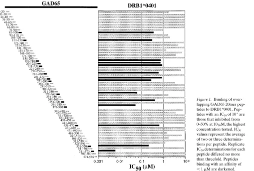
Binding of overlapping 20mer peptides to diabetes-associated human class II alleles, DRB1*0401 and DQ3.2. A 10-residue overlapping 20mer panel of peptides derived from the human GAD65 sequence was tested for binding to DRB1*0401 and DQ3.2 (Figs. 1 and 2). Of the 57 peptides tested, 14 inhibited binding of a labeled ligand to DRB1*0401 with an IC50, 1 mM. Another 19 of 57 peptides had IC50 values ranging from 1 to
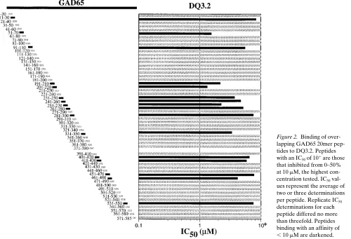
, 10 mM. A large proportion of the 20mer peptides (16 of 57) also bound to DQ3.2 with an IC50, 10 mM (Fig. 2). Although some of the 20mer peptides bound to both class II molecules,
other peptides were relatively specific for either DRB1*0401 or DQ3.2. The high percentage of binding by the 20mer panel of peptides to DRB1*0401 and DQ3.2 is not unexpected due to the known promiscuity of the class II MHC peptide-binding groove. Although a particular motif is preferred, binding to the class II MHC peptide-binding groove is quite flexible in terms of amino acid residues tolerated within each particular posi-tion of the peptide. The generally higher affinity of peptides for DRB1*0401 as compared to DQ3.2 may only be an appar-ent affinity difference caused by differappar-ent intrinsic stabilities of the two class II molecules in detergent (19, 20).
[image:4.612.60.569.395.738.2]Because our goal was to define naturally processed ep-itopes from GAD65 that associate with a diabetes-associated class II molecule on the surface of APC, we determined which of the binding peptides could be immunogenic in a transgenic B10.M strain expressing the DRB1*0401 molecule. We previ-ously demonstrated that the B10.M/DRB1*0401 transgenic strain utilizes the DRB1*0401 molecule as a restriction ele-ment (9). Because a relatively large number of the GAD65 peptides were found to bind to DRB1*0401 (Fig. 1), we used two approaches to narrow the number of peptides tested for in vivo immunogenicity. First, we recently demonstrated that DRB1*0401-binding 13mer peptides from a protein can be quite accurately predicted using an experimentally derived peptide-binding algorithm (8). Second, to avoid the generation of an immune response to a foreign peptide, we chose to test only those DRB1*0401-binding peptides that had an identical
Figure 1. Binding of over-lapping GAD65 20mer pep-tides to DRB1*0401. Pep-tides with an IC50 of 101 are
those that inhibited from 0–50% at 10 mM, the highest concentration tested. IC50
values represent the average of two or three determina-tions per peptide. Replicate IC50 determinations for each
peptide differed no more than threefold. Peptides binding with an affinity of
sequence to that found in murine GAD65. 10 13mer peptides predicted to be among the best binding peptides were synthe-sized and found to bind DRB1*0401 with IC50 values ranging from 18 to 1193 nM (Table I). It is interesting to note that the sequences of all but one of the 13mer peptides are included within 20mer peptides which bind well to DRB1*0401 (, 1
mM). The exception is peptide 339–351 (IC505 778 nM) which is not completely contained in any of the 20mer peptides. The presence or absence of a single residue at the amino or car-boxy terminus of a peptide can significantly alter the binding of a T cell epitope (21).
Generation of GAD65-specific T cell hybridomas. The 10 13mer DRB1*0401-binding peptides were used to immunize B10.M/DRB1*0401 amd B10.M mice (Table I). Two peptides were found to produce responses only in the transgenic B10.M/DRB1*0401 strain, GAD65 274–286 and GAD65 115– 127. These two peptides were nonimmunogenic in B10.H2g7 and NOD mice, two strains that have the diabetogenic H2g7 murine MHC haplotype. In addition to the two DRB1*0401-restricted peptide responses, GAD65 554–566 elicited an im-mune response in both the B10.M and B10.M/DRB1*0401 strains. The remaining seven GAD65 peptides showed low or no reactivity following in vivo immunization. The lack of a proliferative response to the seven DRB1*0401-binding pep-tides could be attributed: (a) to the development of central or peripheral tolerance to these self peptides; (b) to a low precur-sor frequency of T cells that recognize these
DRB1*0401/pep-Figure 2. Binding of over-lapping GAD65 20mer pep-tides to DQ3.2. Peppep-tides with an IC50 of 101 are those
that inhibited from 0–50% at 10 mM, the highest con-centration tested. IC50
val-ues represent the average of two or three determinations per peptide. Replicate IC50
determinations for each peptide differed no more than threefold. Peptides binding with an affinity of
[image:5.612.60.560.61.402.2], 10 mM are darkened.
Table I. Immunogenicity of Human GAD65-derived Peptides
GAD65 peptide
Binding assay IC50 (nM)
Lymph node cell proliferation (cpm 3 1023 {3H]thymidine)
B10.M/DR4 B10.M B10.H2g7 NOD
554–566 18, 7 29.3 37.4 0.8 1.1 325–337 22, 27 4.6 3.2 0.3 274–286* 88, 27, 58 36.4 20.3 0.4 0.2
87–99 70, 59 9.8 9.8 0.3
377–389 120, 173 8.7 9.4 0.4 328–340 321, 280 9.3 2.3 1.6
211–223 268, 335 0.9 0.3 1.0 115–127* 416, 280 67.2 1.3 1.7 2.9 339–351 691, 864 20.4 1.1 0.8 203–215 1193, 1306 0.6 0 20.5
Peptides were examined for immunogenicity in various mouse strains by
injecting footpads with 50 mg of peptide emulsified in CFA. Draining
lymph node cells were tested 9–11 d later for an in vitro recall response.
Responses obtained with 10 mM recall peptide are shown. All responses
indicated have background proliferation (1,168 to 5,274 cpm) sub-tracted. Proliferative values that are bolded indicate those responses that are at least fivefold above background. IC50 refers to the concentration
[image:5.612.57.298.457.626.2]tide complexes; or (c) to the instability of the 13mer peptides in the intracellular or extracellular environment of the immu-nized mouse. Thus, it should be noted that lack of immunoge-nicity to a particular 13mer epitope does not eliminate that peptide as an epitope processed by human APC and recog-nized by self-reactive T cells from DRB1*0401 humans.
DRB1*0401-restricted T cell hybridomas specific for GAD65 274–286 and GAD65 115–127 were developed to assess whether either of these peptide epitopes is naturally processed from the whole GAD65 molecule. Peptide-specific responses by these two hybridomas required presentation on spleen cells obtained from B10.M/DRB1*0401 mice; spleen cells from B10.M or B10.H2g7 mice did not provide appropriate APC since they lacked the DRB1*0401 molecule (Fig. 3).
Processing and presentation of GAD65 by DRB1*0401 APC. Peptide-specific, DRB1*0401-restricted T cell hybrido-mas secreted IL-2 following incubation with GAD65-trans-fected Preiss cells, but not with vector-transGAD65-trans-fected Preiss cells, indicating that GAD65 can be naturally processed to the GAD65 274–286 and GAD65 115–127 epitopes by human
APC (Fig. 4). DRB1*0401/GAD65 274–286 and DRB1*0401/ GAD65 115–127 complexes were both limiting on the surface of GAD65-transfected Preiss cells since addition of 5 mM of the appropriate peptide caused a 10- to 20-fold increase in IL-2 secretion (data not shown). The IL-2 secretion observed in these experiments also demonstrates that the T cell hybrido-mas recognize the GAD65-derived epitopes when presented by the normal, nonchimeric DRB1*0401 molecule expressed on Preiss cells.
More IL-2 was secreted by the T cells recognizing GAD65 274–286 compared to those recognizing GAD65 115–127 when GAD65-transfected Preiss cells were used as a source of APC (Fig. 4). The data are consistent with the hypothesis that GAD65-transfected Preiss cells produce more DRB1*0401/ GAD65 274–286 complexes than DRB1*0401/GAD65 115– 127 complexes. An alternate hypothesis is that GAD65 is processed to an overlapping but different peptide from the 115–127 epitope and is not recognized as well by the 115–127 specific T cell hybridoma. As a control to eliminate the possi-bility that IL-2 secretion is nonspecifically induced by GAD65-transfected Preiss cells, a HA 307–319-specific T cell hybridoma T84.17 (22) was shown to secrete IL-2 when HA 307–319 was added to the culture (196,354 fluorescence units) but not in re-sponse to either transfected Preiss line alone (Fig. 4).
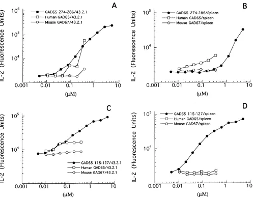
[image:6.612.57.298.59.422.2]To confirm that exogenously supplied GAD65 can be pro-cessed to the GAD65 274–286 and GAD65 115–127 epitopes, soluble human GAD65 (residues 90–585) was used to stimu-late the two hybridomas (Fig. 5). When the APC source was the murine 43.1/DRB1*0401 B cell hybridoma, both T cell hy-bridomas responded in a dose-dependent manner to human GAD65 (Fig. 5, A and C). In contrast, neither hybridoma re-sponded to murine GAD67. The lack of response to murine GAD67 is expected since both hybridomas are GAD65 spe-cific in that they do not respond to homologous peptides de-rived from murine GAD67 (data not shown). Interestingly, when transgenic spleen cells were used as a source of APC, only the GAD65 274–286-specific T cell hybridoma produced IL-2 in response to increasing concentrations of GAD65 (Fig. 5, B and D). The failure of GAD65 115–127-specific T cells to
Figure 3. Peptide-specific T cell hybridomas are restricted by DRB1*0401. The GAD65 274–286-specific T33.1 T cell hybridoma (A) and the GAD65 115–127-specific T35.15 T cell hybridoma (B) were incubated at 105/well with 5 3 105 spleen cells as a source of
[image:6.612.311.556.59.212.2]APC. The data are representative of the results of three experiments.
Figure 4.T cells recognize naturally processed human GAD65. T cell hybridomas (105) specific for GAD65 115–127, GAD65 274–286, and
HA 307–319 were incubated with 105 GAD65-expressing transfected
respond to splenic APC plus GAD65 again suggests that the GAD65 115–127 epitope may be selected less frequently dur-ing processdur-ing than the GAD65 274–286 epitope. Compared to the 43.1/DRB1*0401 B cell hybridoma, splenic APC were gen-erally less efficient at presenting peptide to both T cell hybri-domas, possibly due to the lower expression of DRB1*0401 on splenic APC compared to the 43.1/DRB1*0401 B cell hybri-doma (data not shown).
In summary, we identified two, naturally processed DRB1-*0401-restricted T cell epitopes from human GAD65, GAD65 274–286, and GAD65 115–127. Our experimental strategy rep-resents a general approach to the identification of distinct im-munogenic peptides derived from complex macromolecules in which we first use HLA binding studies to identify a set of po-tentially immunogenic peptides, followed by the use of trans-genic mice to further reduce this set to immunotrans-genic peptides, which are subsequently validated by using human APC to doc-ument the natural processing and presentation of these
ep-itopes. Although not all epitopes relevant in human diabetics can be found using this paradigm because of issues of self-tol-erance or species-specific processing, it is interesting to note that Endl et al. (23) reported the isolation of a DRB1*0401-restricted T cell clone from a recently diagnosed IDDM pa-tient which recognizes a GAD65 peptide (266–285) that over-laps one of those described in the current study.
[image:7.612.59.558.56.445.2]The identification of GAD-derived peptides has potential utility for directing the analysis of islet-specific immune re-sponses in type 1 diabetics. From these studies, it seems likely that the GAD65 274–286 and GAD65 115–127 epitopes are displayed on the surface of DRB1*0401 APC located in hu-man islets. These two epitopes therefore have the potential for being targets of regulatory T cells that have been expanded by peptide immunization. Antigen-specific recruitment of regula-tory cells to the islets following parenteral or oral peptide ex-posure may be a general strategy to downmodulate the au-toimmune response in the prediabetic islet.
Figure 5. T cells recognize naturally processed human GAD65. T cell hybridomas (105) specific for GAD65 115–127 or GAD65 274–286 were
incubated with either 105 murine B cell hybridoma cells transfected with DRB1*0401 (43.2.1), (A and C) or 5 3 105 spleen cells from B10.M/
Acknowledgments
We thank Dr. Janice Blum for her critical comments on the manu-script.
J.F. Elliott is suppported by the Juvenile Diabetes Foundation In-ternational Diabetes Interdisciplinary Research Program.
References
1. Atkinson, M.A., M.A. Bowman, L. Campbell, B.L. Darrow, D.L. Kauf-man, and N.K. Maclaren. 1994. Cellular immunity to a determinant common to
glutamate decarboxylase and coxsackie virus in insulin-dependent diabetes. J.
Clin. Invest. 94:2125–2129.
2. Harrison, L.C., M.C. Honeyman, H.J. DeAizpurua, R.S. Schmidli, P.G. Colman, B.D. Tait, and D.S. Cram. 1993. Inverse relation between humoral and cellular immunity to glutamic acid decarboxylase in subjects at risk of
insulin-dependent diabetes. Lancet. 341:1365–1369.
3. Kaufman, D.L., M. Clare-Salzler, J. Tian, T. Forsthuber, G.S.P. Ting, P. Robinson, M.A. Atkinson, E.E. Sercarz, A.J. Tobin, and P.V. Lehmann. 1993. Spontaneous loss of T-cell tolerance to glutamic acid decarboxlyase in murine
insulin-dependent diabetes. Nature (Lond.). 366:69–72.
4. Tisch, R., X.-D. Yang, S. Singer, R. Liblau, L. Fugger, and H. McDevitt. 1993. Immune response to glutamic acid decarboxylase correlates with insulitis
in non-obese diabetic mice. Nature (Lond.). 366:72–75.
5. Elliott, J.F., H.-Y. Qin, S. Bhatti, D.K. Smith, R.J. Singth, T. Dillon, J. Lauzon, and B. Singth. 1994. Immunization with the larger isoform of mouse glutamic acid decarboxylase (GAD67) prevents autoimmune diabetes in NOD
mice. Diabetes. 43:1494–1499.
6. Atkinson, M.A., and N.K. Maclaren. 1994. The pathogenesis of
insulin-dependent diabetes mellitus. N. Engl. J. Med. 331:1428–1436.
7. Wucherpfennig, K.W., and J.L. Strominger. 1995. Selective binding of self peptides to disease-associated major histocompatibility complex (MHC) molecules: a mechanism for MHC-linked susceptibility to human autoimmune
diseases. J. Exp. Med. 181:1597–1601.
8. Marshall, K.W., K.J. Wilson, J. Liang, A. Woods, D. Zaller, and J.B.
Rothbard. 1995. Prediction of peptide affinity to HLA DRB1*0401. J Immunol.
154:5927–5933.
9. Woods, A., H.Y. Chen, M.E. Trumbauer, A. Sirotina, R. Cummings, and D.M. Zaller. 1994. Human major histocompatibility complex class II-restricted
T cell responses in transgenic mice. J. Exp. Med. 180:173–181.
10. Chang, Y.-C., and D.I. Gottlieb. 1988. Characterization of the proteins
purified with monoclonal antibodies to glutamic acid decarboxylase. J.
Neuro-sci. 8:2123–2130.
11. Karlsen, A.E., W.A. Hagopian, C.E. Grubin, S. Dube, C.M. Disteche, D.A. Adler, H. Barmeier, S. Mathewes, F.J. Grant, D. Foster, et al. 1991.
Clon-ing and primary structure of a human isoform of glutamic acid decarboxylase
from chromosome 10. Proc. Natl. Acad. Sci. USA. 88:8337–8341.
12. Denzin, L.K., N.F. Robbins, C. Carboy-Newcomb, and P. Cresswell. 1994. Assembly and intracellular transport of HLA-DM and correction of the
class II antigen-processing defect in T2 cells. Immunity. 1:595–606.
13. de la Luna, S., I. Soria, D. Pulido, J. Ortin, and A. Jiminez. 1988. Effi-cient transformation of mammalian cells with constructs containing a
puromy-cin-resistance marker. Gene (Amst.). 62:121–126.
14. Novak, T.J., D. Farber, D. Leitenberg, S.-C. Hong, P. Johnson, and K. Bottomly. 1994. Isoforms of the transmembrane tyrosine phosphatase CD45
differentially affect T cell recognition. Immunity. 1:109–119.
15. Takebe, Y., M. Seiki, J.-I. Fujisawa, P. Hoy, K. Yokota, K.-I. Arai, M. Yoshida, and N. Arai. 1988. Sra promotor: an efficient and versatile mamma-lian cDNA expression system composed of the simian virus 40 early promotor and the R-U5 segment of human T-cell leukemia virus type 1 long terminal
re-peat. Mol. Cell. Biol. 8:466–472.
16. Hill, C.M., A. Liu, K.W. Marshall, J. Mayer, B. Jorgensen, B. Yuan, R.M. Cubbon, E.A. Nichols, L.S. Wicker, and J.B. Rothbard. 1994. Exploration
of requirements for peptide binding to HLA DRB1*0101 and DRB1*0401. J.
Immunol. 152:2890–2898.
17. Wicker, L.S., N.H. DeLarato, A. Pressey, and L.B. Peterson. 1993. Ge-netic control of diabetes and insulitis in the nonobese diabetic mouse: analysis
of the NOD.H-2b and B10.H-2nod strains. In Molecular Mechanisms of
Immu-nological Self-Recognition. F.W. Alt, and H.J. Vogel, editors. Academic Press, Inc., New York. 173–181.
18. Lee, D.S., J. Tian, T. Phan, and D.L. Kaufman. 1993. Cloning and sequence
analysis of a murine cDNA encoding glutamate decarboxylase (GAD65).
Bio-chim. Biophys. Acta. 1216:157–160.
19. Tampé, R., and H.M. McConnell. 1991. Kinetics of antigenic peptide
binding to the class II major histocompatibility molecule I-Ad. Proc. Natl. Acad.
Sci. USA. 88:4661–4665.
20. Witt, S.N., and H.M. McConnell. 1992. Antigenic peptide binding to the
mouse major histocompatibility complex class II protein I-Ek. Peptide
stabiliza-tion of the quaternary structure of I-Ek. J. Am. Chem. Soc. 114:3506–3511.
21. Grewal, I.S., K.D. Moudgil, and E.E. Sercarz. 1995. Hindrance of bind-ing to class II major histocompatibility complex molecules by a sbind-ingle amino acid residue contiguous to a determinant leads to crypticity of the determinant
as well as lack of response to the protein antigen. Proc. Natl. Acad. Sci. USA.
92:1779–1783.
22. Kovats, S., G.T. Nepom, M. Coleman, B. Nepom, W.W. Kwok, and J.S. Blum. 1995. Deficient antigen-presenting cell function in multiple genetic
com-plementation groups of type II bare lymphocyte syndrome. J. Clin. Invest. 96:
217–223.