Thrombus formation in vivo
Bruce Furie, Barbara C. Furie
J Clin Invest.
2005;
115(12)
:3355-3362.
https://doi.org/10.1172/JCI26987
.
To examine thrombus formation in a living mouse, new technologies involving intravital
videomicroscopy have been applied to the analysis of vascular windows to directly
visualize arterioles and venules. After vessel wall injury in the microcirculation, thrombus
development can be imaged in real time. These systems have been used to explore the role
of platelets, blood coagulation proteins, endothelium, and the vessel wall during thrombus
formation. The study of biochemistry and cell biology in a living animal offers new
understanding of physiology and pathology in complex biologic systems.
Review Series
Find the latest version:
Thrombus formation in vivo
Bruce Furie1,2 and Barbara C. Furie1,21Division of Hemostasis and Thrombosis, Center for Vascular Biology Research, Beth Israel Deaconess Medical Center, Boston, Massachusetts, USA. 2Department of Medicine, Harvard Medical School, Boston, Massachusetts, USA.
To examine thrombus formation in a living mouse, new technologies involving intravital videomicroscopy have
been applied to the analysis of vascular windows to directly visualize arterioles and venules. After vessel wall
injury in the microcirculation, thrombus development can be imaged in real time. These systems have been used
to explore the role of platelets, blood coagulation proteins, endothelium, and the vessel wall during thrombus
formation. The study of biochemistry and cell biology in a living animal offers new understanding of physiology
and pathology in complex biologic systems.
History of the study of hemostasis and thrombosis
The study of blood coagulation and thrombosis has moved through many stages. The classic studies of thrombus morphol-ogy and pathology were followed by a period in which blood coagulation proteins were identified based on the character- ization of patients with hereditary clotting disorders. The fun-damentals of platelet physiology, including platelet adhesion and aggregation, were defined by turbidimetric assay reporting platelet-platelet interaction and the adherence of platelets to glass beads, which reflects the ability of platelets to stick to sur-faces that mimic the injured vessel wall. Only 40 years ago were the techniques of protein biochemistry applied to the study of hemostasis and thrombosis. Proteins were purified, their amino acid sequences determined, their genes isolated, and their inter-action with other components analyzed in vitro. Protein domain structures were defined and their functional and structural relationship to other protein families determined. Site-specific mutagenesis and definition of the molecular basis of hemophil- ia identified functionally critical amino acids. From the analy-sis of the many enzymes, cofactors, structural proteins, ligands, and receptors involved in hemostasis and thrombosis, descrip-tive models emerged that explained the in vitro experimental results and the in vivo clinical observations in humans (1). These models have changed over the years, with continued refinement based on new information and fresh thinking (2–9). Still, many questions remain. We have learned what reactions and interac-tions can happen in vitro, but we need to understand what does happen in vivo. We are entering a new phase of discovery — the study of thrombosis in living animals to understand the mecha-nisms involved in this complex system.
Mechanisms of thrombus induction in animal models
Although the pathogenesis of thrombus formation can be both an acute and a chronic process in the natural condition, direct experimental observation of this process in animal models requires artificial methods. These methods take many forms, each with advantages and disadvantages, as they relate to the physi- ologic mechanisms of thrombus formation. With photochemi-cal injury, dye (e.g., rose bengal) is infused into the circulation. Photo-excitation leads to oxidative injury of the vessel wall and
subsequent thrombus formation (10). Mechanical (11) or elec-trical trauma (12) directly injures the endothelium and leads to thrombus formation. Vessel ligation causing stasis also initiates thrombus formation (13). Ferric chloride, introduced to initiate arterial thrombosis in small-animal models (14), generally initi-ates severe endothelial damage and vessel occlusion, monitored by the decrease in temperature distal to the developing thrombus. This model system has been widely used (15–17), modified with a Doppler flow probe to monitor vessel occlusion or with direct blood vessel visualization by intravital microscopy. Laser-induced injury causes heat damage to a limited region of the endothelium, with little morphologic change to the vessel wall (18). Because endothelial damage is limited and does not involve denudation of the endothelium, in contrast to ferric chloride–induced injury, the laser-induced injury appears to be a model of thrombosis more akin to the injury caused by inflammation than to that caused by trauma. Using the laser, both temporal and spatial resolution for thrombus generation is obtained, since the location and the precise time of injury are operator-controlled. The laser pulse, applied through the microscope optics, is targeted at the vessel wall. Upon injury, thrombus formation initiates with the rapid accumulation of platelets and the expression of tissue factor at the thrombus–vessel wall interface.
Intravital microscopy of thrombus formation
Intravital microscopy was introduced to study leukocyte interac-tion with the vessel wall in a living animal (19, 20). Since then, many investigators have captured in vivo images in real time using analog videomicroscopy (21, 22). Oude Egbrink et al. were among the first to combine experimental thrombosis, induced by mechanical puncture with glass micropipettes, with intravital vid-eomicroscopy in a living animal (23). Others applied experimental thrombosis with in vivo microscopic analysis (24, 25), adopting the methods used for leukocyte rolling. Photochemical injury of microvessels in the mouse ear allowed analysis of the kinetics of platelet accumulation and vessel occlusion, leading to the obser-vation that hirudin inhibited thrombus formation and promoted vessel recanalization (26). Denis et al. demonstrated that geneti- cally altered mice lacking vWF showed defects in platelet accumu-lation following ferric chloride injury (16).
The discovery that P-selectin is an adhesion molecule that binds platelets to leukocytes (27) and the identification of the P-selectin counterreceptor PSGL-1 (28) prompted us to gener- ate a PSGL-1 knockout mouse to better understand the biol-ogy of PSGL-1 (29). Methods of analog intravital microscopy to examine leukocyte rolling in living mice had been developed by
Conflict of interest: The authors have declared that no conflict of interest exists.
review series
other laboratories (30). However, the ability to directly observe complex events in a living animal by optical microscopy offered new opportunities to understand thrombosis. The combination of available knockout mice, progress in optical spectroscopy, and advances in computer software and hardware supported the possibility of developing novel intravital imaging technol- ogy to study thrombus formation. An imaging system specifi- cally designed for visualizing events in the vasculature of a liv-ing mouse required real-time imaging of thrombus formation using high image acquisition rates of 10–20 images per second in up to 3 fluorescent channels as well as a bright-field channel to appreciate the fluorescent image in a histologic context. The system needed the flexibility of confocal imaging to support 3D image reconstruction and a digital capture system for quantita-tive image analysis. A system that meets these criteria has been developed: an intravital microscopy system that supports high-speed wide-field and confocal imaging of the microcirculation of a living mouse (31, 32). In most experiments, vessel wall injury that initiates thrombosis is induced using a laser focused on the endothelium via the microscope optics. This technology has been applied to the study of thrombus formation in a living mouse, in order to reexamine many of the accepted tenets of thrombus formation and to determine whether or not the predictions of in vitro or ex vivo experiments are valid for understanding the in vivo system. This work, coupled with the in vivo experiments of others, promises to provide more detailed descriptions of the mechanisms involved in thrombus formation. The current
description of in vivo thrombus formation remains incomplete, but the emerging concepts will be reviewed.
Initiation of thrombus formation
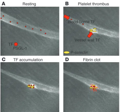
Laser injury initiates the expression of tissue factor activity on or near the vessel wall. Platelets rapidly accumulate on the endo-thelium, and fibrin can be observed at the platelet thrombus– vessel wall interface, at the leading edge of the thrombus, and, over seconds, within and throughout the thrombus (Figure 1). In contrast to classical models of thrombus formation in which it has been thought that the platelet thrombus forms and is then stabilized by the subsequent formation of a fibrin clot, these experiments demonstrate that platelet thrombus formation and fibrin clot formation overlap temporally and occur nearly simultaneously. Similar results are obtained with other forms of injury, including those that lead to thrombus formation through subendothelial matrix exposure.
Platelet adhesion
vWF plays an important role in thrombus formation. vWF-null mice displayed prolonged bleeding times and spontaneous bleed- ing in about 10% of neonates (16). These results simulate the phe- notype of severe von Willebrand disease in humans. Via intravi-tal microscopy using the ferric chloride injury model, significant impairment of platelet–vessel wall interaction was observed, lead-ing to defective thrombus formation. Also, in the complete absence of vWF, platelet accumulation on the vessel wall was significantly decreased but not absent, and thrombi formed in some vessels. These results argue both for the importance of vWF in platelet deposition on the vessel wall and for a vWF-independent mecha-nism for arterial thrombus formation.
[image:3.585.78.255.81.420.2]Fibrinogen is thought to play an important role in platelet–ves- sel wall interaction, particularly at the low shear rates charac-teristic of the venous circulation. To evaluate the contribution of fibrinogen to arterial thrombus formation, Ni et al., using fibrinogen-null mice, demonstrated that platelet deposition and the onset of thrombus formation were the same as in WT mice (33). However, thrombi in fibrinogen-null mice were unstable and embolized as they became larger. Mice deficient in both vWF and fibrinogen formed thrombi that proved unstable. Given the accepted tenet that these 2 adhesive proteins are critical for platelet–vessel wall interaction, the fact that thrombi formed in doubly deficient mice argues for additional adhesive molecules that participate in the platelet–vessel wall synapse. A role for fibronectin in this process remains plausible, since fibronectin
Figure 1
deficiency delays thrombus formation in vivo through putative diminished platelet-platelet interaction (34). It seems that there are multiple participants in platelet–vessel wall interaction, some of which have yet to be directly implicated.
Platelet activation
The tissue factor pathway dominates platelet activation in the laser-induced thrombosis model. PAR4, the sole signaling protease-activated thrombin receptor on mouse platelets, is critical for thrombin-mediated platelet activation (35). Laser-induced injury in mice lacking PAR4 leads to a small, rapid accumulation of platelets (36). This initial thrombus is unstable and subsequently reduces in size. This initial phase of platelet accumulation may be mediated by vWF or another adhesive molecule. After 3–4 minutes, thrombus size in the PAR4-null mice is less that 10% of that in WT mice. During the first several minutes, there is no evidence of platelet activation, as monitored by the surface expression of P-selectin. However, although these thrombi in PAR4-null mice contain markedly reduced numbers of activated platelets, fibrin generation is normal. These results emphasize the importance of thrombin generation and PAR4 for platelet activation and thrombus formation in this model. A major unanswered question is the cellular or subcellular local- ization of the membrane surface that supports thrombin gen-eration. Are the few activated platelets sufficient to supply the necessary membrane surfaces, or do other cells and cell-derived membranes support thrombin generation? Thrombin genera- tion appears critically important to laser-induced thrombus for- mation. For example, in the absence of exposed collagen, laser-induced thrombus formation was normal in FcRγ mice lacking the collagen receptor glycoprotein VI (37). If there is collagen exposure, it is below the sensitivity of this imaging system. Thus, in the laser-induced model of thrombosis, platelets are likely activated by a mechanism independent of interaction of colla-gen with platelet collagen receptors.
Collagen exposure can play an important role in platelet acti-vation during hemostasis, and the importance of this role varies with the type of vascular injury. Ferric chloride induces an oxida- tive injury exposing the subendothelial matrix (33), and type I col-lagen can be stained with anti-collagen antibodies in the blood in vivo after ferric chloride injury (37). Following ferric chloride injury, FcRγ-null mice lacking expression of glycoprotein VI failed to generate platelet thrombi in vivo, in contrast to WT mice. Fur-thermore, blocking antibodies against glycoprotein VI infused into WT mice also inhibited platelet thrombus formation. These results demonstrate the requirement for glycoprotein VI in col-lagen-mediated platelet activation in vivo.
Platelet activation can be monitored by P-selectin expression on the platelet surface. This marker of platelet activation correlates directly with exocytosis of α-granules. Although in vitro studies of P-selectin have established the kinetics of P-selectin expres-sion following activation with various agonists, the kinetics of P-selectin expression in vivo has not been previously character-ized. To this end, P-selectin expression on platelets incorporated into the developing thrombus was studied in vivo after laser- induced injury (38). P-selectin first appears on platelets at the ves- sel wall–thrombus interface. Over 3–4 minutes, a wave of P-selec-tin expression can be monitored through the thrombus from the vessel wall to the luminal surface of the thrombus. However, only when the P-selectin density on the luminal surface is sufficiently
high to support leukocyte rolling on the arterial thrombus can any leukocyte-thrombus interaction be observed. Indeed, direct observation of leukocyte rolling on the developing thrombus can-not be appreciated until after about 3 minutes from the initiation of thrombus formation. These results emphasize a delayed role for leukocyte-thrombus interaction and tissue factor delivery via leukocytes during thrombus formation.
Calcium mobilization in vivo
Platelet activation leads to intracellular calcium mobilization, where increased calcium serves as a second messenger in initi-ating numerous signaling pathways. These calcium transients have been extensively studied in vitro using flow chambers to monitor platelet adherence (39–41). High-speed wide-field intravital microscopy has been used to image this calcium spike in living mice during thrombus formation (42). Platelets were isolated from a donor mouse and loaded with fura-2, a fluoro- chrome that is sensitive to calcium concentration. These plate-lets were infused into a recipient mouse, and the fura-2–labeled platelets were analyzed for significant changes in intracellular calcium during their circulation in blood, their interaction with the thrombus, and their incorporation into a stable platelet thrombus. These studies have revealed that platelet activation, as monitored by calcium mobilization, does not take place in the circulation (42). Rather, platelets bind transiently to the developing thrombus. After a short period, these platelets either undergo calcium mobilization and become stably incorporated into the thrombus or they disengage from the thrombus and float downstream. Calcium mobilization is required for stable platelet incorporation into the developing thrombus.
Other proteins in thrombus formation
The tissue factor–factor VIIa complex initiates thrombin genera-tion and fibrin formation, and deficiency of any of the proteins within this pathway (e.g., factor IX, factor VIII, factor X, factor V, and prothrombin) decreases thrombin generation and thus thrombus formation. However, factor XII–null mice are char-acterized by defective arterial thrombus formation in vivo (43), as are factor XI–null mice (44). A role for factor XII in normal hemostasis has been long dismissed, since patients with factor XII deficiency have no bleeding phenotype. Recently a role for both factor XII and factor XI in thrombus formation has been demon-strated, although the actual mechanism for the participation of the intrinsic pathway in blood coagulation in vivo remains specu-lative. This raises the intriguing possibility that factor XII and factor XI are important for thrombosis but not hemostasis (43).
PECAM-1 is a cell adhesion molecule found on endothelial cells and platelets. PECAM-1–null mice formed larger arterial throm-bi more rapidly than WT mice in the laser-induced thrombosis model (45). Using chimeric mice prepared by reciprocal bone mar- row transplantation, platelet PECAM-1 was shown to be the criti-cal component. These results suggest that PECAM-1 plays a role in negative regulation of thrombus formation.
Gas6, a γ-carboxyglutamic acid–containing membrane protein homologous to protein S, is present on the platelet membrane and binds to several receptor tyrosine kinases, including Axl. Gas6 amplified platelet aggregation and secretion responses to plate-let agonists (46). Deficiency of Gas6, either in a Gas6–/– mouse or
review series
that Gas6 plays a role in amplifying signaling events induced by agonists and does not directly participate in platelet-platelet syn-apse formation of significant affinity.
CD40L, a transmembrane platelet granule protein, is expressed on the plasma membrane of activated platelets, where it can interact with CD40 that is widely distributed on vascular cells. Mice deficient in CD40L but not CD40 showed an in vivo defect in thrombus formation initiated by ferric chloride (47). These
CD40L–/– mice showed delayed arterial occlusion and thrombus
instability. CD40L appears to be an αIIbβ3
ligand required for sta-ble formation of arterial thrombi.
Human platelets express 2 Eph kinases, Eph4 and EphB1, and at least 1 ligand, ephrinB1. During αIIbβ3
-mediated plate-let aggregation, Eph-ephrin interactions on adjacent platelet surfaces contribute to high-affinity platelet-platelet contact (48). These interactions favor thrombus growth and stability, sustain contact-facilitated signaling via complex formation, and promote clot retraction. Inhibition of Eph-ephrin interaction, evaluated in vitro in a flow chamber, showed a 40% decrease in mean thrombus volume (48).
Outside-in αIIbβ3
signaling is required for normal platelet throm-bus formation and is triggered by c-Src activation via PTP-1B (49). Studies of PTP-1B–deficient mouse platelets in vitro indicate that PTP-1B is required for fibrinogen-dependent platelet spreading and clot retraction. Thrombus formation in vivo is reduced in PTP-1B–null mice, a manifestation of ineffective calcium mobili-zation during platelet activation. PTP-1B is a positive regulator for the initiation of outside-in αIIbβ3 signaling.
CD39, the vascular ATP diphosphohydrolase, is largely expressed on endothelial cells. This enzyme converts ATP and ADP to AMP. Tail bleeding times were prolonged and platelet thrombus formation in vivo was delayed in CD39-null mice sub- jected to ferric chloride injury (50). These results appear consis- tent with the importance of ADP release during platelet activa-tion for activation of adjacent platelets. However, interpretation of these results is complicated by the desensitization of the P2Y1 receptor on CD39-null platelets.
The SLAM family of adhesion receptors, a subset of the CD2 Ig superfamily, is expressed on platelets (51). SLAM phosphor-ylation occurs during platelet aggregation. SLAM-deficient platelets showed defective aggregation, and SLAM-null mice were characterized in vivo by delayed thrombus formation but normal tail bleeding times. SLAM may play a secondary role in the formation of the platelet-platelet synapse that is otherwise dominated by αIIbβ3.
Tissue factor–bearing microparticles
P-selectin functions as an adhesion molecule (27) but was subse-quently shown to have a role in fibrin formation (52). In a baboon arteriovenous shunt model of thrombosis, blocking antibodies against P-selectin not only inhibited leukocyte accumulation in the developing thrombus but also decreased fibrin formation. The molecular and cellular basis for this experimental observa-tion was not clear at the time, but it was thought that leukocytes might generate tissue factor upon stimulation more rapidly in vivo than in the in vitro systems used to explore de novo tissue factor biosynthesis in stimulated cells (53, 54). Although a rela-tionship of P-selectin to fibrin formation was secure, the basis for the inhibition of fibrin formation by anti–P-selectin antibodies remained unknown.
[image:5.585.45.286.81.306.2]Using genetically altered mice and digital intravital micros-copy imaging, this question was revisited (55). Tissue factor antigen and fibrin were observed throughout the thrombus generated in WT mice (Figure 1), a result that confirmed ear-lier in vitro experiments (56). However, minimal tissue factor antigen or fibrin was observed in thrombi generated in either P-selectin–null mice or PSGL-1–null mice (55). These results were similar to those obtained in the baboon thrombosis model using anti–P-selectin antibodies to block P-selectin action. We hypothesized that tissue factor and PSGL-1 must be physically coupled. Although this is true of activated monocytes, where tissue factor and PSGL-1 reside on the plasma membrane (57), there is no evidence that such monocytes circulate constitutive-ly in blood (58). Furthermore, leukocytes do not interact with developing thrombi as rapidly as fibrin deposition begins (38). Rather, leukocyte microparticles might provide the basis for this observation. Leukocyte microparticles, first identified in 1994 (59), could express both tissue factor and PSGL-1 if derived from monocytes. Indeed, a population of microparticles exists in the circulation that is positive for both tissue factor antigen and PSGL-1 antigen. Using a monocyte-like cell line, fluores-cently labeled microparticles were generated and infused into mice. During thrombus formation, microparticles accumulated
Figure 2
Figure 3
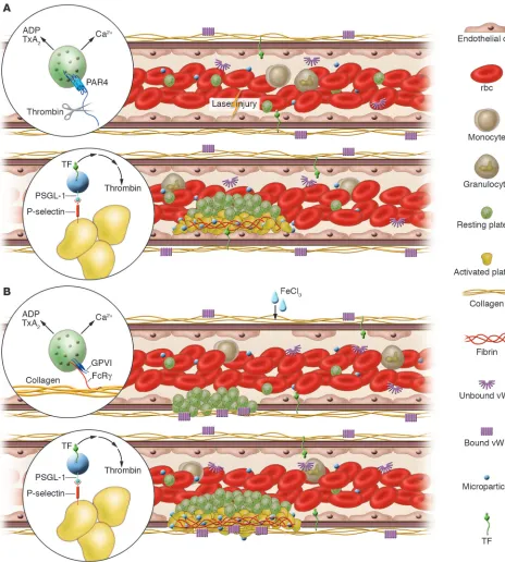
Experimental models of thrombosis. Platelets, red blood cells, monocytes, and granulocytes circulate in blood whereas endothelial cells line the vessel wall. Plasma proteins, including vWF, fibrinogen and other coagulation proteins, and microparticles are also present in the circulation. (A) Upon laser-induced injury of the vessel wall, vWF mediates the interaction of platelets with the endothelium. Tissue factor in the vessel wall leads to thrombin generation. Thrombin activates mouse platelets via the PAR4 receptor (inset). Activated platelets undergo calcium mobilization and the release of ADP and thromboxane A2 (TxA2) to accelerate platelet recruitment and activation and the formation of a platelet thrombus.
These platelets express P-selectin, and leukocyte microparticles expressing PSGL-1 and tissue factor accumulate in the thrombus through the interaction of P-selectin with PSGL-1 (inset). The concentration of tissue factor initiates coagulation, the generation of more thrombin, and the propagation of a fibrin clot. (B) Upon vessel wall oxidative injury with ferric chloride, the endothelium is denuded and the subendothelial matrix exposed. Platelets interact with the matrix via GPIb-V-IX and αIIbβ3 on the platelet membrane and collagen and vWF in the matrix. Glycoprotein
VI (GPVI) binding to collagen is required for platelet activation, and activated platelets undergo calcium mobilization and the release of ADP and thromboxane A2 (inset) to accelerate platelet recruitment and activation and the formation of a thrombus. These platelets express P-selectin, and
review series
in the thrombi of WT mice. In contrast, no accumulation was observed in P-selectin–null mice.
These results are consistent with a model in which circulating microparticles expressing tissue factor and PSGL-1 accumulate in the developing thrombus via the interaction of P-selectin with PSGL-1. This delivers and concentrates tissue factor in the thrombus, leading to a critical concentration that can initiate blood coagulation (Figure 2). Numerous groups have reported tissue factor antigen in platelet-poor plasma, with levels varying from 100 to 150 pg/ml. However, Butenas et al. have recently reopened this issue (58). They report no detectable tissue fac-tor activity in whole blood, no tissue factor antigen associated with unstimulated mononuclear cells in whole blood, and a level of tissue factor activity that cannot exceed 20 fM, equivalent to about 1 pg/ml, and is more likely lower. Since these authors demonstrate that 1 pg/ml of active tissue factor rapidly clots whole blood, it would seem that blood tissue factor concentra- tion is much lower than 1 pg/ml and that a manyfold concentra-tion of tissue factor within the thrombus is a critical component for the initiation of blood coagulation. Alternatively, an inactive form of tissue factor may undergo some form of activation to its biologically functional form.
Tissue factor resides in 3 distinct compartments: (a) the surface of extravascular cells, (b) the vessel wall, and (c) blood micropar-ticles. Upon stimulation, both endothelial cells and monocytes have the capacity to express tissue factor. To determine whether tissue factor associated with blood microparticles contributes to fibrin formation during thrombosis in vivo, 1 strain of chimeric mice in which tissue factor was associated with the vessel wall but not the blood microparticles and another strain of chimeric mice in which tissue factor was associated with the blood mic-roparticles but not the vessel wall were prepared (60). Such mice were generated by bone marrow transplantation of WT mice, with normal levels of tissue factor in both the vessel wall and blood microparticles, and low–tissue factor mice, with about 1% of the normal level of tissue factor (61). Chimeras generated by transplantation of low–tissue factor bone marrow into WT mice showed platelet thrombi containing markedly reduced tissue factor and fibrin (60). Conversely, chimeras generated by transplantation of WT bone marrow into low–tissue factor mice rescued tissue factor accumulation and fibrin generation in the platelet thrombus. These results emphasize that within the context of this in vivo model, fibrin propagation is depen-dent on tissue factor derived from blood microparticles. Both vessel wall tissue factor and microparticle tissue factor appear to contribute to thrombus formation. In thrombosis models where there is no vessel wall tissue factor (56), where vessel wall injury causes vessel wall tissue factor to predominate (62), or where there is no blood flow and thus the deposition of microparticles is eliminated (62), the balance between the contribution of ves-sel wall tissue factor and that of microparticle tissue factor can be altered, giving varying results. Likely, different pathologies associated with thrombosis may also differentially impact on the contributions of tissue factor from the vessel wall and from blood microparticles.
Animal models and their relevance to human disease
The laser-injury thrombosis model has numerous advantages for the study of thrombosis in vivo. This model permits the exam-ination of thrombus formation in a living animal that is not
anticoagulated and that has an intact vessel wall, all circulating cellular elements, and all circulating plasma proteins. Second, the precise location of the injury is known — within a micron or two — and the exact time of injury is known — within a second or two. This temporal and spatial resolution is in contrast to the ferric chloride model, where oxidative injury is generalized. Third, the laser injury is a heat injury, which does not induce morphologic changes to the vessel wall if the appropriate energy level is used. However, if excessive energy is used, tissue disrup-tion and hemorrhage are observed.
Nonetheless, all of the models of thrombus formation are just models. For example, laser-induced injury, like mechanical disrup- tion, electrical stimulation, chemical oxidation, or stasis, is non-physiologic. The microcirculation of the cremaster muscle offers an ideal transparent vascular window for optical microscopy. However, atherothrombosis and peripheral arterial thrombosis are diseases of large arteries that are too thick to study by the cur-rent methods. Furthermore, the shear rates and flow dynamics within the microcirculation are different from those in large ves- sels. Lastly, mice are poor animal models for human atheroscle-rosis, although thrombosis may be more parallel for comparing with the human system. Insofar as in vitro studies have allowed the construction of an understanding of thrombosis, vascular injury models, albeit not perfect, are yet another step closer to studying thrombosis in the real thing: human arteries.
Summary
opportunities for targets for novel antithrombotics but also indi- 1.opportunities for targets for novel antithrombotics but also indi- Furie,opportunities for targets for novel antithrombotics but also indi- B.,opportunities for targets for novel antithrombotics but also indi- andopportunities for targets for novel antithrombotics but also indi- Furie,opportunities for targets for novel antithrombotics but also indi- B.C.opportunities for targets for novel antithrombotics but also indi- 1988.opportunities for targets for novel antithrombotics but also indi- Theopportunities for targets for novel antithrombotics but also indi- molecularopportunities for targets for novel antithrombotics but also indi- basisopportunities for targets for novel antithrombotics but also indi- of blood coagulation. Cell. 53:505–518.
2. Goto, S., Ikeda, Y., Saldivar, E., and Ruggeri, Z.M. 1998. Distinct mechanisms of platelet aggregation as a consequence of different shearing flow conditions.
J. Clin. Invest. 101:479–486.
3. Ruggeri, Z.M. 2002. Platelets in atherothrombosis.
Nat. Med. 8:1227–1234.
4. Goto, S. 2004. Understanding the mechanism of platelet thrombus formation under blood flow conditions and the effect of new antiplatelet agents. Curr. Vasc. Pharmacol. 2:23–32.
5. Andrews, R.K., and Berndt, M.C. 2004. Plate-let physiology and thrombosis. Thromb. Res.
114:447–453.
6. Gibbins, J.M. 2004. Platelet adhesion signalling and the regulation of thrombus formation. J. Cell Sci.
117:3415–3425.
7. Furie, B., and Furie, B.C. 2004. Role of platelet P-selectin and microparticle PSGL-1 in thrombus formation. Trends Mol. Med. 10:171–178. 8. Huo, Y., and Ley, K.F. 2004. Role of platelets in the
development of atherosclerosis. Trends Cardiovasc. Med. 14:18–22.
9. Robbie, L., and Libby, P. 2001. Inflammation and atherothrombosis. Ann. N. Y. Acad. Sci. 947:167–179; discussion 179–180.
10. Watson, B.D., Dietrich, W.D., Busto, R., Wachtel, M.S., and Ginsberg, M.D. 1985. Induction of repro- ducible brain infarction by photochemically initi-ated thrombosis. Ann. Neurol. 17:497–504.
11. Le Menn, R., Bara, L., and Samama, M. 1981. Ultra-structure of a model of thrombogenesis induced by mechanical injury. J. Submicrosc. Cytol. 13:537–549. 12. Carmeliet, P., et al. 1997. Vascular wound healing
and neointima formation induced by perivascular electric injury in mice. Am. J. Pathol. 150:761–776. 13. Gitel, S.N., and Wessler, S. 1983. Dose-dependent
antithrombotic effect of warfarin in rabbits. Blood.
61:435–438.
14. Kurz, K.D., Main, B.W., and Sandusky, G.E. 1990. Rat model of arterial thrombosis induced by ferric chloride. Thromb. Res. 60:269–280.
15. Farrehi, P.M., Ozaki, C.K., Carmeliet, P., and Fay, W.P. 1998. Regulation of arterial thrombolysis by plas-minogen activator inhibitor-1 in mice. Circulation.
97:1002–1008.
16. Denis, C., et al. 1998. A mouse model of severe von Willebrand disease: defects in hemostasis and thrombosis. Proc. Natl. Acad. Sci. U. S. A.
95:9524–9529.
17. Fay, W.P., Parker, A.C., Ansari, M.N., Zheng, X., and Ginsburg, D. 1999. Vitronectin inhibits the thrombotic response to arterial injury in mice.
Blood. 93:1825–1830.
18. Rosen, E.D., et al. 2001. Laser-induced noninvasive vascular injury models in mice generate platelet- and coagulation-dependent thrombi. Am. J. Pathol.
158:1613–1622.
19. Atherton, A., and Born, G.V. 1972. Quantitative
investigations of the adhesiveness of circulating polymorphonuclear leucocytes to blood vessel walls. J. Physiol. 222:447–474.
20. Schmid-Schonbein, G.W., Usami, S., Skalak, R., and Chien, S. 1980. The interaction of leukocytes and erythrocytes in capillary and postcapillary vessels.
Microvasc. Res. 19:45–70.
21. Tangelder, G.J., and Arfors, K.E. 1991. Inhibition of leukocyte rolling in venules by protamine and sulfated polysaccharides. Blood. 77:1565–1571. 22. Sriramarao, P., Languino, L.R., and Altieri, D.C.
1996. Fibrinogen mediates leukocyte-endothe-lium bridging in vivo at low shear forces. Blood.
88:3416–3423.
23. oude Egbrink, M.G., Tangelder, G.J., Slaaf, D.W., and Reneman, R.S. 1988. Thromboembolic reac-tion following wall puncture in arterioles and venules of the rabbit mesentery. Thromb. Haemost.
59:23–28.
24. Roesken, F., et al. 1997. A new model for quanti-tative in vivo microscopic analysis of thrombus formation and vascular recanalisation: the ear of the hairless (hr/hr) mouse. Thromb. Haemost.
78:1408–1414.
25. Rucker, M., Roesken, F., Vollmar, B., and Menger, M.D. 1998. A novel approach for comparative study of periosteum, muscle, subcutis, and skin micro-circulation by intravital fluorescence microscopy.
Microvasc. Res. 56:30–42.
26. Roesken, F., Vollmar, B., Rucker, M., Seiffge, D., and Menger, M.D. 1998. In vivo analysis of anti-thrombotic effectiveness of recombinant hirudin on microvascular thrombus formation and recana-lization. J. Vasc. Surg. 28:498–505.
27. Larsen, E., et al. 1989. PADGEM protein: a recep-tor that mediates the interaction of activated platelets with neutrophils and monocytes. Cell.
59:305–312.
28. Sako, D., et al. 1993. Expression cloning of a functional glycoprotein ligand for P-selectin. Cell.
75:1179–1186.
29. Yang, J., et al. 1999. Targeted gene disruption dem-onstrates that P-selectin glycoprotein ligand 1 (PSGL-1) is required for P-selectin-mediated but not E-selectin-mediated neutrophil rolling and migration. J. Exp. Med. 190:1769–1782.
30. Ley, K., et al. 1995. Sequential contribution of L- and P-selectin to leukocyte rolling in vivo. J. Exp. Med. 181:669–675.
31. Falati, S., Gross, P., Merrill-Skoloff, G., Furie, B.C., and Furie, B. 2002. Real-time in vivo imaging of platelets, tissue factor and fibrin during arte-rial thrombus formation in the mouse. Nat. Med.
8:1175–1181.
32. Celi, A., et al. 2003. Thrombus formation: direct real time observation and digital analysis of throm-bus assembly in a living mouse by confocal and widefield intravital microscopy. J. Thromb. Haemost.
1:60–68.
33. Ni, H., et al. 2000. Persistence of platelet thrombus
formation in arterioles of mice lacking both von Willebrand factor and fibrinogen. J. Clin. Invest.
106:385–392.
34. Ni, H., et al. 2003. Plasma fibronectin promotes thrombus growth and stability in injured arterioles.
Proc. Natl. Acad. Sci. U. S. A. 100:2415–2419. 35. Kahn, M.L., et al. 1998. A dual thrombin receptor
system for platelet activation. Nature. 394:690–694. 36. Vandendries, E., Hamilton, J.R., Coughlin, S.R., Furie, B.C., and Furie, B. 2004. Protease-activa-tor receptor 4 is required for maximal thrombus growth, but not for fibrin generation in thrombi after laser injury [abstract]. Blood. 104(Suppl.):624. 37. Dubois, C., Panicot-Dubois, L., Furie, B., and Furie, B.C. 2004. Importance of GPVI in platelet activa-tion and thrombus formation in vivo [abstract].
Blood. 104(Suppl.):842.
38. Gross, P., Furie, B.C., Merrill-Skoloff, G., Chou, J., and Furie, B. 2005. Leukocyte versus microparticle-mediated tissue factor transfer during arteriolar thrombus development. J. Leukoc. Biol. doi:10.1189/ jlb.0405193.
39. Mazzucato, M., Pradella, P., Cozzi, M.R., De Marco, L., and Ruggeri, Z.M. 2002. Sequential cytoplasmic calcium signals in a 2-stage platelet activation pro- cess induced by the glycoprotein Ibalpha mechano-receptor. Blood. 100:2793–2800.
40. Mazzucato, M., Cozzi, M.R., Pradella, P., Ruggeri, Z.M., and De Marco, L. 2004. Distinct roles of ADP receptors in von Willebrand factor-mediated plate-let signaling and activation under high flow. Blood.
104:3221–3227.
41. Nesbitt, W.S., et al. 2003. Intercellular calcium communication regulates platelet aggregation and thrombus growth. J. Cell Biol. 160:1151–1161. 42. Dubois, C., Panicot-Dubois, L., Furie, B.C., and
Furie, B. 2004. Direct real time visualization of platelet calcium signaling in vivo: role of platelet activation and thrombus formation in a living mouse [abstract]. Blood. 104(Suppl.):325.
43. Renne, T., et al. 2005. Defective thrombus forma-tion in mice lacking coagulation factor XII. J. Exp. Med. 202:271–281.
44. Wang, X., et al. 2005. Effects of factor IX or factor XI deficiency on ferric chloride-induced carotid artery occlusion in mice. J. Thromb. Haemost.
3:695–702.
45. Falati, S., et al. 2005. Platelet PECAM-1 inhibits thrombus formation in vivo. Blood. doi:10.1182/ blood-2005-04-1512.
46. Angelillo-Scherrer, A., et al. 2001. Deficiency or inhibition of Gas6 causes platelet dysfunction and protects mice against thrombosis. Nat. Med.
7:215–221.
47. Andre, P., et al. 2002. CD40L stabilizes arterial thrombi by a beta3 integrin–dependent mechanism.
Nat. Med. 8:247–252.
review series
Natl. Acad. Sci. U. S. A. 102:9820–9825.
49. Arias-Salgado, E.G., et al. 2005. Protein tyrosine phosphatase PTP-1B is an essential positive regula-tor of platelet thrombus formation and outside-in IIb 3 signaling. J. Cell Biol. 170:837–845.
50. Enjyoji, K., et al. 1999. Targeted disruption of cd39/ ATP diphosphohydrolase results in disordered hemostasis and thromboregulation. Nat. Med.
5:1010–1017.
51. Nanda, N., et al. 2005. Platelet aggregation induc-es platelet aggregate stability via SLAM fam-ily receptor signaling. Blood . doi:10.1182/blood-2005-01-0333.
52. Palabrica, T., et al. 1992. Leukocyte accumula-tion promoting fibrin deposition is mediated in vivo by P-selectin on adherent platelets. Nature.
359:848–851.
53. Semeraro, N., et al. 1983. Direct induction of tissue
factor synthesis by endotoxin in human macrophages from diverse anatomical sites. Immunology.
50:529–535.
54. Celi, A., et al. 1994. P-selectin induces the expres-sion of tissue factor on monocytes. Proc. Natl. Acad. Sci. U. S. A. 91:8767–8771.
55. Falati, S., et al. 2003. Accumulation of tissue factor into developing thrombi in vivo is dependent upon microparticle P-selectin glycoprotein ligand 1 and platelet P-selectin. J. Exp. Med. 197:1585–1598. 56. Giesen, P.L., et al. 1999. Blood-borne tissue factor:
another view of thrombosis. Proc. Natl. Acad. Sci. U. S. A.
96:2311–2315.
57. Moore, K., et al. 1992. Identification of a spe-cific glycoprotein ligand for P-selectin (CD62) on myeloid cells. J. Cell Biol. 118:445–456.
58. Butenas, S., Bouchard, B.A., Brummel-Ziedins, K.E., Parhami-Seren, B., and Mann, K.G. 2005.
Tissue factor activity in whole blood. Blood.
105:2764–2770.
59. Satta, N., et al. 1994. Monocyte vesiculation is a possible mechanism for dissemination of mem- brane-associated procoagulant activities and adhe- sion molecules after stimulation by lipopolysac-charide. J. Immunol. 153:3245–3255.
60. Chou, J., et al. 2004. Hematopoietic cell-derived microparticle tissue factor contributes to fibrin formation during thrombus propagation. Blood.
104:3190–3197.
61. Parry, G.C., Erlich, J.H., Carmeliet, P., Luther, T., and Mackman, N. 1998. Low levels of tissue factor are compatible with development and hemostasis in mice. J. Clin. Invest. 101:560–569.