Inhibition of platelet function by recombinant
soluble ecto-ADPase/CD39.
R B Gayle 3rd, … , M J Broekman, A J Marcus
J Clin Invest.
1998;
101(9)
:1851-1859.
https://doi.org/10.1172/JCI1753
.
Excessive platelet accumulation and recruitment, leading to vessel occlusion at sites of
vascular injury, present major therapeutic challenges in cardiovascular medicine.
Endothelial cell CD39, an ecto-enzyme with ADPase and ATPase activities, rapidly
metabolizes ATP and ADP released from activated platelets, thereby abolishing
recruitment. Therefore, a soluble form of CD39, retaining nucleotidase activities, would
constitute a novel antithrombotic agent. We designed a recombinant, soluble form of human
CD39, and isolated it from conditioned media from transiently transfected COS-1 cells and
from stably transfected Chinese hamster ovary (CHO) cells. Conditioned medium from CHO
cells grown under serum-free conditions was subjected to anti-CD39 immunoaffinity column
chromatography, yielding a single approximately 66-kD protein with ATPase and ADPase
activities. Purified soluble CD39 blocked ADP-induced platelet aggregation in vitro, and
inhibited collagen-induced platelet reactivity. Kinetic analyses indicated that, while soluble
CD39 had a Km for ADP of 5.9 microM and for ATP of 2.1 microM, the specificity constant
kcat/Km was the same for both substrates. Intravenously administered soluble CD39
remained active in mice for an extended period of time, with an elimination phase half-life of
almost 2 d. The data indicate that soluble CD39 is a potential therapeutic agent for inhibition
of platelet-mediated thrombotic diatheses.
Research Article
Find the latest version:
The Journal of Clinical Investigation Volume 101, Number 9, May 1998, 1851–1859 http://www.jci.org
Inhibition of Platelet Function by Recombinant Soluble Ecto-ADPase/CD39
Richard B. Gayle III,* Charles R. Maliszewski,* Steven D. Gimpel,* Michael A. Schoenborn,* R. Guy Caspary,*
Cheryl Richards,* Kenneth Brasel,* Virginia Price,* Joan H.F. Drosopoulos,‡§ Naziba Islam,‡§ Tatiana N. Alyonycheva,‡§
M. Johan Broekman,‡§ and Aaron J. Marcus‡§i
*Immunex Corporation, Seattle, Washington 98101; ‡Division of Hematology and Medical Oncology, Department of Medicine, Veterans
Affairs Medical Center, New York 10010-5050; §Division of Hematology and Medical Oncology, Department of Medicine, and
iDepartment of Pathology, Cornell University Medical College, New York 10010
Abstract
Excessive platelet accumulation and recruitment, leading to vessel occlusion at sites of vascular injury, present major therapeutic challenges in cardiovascular medicine. Endo-thelial cell CD39, an ecto-enzyme with ADPase and ATPase activities, rapidly metabolizes ATP and ADP released from activated platelets, thereby abolishing recruitment. There-fore, a soluble form of CD39, retaining nucleotidase activi-ties, would constitute a novel antithrombotic agent. We de-signed a recombinant, soluble form of human CD39, and isolated it from conditioned media from transiently trans-fected COS-1 cells and from stably transtrans-fected Chinese hamster ovary (CHO) cells. Conditioned medium from CHO cells grown under serum-free conditions was sub-jected to anti-CD39 immunoaffinity column chromatogra-phy, yielding a single z 66-kD protein with ATPase and ADPase activities. Purified soluble CD39 blocked ADP-induced platelet aggregation in vitro, and inhibited col-lagen-induced platelet reactivity. Kinetic analyses indicated that, while soluble CD39 had a Km for ADP of 5.9 mM and
for ATP of 2.1 mM, the specificity constant kcat/Km was the
same for both substrates. Intravenously administered solu-ble CD39 remained active in mice for an extended period of time, with an elimination phase half-life of almost 2 d. The data indicate that soluble CD39 is a potential therapeutic agent for inhibition of platelet-mediated thrombotic diathe-ses. (J. Clin. Invest. 1998. 101:1851–1859.) Key words: plate-let aggregation inhibitors • CD39 antigen • adenosine
diphosphate • recombinant proteins • apyrase
Introduction
Platelets adhere to sites of vascular injury, undergo activation, and subsequently release ADP, thromboxane A2, serotonin, and several other biologically active substances. The ADP in this releasate is mainly responsible for activation, recruitment, and induction of aggregation of additional platelets in the mi-croenvironment (1). These events constitute primary hemo-stasis and are followed by interactions between coagulation proteins and the activated platelet surface, culminating in
thrombin generation, further platelet activation and recruit-ment, finally resulting in formation of an insoluble, consoli-dated fibrin plug (secondary hemostasis). Under normal circumstances, this hemostatic process is tightly regulated to prevent excessive clot formation and vessel occlusion. In pathologic conditions, such as a fissured atherosclerotic plaque, the normal hemostatic process can escape control by normal regulatory systems, resulting in irreversible vessel oc-clusion and excessive morbidity.
Vascular endothelial cells constitutively express a cell-sur-face ADPase (ecto-ATP diphosphohydrolase, apyrase, EC 3.6.1.5), one of at least three thromboregulatory systems which function in the maintenance of blood fluidity (2, 3). This ecto-ADPase rapidly metabolizes ADP in the platelet releasate, terminating further platelet recruitment and aggregation (3, 4). The enzyme belongs to the E-type ATP diphosphohydrolase (ATPDase)1 family, members of which degrade nucleotide tri-and/or diphosphates, but not monophosphates (5).
In 1996, an ATPDase from potato tubers was cloned (6). The amino acid sequences of this and several other NTPases demonstrated a high degree of similarity, particularly within several small apyrase-conserved regions (ACR). CD39 (6, 7), a membrane-bound 95-kD glycoprotein originally identified on activated human B lymphocytes (8–10), also contains these ACR sequences. Recently, we and others demonstrated that native and recombinant full-length CD39 possess ATPDase activity (4, 11, 12). This was further supported by protein puri-fication studies on ecto-ATPDases from different cell sources (11, 13).
Immunoprecipitation of human umbilical vein endothelial cell (HUVEC) detergent lysates with anti-CD39 mAb resulted in completex capture of cell-associated ADPase activity, sug-gesting that CD39 is the only ecto-ADPase on endothelial cells (4). COS cell transfectants expressing recombinant CD39 at the cell surface inhibited ADP-induced platelet aggregation (4, 11). Cells expressing CD39 reacted to oxidative stress with a loss of ATPDase activity, similar to what is seen on HUVEC (11). Thus, CD39 plays a prominent role in thromboregula-tion. These observations suggested that a soluble form of CD39 with full ADPase activity might constitute a novel ap-proach to prevention and/or treatment of thromboembolic disease. In the present study we describe development of a novel recombinant, soluble form of human CD39 with potent
Address correspondence to Richard B. Gayle III, Immunex Corpora-tion, 51 University Street, Seattle, WA 98101. Phone: 206-587-0430; FAX: 206-233-9733; E-mail: [email protected]
Received for publication 18 September 1997 and accepted in re-vised form 26 February 1998.
ADPase and ATPase activities and strong inhibitory effects on platelet aggregation in vitro.
Methods
Reagents.The B73 mAb, a murine IgG1 recognizing human CD39, was kindly provided by Dr. Guy Delespesse (University of Montreal, Montreal, Canada). The M2 mAb recognizing the Flag peptide (DYKDDDDK), a murine IgG1, was prepared at Immunex Corp. Affigel 10 (Bio-Rad, Hercules, CA) and CNBr-activated Sepharose 4B (Pharmacia Biotech, Piscataway, NJ) immunoaffinity columns were prepared according to manufacturers’ instructions. Typically, coupling efficiencies in the range of 3–5 mg mAb per milliliter of af-finity gel slurry were obtained.
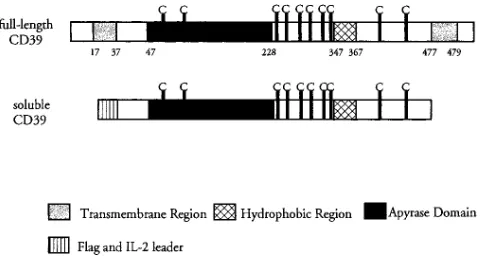
Expression plasmid construction.Using oligonucleotide cassettes and PCR primers, a CD39 sequence was constructed comprising the human coding sequence from Thr 38 to Thr 476. This removed the nucleotides coding for hydrophobic regions at the amino terminus and the carboxyl terminus (7). The remaining CD39 sequence was fused to a sequence coding for the Flag peptide and ligated into the pDC206 vector (14) with an IL-2–derived leader sequence (15), creat-ing pIL-2L-FlagsolCD39. The pDC206 vector allows expression of re-combinant protein in a variety of mammalian systems. The Flag se-quence was flanked by unique restriction enzyme sites, permitting its removal to create pIL-2L-solCD39.
Preparation of conditioned medium from solCD39 transient trans-fectants.Plasmid DNA was transfected into subconfluent layers of COS-1 cells using DEAE dextran followed by chloroquine as previ-ously described (16). Transfected cells were incubated (378C, 5% CO2) in 0.5% FCS-supplemented DMEM-F12 medium in 10-cm2
Petri dishes or 175-cm2 tissue culture flasks. After 5 d (unless
other-wise indicated), conditioned medium (CM) from these cultures was collected, purified of cells and debris by centrifugation, and concen-trated 4–10-fold using a pressurized, stirred cell fitted with a YM-10 (10 kD cutoff) membrane (Amicon Corp., Danvers, MA).
Immunoaffinity depletion of solCD39 from COS-1 CM. CM was collected from COS-1 cells transfected with pIL-2L-FlagsolCD39, which had been cultured for 5 d in DMEM/F12 supplemented with 5% FCS. 100 ml of drained, conjugated AG slurry was added per mil-liliter of CM. CM was subjected to one or two cycles of binding with one of the following: ovalbumin-conjugated AG, M2 mAb-conju-gated AG, or B73 mAb-conjumAb-conju-gated AG. Each cycle involved continu-ous gentle agitation of the slurry for 14 h at 48C followed by centrifu-gation to recover supernatants for a subsequent binding cycle or for ATPDase activity measurements.
Development of a stably transfected cell line secreting solCD39. The solCD39 cDNA insert, containing the recombinant solCD39 se-quence and the IL-2 leader, was excised from the pIL-2L-solCD39 plasmid by XmaI/BglII digestion, then inserted into 2A5Ib, an ex-pression vector optimized for stable Chinese hamster ovary (CHO) cell lines (17). The solCD39-2A5Ib plasmid was transfected into CHO cells using Lipofectamine (GIBCO BRL; Gaithersburg, MD) according to manufacturer’s recommendations. The CHO cell line used in these studies, DX B-11, had been adapted to serum-free sus-pension culture conditions.
Transfected cells were grown in modified DMEM-F12 medium, supplemented with peptone, glutamine, glucose, transferrin, lipids, and IGF-1 (used solely when cultures were induced for protein ex-pression). After 3 d growth, the cells were transferred to selective me-dium lacking hypoxanthine and thymidine. Stock cultures were grown at 378C in suspension, and passaged every 2–3 d. Induction cul-tures were grown at 318C in suspension, with IGF-1 and sodium bu-tyrate (1–2 mM). Cell density at start of induction cultures was 1.5– 2.0 3 106 cells/ml. Average induction culture period was 7 d, at which
time CM was collected for further analyses.
Radioimmunoprecipitation and SDS-PAGE autoradiography. COS-1 cells were transfected with mammalian expression vectors
en-coding cell surface CD39 (pHuCD39) (4), soluble CD39 (pIL-2L-FlagsolCD39), or soluble CD40 ligand (pIL-2L-CD40L) (18) and grown in 5% FCS-supplemented DMEM/F12 medium in 10-cm2
dishes. 2 d after transfection, the medium was replaced with Cys/Met-free medium and cells were incubated for 1 h at 378C. The culture me-dium was replaced with fresh Cys/Met-free meme-dium supplemented with 5 ml of [35S]-Cys/Met (Amersham Corp., Arlington Heights, IL),
and cells were cultured for 5 h at 378C. CM from the metabolically ra-diolabeled cells was collected, purified of cells and debris by centrifu-gation and sterile filtration, and stored at 48C until further use. For radioimmunoprecipitation, 500 ml of 35S-labeled CM was added to
250 ml of 3% BSA in Tris-buffered saline (TBS), pH 7.7, followed by addition of 50 ml of a 80% slurry of mAb-coated AG beads. (In some cases, 35S-labeled CM was incubated with ovalbumin-coated AG
beads to remove nonspecifically binding materials before addition of Ab-coated beads). After incubation for 14 h at 48C, beads were re-moved by centrifugation and washed three times in cold TBS. For SDS-PAGE analysis, 35 ml of fourfold concentrated reducing sample buffer (250 mM Tris/HCl, pH 6.8, 8% [wt/vol] SDS, 40% [vol/vol] glycerol, 20% [vol/vol] 2-mercaptoethanol, 0.05% bromophenol blue dye) was added to each AG pellet, boiled for 5 min, and loaded onto a 8–16% Novex (San Diego, CA) polyacrylamide gel. Gels were elec-trophoresed at 25 mA (19), prepared for autoradiography by soaking in Enhance (NEN Life Science Products, Boston, MA) for 1 h and in H2O for 20 min, followed by vacuum drying at 808C. Gels were
ex-posed to X-omat AR film (Eastman Kodak Co., Rochester, NY) for 2 h, then developed.
Affinity purification of solCD39 protein. 30 ml of 10-fold concen-trated CM from solCD39-secreting CHO cells was added to 3 ml of B73 mAb-coated Sepharose 4B gel slurry and mixed overnight at 48C. The affinity matrix was pelleted by centrifugation, washed three times with PBS, and added to a plastic column. Specifically bound protein was eluted by the addition of 0.1 M triethylamine, pH 11.5. Fractions (1.2 ml each) were collected in tubes containing 120 ml of neutralizing solution (1 M sodium phosphate, monobasic; pH 4.3) and analyzed for protein content by SDS-PAGE, followed by Coomassie blue staining. Biological activity was determined using an ATPase as-say, so that peak fractions could be pooled, buffer exchanged into PBS, and concentrated fivefold. N-linked sugars were removed from purified protein using N-glycanase according to manufacturer’s in-structions (Oxford Glycosystems, Rosedale, NY).
Phosphate release assay for ATPase activity. Samples (z 100 ml of either concentrated CM or purified protein) were combined with 20 ml of 103 assay buffer (200 mM Hepes, 1.2 M NaCl, 50 mM KCl, 15 mM CaCl2, 15 mM MgCl2, and 3 mM ATP) and sterile water was
added to a final volume of 200 ml. Radiolabeled ATP (0.8 mCi g [32P]
ATP; Amersham Corp.) was added and the mixture incubated for 20 min at 378C. Stop mix (0.5 ml of 20% activated charcoal/1 M HCl) was added and the reaction was placed on ice for 10 min. After cen-trifugation (14 g for 10 min), the supernatant was assayed for free 32P
using a MINAXI beta 4000 scintillation counter (Packard, Meriden, CT). Data are expressed as picomoles of ATP degraded per minute.
Radio-TLC assays for ADPase and ATPase activities. ADPase (3) assays were primarily used in determining enzyme kinetics and phar-macokinetics. Test samples were incubated with 50 mM [14C] ADP
(NEN Life Science Products, Boston, MA) in assay buffer (15 mM Tris, 134 mM NaCl, 5 mM glucose, pH 7.4, containing 10 mM Ap5A (P1,P5-di[adenosine-59]pentaphosphate, 1 mM ouabain, 10 mM
dipy-ridamole, and 3 mM CaCl2) in a total volume of 50 ml (5 min, 378C).
Reactions were stopped by placement on ice and addition of 10 ml “stop solution” (160 mM disodium EDTA, pH 7.0, 17 mM ADP, 0.15 M NaCl) to block further metabolism of ADP (3). Nucleotides, nucleosides, and bases were separated by radio-TLC using isobu-tanol/1-pentanol/ethylene glycol monoethyl ether/NH4OH/H2O (90:
expressed as percentage of ADP metabolized or as picomoles ADP metabolized per min. A unit of activity is the quantity of enzyme which will degrade 1 mmol of ADP in 1 min at 378C. Identical assays were performed using ATP as a substrate to examine the kinetics of the ATPase activity of CD39.
Preparation of platelet-rich plasma. After obtaining informed consent from volunteers, blood was collected via plastic tubing using acid citrate-dextrose (38 mM citric acid; 75 mM sodium citrate; 135 mM glucose) as anticoagulant (3, 20). Where indicated, volunteers had in-gested 650 mg acetylsalicylic acid (ASA) 18 h before blood donation. Platelet-rich plasma (PRP) was prepared with an initial whole blood centrifugation (200 g, 15 min, 258C), and a second centrifugation of the PRP (90 g, 10 min) to eliminate residual erythrocytes and leuko-cytes. The stock suspension of PRP was maintained at room tempera-ture under 5% CO2-air.
Platelet aggregation studies. PRP containing 1.22 3 108 platelets
was preincubated (3 min, 378C) in an aggregometer cuvette (Lumiag-gregometer; Chrono-Log, Havertown, PA) alone or in combination with test samples containing solCD39. Total volumes were adjusted to 300 ml with TSG buffer (3, 4). After the 3-min preincubation, platelet agonists (ADP or collagen) were added at the concentrations indicated, and the aggregation response recorded for 4–5 min. Where indicated, 10 mM indomethacin (Sigma Chemical Co., St. Louis, MO) was added to PRP to inhibit endogenous cyclooxygenase activity.
Preparation of FSBA-modified solCD39. Fluorosulfonylbenzoyl-adenosine (FSBA; Sigma Chemical Co.) was used to inactivate the catalytic activity of solCD39 as follows. solCD39 (2 nmol) was com-bined with 2 ml labeling buffer (100 mM Hepes, pH 7.4, 200 mM NaCl, 4% dimethylformamide [vol/vol]), 400 ml 5 mM FSBA (dis-solved in ethanol), and 1.52 ml H2O. A mock-treated sample was also
prepared in which the FSBA solution was replaced with H2O. After
incubating at 378C for 90 min, the samples were centrifuged in a Cen-tricon-10 filter unit (Amicon Corp.) for 1 h at 5,500 rpm and buffer exchanged into PBS to remove unreacted material.
Biochemical analyses. Using the radio-TLC assay system as de-scribed previously (4), the ADPase activity of the membrane-bound HUVEC ecto-ADPase was determined at different pHs in buffers containing 100 mM bis-trispropane (Sigma Chemical Co.). This was compared to the ADPase activity of purified solCD39 at these pHs. Kinetic constants for solCD39 metabolism of ATP and ADP were de-termined by measuring the initial rates of reaction as analyzed in the radio-TLC system. ADP or ATP at 2.5–150 mM were incubated sepa-rately with 2 3 1029 M solCD39.
Pharmacokinetic analysis. Balb/c mice (6–8 wk of age; main-tained under specific pathogen-free conditions; Jackson Laboratory, Bar Harbor, ME) were intravenously injected with 50 mg recombi-nant solCD39 in 100 ml sterile saline (0.9% NaCl). At various times after injection (5, 10, 30 min, 1, 2, 4, 8, 24 h), pairs of mice were bled by cardiac puncture and killed. Serum was prepared from each blood sample and frozen until assay. The presence of biologically active solCD39 in serum samples was measured in ATPase and ADPase as-says. The data were fit using Deltagraph (Deltapoint, Monterey, CA). The best fit was derived using double exponential decay. Where indicated, specificity of enzyme activity was determined by incubating serum samples with anti-CD39 mAb-coated beads to remove solCD39 before testing for ATPase activity.
Results
Construction of a soluble CD39 (solCD39) expression plas-mid. The purpose of our experiments was to generate a solu-ble molecule with the properties of human ADPase, which would function as an agent with antithrombotic properties. To accomplish this, it was necessary to remove the NH2-terminal and COOH-terminal portions of CD39, including the two transmembrane regions (Fig. 1). To allow proper transport of soluble CD39 to the medium, an appropriate leader sequence
providing for secretion was required at the amino terminus of the protein. Thus, the amino terminal transmembrane region was replaced by a leader derived from human proinsulin/IL-2 sequences (15) (IL-2L) and a sequence coding for a small peptide tag, Flag. A stop codon was placed immediately up-stream of the COOH-terminal transmembrane coding region (Fig. 1). This cDNA construct was ligated into the pDC206 mammalian expression plasmid, generating the pIL-2L-FlagsolCD39 vector.
Production of recombinant solCD39 protein. Conditioned media (CM) from cultures of COS-1 cells transfected with pIL-2L-FlagsolCD39 were assayed for the presence of recom-binant solCD39 protein. ATPase activity was detectable in CM from pIL-2L-FlagsolCD39 transfected cells, but not in CM from COS-1 cells transfected with control vector (data not shown). These ATPase levels increased with time in culture over at least 4 d after transfection. To confirm that recombi-nant solCD39 accounted for the ATPase activity observed, CM from COS-1 transfectants were incubated with immunoaf-finity beads before enzyme assay. Immunoprecipitation with anti-CD39 mAb-conjugated beads resulted in removal of
. 80% of ATPase activity from CM. Over 95% was removed with a second antibody adsorption step (Fig. 2). Immunopre-cipitation (two cycles) with anti-Flag mAb-coated beads also resulted in substantial depletion of enzyme activity. Two rounds of preincubation with a control (ovalbumin-conjugated beads) did not remove significant ATPase activity from the su-pernatants (Fig. 2).
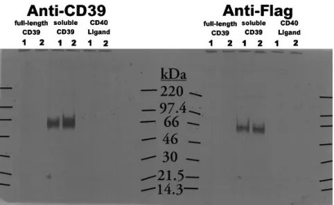
Immunoprecipitation of recombinant solCD39. To charac-terize recombinant solCD39 protein expression, COS-1 cells were transfected with pIL-2L-FlagsolCD39, grown for 2 d, and then cultured for an additional 5 h in medium containing [35 S]-cysteine/methionine in order to label newly synthesized pro-teins. CM was collected and incubated with CD39 or anti-Flag mAb-coated beads. Bound proteins were analyzed by SDS-PAGE and autoradiography (Fig. 3). IL-2L-FlagsolCD39 transfectants secreted a radiolabeled protein of z 66 kD that was recognized by CD39. This was not present in
[image:4.612.316.556.58.188.2]CD39 immunoprecipitated CM from COS-1 cells transfected with a vector encoding full-length CD39 (including NH2 -termi-nal and COOH-termi-termi-nal hydrophobic regions), or with a vec-tor encoding a secreted protein, CD40 ligand. Anti-Flag mAb immunoprecipitated a similar-sized band from CM of the pIL-2L FlagsolCD39 transfectant, consistent with the presence of the Flag peptide in recombinant solCD39 (Fig. 3). Preclearing radiolabeled culture supernatants with antiovalbumin-coated beads failed to remove the 66-kD band, indicating that binding to anti-CD39 and anti-Flag was specific. Beads coated with an
irrelevant, isotype matched control antibody failed to immu-noprecipitate the 66-kD band from solCD39 containing CM (data not shown).
Affinity purification of solCD39 from stably transfected CHO cells. A CHO cell line expressing solCD39 was gener-ated to improve recombinant solCD39 protein production. Dihydrofolate reductase (DHFR)-deficient CHO cells were transfected with an expression plasmid, consisting of the 2A5Ib vector backbone, containing the DHFR gene (17), into which IL-2L-solCD39 was inserted without the Flag sequence. After growth in selective medium, CM from CHO cell cultures was analyzed for ATPase and ADPase activities. Compared to CM from transfected COS-1 cells, the stably transfected CHO cells secreted 20-fold higher levels of both enzyme activities, consistent with the presence of solCD39 (Table I).
CM from the stably transfected CHO cells was affinity pu-rified and analyzed by SDS-PAGE for the presence of recom-binant solCD39 (see Methods). A prominent band of z 66 kD was present in early eluted fractions, with a peak of Coomassie Blue staining material around fraction 5 (Fig. 4 A). Over 90% of the protein present was found as this major band, although overloading the polyacrylamide gel did reveal some smaller molecular weight contaminants. ATPDase activity of the affin-ity column fractions correlated with the intensaffin-ity of protein bands on SDS-PAGE (Fig. 4, A and B). ATPDase activity was barely detectable in the anti-CD39 column flowthrough, indi-cating that affinity purification is an effective means of isolat-ing biologically active recombinant solCD39. Treatment of the purified protein with N-glycanase to remove NH2-linked oli-gosaccharides caused the broad band of protein at 66 kD to re-solve into a much tighter band of protein at z 52 kD, the pre-dicted peptide size for solCD39 (Fig. 4 C).
Quantitative amino acid analysis of peak fractions (3–9) from the affinity column yielded a ratio of amino acid residues consistent with calculated values for solCD39. Protein se-quencing verified that the amino terminus of the isolated pro-tein contained the CD39 sequence (data not shown). Total protein yield from 1 liter of CHO-solCD39 CM was z 2 mg.
Biochemical properties of affinity-purified solCD39. The pH
optima for the ecto-ADPase on the surface of HUVEC and for affinity-purified recombinant solCD39 ADPase activities were between pH 8 and 8.5 (Fig. 5 A). This indicated that re-combinant solCD39 would be maximally active under the same physiological conditions as native CD39/ecto-ADPase.
[image:5.612.59.298.59.189.2]Initial rates of ATP and ADP metabolism by recombinant solCD39 were determined (Fig. 5 B), and kinetic constants de-rived. The Km and Vmax for ADP were 5.9 mM and 72 pmol/ min, respectively; for ATP a Km of 2.1 mM and Vmax of 26
Figure 2. Identification of COS-1 CM-derived ATPase as solCD39. 5-d CM from COS-1 cells transfected with the pIL-7L-FlagsolCD39 plasmid was incubated with Affigel beads conjugated with either chicken ovalbumin, anti-Flag mAb, or anti-CD39 mAb (see Methods). Beads and CM were separated by low speed centrifugation. Follow-ing one (1X) or two (2X) rounds of adsorption, samples were assayed by the radioactive phosphate release assay for ATPase activity. Data are expressed as picomoles of ATP degraded per min.
Figure 3. Immunoprecipitation of solCD39 from COS-1 CM. COS-1 cells transfected with plasmids for expression of full-length human CD39, solCD39 2L-FlagsolCD39), or CD40 ligand (pIL-2LCD40L) were metabolically labeled by culturing for 5 h in [35
[image:5.612.314.556.83.152.2]S]-Cys/Met–containing medium. CM from the cultures was incubated in the presence of immunoaffinity beads coated with either anti-CD39 mAb or anti-Flag mAb. Material specifically bound to the antibody-coated beads was released and subjected to SDS-PAGE and autora-diography as described in Methods (lane 2). Where indicated, some of the material was preincubated with ovalbumin-coated beads to re-move nonspecifically bound material, then treated as described for lane 2 of each set (lane 1). Migration of molecular weight standards is indicated in kilodaltons.
Table I. Comparison of ADPase and ATPase Activities in CM Containing solCD39
Cell type ADPase ATPase
pmol/min/per ml pmol/min/per ml
solCD39 (CHO) 1403 970
solCD39 (COS-1) 70 44
Radio-TLC assays were performed on CM (see Methods). Results are
[image:5.612.57.298.452.600.2]pmol/min were determined. The assays were performed with 2 3 1029 M solCD39, yielding k
cat of 720 min21 (ADP) and 260 min21 (ATP). Thus, the specificity constant k
cat/Km (1.2 3 108 min21 M21) was identical for ADP and ATP. The specific activity for purified recombinant solCD39 was 11 U/mg for ADP and 4 U/mg for ATP.
Platelet inhibitory properties of solCD39. The
aforemen-tioned biochemical properties of recombinant affinity purified solCD39 suggested that it might be effective as an inhibitor of platelet reactivity. As shown in Fig. 6, this was indeed the case. Whereas addition of 10 mM ADP to PRP alone resulted in a
[image:6.612.58.334.51.640.2]Figure 4. Immunoaffinity purification and character-ization of solCD39. 5-d CM from CHO cells stably transfected with the solCD39 expression plasmid was concentrated and incubated overnight with anti-CD39 immunoaffinity beads. After extensive wash-ing, specifically bound protein was eluted from the beads with triethanolamine, pH 11.5 and immediately neutralized in 1 M sodium phosphate, monobasic; pH 4.3. Fractions from the immunoaffinity column were analyzed for (A) protein content by SDS-PAGE, and (B) enzyme activity by the radioactive phosphate re-lease assay (see Methods). (C) Purified solCD39 (lane 1) was treated with N-glycanase (see Methods) for 18 h (lane 2). The protein band at z 35 kD in lane 2 corresponds to the N-glycanase used in the reaction.
[image:6.612.315.506.483.661.2]Figure 5. Biochemical characteristics of solCD39. (A) pH optimum profiles of HUVEC membrane ecto-ADPase and recombinant solCD39. HUVEC membrane ecto-ADPase (4) and purified solCD39 from CHO cell CM were assayed for ADPase activity in bis-tris propane buffer at the indicated pH (see Methods). (B) Eadie-Hofstee plot of rates of metabolism at different concentrations of ATP or ADP using purified solCD39 (6.5 ng). Increasing quantities of substrates were reacted with solCD39, and ADPase activity mea-sured by radio-TLC (see Methods).
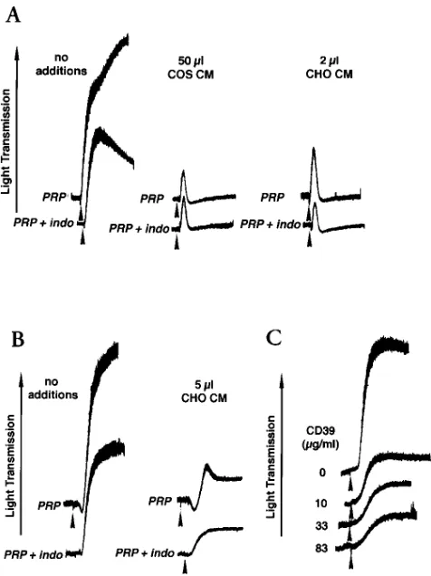
full, irreversible aggregation response, partially reversible ag-gregation occurred at lower ADP concentrations. However, in the presence of only 3.3 mg/ml solCD39, platelet aggregation induced by 10 mM ADP was abruptly terminated and the curve rapidly returned to baseline (Fig. 6). Importantly, the ex-tent of aggregation was reduced to levels below those ob-served with 1 mM ADP. Higher concentrations of solCD39 had an even more profound inhibitory effect, virtually elimi-nating the initial burst of aggregation elicited by 10 mM ADP. Thus, these results indicate that purified solCD39 is indeed an effective inhibitor of platelet aggregation in vitro.
Platelet responsiveness to 5 mM ADP was examined in PRP treated with and without the cyclooxygenase inhibitor in-domethacin (10 mM), in the presence of CM containing solCD39 from either COS-1 and CHO cells (Fig. 7 A). In-domethacin treatment resulted in partial reversal of ADP-induced platelet aggregation in the absence of solCD39. In contrast, CM containing solCD39 were capable of completely abrogating platelet responses to ADP, whether PRP was in-domethacin treated or not.
Inhibition of platelet reactivity by solCD39 was not limited to blocking the agonistic effects of ADP. Collagen, another critical platelet agonist, was used at 1 mg/ml to induce platelet aggregation. The presence of solCD39 markedly reduced the response to collagen as compared to control (Fig. 7 B, upper
curves). A similar inhibitory effect of solCD39 was observed in
PRP treated with indomethacin (Fig. 7 B, lower curves), when collagen was used at 3.3 mg/ml. The effect of solCD39 on col-lagen-induced aggregation was dose dependent (Fig. 7 C).
Inactivation of enzymatic activity of solCD39 and the effect on inhibition of platelet activation. To demonstrate that the
[image:7.612.317.535.55.422.2]ability of solCD39 to inhibit platelet activation was due to the enzymatic activity of solCD39 and not to some other property,
[image:7.612.58.296.313.631.2]Figure 7. Comparison of platelet reactivity as modulated by different agonists and inhibitors. The effects of CM from COS-1 or CHO cells expressing solCD39 on platelet aggregation induced by (A) ADP (5 mM) and (B) collagen were compared in PRP, and PRP treated with 10 mM indomethacin (a cyclooxygenase inhibitor). In B, 1 mg/ml collagen was used in the upper samples, and 3.3 mg/ml in the lower (indo-methacin-treated) samples. (C) The inhibition of collagen-induced aggregation by increasing quantities of solCD39 in PRP from a donor who had ingested aspirin. Arrows indicate addition of agonist. Data are presented as relative light transmission vs time (4 min).
the molecule was reacted with FSBA, an ATP analogue that inhibits collagen-induced platelet activation (21) and binds ir-reversibly with ATPDases found on several cell types (22, 23). Induction of platelet activation by ADP (Fig. 8 A) or collagen (Fig. 8 B) was significantly inhibited by either purified solCD39 or mock FSBA-treated solCD39. In contrast, incuba-tion with FSBA-treated solCD39 did not have a significant effect on platelet activation. A comparative titration of mock-treated solCD39 versus FSBA-mock-treated solCD39 (Fig. 8 C) indi-cated that 22.0 mg/ml of FSBA-treated solCD39 gave a similar aggregation profile as 0.88 mg/ml of mock-treated solCD39. This indicated that 96% of the aggregation inhibitory activity of solCD39 was lost after FSBA derivitization. Analyses of re-sidual ADPase activity of FSBA-treated solCD39 by the ra-dio-TLC assay system demonstrated that z 94% of the enzy-matic activity was blocked, while the phosphate release assay indicated that a similar percentage of the ATPase activity was lost as well (data not shown).
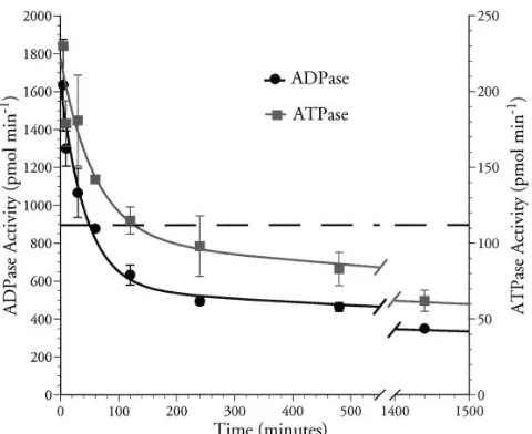
Persistence of solCD39 after in vivo administration. Mice
were injected with solCD39 (50 mg) intravenously and bled at specified time intervals thereafter. Serum samples were exam-ined for both ATPase and ADPase activities. The data ob-tained best fit a biphasic exponential curve. The amount of ATPase activity from 25 mg/ml of solCD39 placed in murine serum is presented for comparison. The t1/2a (distribution phase) was calculated to be 59 min in the ATPase assay and 43 min in the ADPase assay (Fig. 9). Approximately 55–65% of apyrase activity was cleared from the circulation during this
phase. The elimination phase had a t1/2b of z 40 h in both as-says. Preclearing the 10 min, 2, and 24 h time point samples with anti-CD39 mAb-coated beads completely eliminated se-rum ATPase/ADPase activities (data not shown). These data also demonstrated that the assays specifically detected recom-binant human solCD39.
Discussion
Excessive platelet activation is a contributing factor to several clinical disorders, including myocardial infarction, restenosis after angioplasty or bypass surgery, and stroke. Thus, it follows that therapeutic strategies wherein platelet reactivity is phar-macologically neutralized (24, 25) would be clinically benefi-cial. This creates an impetus to expand development of poten-tial therapeutic agents which show promise in vitro. In vitro studies demonstrated that ADP secreted from stimulated platelets is rapidly metabolized by endothelial ecto-ADPase/ CD39, and thereby becomes unavailable for promotion of fur-ther platelet activation and recruitment (3, 4, 11). This indi-cated that CD39 might be useful as an antithrombotic agent. For in vivo evaluation of ecto-ADPase/CD39, a recombinant soluble form was deemed necessary. Research presented in this paper describes a novel soluble, recombinant ADPase/ CD39, solCD39, which is capable of returning stimulated platelets to the resting state, confirming its potential as an anti-thrombotic agent.
CD39 contains two putative transmembrane regions, near the amino and carboxyl termini, respectively, which may serve to anchor the native protein in the cell membrane (Fig. 1) (7). Modeling studies, antibody epitope analyses, and sequence ho-mology have shown that the portion of the molecule between the transmembrane regions is external to the cell (7), and con-tains the four ACRs characteristic of members of the apyrase family (6, 7). This suggested that the external portion of CD39 is critical for its ecto-ADPase activity. The extracellular do-main, which encodes a 439 amino acid polypeptide, was iso-lated using oligonucleotide cassettes and PCR and placed in a mammalian expression vector. Secretion of the recombinant molecule was ensured by addition of a mammalian leader se-quence. After transfection with solCD39-encoding plasmid, COS-1 cells secreted readily detectable levels of ATPase activ-ity (Fig. 2). This activactiv-ity was solCD39 specific, since: (a) cells transfected with unrelated plasmids failed to secrete nucleoti-dase activity, and (b) solCD39 CM was depleted of apyrase ac-tivity by specific immunoadsorption (Fig. 3). The molecular size of solCD39 immunoprecipitated from COS-1 CM (z 66 kD, Fig. 4 C) was larger than the predicted size of 52 kD. However, incubation of the purified protein with N-glycanase to remove
N-linked oligosaccharides yielded protein with the appropriate
molecular weight.
To increase protein production, a modified CHO-based solCD39 expression system was used. This could be main-tained as a stably expressing line, and could also be grown in a defined, serum-free medium to facilitate protein purification. CM from these CHO cells contained 20-fold more ATPase and ADPase activity than that from COS cells (Table I). Affin-ity purification (see Methods) resulted in yields of 1–3 mg solCD39/liter CM, which remained biologically active upon extended storage at 48C (data not shown).
The pH optimum of solCD39 ADPase activity was
be-Figure 9. Pharmacokinetic analysis of solCD39 in mice. SolCD39 (50
mg in 100 ml sterile saline) was injected intravenously into Balb/c mice. At various time points (5 min to 24 h after injection), mice were bled by cardiac puncture and serum was prepared. ATPDase in se-rum was measured in the radioactive phosphate release ATPase as-say (j) or the ADPase assay (d) (see Methods). Both activities are expressed as picomoles nucleotide degraded per minute. The dashed line, which indicates the ATPase activity of 25 mg/ml of solCD39 in murine serum, is provided as a comparison. The data were fit to a bi-phasic curve (see Methods) resulting in z 65% of the activity being present in the distribution phase (t1/2a5 59 min [ATP]; 43 min
[image:8.612.58.298.402.598.2]tween 8 and 8.5, similar to that of HUVEC membrane-bound ecto-ADPase (4), and other reported ATPDases (13, 26). The specific activities of solCD39 (4 U/mg for ATP and 11 U/mg for ADP) are of the same order as those seen for other mam-malian ecto-apyrases (13, 27). Care must be taken when com-paring these values, however, as there are many conditions used in the purification process that could affect the specific activity of solCD39. In fact, recent modifications in the purifi-cation protocols have increased the specific activity two- to fivefold (data not shown).
It is important to examine the kinetic constants for solCD39 in some detail. The Km reveals the concentration of substrate at which the enzymatic degradation delineated by kcat is most efficient. The Km of solCD39 for its substrates (5.9 mM for ADP, 2.1 mM for ATP) is quite low, similar to that of other mammalian ecto-ATPDases. For example, an ATPDase from bovine lung had a Km for ATP of 7 mM (28), and an ATPDase from bovine spleen had a Km for ADP of 9 mM (26). A re-cently reported ATPDase, purified from bovine heart and hav-ing antigenic similarities to CD39, has been found to have a Km for ADP of 29 mM (27). Importantly, the Km for ADP of solCD39 is in the range at which ADP exerts its biological ac-tivity on platelets, as ADP and ATP concentrations present in serum after platelet activation are 3–5 mM (29, 30). Thus solCD39 should degrade ATP and ADP very efficiently at bio-logically relevant concentrations of these nucleotides, with vir-tually every molecule of solCD39 participating in the reaction. The potato apyrase, which has a higher kcat than solCD39 for ATP, also has a significantly higher Km, thus resulting in a kcat/
Km with a similar magnitude as solCD39 (6). Potato apyrase, while capable of degrading bound nucleotide faster than solCD39, will, at biologically relevant concentrations, have far fewer protein molecules participating in the reaction. The sim-ilarities in kcat/Km for both solCD39 and potato apyrase suggest that, at the low concentrations of extracellular ADP observed after platelet activation, equal concentrations of solCD39 and potato apyrase will have comparable rates of nucleotide degra-dation.
Examination of the specificity constants (kcat/Km) of solCD39 for the two substrates shows no preference for one over the other. As a consequence, when ATP and ADP are both present, the ratio of degradation rates (nADP/nATP) will al-ways equal the ratio of substrate concentrations ([ADP]/ [ATP]). This may have important implications not only for thrombosis, but for other settings as well. For example, CD39 is expressed on alloactivated cytotoxic T lymphocytes (CTL) (31), and an ecto-ATPase activity on CTL is required for effec-tor functions (32). Inhibition of ATPase activity blocks secre-tion of relevant cytokines, such as TNF-a and IFN-g, as well as cell-mediated killing. Incubation of PMN with ATP and ADP inhibits the biological activities of these cells (33). Interest-ingly, this inhibition is due to accumulation of AMP. Investiga-tions that the role of CD39 plays in this and other immunolog-ically relevant processes may yield valuable insights.
Significant quantities of recombinant solCD39 can now be prepared in order to investigate details of its biological activity in vitro and in vivo. As depicted in Figs. 6–8, solCD39 strongly inhibits platelet aggregation and recruitment in an aggregome-try system, an in vitro model of thrombosis. In the absence of solCD39, addition of ADP or collagen to PRP results in rapid, irreversible aggregation (Figs. 6 and 7). Results with collagen are of particular interest. This strong platelet agonist promotes
platelet aggregation via pathways independent of release of ADP and other dense body constituents from platelets (34). As seen in Fig. 7, B and C, solCD39 inhibition of collagen-induced platelet aggregation was very pronounced. This could indicate that ADP contributes more to collagen-induced ag-gregation than previously appreciated (35, 36).
Our data indicate that solCD39 leads to more effective blockade of platelet responsiveness than cyclooxygenase inhi-bition (Fig. 7, A and B). Cyclooxygenase inhibitors, such as as-pirin or indomethacin, reduced the extent of platelet aggrega-tion, but disaggregation did not occur, even over a period of several minutes. In contrast, solCD39 significantly decreased platelet aggregation to collagen (Fig. 7 B), and completely in-hibited the platelet response to 10 mM ADP (Fig. 7 A). Inhibi-tion of platelet reactivity by solCD39 is independent of cy-clooxygenase inactivation. This indicates that aspirin and solCD39 could be utilized concurrently as therapeutic modali-ties.
To verify that the inhibition of collagen-induced platelet activation by solCD39 was due to its enzymatic activity and not due to another aspect of the molecule (e.g., binding to reactive sites on collagen or on platelets), solCD39 was reacted with a substrate analogue, FSBA. This molecule binds near the active site and prevents further catalysis (37). After treatment, the molecule could no longer metabolize ADP efficiently, having lost 94% of its activity. The loss in catalytic activity paralleled the inability of the material to inhibit platelet activation after addition of ADP or collagen (Fig. 8). Mock-treated solCD39 continued to inhibit platelet reactivity. Therefore, the ability of solCD39 to inhibit platelet aggregation is due solely to its en-zymatic activity.
In vivo analyses in mice demonstrated that intravenously administered solCD39 remained active in the circulation for extended periods of time and was not inactivated during serum preparation (Fig. 9). Two different assays, examining both ATPase and ADPase activities of solCD39, produced very similar results. The initial reduction in solCD39 activity (distri-bution phase) was followed by a considerably slower clearance phase. A comparison between the ATPase activity of a known amount of solCD39 placed into murine serum (Fig. 9, dashed
line) with the activity seen in intravenously administered
solCD39, coupled with the half-life of almost 2 d seen during the clearance phase, indicates that potentially therapeutic lev-els of solCD39 may be sustainable for long periods. This would obviate the need for complicated administration protocols such as continuous infusion. Furthermore, it is noteworthy that mice treated with 50 mg of solCD39 displayed no overt exter-nal difficulties over the observation period. Experiments are currently under way to examine the biological effects of solCD39 in relevant animal models of vascular diseases.
Acknowledgments
The authors wish to thank Norman Boiani for technical support; Drs. Doug Williams, Carl March, and Ray Paxton for critical review of the manuscript; and Christine Jones for editorial assistance.
This work was supported in part by a Merit Review grant from the Department of Veterans Affairs and by National Institutes of Health grants HL-47073, HL-46403, and HL-07423.
References
1. Marcus, A.J. 1996. Platelets and their disorders. In Disorders of Hemo-stasis. O.D. Ratnoff and C.D. Forbes, editors. W.B. Saunders, Philadelphia. 79– 137.
2. Marcus, A.J., and L.B. Safier. 1993. Thromboregulation: multicellular modulation of platelet reactivity in hemostasis and thrombosis. FASEB J. 7: 516–522.
3. Marcus, A.J., L.B. Safier, K.A. Hajjar, H.L. Ullman, N. Islam, M.J. Broekman, and A.M. Eiroa. 1991. Inhibition of platelet function by an aspirin-insensitive endothelial cell ADPase. Thromboregulation by endothelial cells. J.
Clin. Invest. 88:1690–1696.
4. Marcus, A.J., M.J. Broekman, J.H.F. Drosopoulos, N. Islam, T.N. Aly-onycheva, L.B. Safier, K.A. Hajjar, D.N. Posnett, M.A. Schoenborn, K.A. Schooley, R.B. Gayle, III, and C.R. Maliszewski. 1997. The endothelial cell ecto-ADPase responsible for inhibition of platelet function is CD39. J. Clin.
In-vest. 99:1351–1360.
5. Plesner, L. 1995. Ecto-ATPases: identities and functions. Int. Rev. Cytol. 158:141–214.
6. Handa, M., and G. Guidotti. 1996. Purification and cloning of a soluble ATP-diphosphohydrolase (apyrase) from potato tubers (Solanum tuberosum).
Biochem. Biophys. Res. Commun. 218:916–923.
7. Maliszewski, C.R., G.J. Delespesse, M.A. Schoenborn, R.J. Armitage, W.C. Fanslow, T. Nakajima, E. Baker, G.R. Sutherland, K. Poindexter, C. Birks, et al. 1994. The CD39 lymphoid cell activation antigen. Molecular clon-ing and structural characterization. J. Immunol. 153:3574–3583.
8. Rowe, M., J.E. Hildreth, A.B. Rickinson, and M.A. Epstein. 1982. Mono-clonal antibodies to Epstein-Barr virus-induced, transformation-associated cell surface antigens: binding patterns and effect upon virus-specific T-cell cytotox-icity. Int. J. Cancer. 29:373–381.
9. Ling, N.R., D. Hardie, J. Lowe, G.D. Johnson, M. Khan, and I.C. Mac-Lennan. 1989. A phenotypic study of cells from Burkitt lymphoma and EBV-B-lymphoblastoid lines and their relationship to cells in normal lymphoid tis-sues. Int. J. Cancer. 43:112–118.
10. Kansas, G.S., G.S. Wood, and T.F. Tedder. 1991. Expression, distribu-tion, and biochemistry of human CD39. Role in activation-associated homo-typic adhesion of lymphocytes. J. Immunol. 146:2235–2244.
11. Kaczmarek, E., K. Koziak, J. Sévigny, J.B. Siegel, J. Anrather, A.R. Beaudoin, F.H. Bach, and S.C. Robson. 1996. Identification and characteriza-tion of CD39 vascular ATP diphosphohydrolase. J. Biol. Chem. 271:33116– 33122.
12. Wang, T.F., and G. Guidotti. 1996. CD39 is an ecto-(Ca21,Mg21 )-apy-rase. J. Biol. Chem. 271:9898–9901.
13. Christoforidis, S., T. Papamarcaki, D. Galaris, R. Kellner, and O. Tso-las. 1995. Purification and properties of human placental ATP diphosphohydro-lase. Eur. J. Biochem. 234:66–74.
14. Kozlosky, C.J., E. Maraskovsky, J.T. McGrew, T. VandenBos, M. Teepe, S.D. Lyman, S. Srinivasan, F.A. Fletcher, R.B. Gayle, III, and D.P. Cer-retti. 1995. Ligands for the receptor tyrosine kinases hek and elk: isolation of cDNAs encoding a family of proteins. Oncogene. 10:299–306.
15. Cullen, B.R. 1988. Expression of a cloned human interleukin-2 cDNA is enhanced by the substitution of a heterologous mRNA leader region. DNA (NY). 7:645–650.
16. Cosman, D., D.P. Cerretti, A. Larsen, L. Park, C. March, S. Dower, S. Gillis, and D. Urdal. 1984. Cloning, sequence and expression of human inter-leukin-2 receptor. Nature. 312:768–771.
17. Morris, A.E., C. Lee, K. Hodges, T.L. Aldrich, C. Krantz, P.S. Smidt, and J.N. Thomas. 1997. Expression augmenting sequence element (EASE) iso-lated from chinese hamster ovary cells. In Animal Cell Technology: from Vac-cines to Genetic Medicine. M.J.T. Carrondo, B. Griffiths, and J.L.P. Moreira, editors. Kluwer Academic Publishers, Boston. 529–534.
18. Spriggs, M.K., R.J. Armitage, L. Strockbine, K.N. Clifford, B.M. Macduff, T.A. Sato, C.R. Maliszewski, and W.C. Fanslow. 1992. Recombinant human CD40 ligand stimulates B cell proliferation and immunoglobulin E se-cretion. J. Exp. Med. 176:1543–1550.
19. Laemmli, U.K. 1970. Cleavage of structural proteins during the assem-bly of the head of bacteriophage T4. Nature. 227:680–685.
20. Marcus, A.J. 1990. Eicosanoid interactions between platelets, endothe-lial cells, and neutrophils. Methods Enzmol. 187:585–598.
21. Colman, R.W., W.R. Figures, L.M. Scearce, A.M. Strimpler, F.X. Zhou, and A.K. Rao. 1986. Inhibition of collagen-induced platelet activation by 59-p-fluorosulfonylbenzoyl adenosine: evidence for an adenosine diphosphate requirement and synergistic influence of prostaglandin endoperoxides. Blood. 68:565–570.
22. Sevigny, J., F.P. Levesque, G. Grondin, and A.R. Beaudoin. 1997. Puri-fication of the blood vessel ATP diphosphohydrolase, identiPuri-fication and locali-sation by immunological techniques. Biochim. Biophys. Acta. 1334:73–88.
23. Sevigny, J., Y.P. Cote, and A.R. Beaudoin. 1995. Purification of pan-creas type-I ATP diphosphohydrolase and identification by affinity labelling with the 59-p-fluorosulphonylbenzoyladenosine ATP analogue. Biochem. J. 312:351–356.
24. Coller, B.S. 1997. Platelet GPIIb/IIIa antagonists: the first anti-integrin receptor therapeutics. J. Clin. Invest. 99:1467–1471.
25. Hennekens, C.H., C.M. Albert, S.L. Godfried, J.M. Gaziano, and J.E. Buring. 1996. Adjunctive drug therapy of myocardial infarction—Evidence from clinical trials. N. Engl. J. Med. 335:1660–1667.
26. Moodie, F.D.L., H. Baum, P.J. Butterworth, and T.J. Peters. 1991. Puri-fication and characterisation of bovine spleen ADPase. Eur. J. Biochem. 202: 1209–1215.
27. Beaudoin, A.R., J. Sevigny, G. Grondin, S. Daoud, and F.P. Levesque. 1997. Purification, characterization and localization of two ATPdiphosphohy-drolase isoforms in bovine heart. Am. J. Physiol. 273:H673–H681.
28. Picher, M., Y.P. Côté, R. Béliveau, M. Potier, and A.R. Beaudoin. 1993. Demonstration of a novel type of ATP-diphosphohydrolase (EC 3.6.1.5) in the bovine lung. J. Biol. Chem. 268:4699–4703.
29. Holmsen, H., and C.A. Dangelmaier. 1989. Measurement of secretion of adenine nucleotides. Methods Enzymol. 169:195–205.
30. McGarrity, S.T., A.H. Stephenson, T.M. Hyers, and R.O. Webster. 1988. Inhibition of neutrophil superoxide anion generation by platelet products: role of adenine nucleotides. J. Leukocyte Biol. 44:411–421.
31. Gouttefangeas, C., I. Mansur, M. Schmid, H. Dastot, C. Gelin, G. Ma-houy, L. Boumsell, and A. Bensussan. 1992. The CD39 molecule defines dis-tinct cytotoxic subsets within alloactivated human CD8-positive cells. Eur. J.
Immunol. 22:2681–2685.
32. Dombrowski, K.E., Y. Ke, L.F. Thompson, and J.A. Kapp. 1995. Anti-gen recognition by CTL is dependent upon ectoATPase activity. J. Immunol. 154:6227–6237.
33. Aziz, K.A., J.C. Cawley, A.T. Treweeke, and M. Zuzel. 1997. Sequential potentiation and inhibition of PMN reactivity by maximally stimulated plate-lets. J. Leukocyte Biol. 61:322–328.
34. Holmsen, H. 1994. Platelet secretion and energy metabolism. In Hemo-stasis and Thrombosis: Basic Principles and Clinical Practice. R.W. Colman, J. Hirsh, V.J. Marder, and E.W. Salzman, editors. J.B. Lippincott Co., Philadel-phia. 524–545.
35. Bressler, N.M., M.J. Broekman, and A.J. Marcus. 1979. Concurrent studies of oxygen consumption and aggregation in stimulated human platelets.
Blood. 53:167–178.
36. Packham, M.A., R.L. Kinlough-Rathbone, H.-J. Reimers, S. Scott, and J.F. Mustard. 1977. Mechanisms of platelet aggregation independent of adeno-sine diphosphate. In Prostaglandins in Hematology. M.J. Silver, J.B. Smith, and J.J. Kocsis, editors. Spectrum Publications, New York. 247–276.