2561
Introduction
Destruction of intracellular proteins at the 26S proteasome is a finely regulated process that determines the half-life of most key cell regulators (Hershko and Ciechanover, 1998). The proteasome is a 25 MDa multi-subunit protease complex that recognizes and degrades proteins that have been tagged with a poly-ubiquitin chain (Voges et al., 1999). The poly-ubiquitin chain is synthesized on the substrate that must be degraded by the sequential activities of three enzymes: an ubiquitin-activating enzyme (E1), an ubiquitin-conjugating enzyme (E2) and an E3 ubiquitin ligase. It is now widely accepted that specificity of the ubiquitination process relies on the selective recognition of substrates by specific E3 enzymes (Pickart, 2001).
SCF complexes are a family of E3 ubiquitin ligases that have been reported to target different signaling molecules and cell cycle regulators (Deshaies, 1999; Zheng et al., 2002). They are composed of three relatively constant polypeptides – Skp1, Cullin1/3 and a Ring finger domain protein, Roc1/Rbx1 (Kamura et al., 1999; Ohta et al., 1999) – and one variable component – the F-box protein – that is specific for a particular substrate or small group of substrate proteins (Patton et al., 1998; Winston et al., 1999). The Drosophila F-box protein
Slimb (Slmb) fulfills several functions in development and cell physiology: it participates in E2f destruction at the beginning of the S phase of the cell cycle (Heriche et al., 2003); it is necessary for normal circadian rythmicity by targeting the clock protein Period (Grima et al., 2002; Ko et al., 2002); it plays a role in limiting centrosome duplication (Wojcik et al., 2000) by destroying the inhibitor of the Anaphase Promoting Complex Emi1 (Margottin-Goguet et al., 2003); it represses the immune response through downregulation of the transcription factor Relish (Khush et al., 2002); and it participates in imaginal wing, limb and eye development through the modulation of Wingless, Hedgehog and Dpp/TGF-βpathways (Jiang and Struhl, 1998; Miletich and Limbourg-Bouchon, 2000; Ou et al., 2002; Theodosiou et al., 1998). In order to assess new functions of the Slmb-containing SCF complex in Drosophila development, we have begun to study the role of Slmb during oogenesis.
The Drosophila ovary is made up of 16-20 chains of egg chambers of progressive age, called ovarioles. Egg chambers are formed at the anterior end of each ovariole in a structure called germarium that harbors both germline and somatic stem cells (Margolis and Spradling, 1995; Wieschaus and Szabad, 1979). The germarium can be divided into three regions. In Substrate-specific degradation of proteins by the
ubiquitin-proteasome pathway is a precise mechanism that controls the abundance of key cell regulators. SCF complexes are a family of E3 ubiquitin ligases that target specific proteins for destruction at the 26S-proteasome. These complexes are composed of three constant polypeptides – Skp1, Cullin1/3 and Roc1/Rbx1 – and a fourth variable adapter, the F-box protein. Slimb (Slmb) is a Drosophila F-Box protein that fulfills several roles in development and cell physiology. We analyzed its participation in egg chamber development and found that slmbis required in both the follicle cells and the germline at different stages of oogenesis. We observed that in slmb somatic clones, morphogenesis of the germarium and encapsulation of the cyst were altered, giving rise to egg chambers with extra germline cells and two oocytes. Furthermore, in slmb somatic clones, we observed ectopic Fasciclin 3 expression, suggesting a delay in follicle cell differentiation, which correlated with the occurrence of ectopic polar cells, lack of interfollicular stalks and
mislocalization of the oocyte. Later in oogenesis, Slmb was required in somatic cells to specify the position, size and morphology of dorsal appendages. Mild overactivation of the Dpp pathway caused similar phenotypes that could be antagonized by simultaneous overexpression of Slmb, suggesting that Slmb might normally downregulate the Dpp pathway in follicle cells. Indeed, ectopic expression of a dad-LacZenhancer trap revealed that the Dpp pathway was upregulated in slmbsomatic clones and, consistent with this, ectopic accumulation of the co-Smad protein, Medea, was recorded. By analyzing slmbgermline clones, we found that loss of Slmb provoked a reduction in E2f2 and Dp levels, which correlated with misregulation of mitotic cycles during cyst formation, abnormal nurse cell endoreplication and impairment of dumping of the nurse cell content into the oocyte.
Key words: Drosophila, Slmb, Oogenesis, Egg chamber, Eggshell
Summary
Multiple roles of the F-box protein Slimb in Drosophila egg chamber
development
Mariana Muzzopappa and Pablo Wappner*
Instituto Leloir and IIB, FCEyN-Universidad de Buenos Aires, Patricias Argentinas 435, Buenos Aires, 1405, Argentina
*Author for correspondence (e-mail: [email protected])
Accepted 29 March 2005
Development 132, 2561-2571
Published by The Company of Biologists 2005 doi:10.1242/dev.01839
Research article
De
region I, at the anterior part, a germline stem cell divides asymmetrically producing one daughter stem cell and one cystoblast. The cystoblast undergoes four rounds of mitosis with incomplete cytokinesis, giving rise to a cyst formed by 16 germ cells interconnected by ring canals (de Cuevas et al., 1997; Spradling, 1993). At germarium region IIa, the two germ cells with four ring canals enter meiosis and become pro-oocytes; afterwards, one of them is selected to become the oocyte while the other 15 cells develop as nurse cells. In region IIb, the cyst is contacted posteriorly and surrounded by somatically originated follicle cells (FC), thus acquiring a lens shape. Later on, in region III, the cyst re-shapes into a sphere, giving rise to an egg chamber that buds off from the germarium covered by a monolayer of FC (van Eeden and St Johnston, 1999). At this stage, the oocyte localizes to the posterior of the cyst, attaching through specific interactions to posterior FC of the egg chamber (Godt and Tepass, 1998; Gonzalez-Reyes and St Johnston, 1998).
As the egg chamber leaves the germarium, three types of FC have differentiated: stalk cells, polar cells and cuboidal FC (Dobens and Raftery, 2000; Torres et al., 2003). Stalk cells are a subtype of five to eight FC that separate adjacent egg chambers; polar cells are two pairs of FC located at the anterior and posterior termini of the follicle that induce a terminal cell fate on their neighboring FC (Beccari et al., 2002; Xi et al., 2003). The third subtype, the cuboidal FC, form an epithelium that surrounds the cyst. At mid-oogenesis, cuboidal FC are patterned by the combined activities
of Epidermal Growth Factor Receptor (EGFR) and
Decapentaplegic (Dpp) pathways that determine the size, shape and position of specific chorion structures such as the operculum and dorsal appendages (Dobens and Raftery, 2000; Peri and Roth, 2000).
In this paper, we report that Slmb plays several different roles in oogenesis: it is required in FC for normal morphogenesis of the germarium, for cyst encapsulation, for timely differentiation of FC into different subpopulations and for chorion patterning. We present evidences that Slmb downregulates the Dpp pathway in FC, suggesting that most of the above phenotypes might be caused by overactivation of this pathway. In addition, we show that slmbloss of function in the germline provokes misregulation of cystocyte divisions, nurse cell endoreplication defects and incomplete dumping. These germline phenotypes correlate with a sharp decrease in the levels of the E2f subunits E2f2 and Dp, suggesting that Slmb participates in the regulation of the network of cell cycle modulators.
Materials and methods
Fly strains
The following slmballeles were used: slmb00295(provided by T. Xu), slmb1and slmb2(provided by J. Jiang). The following markers were incorporated or recombined into slmb2 P{neoFRT82B}/TM3B Sb genotype: slbo01310(Montell et al., 1992), PZ80 and dad1883(obtained from A. Spradling). For overexpression studies, the following lines were used: K25sevHs-gal4 (strong); Hs-gal4 (weak); CY2-gal4 (Queenan et al., 1997); T155-gal4,e22C-gal4(Duffy et al., 1998); UAS-dpp(strong) (Ruberte et al., 1995); UAS-dpp(weak) (Staehling-Hampton and Hoffmann, 1994); UAS-medea (Marquez et al., 2001); UAS-activated tkv(provided by S. Cohen) and UAS-slmb (Grima et al., 2002). sogU2,dadj1E4,CSN5L4032, dCul112764 and dCul110494lines
were obtained from the Bloomington Drosophila Stock Center. Canton-S was used as wild type.
Induction of constructs and mosaic clones
For Gal4/UAS induction, flies were grown at 18°C until eclosion and then transferred to 25°C. Heat shocks were performed for 20 minutes at 37°C on three consecutive days. Eggs from these flies were collected or females were dissected 2-6 days after the last heat shock. Mutant clones were generated by FLP-mediated mitotic recombination (Xu and Rubin, 1993). Briefly, females y w; slmb00295, 1 or 2P{neoFRT82B}/TM3B Sb;were mated with males y w P{Hs-FLP}; P{neoFRT82B} P{w[+mC]=ovoD1}3R/TM3to generate germline clones (Chou and Perrimon, 1996); with males w; P{en2.4-GAL4}e22c P{UAS-FLP1.D}JD1/CyO; P{neoFRT}82B ry506 to generate follicle cell clones; or with males w P{GawB}elav[C155], P{UAS-eGFP} P{Hs-FLP}; P{neoFRT}82B P{tubP-GAL80}LL3/TM6Bto generate positively marked GFP somatic clones (Lee et al., 2000).
Antibodies and cDNAs
For immunostaining, the following primary antibodies were used: rabbit anti-β-galactosidase (1/1200); anti-GFPmAB 3E6 (1/500) and rabbit anti-GFP (1/200; Molecular Probes); 7G10 anti-Fasciclin 3 (1/100, Developmental Studies Hybridoma Bank); and rabbit anti-Medea (1/500; a gift from Laurel Raftery). For western blot, the following antibodies were used: guinea pig anti-E2f1 (1/2000; a gift from Terry Orr-Weaver); anti-E2f2 mei8 (1/2) and anti-Dp yun-6 (1/3) (Frolov et al., 2001); mouse monoclonal anti-CycE (1/10; a gift from Helena Richardson); and mouse anti-hsp70 (1/5000, Sigma). The secondary antibodies Cy3-conjugated donkey anti-mouse, Cy2-conjugated donkey anti-rabbit, donkey anti-mouse-HRP and goat rabbit-HRP were from Jackson ImmunoResearch; anti-guinea pig-HRP was from Sigma. The Broad-Complexcore domain cDNA (Deng and Bownes, 1997) was kindly provided by Mary Bownes.
Results
Slimb expression during oogenesis
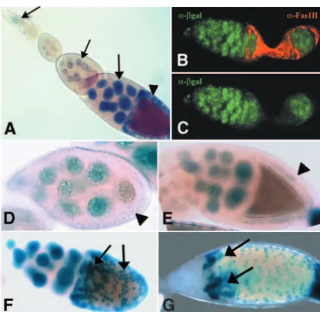
As a first step in the analysis of Slmb function in oogenesis, we wanted to assess the cell types and stages in which the gene is expressed. We failed to detect the transcript by in situ hybridization, suggesting that slmbis expressed at very low levels. Although enhancer traps do not always reproduce all aspects of endogenous transcription, they might provide a useful and often more sensitive method for studying gene expression when transcripts cannot be detected directly. We used the slmb00295 enhancer trap line (Jiang and Struhl, 1998) that revealed a widespread and dynamic expression profile of slmbduring oogenesis. High levels of expression were observed in nurse cells all throughout oogenesis (Fig. 1A). From the germarium until stage 8 of oogenesis, expression was excluded from FC (Fig. 1B-D) and at stage 9, low β-galactosidase levels could be detected (Fig. 1E). From stage 10 onwards, strong expression was recorded in patches of FC surrounding the oocyte (Fig. 1F), being still detected by the end of oogenesis (not shown). Remarkably, at stage 11 very strong expression was observed in two dorsal patches of FC that give rise to dorsal appendages (Fig. 1G) (Deng and Bownes, 1997; Sapir et al., 1998; Wasserman and Freeman, 1998). As the expression pattern suggested a function for slmbin both the germline and FC, we wanted to determine whether slmbloss of function provokes defects in oogenesis.
De
Slimb is required in both germline and follicle cells for egg chamber development
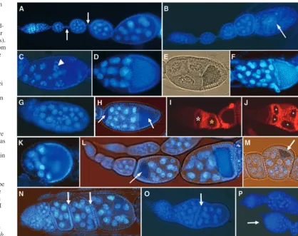
We tested three different slmb alleles: slmb1, previously described as a hypomorphic allele; and slmb2 and slmb00295, reported to be null alleles (Jiang and Struhl, 1998; Theodosiou et al., 1998). As in all cases homozygous or heteroallelic combinations were lethal at different developmental stages (Jiang and Struhl, 1998), we decided to examine the effect of slmb loss of function in homozygous germline clones generated by the FLP/OvoDmethod (Chou et al., 1993; Chou and Perrimon, 1996) or in somatic clones induced with the FC driver e22C (Duffy et al., 1998) (see Materials and methods). slmb00295clones were recovered at low rates, probably owing to poor cell viability (Jiang and Struhl, 1998); slmb1and slmb2 alleles exhibited similar highly reproducible egg chamber defects and were used in most of the experiments throughout this study. DAPI staining of egg chambers bearing germline clones revealed that 17.4% of the follicles (n=483) exhibited different kinds of abnormalities. To rule out the possibility that some of the observed defects arise from an effect of the OvoD itself, we focused our phenotypic analysis on ovarioles with vitellogenic cysts, a situation never found in ovaries from females carrying one copy of the OvoD transgene (n=100 ovarioles). Germline phenotypes included follicles that degenerated, exhibiting features of apoptosis (3.7%, Fig.
2A,B), such as germ cell nuclear fragmentation (Fig. 2C); consistent with this, TUNEL-positive staining at early stages of oogenesis was recorded (data not shown). Another frequently observed phenotype consisted of follicles displaying either increased (9.9%) or reduced (1.7%) numbers of germ cells (Fig. 2D-F), as well as germ cell nuclei that were heterogeneous in size (2.9%, Fig. 2G). These results suggest that mitotic divisions, as well as the switch from mitosis to endoreplication, might be misregulated in these cysts (see below). Remarkably, in egg chambers with a reduced number of germ cells, the nuclei were bigger than normal (see below Fig. 3E), suggesting that these cells failed to divide and, instead, entered endoreplication prematurely. In another set of experiments, we analyzed the effect of slmbloss of function in FC clones. Although slmb00295enhancer trap analysis indicated that expression in FC apparently starts at stage 9 (Fig. 1E), we frequently observed two oocytes and extra nurse cells within a single follicular epithelium (Fig. 2H). At vitellogenic stages, we never found supernumerary follicles exhibiting one single oocyte, suggesting that the phenotype arise from encapsulation defects rather than from an extra round of mitosis in the cyst. Consistent with encapsulation defects, we observed that morphology of mutant germaria was altered: in wild-type germaria region IIb, the cyst has acquired a lens shape, spanning the whole width of the germarium (Fig. 2I); instead, slmb mutant ovarioles often exhibited germaria in which two or more cysts were placed side by side in regions IIb and III (Fig. 2J). In addition, egg chambers with somatic clones often displayed an aberrant shape (Fig. 2K), suggesting that normal formation of the follicular epithelium was impaired. Moreover, we observed that oocytes were frequently mispositioned within the follicle, adopting an anterior (Fig. 2L) or lateral (Fig. 2M) location, indicating that axial polarity of the egg chamber was altered. Finally, another category of phenotypes consisted of ovarioles lacking interfollicular stalks between adjacent follicles but separated, instead, by a layer of epithelial cells (Fig. 2N,O). Previous work has shown that posterior localization of the oocyte depends on the presence of a stalk that links the egg chamber with the neighboring older follicle (Torres et al., 2003). We observed that in some of the follicles with mislocalized oocytes, posterior interfollicular stalks were missing (9/15), while in others posterior stalks were present (6/15), suggesting that alterations in axial polarity in the latter egg chambers involved a different mechanism.
As Slmb is a substrate recognition subunit of an SCF complex (Bocca et al., 2001; Feldman et al., 1997; Skowyra et al., 1997; Yaron et al., 1998), mutations affecting other components of the complex or molecules required for its ubiquitin ligase activity should render overlapping biochemical effects and similar phenotypes in oogenesis. We induced cullin1 (lin19 – FlyBase) (dCul110494) (Bocca et al., 2001; Filippov et al., 2000; Heriche et al., 2003; Khush et al., 2002) general clones by the Hs-FLP/FRT method that rendered ovarioles with cyst encapsulation defects, lack of interfollicular stalks and nurse cell nuclei of heterogeneous size (data not shown). Moreover, mutations affecting the subunits CSN4 and CSN5 of the COP9 signalosome, a highly conserved complex that regulates the activity of SCF complexes (Bech-Otschir et al., 2002; Schwechheimer and Deng, 2001; Seeger et al., 2001), have recently been reported to cause phenotypes that are also very similar to those provoked by slmb mutations Fig. 1.slmbshows a dynamic expression pattern during egg chamber
development. Ovaries from females carrying the slmb00295enhancer trap were analyzed by X-gal staining or by anti-β-galactosidase
(anti-β-gal) immunofluorescence. (A) In the ovariole, nurse cells stain positive for X-gal throughout oogenesis (arrows). X-gal signal in FC surrounding the oocyte appears at mid-oogenesis (arrowhead). (B,C) In the germarium, expression of β-gal (green) is restricted to the germline and is excluded from FC that express Fasciclin 3 (red). (D) Expression of β-gal cannot be detected in FC at stage 8 (arrowhead) and starts at stage 9 (E, arrowhead). (F) At oogenesis stage 10, strong expression of the enhancer trap occurs in a scattered pattern in the follicular epithelium (arrows). (G) At stage 11, expression becomes stronger in two patches of FC that will form the dorsal appendages (arrows).
De
[image:3.612.53.283.71.295.2](Doronkin et al., 2002; Doronkin et al., 2003). To study if Slmb might be a component of an SCF complex in the ovary, we looked for genetic interactions in egg chambers with slmb2 germline clones that were at the same time heterozygous for dCul112764 or CSN5L4032 alleles. Strong interactions were detected, as the proportion of egg chambers with extra germ cells increased from 9.9% (n=483) in ovaries with slmb2 homozygous clones to 32.1% in those with dCul112764/+; slmb2/slmb2 clones (n=156) and to 25.4% in ovaries with CSN5L4032/+; slmb2/slmb2 clones (n=130). Moreover, the phenotypes were enhanced in these genotypic combinations, as egg chambers with more than 32 germ cells and enlarged germaria (Fig. 2P) were observed in addition to the phenotypes described above. These results suggest that Slmb functions as a component of an SCF complex in oogenesis.
Control of germ cell divisions is impaired in slimb germline clones
Phenotypes described in Fig. 2E,F suggested that slmbloss of function in the germline might cause an impairment in the regulation of mitotic divisions that originate the cyst (de Cuevas et al., 1997). Among follicles with extra germ cells (n=38), two types of egg chambers could be recognized: those with one single oocyte (16/38) and those with two oocytes (22/38). In order to determine if extra gem cells arise from an
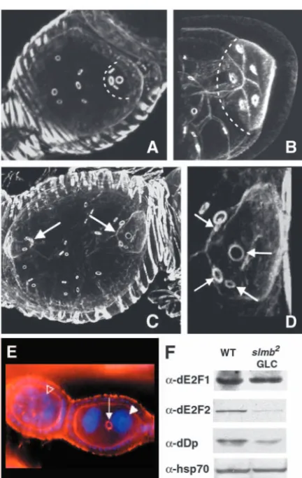
additional round of mitotic divisions, we analyzed the pattern of ring canals in these cysts. In wild-type cysts, four rounds of mitotic divisions take place, giving rise to oocytes with four ring canals clearly visible by phalloidin staining (Fig. 3A) (Hawkins et al., 1996). By contrast, in slmbgermline clones, all oocytes from follicles with extra nurse cells and one single oocyte exhibited five ring canals instead of four (16/16; Fig. 3B), indicating that an extra round of mitosis occurred. However, all the observed egg chambers bearing two oocytes exhibited four ring canals per oocyte (22/22; Fig. 3C,D), suggesting that in these cases mitotic divisions were normal. We believe that the latter phenotype did not reflect slmbloss of function in the germline but instead was due to the occurrence of slmb FC clones that provoked defects in cyst encapsulation as described above. Interestingly, egg chambers with fewer than 16 germ cells (n=8) exhibited a number of ring canals that was always consistent with the predicted rounds of cell divisions (i.e. two cells=one ring canal; four cells=no more than two ring canals per cell, etc.) (Fig. 3E). From these experiments, we hypothesized that Slmb is required in the germline for the regulation of mitotic cycles during cyst formation.
[image:4.612.142.561.73.404.2]It has been reported that DrosophilaDp (Dp), which forms heterodimers with E2f transcription factors (E2f1 and E2f2), is required for essential processes during oogenesis. Dp mutant Fig. 2.slmbloss of function
provokes a wide array of ovarian phenotypes. (A) DAPI staining of a wild-type ovariole (interfollicular stalks are marked by arrows). (B) Some egg chambers from females with slmbgermline clones exhibit premature apoptosis (arrow) indicated by the occurrence of fragmented nurse cell nuclei (C, arrowhead). (D) Wild-type follicles always contain 15 nurse cells and one oocyte; in slmb germline clones, egg chambers with decreased (E) or increased (F) number of germ cells are frequently observed, whereas in other follicles germ cell nuclei were heterogeneous in size (G). (H) In females bearing FC clones, egg chambers with two oocytes occur (arrows). (I) Wild-type germarium in which FC are stained for Fas3; the cyst in region IIb shows the typical lens shape (white asterisk) and once in region III the cyst re-shapes into a sphere (yellow asterisk). (J) In slmb somatic clones, the general
shape of the germarium is frequently altered and cysts are placed side by side in regions II and III (asterisks). slmbFC clones exhibit a wide array of defects, including abnormal morphology of egg chambers (K; compare with D), oocytes that are mispositioned (L,M, arrows) and lack of interfollicular stalks between adjacent follicles (N,O, arrows; compare with A). (P) slmb/CSN5genetic interactions; slmb2germline clones that are heterozygous for CSN5L4032display enhanced phenotypes such as enlarged germaria (arrow).
De
germline clones often execute an extra round of mitosis, giving rise to follicles with extra nurse cells (Myster et al., 2000; Royzman et al., 2002; Taylor-Harding et al., 2004). To gain insights about the mechanism underlying misregulation of the cell cycle in slmb germline clones, we performed western blot analysis of Dp, E2f1 and E2f2. As can be seen in Fig. 3F, E2f1 protein levels were not affected in slmbmutant ovaries but, by contrast, a major decrease in E2f2 and Dp levels occurred. As a Slmb direct target is expected to be increased in slmbmutant clones, we believe that the reduction of Dp and E2f2 probably occurs through an indirect mechanism.
Slimb somatic clones cause defects in
differentiation of follicle cells and generate ectopic polar cells
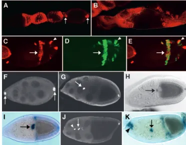
In the germarium, a lineage of intercyst FC differentiates into two subpopulations that stop dividing and originate stalk and polar cells (Bai and Montell, 2002; Margolis and Spradling, 1995; Tworoger et al., 1999). The lack of interfollicular stalk cells observed in slmbloss-of-function ovarioles suggested that differentiation of FC into different subpopulations might be impaired. In wild-type germaria, undifferentiated FC express high levels of Fasciclin 3 (Fas3), and by oogenesis stage 4, expression becomes restricted to polar cells (Fig. 4A). In slmb somatic clones refinement of Fas3 expression was often delayed (Fig. 4B), being still detected in large patches of cells beyond oogenesis stage 9 (Fig. 4C). In order to examine cell autonomy of this effect, clones positively marked with GFP were generated by using the MARCM system (Lee et al., 2000). As can be seen in Fig. 4C-E, Fas3 labeling largely overlaps with cell patches expressing GFP, indicating that the effect is cell autonomous. In some mosaic egg chambers, Fas3 expression refined into a pattern that seemed to correspond to ectopic polar cells and, interestingly, these cells were mutant for slmb, as indicated by GFP positive staining (Fig. 4C-E, arrowheads). As Fas3 is not a specific polar cell marker (Lopez-Schier and St Johnston, 2001; Zhang and Kalderon, 2000), we used the PZ80 enhancer trap that is specifically expressed in these cells (Fig. 4F-H) (Karpen and Spradling, 1992; Spradling, 1993). In wild-type follicles, anterior polar cells induce differentiation of five to eight FC into a border cell fate. The whole border cell cluster, including polar cells, expresses the slow border cells gene (slbo), as revealed by expression of the slbo-lacZenhancer trap (Fig. 4I) (Grammont, 2002; Liu and Montell, 1999; Montell et al., 1992; Rorth et al., 2000). At oogenesis stage 9, polar cells surrounded by the whole cluster start migrating posteriorly between nurse cells (Fig. 4G), and by stage 10 they have reached the anterior border of the oocyte (Fig. 4H). In slmb mutant egg chambers we clearly observed the occurrence of ectopic polar cells (Fig. 4J) and consistent with this, ectopic clusters of border cells differentiated (Fig. 4K). Interestingly, migration of border cell clusters was often impaired in these follicles (Fig. 4J,K).
Slimb is required for chorion patterning
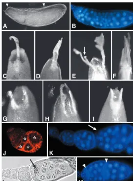
[image:5.612.60.276.69.410.2]Eggs laid by females in which slmb mutant clones were induced with the Hs-FLP driver exhibited diverse chorion patterning defects. Some of them, like a smaller size or abnormal shape of the egg (Fig. 5A; data not shown) probably derive from dumping defects or from alterations in the general morphology of the egg chamber (Fig. 2K). Interestingly, we also observed a wide array of eggshell phenotypes in which the size, shape and/or position of dorsal appendages (DA) was altered. In wild-type eggs, the dorsoanterior region of the chorion displays a pair of filaments (DA) separated by a gap (Fig. 5B). Some eggshells derived from slmb mosaic ovaries had a dorsalized phenotype (Sapir et al., 1998; Wasserman and Freeman, 1998) characterized by laterally expanded DA (Fig. 5C) or in more severe cases, by DA surrounding all the anterior circumference of the egg (Fig. 5D). In other cases, ectopic DA formed (Fig. 5E) or DA deranged, giving rise to ramified structures (Fig. 5F). Another category of phenotypes was that of eggshells apparently ventralized (Schupbach, 1987; Fig. 3.Germ cell mitosis is misregulated in slmbgermline clones.
Ring canals are visualized by TRITC-Phalloidin staining. (A) In wild-type egg chambers, the oocyte (marked with a broken line) connects to neighboring nurse cells through four ring canals. (B) An oocyte (broken line) from a slmbgermline clone exhibits five ring canals, indicating that an extra round of mitosis has occurred. (C)slmbmutant egg chamber with extra germ cells and two oocytes (arrows). (D) Higher magnification image of the oocyte on the right in C, showing that in these follicles, oocytes exhibit four ring canals (arrows). (E) In a slmbmutant ovariole, the egg chamber on the right has only two germ cells connected by a single ring canal (arrow) and exhibit very big nuclei (white arrowhead); in the same ovariole, the egg chamber on the left has the normal complement of germ cells and their nuclei are much smaller (open arrowhead). (F) Western blot analysis of E2f complex components in ovarian extracts with slmb germline clones (GLC), compared with those of wild-type ovaries; whereas E2f1 does not show obvious differences, Dp and E2f2 were clearly reduced in slmbmutant ovaries.
De
Wasserman and Freeman, 1998), with DA partially or totally fused (Fig. 5G,H). Finally, in the last category, eggshells exhibited a slightly enlarged operculum [with DA shifted to a more posterior site (Fig. 5I)] or a greatly reduced amount or absence of DA material (Fig. 5J,K).
In order to distinguish which chorion phenotypes arise from slmb loss of function in the germline and which ones from clones in the follicular epithelium, we induced clones through the OvoD method or the FC driver e22C. The latter method caused the same DA phenotypes described above, indicating that abnormalities reflected slmb loss of function in the follicular epithelium. By contrast, eggs of smaller size, which derived from dumping defects (data not shown), were not observed upon induction of somatic cell clones but rather, were
laid by females with germline clones (8%; n=902), indicating that this phenotype was due to Slmb loss of function in the germline.
Mild overexpression of Dpp in follicle cells mimics slimb loss-of-function phenotypes
[image:6.612.187.560.75.365.2]It has been reported that high levels of Dpp at the anterior-most region of the chorion prevent DA formation, leading to differentiation of the operculum (Peri and Roth, 2000; Twombly et al., 1996). As eggshells derived from follicles with slmb somatic clones exhibited phenotypes resembling those that occur upon overactivation of the Dpp pathway (Fig. 5I), we decided to perform further ectopic Dpp expression experiments and look for novel eggshell phenotypes and for abnormalities Fig. 4.Delayed FC differentiation
and ectopic polar cells occur in slmb somatic clones. (A) In wild-type ovarioles, Fas3 expression (red) refines as oogenesis progresses and from stage 4 onwards becomes restricted to polar cells (arrows). (B) In slmbFC clones, Fas3 remains widespread (egg chambers are outlined). (C) In egg chambers bearing slmbFC clones, large patches of cells expressing Fas3 are still detected at stage 9 (arrow). Mutant clones from the egg chamber shown in C were positively marked with GFP (D, arrow) and the merged confocal image shows that ectopic Fas3 correlates with the position of the clones (E, arrow). Arrowheads in C-E mark a pair of cells that are probably ectopic polar cells. (F-H,J) The PZ80 enhancer trap is specifically expressed in polar cells. In wild-type ovaries at stage 8, the two pairs of polar cells are localized at the anterior and posterior termini of the follicle (F, arrows) at stage 8; at stage 9, anterior polar cells migrate posteriorly between nurse cells (G,
arrow); and at stage 10, they have reached the anterior border of the oocyte (H, arrow). (I,K) The slbo-LacZ element is expressed in border cells. In stage 10 wild-type follicles, border cells can be seen at the anterior end of the oocyte (I, arrow). Egg chambers bearing slmbFC clones often exhibit extra polar cells (J, arrowhead) and differentiate ectopic clusters of border cells (K, arrowhead). In these follicles, border cell migration is often delayed and by stage 10, they are located between nurse cells (J,K, arrows).
Fig. 5.slmbmutant clones cause chorion patterning defects. Eggshells were visualized by reflected light or dark-field microscopy. (A) Eggs laid by females with slmbgermline clones (right) are often much smaller than wild-type eggs (left). (B) Wild-type dorsal appendage (DA) pattern. (C-F) Egg chambers with slmbsomatic clones generate eggshells with variable DA abnormalities, including dorsalized phenotypes (C-E), ramified DA (F), ventralized phenotypes (G,H), and DA shifted to a more posterior position (I), reduced (I,J) or absent (K). In cases where DA are shifted posteriorly (I), the operculum appears enlarged when compared with wild-type eggshells (B); the limits of the opercula are indicated with broken lines.
De
[image:6.612.264.561.600.740.2]in egg chamber development. We used various Gal4 drivers and UAS constructs for inducing the Dpp pathway at different levels. At 25°C all crosses were lethal at pre-imaginal stages and only at 18°C some of the tested combinations rendered viable adults that allowed ovary and chorion analysis. As depicted in Table 1, in the strongest viable combinations overexpressing Dpp, opercula expanded dramatically and DA were absent (Fig. 6A) as was previously reported (Peri and Roth, 2000; Twombly et al., 1996). In these cases, we observed germaria with tumorous arrays of germline stem cells (not shown) (Xie and Spradling, 1998) as well as aberrant follicles with large numbers of germ cells engulfed in the same follicular epithelium (Fig. 6B). Interestingly, upon milder induction of the Dpp pathway, we observed variable DA phenotypes, very similar to those caused by slmbloss of function in somatic cells (Table 1). These included eggshells exhibiting one thin dorsal filament (Fig. 6C), fused or ramified DA (Fig. 6D,E), DA spanning the whole anterior circumference of the egg (Fig. 6F) and reduced DA (Fig. 6G). Similar phenotypes were observed (1) upon overexpression of an activated form of the type I receptor Thick veins (not shown); (2) by overexpressing the co-Smad protein Medea (Fig. 6H); and, (3) in low proportion, in eggs laid by females heterozygous for short gastrulation (sog) or for daughters against dpp (dad) genes (Fig. 6I; Table 1), which encode negative regulators of the pathway (Araujo and Bier, 2000; Francois et al.,
[image:7.612.44.573.83.212.2]1994; Tsuneizumi et al., 1997). When we analyzed ovarioles from females subjected to the same mild overexpression conditions of Dpp, we observed a wide array of phenotypes similar to those observed in slmbmutant somatic clones. These included abnormal germaria, two complete cysts encapsulated within a single follicular epithelium, mislocalized oocytes and lack of interfollicular stalks between adjacent follicles (Fig. 6J-M). In order to determine whether loss of Slmb might cause increased activation of the Dpp pathway leading to the above phenotypes, we initially tested whether overexpression of Slmb was able to antagonize the effect of ectopic Dpp on eggshell morphogenesis. Indeed, Slmb overexpression rendered full Table 1. Chorion phenotypes obtained upon overactivation of the Dpp pathway
Maternal genotype Chorion phenotypes (n≥120)
Hs-gal4(strong)/+; UAS-dpp(weak)/+ No viable adults were obtained
Hs-gal4(weak)/+; UAS-dpp(strong)/+ 89% of eggshells with giant opercula and reduced or absent dorsal appendages (DA).
CY2-gal4/+; UAS-dpp(weak)/+ 80% of eggshells with enlarged opercula and reduced DA.
T155-gal4/+; UAS-dpp(weak)/+ 76% of eggshells with enlarged opercula and reduced or malformed DA. Weaker phenotypes than with the CY2 driver.
Hs-gal4(weak)/UAS-dpp(weak) 38% of eggshells with variable phenotypes, including ventralized or fused DA and ectopic DA material.
e22C-gal4/UAS-dpp(weak) 17% of eggshells displaying abnormal DA morphology, such as ramified, fused or expanded DA.
Hs-gal4(weak)/UAS-dpp(weak) 29% of eggshells with ectopic DA material deposited at the base, often displaying shorter or malformed DA. (no heat shock, leaky overexpression)
sogU2/+ 2% of eggshells with shorter DA and ectopic material deposited at the base and 9% with fused or partially fused DA.
dadj1E4/+ 3% of eggshells with shorter DA and ectopic material deposited at the base.
CY2-gal4/UAS-slmb;+/+ All wild-type egg chambers.
CY2-gal4/UAS-slmb; UAS-dpp(weak)/+ All wild-type egg chambers.
Fig. 6.Mild overactivation of the Dpp pathway mimics slmb mutant phenotypes. (A) Eggs laid by females bearing the weak Hs-gal4driver and the strong UAS-dppelement that were subjected to heat shock show a dramatic expansion of the operculum (marked by arrowheads) and lack of DA. (B) DAPI staining of an ovariole dissected from one of these females reveals that many germ cells are surrounded by a single follicular epithelium. (C,D) Upon milder overexpression using the weak UAS-dppelement and the weak Hs-gal4driver, ventralized eggshells with a single DA occurred. (E,F) Variable chorion phenotypes resulting from overexpression with the same UAS-dppelement driven by e22C-gal4: DA can be ramified (E, arrow), expanded (F) or greatly reduced (G). DA reduction was also observed upon overexpression of Medea with the weak Hs-gal4 driver (H) and in eggs laid by heterozygous sogU2females (I). Mild overexpression of Dpp with the weak Hs-gal4or e22C-gal4drivers (J-M) produced germaria with abnormal morphology (J), egg chambers with extra germ cells (K, arrow), mispositioned oocytes (L, arrow) and ovarioles lacking interfollicular stalks (M, arrowheads).
De
[image:7.612.287.559.368.737.2]reversion of the ‘expanded operculum’ phenotype (n=376; Fig. 6A) of eggs laid by females overexpressing Dpp under control of a CY2-gal4driver (Table 1); no rescue was observed when an UAS-EGFP transgene was overexpressed instead. Thus, based on similarities of slmbloss-of-function phenotypes with those caused by Dpp mild overexpression and, considering that the strong ectopic Dpp eggshell phenotype could be rescued by simultaneous overexpression of Slmb, we hypothesized that Slmb negatively regulates Dpp pathway in ovarian somatic cells.
Ectopic activation of the Dpp pathway occurs in slmb loss-of-function follicles
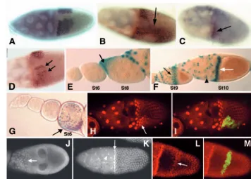
In stage 10 wild-type egg chambers, the intersection of Dpp and EGFR pathways induces the expression of the Broad-Complex(BR-C) gene in two patches of dorsoanterior FC (Fig. 7A), promoting DA specification and positioning (Deng and Bownes, 1997; Peri and Roth, 2000). We found that in egg chambers bearing slmbFC clones the BR-Cpattern was altered, being in some cases expanded (Fig. 7B), whereas in others it was reduced (Fig. 7C) or partitioned (Fig. 7D). As we did not detect alterations in the EGFR pathway (data not shown), we analyzed a possible effect of slmbmutations on Dpp signaling, by using the dad1883enhancer trap (dad-LacZ) (Casanueva and Ferguson, 2004). In wild-type ovarioles, expression of dad-lacZ is first detected at stage 8 in terminal anterior FC that migrate towards the oocyte (Fig. 7E). By oogenesis stage 10, dad-LacZ expression is detected in centripetal and border cells located at the anterior border of the oocyte, as well as in stretched cells which surround the nurse cells (Fig. 7F). Analysis of dad-lacZ in ovaries bearing slmb mutant clones
revealed that expression started prematurely at stage 6 (Fig. 7G) and later on, at stages 9-10, the pattern was often expanded posteriorly (Fig. 7H,I). These results suggest that loss of slmb might cause ectopic activation of the Dpp pathway. The ubiquitin/proteasome system regulates the stability of various Smad proteins through the activity of different E3 ubiquitin ligases (Casanueva and Ferguson, 2004; Fukasawa et al., 2004; Fukuchi et al., 2001; Li et al., 2004; Liang et al., 2004; Wan et al., 2004). Because in mammalian cell culture proteasomal degradation of Smad4, a common signal transducer in the TGFβ signaling pathway, is regulated by the Slmb ortholog,
βTrcp1 (Wan et al., 2004), we decided to explore whether a similar mechanism operates in the Drosophila ovary. We initially analyzed the expression of the Drosophila Smad4 homologue Medea (Sutherland et al., 2003) in wild-type follicles. At stage 8-9, expression was restricted to migrating anterior cells (Fig. 7J) and at stage 10 expression could be observed in stretched cells as well as in all FC surrounding the oocyte, being remarkably stronger in the centripetally migrating FC, located at the anterior border of the oocyte (Fig. 7K). Interestingly, in egg chambers bearing slmb clones, ectopic patches expressing high levels of Medea were observed (Fig. 7L,M). Taken together, these results suggest that at least some of the slmbmutant phenotypes might have been caused by increased levels of Medea protein, which led to overactivation of the Dpp pathway.
Discussion
Slimb function in the germline
[image:8.612.203.560.437.692.2]We have shown that the F-box protein Slmb is required for
Fig. 7.Broad-Complex(BR-C) expression and ectopic activation of the Dpp pathway. (A) mRNA in situ hybridization in a stage 10 wild-type egg chamber showing that BR-Cis expressed in two patches of dorsoanterior FC. (B-D) In follicles bearing slmbsomatic clones, BR-C expression is in some cases expanded (B, arrow) and in others reduced (C, arrow) or split (D, arrows). (E,F) Expression of the dad-lacZenhancer trap in wild-type ovarioles. (E) In the follicular epithelium, expression is first observed at stage 8 in anterior FC (arrow), being undetectable at stage 6. (F) At stage 9, dad-lacZcan be seen in migrating anterior FC (black arrow) and at stage 10 in stretched cells (arrowhead) and in centripetal cells localized at the anterior border of the oocyte (white arrow). (G) In ovarioles bearing slmbmutant clones, expression of dad-lacZoccurs prematurely at stage 6 (arrow); at stage 10 (H), expression of the enhancer trap often expands posteriorly (arrow). (I) Positive GFP labeling of a slmbmutant clone showing that GFP signal overlaps with ectopic expression of
dad-lacZin the clone. (J,K) In wild-type ovaries, expression of Medea occurs at stage 9 in anterior FC that migrate towards the oocyte (J, arrow) and at stage 10 (K) in all FC, being much stronger in stretched cells (arrowhead) and in centripetal cells localized at the anterior border of the oocyte (arrow). (L) In slmbmutant follicles, strong Medea signal was detected in ectopic patches of columnar FC, at stage 10 (arrow). (M) Positive GFP labeling of the slmbmutant clone overlaps with this patch.
De
oogenesis in both the germline and FC. In the germline, we found that Slmb plays a role in the control of mitotic cycles during cyst formation, in regulation of nurse cell endoreplication and in nurse cell dumping. Recent reports have demonstrated that Slmb can control cell cycle progression in different experimental settings. It has been reported that, following DNA replication, Slmb is required in larval wing discs for proteolysis of a specific cell cycle modulator: the transcription factor E2f1 (Heriche et al., 2003). Remarkably, the E2f complex was implicated in cell cycle control of ovarian germ cells, in nurse cell transition from polyteny to polyploidy and in dumping of the nurse cell content into the oocyte (Myster et al., 2000; Royzman et al., 2002). In this study, we found that two subunits of the E2f complex, Dp and E2f2, were downregulated in ovaries bearing slmbgermline clones, while E2f1 did not change. Differences in Cyclin E levels, another cell cycle regulator involved in cyst formation (Doronkin et al., 2003; Lilly and Spradling, 1996), could not be detected in these clones (data not shown). We want to stress the good correlation that exists between the phenotypes observed in slmbgermline clones (this study) and in Dp germline clones (Myster et al., 2000), as in both cases an additional round of cystocyte mitotic divisions occurs. In order to understand the molecular mechanism causing Dp and E2f2 reduction in slmb germline clones, a detailed analysis of the alterations of the network regulating the cell cycle is required.
Slimb function in follicle cells
Although expression levels in somatic cells in the germarium are too low to be detected through an enhancer trap or by in situ hybridization, loss-of-function experiments suggest that slmbis needed in these cells for normal morphogenesis of the egg chamber and for encapsulation of the cyst. In addition, our results suggest that Slmb is required for timely differentiation of FC that is reflected by the refinement of Fas3 expression; this is accompanied by the occurrence of ectopic polar cells, lack of interfollicular stalks and disruption of normal egg chamber polarity. Later in oogenesis, Slmb is expressed at high levels in FC surrounding the oocyte and participates in chorion patterning, contributing to define the shape and position of DA. It has been reported that slmbmutant clones induce ectopic activation of the Hedgehog (Hh) pathway in limb discs (Jiang and Struhl, 1998; Theodosiou et al., 1998). Notably, some of the phenotypes observed upon slmb somatic clone induction were similar to those originated by overactivation of the Hh pathway in FC. These include a delay in FC differentiation, development of ectopic polar cells and mislocalization of the oocyte (Forbes et al., 1996a; Zhang and Kalderon, 2000). Nevertheless, excessive activation of the Hh pathway also causes FC over-proliferation that results in excess of undifferentiated somatic cells that form very long interfollicular stalks between egg chambers (Forbes et al., 1996a; Forbes et al., 1996b; Zhang and Kalderon, 2000). By contrast, we observed that slmbloss of function in FC caused a lack rather than an excess of interfollicular cells. Finally, dominant genetic interactions were not detected between slmb and negative regulators of Hh pathway and the ptc-LacZ enhancer trap, which was reported to be activated in FC by the Hh pathway (Forbes et al., 1996b), was not induced ectopically in slmbmutant clones (data not shown). These results indicate that, despite some similarities between slmb loss-of-function
and hhgain-of-function phenotypes, Slmb is unlikely to be a negative regulator of Hh pathway during oogenesis.
In limb discs, Slmb was also reported to be a negative regulator of the Dpp pathway, although the molecular mechanism involved is still unclear (Miletich and Limbourg-Bouchon, 2000; Theodosiou et al., 1998). We have shown that mild overexpression of Dpp caused a wide spectrum of phenotypes that were largely coincident with those caused by slmb loss of function in FC. Supporting the idea that loss of slmb might cause hyperactivation of the Dpp pathway, the strongest chorion phenotypes originated by overexpression of Dpp were completely antagonized by simultaneous overexpression of Slmb in FC. Moreover, expansion of dad-lacZ expression occurred in slmb mutant follicles, further suggesting that ectopic induction of the Dpp pathway indeed occurs as a consequence of slmbloss of function. Consistent with this, we found that a downstream component of the Dpp pathway, the co-Smad protein Medea, was upregulated in slmb mutant egg chambers. Because in mammalian cell culture it was demonstrated that Smad4 is a direct target of the mammalian Slmb ortholog, βTrcp1 (Wan et al., 2004), we believe that Medea could be a direct target of Slmb. Further molecular work is required to assess whether this is indeed the case or if alternatively, the effect of Slmb on Medea is indirect.
We thank Maki Asano, Konrad Basler, Mary Bownes, Stephen Cohen, Nick Dyson, Jing Jiang, Bernardette Limbourg-Bouchon, Stuart Newfeld, Terry Orr-Weaver, Laurel Raftery, Helena Richardson, Francois Rouyer, Danny Segal, Benny Shilo, Allan Spradling, David Stein, Tian Xu, the Bloomington DrosophilaStock Center and the DSHB for stocks and/or antibodies. We thank Helena Araujo and Amir Sapir for technical advice; and Hernán López-Schier, Fernanda Ceriani, Lázaro Centanín and members of P.W.’s laboratory for discussion. Wellcome Trust Grant 070161/Z/03/Z, Universidad de Buenos Aires X411 and ANPCyT 01-10839. M.M. is a fellow and P.W. is a career investigator of CONICET.
References
Araujo, H. and Bier, E. (2000). sog and dpp exert opposing maternal functions to modify toll signaling and pattern the dorsoventral axis of the
Drosophilaembryo. Development127, 3631-3644.
Bai, J. and Montell, D.(2002). Eyes Absent, a key repressor of polar cell fate during Drosophilaoogenesis. Development129, 5377-5388.
Beccari, S., Teixeira, L. and Rorth, P.(2002). The JAK/STAT pathway is required for border cell migration during Drosophilaoogenesis. Mech. Dev.
111, 115-123.
Bech-Otschir, D., Seeger, M. and Dubiel, W.(2002). The COP9 signalosome: at the interface between signal transduction and ubiquitin-dependent proteolysis. J. Cell Sci. 115, 467-473.
Bocca, S. N., Muzzopappa, M., Silberstein, S. and Wappner, P.(2001). Occurrence of a putative SCF ubiquitin ligase complex in Drosophila.
Biochem. Biophys. Res. Commun. 286, 357-364.
Casanueva, M. O. and Ferguson, E. L.(2004). Germline stem cell number in the Drosophilaovary is regulated by redundant mechanisms that control Dpp signaling. Development131, 1881-1890.
Chou, T. B. and Perrimon, N.(1996). The autosomal FLP-DFS technique for generating germline mosaics in Drosophila melanogaster. Genetics144, 1673-1679.
Chou, T. B., Noll, E. and Perrimon, N. (1993). Autosomal P[ovoD1] dominant female-sterile insertions in Drosophilaand their use in generating germ-line chimeras. Development119, 1359-1369.
de Cuevas, M., Lilly, M. A. and Spradling, A. C.(1997). Germline cyst formation in Drosophila. Annu. Rev. Genet. 31, 405-428.
Deng, W. M. and Bownes, M. (1997). Two signalling pathways specify localised expression of the Broad-Complex in Drosophila eggshell patterning and morphogenesis. Development124, 4639-4347.
De
Deshaies, R. J.(1999). SCF and Cullin/Ring H2-based ubiquitin ligases.
Annu. Rev. Cell Dev. Biol. 15, 435-467.
Dobens, L. L. and Raftery, L. A.(2000). Integration of epithelial patterning and morphogenesis in Drosophilaovarian follicle cells. Dev. Dyn. 218, 80-93.
Doronkin, S., Djagaeva, I. and Beckendorf, S. K. (2002). CSN5/Jab1
mutations affect axis formation in the Drosophilaoocyte by activating a meiotic checkpoint. Development129, 5053-5064.
Doronkin, S., Djagaeva, I. and Beckendorf, S. K. (2003). The COP9 signalosome promotes degradation of Cyclin E during early Drosophila
oogenesis. Dev. Cell. 4, 699-710.
Duffy, J. B., Harrison, D. A. and Perrimon, N.(1998). Identifying loci required for follicular patterning using directed mosaics. Development125, 2263-2271.
Feldman, R. M., Correll, C. C., Kaplan, K. B. and Deshaies, R. J.(1997). A complex of Cdc4p, Skp1p, and Cdc53p/cullin catalyzes ubiquitination of the phosphorylated CDK inhibitor Sic1p. Cell91, 221-230.
Filippov, V., Filippova, M., Sehnal, F. and Gill, S. S.(2000). Temporal and spatial expression of the cell-cycle regulator cul-1in Drosophilaand its stimulation by radiation-induced apoptosis. J. Exp. Biol. 203, 2747-2756.
Forbes, A. J., Lin, H., Ingham, P. W. and Spradling, A. C. (1996a).
hedgehog is required for the proliferation and specification of ovarian somatic cells prior to egg chamber formation in Drosophila. Development
122, 1125-1135.
Forbes, A. J., Spradling, A. C., Ingham, P. W. and Lin, H.(1996b). The role of segment polarity genes during early oogenesis in Drosophila.
Development122, 3283-3294.
Francois, V., Solloway, M., O’Neill, J. W., Emery, J. and Bier, E.(1994). Dorsal-ventral patterning of the Drosophilaembryo depends on a putative negative growth factor encoded by the short gastrulation gene. Genes Dev.
8, 2602-2616.
Frolov, M. V., Huen, D. S., Stevaux, O., Dimova, D., Balczarek-Strang, K., Elsdon, M. and Dyson, N. J.(2001). Functional antagonism between E2F family members. Genes Dev. 15, 2146-2160.
Fukasawa, H., Yamamoto, T., Togawa, A., Ohashi, N., Fujigaki, Y., Oda, T., Uchida, C., Kitagawa, K., Hattori, T., Suzuki, S. et al.(2004). Down-regulation of Smad7 expression by ubiquitin-dependent degradation contributes to renal fibrosis in obstructive nephropathy in mice. Proc. Natl. Acad. Sci. USA 101, 8687-8692.
Fukuchi, M., Imamura, T., Chiba, T., Ebisawa, T., Kawabata, M., Tanaka, K. and Miyazono, K.(2001). Ligand-dependent degradation of Smad3 by a ubiquitin ligase complex of ROC1 and associated proteins. Mol. Cell. Biol.
12, 1431-1443.
Godt, D. and Tepass, U.(1998). Drosophilaoocyte localization is mediated by differential cadherin-based adhesion. Nature395, 387-391.
Gonzalez-Reyes, A. and St Johnston, D.(1998). The DrosophilaAP axis is polarised by the cadherin-mediated positioning of the oocyte. Development
125, 3635-3644.
Grammont, M. and Irvine, K. D.(2002). Organizer activity of the polar cells during Drosophilaoogenesis. Development129, 5131-5140.
Grima, B., Lamouroux, A., Chelot, E., Papin, C., Limbourg-Bouchon, B. and Rouyer, F.(2002). The F-box protein slimb controls the levels of clock proteins period and timeless. Nature420, 178-182.
Hawkins, N. C., Thorpe, J. and Schupbach, T.(1996). Encore, a gene required for the regulation of germ line mitosis and oocyte differentiation during Drosophilaoogenesis. Development122, 281-290.
Heriche, J. K., Ang, D., Bier, E. and O’Farrell, P. H.(2003). Involvement of an SCFSlmb complex in timely elimination of E2F upon initiation of DNA replication in Drosophila. BMC Genet.4, 9.
Hershko, A. and Ciechanover, A.(1998). The ubiquitin system. Annu. Rev. Biochem. 67, 425-479.
Jiang, J. and Struhl, G.(1998). Regulation of the Hedgehog and Wingless signalling pathways by the F-box/WD40-repeat protein Slimb. Nature391, 493-496.
Kamura, T., Koepp, D. M., Conrad, M. N., Skowyra, D., Moreland, R. J., Iliopoulos, O., Lane, W. S., Kaelin, W. G., Jr, Elledge, S. J., Conaway, R. C. et al.(1999). Rbx1, a component of the VHL tumor suppressor complex and SCF ubiquitin ligase. Science284, 657-661.
Karpen, G. H. and Spradling, A. C. (1992). Analysis of subtelomeric heterochromatin in the Drosophilaminichromosome Dp1187by single P
element insertional mutagenesis. Genetics132, 737-753.
Khush, R. S., Cornwell, W. D., Uram, J. N. and Lemaitre, B.(2002). A ubiquitin-proteasome pathway represses the Drosophilaimmune deficiency signaling cascade. Curr. Biol. 12, 1728-1737.
Ko, H. W., Jiang, J. and Edery, I.(2002). Role for Slimb in the degradation of DrosophilaPeriod protein phosphorylated by Doubletime. Nature420, 673-678.
Lee, T., Winter, C., Marticke, S. S., Lee, A. and Luo, L.(2000). Essential roles of DrosophilaRhoA in the regulation of neuroblast proliferation and dendritic but not axonal morphogenesis. Neuron25, 307-316.
Li, L., Xin, H., Xu, X., Huang, M., Zhang, X., Chen, Y., Zhang, S., Fu, X. Y. and Chang, Z.(2004). CHIP mediates degradation of Smad proteins and potentially regulates Smad-induced transcription. Mol. Cell. Biol. 24, 856-864.
Liang, M., Liang, Y. Y., Wrighton, K., Ungermannova, D., Wang, X. P., Brunicardi, F. C., Liu, X., Feng, X. H. and Lin, X.(2004). Ubiquitination and proteolysis of cancer-derived Smad4 mutants by SCFSkp2. Mol. Cell. Biol. 24, 7524-7537.
Lilly, M. A. and Spradling, A. C. (1996). The Drosophilaendocycle is controlled by Cyclin E and lacks a checkpoint ensuring S-phase completion.
Genes Dev. 10, 2514-2526.
Liu, Y. and Montell, D. J.(1999). Identification of mutations that cause cell migration defects in mosaic clones. Development126, 1869-1878.
Lopez-Schier, H. and St Johnston, D.(2001). Delta signaling from the germ line controls the proliferation and differentiation of the somatic follicle cells during Drosophilaoogenesis. Genes Dev. 15, 1393-1405.
Margolis, J. and Spradling, A. (1995). Identification and behavior of epithelial stem cells in the Drosophilaovary. Development121, 3797-3807.
Margottin-Goguet, F., Hsu, J. Y., Loktev, A., Hsieh, H. M., Reimann, J. D. and Jackson, P. K. (2003). Prophase destruction of Emi1 by the SCF(betaTrCP/Slimb) ubiquitin ligase activates the anaphase promoting complex to allow progression beyond prometaphase. Dev. Cell4, 813-826.
Marquez, R. M., Singer, M. A., Takaesu, N. T., Waldrip, W. R., Kraytsberg, Y. and Newfeld, S. J.(2001). Transgenic analysis of the Smad family of TGF-beta signal transducers in Drosophila melanogastersuggests new roles and new interactions between family members. Genetics157, 1639-1648.
Miletich, I. and Limbourg-Bouchon, B.(2000). Drosophilanull slimbclones transiently deregulate Hedgehog-independent transcription of winglessin all limb discs, and induce decapentaplegictranscription linked to imaginal disc regeneration. Mech. Dev. 93, 15-26.
Montell, D. J., Rorth, P. and Spradling, A. C.(1992). slow border cells, a locus required for a developmentally regulated cell migration during oogenesis, encodes DrosophilaC/EBP. Cell71, 51-62.
Myster, D. L., Bonnette, P. C. and Duronio, R. J.(2000). A role for the DP subunit of the E2F transcription factor in axis determination during
Drosophilaoogenesis. Development127, 3249-3261.
Ohta, T., Michel, J. J., Schottelius, A. J. and Xiong, Y.(1999). ROC1, a homolog of APC11, represents a family of cullin partners with an associated ubiquitin ligase activity. Mol. Cell3, 535-541.
Ou, C. Y., Lin, Y. F., Chen, Y. J. and Chien, C. T.(2002). Distinct protein degradation mechanisms mediated by Cul1 and Cul3 controlling Ci stability in Drosophilaeye development. Genes Dev. 16, 2403-2414.
Patton, E. E., Willems, A. R. and Tyers, M.(1998). Combinatorial control in ubiquitin-dependent proteolysis: don’t Skp the F-box hypothesis. Trends Genet. 14, 236-243.
Peri, F. and Roth, S. (2000). Combined activities of Gurken and decapentaplegic specify dorsal chorion structures of the Drosophilaegg.
Development127, 841-850.
Pickart, C. M.(2001). Mechanisms underlying ubiquitination. Annu. Rev. Biochem. 70, 503-533.
Queenan, A. M., Ghabrial, A. and Schupbach, T.(1997). Ectopic activation of torpedo/Egfr, a Drosophilareceptor tyrosine kinase, dorsalizes both the eggshell and the embryo. Development124, 3871-3880.
Rorth, P., Szabo, K. and Texido, G.(2000). The level of C/EBP protein is critical for cell migration during Drosophila oogenesis and is tightly controlled by regulated degradation. Mol. Cell6, 23-30.
Royzman, I., Hayashi-Hagihara, A., Dej, K. J., Bosco, G., Lee, J. Y. and Orr-Weaver, T. L.(2002). The E2F cell cycle regulator is required for
Drosophilanurse cell DNA replication and apoptosis. Mech. Dev. 119, 225-237.
Ruberte, E., Marty, T., Nellen, D., Affolter, M. and Basler, K.(1995). An absolute requirement for both the type II and type I receptors, punt and thick veins, for dpp signaling in vivo. Cell80, 889-897.
Sapir, A., Schweitzer, R. and Shilo, B. Z.(1998). Sequential activation of the EGF receptor pathway during Drosophila oogenesis establishes the dorsoventral axis. Development125, 191-200.
Schupbach, T.(1987). Germ line and soma cooperate during oogenesis to
De
establish the dorsoventral pattern of egg shell and embryo in Drosophila
melanogaster. Cell49, 699-707.
Schwechheimer, C. and Deng, X. W.(2001). COP9 signalosome revisited: a novel mediator of protein degradation. Trends Cell Biol. 11, 420-426.
Seeger, M., Gordon, C. and Dubiel, W.(2001). Protein stability: the COP9 signalosome gets in on the act. Curr. Biol. 11, R643-R646.
Skowyra, D., Craig, K. L., Tyers, M., Elledge, S. J. and Harper, J. W.
(1997). F-box proteins are receptors that recruit phosphorylated substrates to the SCF ubiquitin-ligase complex. Cell91, 209-219.
Spradling, A. (1993). Developmental genetics of oogenesis. In The Development of Drosophila melanogaster, vol. 1 (ed. M.-A. A. Bate), pp. 1-70. Cold Spring Harbor, New York: Cold Spring Harbor Laboratory Press.
Staehling-Hampton, K. and Hoffmann, F. M. (1994). Ectopic decapentaplegic in the Drosophila midgut alters the expression of five homeotic genes, dpp, and wingless, causing specific morphological defects.
Dev. Biol. 164, 502-512.
Sutherland, D. J., Li, M., Liu, X. Q., Stefancsik, R. and Raftery, L. A.
(2003). Stepwise formation of a SMAD activity gradient during dorsal-ventral patterning of the Drosophilaembryo. Development130, 5705-5716.
Taylor-Harding, B., Binne, U. K., Korenjak, M., Brehm, A. and Dyson, N. J.(2004). p55, the Drosophilaortholog of RbAp46/RbAp48, is required for the repression of dE2F2/RBF-regulated genes. Mol. Cell. Biol. 24, 9124-9136.
Theodosiou, N. A., Zhang, S., Wang, W. Y. and Xu, T.(1998). slimb
coordinates wg and dpp expression in the dorsal-ventral and anterior-posterior axes during limb development. Development125, 3411-3416.
Torres, I. L., Lopez-Schier, H. and St Johnston, D.(2003). A Notch/Delta-dependent relay mechanism establishes anterior-posterior polarity in
Drosophila. Dev. Cell5, 547-558.
Tsuneizumi, K., Nakayama, T., Kamoshida, Y., Kornberg, T. B., Christian, J. L. and Tabata, T. (1997). Daughters against dpp modulates dpp organizing activity in Drosophilawing development. Nature389, 627-631.
Twombly, V., Blackman, R. K., Jin, H., Graff, J. M., Padgett, R. W. and Gelbart, W. M.(1996). The TGF-beta signaling pathway is essential for
Drosophilaoogenesis. Development122, 1555-1565.
Tworoger, M., Larkin, M. K., Bryant, Z. and Ruohola-Baker, H.(1999). Mosaic analysis in the Drosophila ovary reveals a common hedgehog-inducible precursor stage for stalk and polar cells. Genetics151, 739-748.
van Eeden, F. and St Johnston, D.(1999). The polarisation of the anterior-posterior and dorsal-ventral axes during Drosophilaoogenesis. Curr. Opin. Genet. Dev. 9, 396-404.
Voges, D., Zwickl, P. and Baumeister, W.(1999). The 26S proteasome: a molecular machine designed for controlled proteolysis. Annu. Rev. Biochem.
68, 1015-1068.
Wan, M., Tang, Y., Tytler, E. M., Lu, C., Jin, B., Vickers, S. M., Yang, L., Shi, X. and Cao, X. (2004). Smad4 protein stability is regulated by ubiquitin ligase SCF beta-TrCP1. J. Biol. Chem. 279, 14484-14487.
Wasserman, J. D. and Freeman, M.(1998). An autoregulatory cascade of EGF receptor signaling patterns the Drosophilaegg. Cell95, 355-364.
Wieschaus, E. and Szabad, J.(1979). The development and function of the female germ line in Drosophila melanogaster: a cell lineage study. Dev. Biol.
68, 29-46.
Winston, J. T., Koepp, D. M., Zhu, C., Elledge, S. J. and Harper, J. W.
(1999). A family of mammalian F-box proteins. Curr. Biol. 9, 1180-1182.
Wojcik, E. J., Glover, D. M. and Hays, T. S.(2000). The SCF ubiquitin ligase protein Slimb regulates centrosome duplication in Drosophila. Curr. Biol.
10, 1131-1134.
Xi, R., McGregor, J. R. and Harrison, D. A.(2003). A gradient of JAK pathway activity patterns the anterior-posterior axis of the follicular epithelium. Dev. Cell4, 167-177.
Xie, T. and Spradling, A. C.(1998). decapentaplegicis essential for the maintenance and division of germline stem cells in the Drosophilaovary.
Cell94, 251-260.
Xu, T. and Rubin, G. M.(1993). Analysis of genetic mosaics in developing and adult Drosophilatissues. Development117, 1223-1237.
Yaron, A., Hatzubai, A., Davis, M., Lavon, I., Amit, S., Manning, A. M., Andersen, J. S., Mann, M., Mercurio, F. and Ben-Neriah, Y. (1998). Identification of the receptor component of the IkappaBalpha-ubiquitin ligase. Nature396, 590-594.
Zhang, Y. and Kalderon, D. (2000). Regulation of cell proliferation and patterning in Drosophilaoogenesis by Hedgehog signaling. Development
127, 2165-2176.
Zheng, N., Schulman, B. A., Song, L., Miller, J. J., Jeffrey, P. D., Wang, P., Chu, C., Koepp, D. M., Elledge, S. J., Pagano, M. et al. (2002).
Structure of the Cul1-Rbx1-Skp1-F boxSkp2 SCF ubiquitin ligase complex.
Nature416, 703-709.