Mouse Disp1 is required in sonic hedgehog-expressing cells for paracrine activity of the
cholesterol-modified ligand
Hua Tian, Juhee Jeong, Brian D. Harfe, Clifford J. Tabin and Andrew P. McMahon
There was a reanalysis of data required to support the findings in Development132, 133-142.
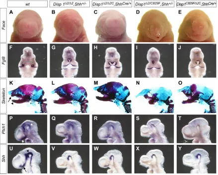
[image:1.612.88.527.261.614.2]We have repeated the facial analysis reported in Fig. 2 to provide the data required to support some of the original findings of this study (see Publisher’s note). Our findings substantiate the original conclusions drawn from Fig. 2 of a dose-related genetic interaction between Disp1 and Shhalleles, and of the function of Disp1within Shh-producing cells. Some differences are reported below, which might reflect slight differences in embryonic staging or increased sensitivity of the whole-mount in situ hybridization procedure here. Importantly, they do not alter the key conclusion that reducing Disp1 levels in Shh-producing cells results in a phenotype similar to that of genetically matched embryos with reduced Disp1 activity throughout the embryo. These data support the overall conclusions of the paper, and, together with other data in the same report, support a model in which the principal requirement for Disp1 activity is in Shh-producing cells.
Fig. 2. Attenuating Disp1activity specifically in Shh-producing cells phenocopies Disp1hypomorphic mutants.Attenuating Disp1 activity specifically in Shh-producing cells produced facial phenotypes that resembled genetically matched embryos with Disp1reduction throughout the embryo. (A-E) External facial views of E18.5 embryos with the indicated genotypes. (F-J) Whole-mount in situ hybridization with Fgf8probes to E10.5 embryos of the indicated genotypes. The separation of Fgf8expression domains in the frontal nasal processes of wild-type embryos (bracket in F) reflects the normal development of midline structures that were lost to varying degrees in embryos with attenuated Disp1and Shhactivity (G-J). (K-O) Alcian Blue (non-mineralized cartilage)-stained and Alizarin Red (mineralized cartilage and bone)-stained head skeletal preparations of E18.5 embryos with indicated genotypes. A variable loss was observed in both upper (arrow in K) and lower (arrowhead in K) jaw structures, including the midline incisors. (P-Y) Whole-mount in situ hybridization with (P-T) Ptch1and (U-Y) Shhprobes to E9.5 embryos of the indicated genotypes. Ptch1andShh expression was evident in midline cell populations rostral to the optic lobes in wild-type embryos (arrows in P and U). Their expression was either markedly reduced or lost, depending on the specific combination of Disp1and Shhalleles (Q-T and V-Y). Ptch1andShh
expression was detected in the midbrain region (indicated by arrowheads in P and U) of all genotypes, albeit at reduced levels.
3472
The following text replaces the paragraph running from p. 135 to p. 137 (‘In both genotypes…in the Disp1mutant background.’)
Disp1Δ2/Δ2C; ShhCre/+embryos in which Disp1activity was specifically knocked down in Shh-producing cells have a facial phenotype with
a narrowing of the face and reduction of the premaxilla. However, the length of the snout is similar to wild type and the mandibular incisors are not fused (Fig. 2A,C,K,M). Thus, the phenotype is, as expected, generally less severe than that of the Disp1Δ2/Δ2; Shh+/–embryos (Fig. 2B,L). The severity of the conditional phenotype is enhanced when Disp1activity is further lowered in Disp1Δ2C/C829F; ShhCre/+ mice (Fig. 2E,O) but the phenotype is slightly weaker than that in Disp1Δ2/C829F(data not shown) or Disp1Δ2/C829F; Shh+/–embryos (Fig. 2D,N); the tubular nasal process was shorter and the premaxillary bone was more extensive. In a proportion of the latter, truncated fused mandibles lack incisors (Fig. 2N); however, mandibular fusion was not observed in Disp1Δ2C/C829F; ShhCre/+embryos. The slightly weaker facial phenotype seen at term with each of the conditional removal combinations was evident at E10.5 when the distance between the Fgf8 -expressing frontal-nasal processes is compared by whole-mount in situ hybridization (Fig. 2F-J). Variable weak midlineShhexpression was observed rostral to the optic stalk in Disp1Δ2/Δ2C; ShhCre/+embryos at E9.5 (Fig. 2U-W). As expected, this resulted in Ptch1expression in adjacent nascent facial structures (Fig. 2P-R). Small, weak domains ofShhand Ptch1expression were observed close to the midline, localized to the region of the optic stalk in Disp1Δ2C/C829F; ShhCre/+embryos (not readily visible in Fig. 2T,Y). Only Disp1Δ2/C829F; Shh+/– embryos, the strongest genetic combination, completely lacked Shhand Ptch1expression rostral to the diencephalon (Fig. 2S,X). The authors apologise to readers for any inconvenience caused by the requirement to replicate these data and thank Mary Duah and Jill McMahon for replicating the experiments, Joe Vaughan and Celia Shneider for help with data acquisition, Renate Hellmiss for generating the figures and the journal Developmentfor encouraging the reanalysis.
Publisher’s note
Re: Tian, H., Jeong, J., Harfe, B. D., Tabin, C. J. and McMahon, A. P.(2005a). Mouse Disp1 is required in sonic hedgehog-expressing cells for paracrine activity of the cholesterol-modified ligand. Development132, 133-142.
In 2005, the McMahon laboratory reported that a re-examination of two papers published by their group in Development(Tian et al., 2004; Tian et al., 2005a) had revealed a duplication of Dr Tian’s data in these papers. Following their analysis, the authors announced, with regret, that they must retract Tian et al. (2004), and this retraction was published by Developmentin November 2005, along with their apology to the editors and readership of the journal (Tian et al., 2005b). With respect to the second paper (Tian et al., 2005a), the authors’ review, overseen by the Committee on Professional Conduct (CPC) for the Faculty of Arts and Sciences at Harvard University, found that the principal conclusions of the paper were supported by appropriate documentation but that the documentation for Fig. 2 was inadequate, requiring a replication of those data. The replicated data have been reviewed by Developmentand are published in this Corrigendum. Developmentand its publishers take very seriously issues relating to the authenticity of data. We acknowledge the dedication and openness of the McMahon laboratory in this matter, and the contributions of additional members of the laboratory, not on the paper’s authorship list, in repeating these extensive experiments and data analyses for the Corrigendum.
References
Tian, H., Tenzen, T. and McMahon, A. P.(2004). Dose dependency of Disp1and genetic interaction between Disp1and other hedgehog signaling components in the mouse. Development131, 4021-4033.
Tian, H., Jeong, J., Harfe, B. D., Tabin, C. J. and McMahon, A. P.(2005a). Mouse Disp1 is required in sonic hedgehog-expressing cells for paracrine activity of the cholesterol-modified ligand. Development132, 133-142.
Tian, H., Tenzen, T. and McMahon, A. P.(2005b). Retraction: Dose dependency of Disp1and genetic interaction between Disp1and other hedgehog signaling components in the mouse. Development132, 5615.
133
Introduction
The hedgehog (Hh) family of secreted proteins control tissue growth and patterning in many key developmental processes in both vertebrates and invertebrates. In mammals, there are three hedgehog genes (Echelard et al., 1993): sonic hedgehog (Shh), Indian hedgehog (Ihh) and desert hedgehog (Dhh). All three can act as potent mitogens, survival factors and inducers of distinct cell types in a dose-dependent manner (reviewed by McMahon et al., 2003). Shhis essential for early patterning of the ventral CNS, and somite and anterior-posterior organization of the limb. Shhalso plays a redundant role with
Ihh in establishment of left-right asymmetry (Zhang et al., 2001). Later in development, Shhis involved in the growth and morphogenesis of several organs, including hair, tooth, lung, gut and kidney, whereas Ihh coordinates growth and differentiation of the endochondral skeleton, Dhh is required for development of the peripheral nerve, testicular organization and spermatogenesis.
As extracellular signals, Hh proteins are synthesized in discrete subsets of cells in many organs and act in short and long-range signaling processes. The best characterized mammalian Hh-target field is the developing ventral neural tube, where progenitors differentiate into several different cell types in response to a morphogen gradient of Shh, issuing from two point sources: the notochord and floorplate (Briscoe and
Ericson, 1999; Briscoe and Ericson, 2001; Jessell, 2000; McMahon et al., 2003). The notochord is a ventral rod of mesoderm that underlies the neural tube while the floorplate is a population of support cells at the ventral midline of the neural tube that is induced by notochordal Shh. Distinct ventral neural progenitors are induced at specific positions with respect to the source, and apparent concentration, of Hh ligand.
All Hh proteins are synthesized as full-length precursors that undergo an autocatalytic cleavage reaction. This removes the C-terminal catalytic domain and attaches a cholesterol molecule to the C terminus of the N-terminal signaling fragment (N-Shhp; ‘p’ stands for ‘processed’ in the N-Shh signaling moiety) (Porter et al., 1996a). The hydrophobic cholesterol moiety is thought to bind Hh to the cell membrane. The hydrophobicity of the Hh molecule is further increased by the addition of a palmitoyl group to a conserved cysteine residue that is exposed at the N terminus after signal peptide cleavage (Pepinsky et al., 1998). These modifications regulate Hh activity, oligomerization, range of action, potency and might in many cases, shape a signaling gradient (Burke et al., 1999; Chamoun et al., 2001; Chen et al., 2004; Chen and Struhl, 1996; Kohtz et al., 2001; Lee et al., 2001; Lewis et al., 2001; Zeng et al., 2001). The requirement for such modifications may depend on the context in which Hh proteins act (Chen and Struhl, 1996; Lewis et al., 2001). As cholesterol
Previous studies have demonstrated that Disp1 function is
essential for Shh and Ihh signaling in the mouse, and Disp1
gene dose regulates the level of Shh signaling activity in vivo. To determine whether Disp1 activity is required in Shh-producing cells for paracrine signaling in Shh target
fields, we used a ShhGFP-Cre (here shortened to ShhCre)
knock-in allele and a Disp1 conditional allele to knock
down Disp1 activity specifically within Shh-producing cells. The resulting facial and neural tube phenotypes support the conclusion that the primary and probably exclusive role for Disp1 is within hedgehog protein-producing cells. Furthermore, using an allele that produces N-Shh (a
non-cholesterol modified form of the Shh protein), we demonstrate that N-Shh is sufficient to rescue most of the
early embryonic lethal defects in a Disp1-null mutant
background. Thus, Disp1 activity is only required for paracrine hedgehog protein signaling by the cholesterol modified form of Shh (N-Shhp), the normal product generated by auto-processing of a Shh precursor protein. In both respects, Disp function is conserved from
Drosophilato mice.
Key words: Dispatched1, Shh, Cholesterol, Paracrine, Autocrine
Summary
Mouse Disp1 is required in sonic hedgehog-expressing cells for
paracrine activity of the cholesterol-modified ligand
Hua Tian1,*, Juhee Jeong2,*, Brian D. Harfe3, Clifford J. Tabin4and Andrew P. McMahon5,†
1One DNA Way, Genentech Incorporated, South San Francisco, CA 94080, USA
21550 Fourth Street, Room 282, UCSF Mission Bay Campus, San Francisco, CA 94158, USA
3University of Florida College of Medicine, Department of Molecular Genetics and Microbiology, 1600 SW Archer Road,
Gainesville, FL 32610-0266, USA
4Department of Genetics, Harvard Medical School, NRB room 360, 77 Avenue Louis Pasteur, Boston, MA 02115, USA
5Department of Molecular and Cellular Biology, The Biolabs, Harvard University, 16 Divinity Avenue, Cambridge, MA 02138, USA
*These authors contributed equally to this work
†Author for correspondence (e-mail: [email protected])
Accepted 4 November 2004
Development 132, 133-142
Published by The Company of Biologists 2005 doi:10.1242/dev.01563
Research article
De
modification is unique to Hh-ligands, the role of cholesterol has attracted considerable attention. Forms of Hh ligand have been engineered that lack the autocatalytic cleavage site and C-terminal cleavage domain, and therefore are not cholesterol-modified [Hh-N in Drosophila(Chen and Struhl, 1996) and N-Shh in mammals (Lewis et al., 2001)]. In both instances, palmitoylation appears to be largely independent of cholesterol addition (Chen et al., 2004; Gallet et al., 2003).
Despite dual lipid modifications that normally retain protein on the cell membrane, DrosophilaHh and mouse Shh and Ihh protein can be detected at significant distances from their expression domains (Chen and Struhl, 1996; Gallet et al., 2003; Gritli-Linde et al., 2001; Lewis et al., 2001; Porter et al., 1996a). This long-distance action is an active process involving a transmembrane protein Dispatched (Disp). Disp was first identified in Drosophilastudies, where its activity is required specifically within Hh-producing cells for movement of cholesterol modified ligand into Hh target fields (Burke et al., 1999). By contrast, Hh-N signaling is Disp independent (Burke et al., 1999).
The mouse has two Disp homologs, Disp1 and Disp2, but only Disp1 is able to rescue a Drosophila disp mutant phenotype (Ma et al., 2002). Genetic studies in zebrafish suggest that Disp2is not involved in Hh signaling (Nakano et al., 2004), and mouse studies support this view (H.T. and A.P.M., unpublished). Three mutant alleles of mouse Disp1
have been described: Disp1∆8, Disp1C829F and Disp1∆2
(Caspary et al., 2002; Kawakami et al., 2002; Ma et al., 2002; Tian et al., 2004). Analysis of these alleles lead to similar general conclusion, that Disp1 is involved in Hh signaling during early embryonic development. Among these mutant alleles, the Disp1∆8deletion allele (Kawakami et al., 2002; Ma et al., 2002) and Disp1C829F missense allele (Caspary et al., 2002) are likely to represent null alleles of Disp1. Homozygous
Disp1∆8and Disp1C829Fmutants do not survive beyond E9.5, and exhibit gross morphological features that are similar to
Smoothened(Smo) mutants in which all Hh signaling activity is abolished (Zhang et al., 2001). In Disp1C829F/C829F and
Disp1∆8/∆8 embryos, Hh signaling is retained but only in midline cells of the notochord that both produce Shh and respond to Shh signals (Ma et al., 2002). Disp1∆2, by contrast, encodes a hypomorphic allele (Tian et al., 2004). Homozygous
Disp1∆2 mutants die at birth with facial midline patterning defects, characteristic of attenuated Shh signaling (Wallis and Muenke, 1999). This hypomorphic allele has permitted us to demonstrate the genetic interaction of Disp1 with specific components of the Hh signaling pathway. By combining a
Disp1∆2 hypomorphic allele with Disp1C829F, Shhnull and patched 1 (Ptch1null) alleles, we created a set of graded facial midline and neural tube phenotypes demonstrating that Disp1
gene dose regulates the level of Shh signaling activity in vivo. Furthermore, rescue of the Disp1∆2/∆2 mutant upon removing one copy of Ptch1suggested that Disp1 functions exclusively in the Hh pathway (Tian et al., 2004).
Although these phenotypic analyses demonstrated a conserved requirement for Disp1 in the Hh signaling pathway and highlighted the importance of Disp1 dose for normal levels of Shh signaling, they did not address the specific cellular or molecular limits to Disp1 action in mammalian Hh signaling. To determine whether Disp1 activity is required in Shh-producing cells or in paracrine signaling in the target field, we
used a ShhCre knock-in allele to remove Disp1 exon 2 specifically from Shh-producing cells. Our results indicate that
Disp1activity in these cells is essential for Hh signaling to the target field. Furthermore, using an allele that produces N-Shh, we demonstrate that Disp1 activity is required only for the paracrine action of the cholesterol modified form of Shh.
Materials and methods
MiceDisp1C829F/+ mice were kindly provided by Kathryn Anderson (Caspary et al., 2002). The Sox2Creand Shhn (Shh–) alleles have been described previously (Hayashi et al., 2002; St-Jacques et al., 1998). The construction of ShhGFP-Cre(here shortened to ShhCre) is described by Harfe et al. (Harfe et al., 2004). N-ShhCalleles will be reported in detail elsewhere (J.J. and A.P.M., unpublished). In short, Cre-mediated recombination leads to the generation of an allele functionally identical to the N-Shhallele reported by Lewis et al. (Lewis et al., 2001). Mutants were studied on a mixed genetic background, principally 129SV, C57BL6/J and SW.
Generation of Disp1∆2C and Disp1∆2alleles
To remove exon 2 of Disp1, a targeting vector was engineered in which exon 2 was flanked by loxP sites. Exon 2 encodes the amino terminal cytoplasmic domain and first transmembrane domain of Disp1 (Tian et al., 2004). After homologous recombination at the
Disp1locus in AV3 ES cells, a heterozygous ES cell line was injected into blastocysts of the C57BL6/J strain to generate chimeras. These were bred with Swiss Webster mice to obtain Disp1∆2C/+ offspring. Chimeric males were also bred with β-actin-Crefemales, Cre activity in the preimplantation embryo allow the recovery of Disp1∆2/+
heterozygous offspring.
RNA in situ hybridization
Embryos were fixed in 4% paraformaldehyde at 4°C overnight. Whole-mount and section in situ hybridization using digoxigenin-labeled RNA probes was performed as described previously (Schaeren-Wiemers and Gerfin-Moser, 1993; Wilkinson, 1992).
Immunofluorescence
Immunofluorescence on embryonic sections was performed as described for sections (Yamada et al., 1991). Antibodies and dilutions were as follows: rabbit αFoxa2, 1:8000 (Ruiz i Altaba et al., 1995);
αNkx6.1, 1:3000 (Cai et al., 2000); αNkx2.2, 1:4000 (Briscoe et al., 1999); αOlig2, 1:5000 (Takebayashi et al., 2000); mouse αNkx2.2, 1:50 (Ericson et al., 1997a); αPax6, 1:20 (Ericson et al., 1997b);
αPax7, 1:20 (Ericson et al., 1996); and αMNR2, 1:20 (DHSB).
Skeletal preparations
For skeletal preparations, 18.5 dpc embryos were processed as described previously (Karp et al., 2000).
Results
Generation of a tissue specific knockout of Disp1 exon 2 in the Shh-expressing cells
To remove Disp1activity specifically from the Shhexpression domain using the Cre/loxP system, we first generated a Disp1 -conditional allele (Disp1∆2C) by flanking the first coding exon with DNA recognition sites (loxP) for Cre recombinase (Cre) (see Tian et al., 2004). Mice homozygous for this allele (Disp1∆2C/∆2C) were viable and fertile with no discernible
phenotype, indicating that Disp1∆2C has a wild-type function (data not shown). Conversion of Disp1∆2Cinto a Disp1∆2allele
De
135 Disp1 activity in Shh producing cell specific for N-Shhp
in the presence of Cre recombinase is highly efficient, as demonstrated by our previous study in which a ubiquitous
Disp1∆2 allele was generated by crossing a β-actin Cre transgene onto a Disp1∆2C/+background (Tian et al., 2004).
To knock down Disp1 activity specifically within Shh-producing cells, we used a Cre knock-in allele in which sequence encoding a GFP-Cre fusion protein was inserted into the Shh locus (ShhGFP-Cre, herein ShhCre) (Harfe et al., 2004). This allele no longer expresses a normal Shh transcript, and is a null allele for Shh activity. When combined with a Cre reporter line R26R (Soriano, 1999), ShhCre gave rise to lacZ
expression that was almost identical to Shh mRNA distribution at E10.5 (Fig. 1A,B), indicating that the GFP-Cre fusion protein is functionally active within all Shh expression domains. To further confirm the functional specificity of GFP-Cre fusion protein, ShhCrewas crossed with a Shh conditional allele (Shh∆2C) (Lewis et al., 2001) to remove Shh in the Shh-producing cell.
[image:5.612.312.559.76.612.2]The specification of ventral progenitor domains in the presumptive spinal cord gives the most detailed read-out of Shh signaling. The ventral half of the mouse neural tube is occupied by five ventricular progenitor populations, from ventral to dorsal, pV3, pMN, pV2, pV1 and pV0, all of which require a direct Hh signaling input for their development (Briscoe et al., 2001; Wijgerde et al., 2002). These cells move laterally and differentiate into V3 interneuron, motoneurons, and V2, V1 and V0 interneurons, respectively (Briscoe and Ericson, 2001; Jessell, 2000; McMahon et al., 2003). Induction of distinct cell types depends on Shh signaling from the notochord and floorplate. A key aspect of these inductive events is that from pV0 to the floorplate (which is also induced by Shh signaling), individual cell identities require a progressively higher concentration of Shh for their induction (floorplate>pV3>pMN>pV2>pV1>pV0) (Ericson et al., 1997a; Roelink et al., 1995). The ShhCre/Cmutants displayed a cyclopic head (compare Fig. 1D with 1E) and ventral neural tube patterning defects (compare Fig. 1G,H,J,K,M,N) very similar to those of Shh mutants, indicating that ShhCre was effective in removing Shh activity from all Shh-producing cells (Fig. 1).
Appropriate crosses were set up to generate mice that were either Disp1∆2/∆2C, ShhCre/+or Disp1C829F/∆2C, ShhCre/+. In both cases, pups died within 1 day of birth. In the former, ShhCre
activity should generate Shh-expressing cells that are homozygous for the Disp1∆2/∆2 allele. In the latter, the only Disp1 activity comes from a single Disp1∆2 hypomorphic allele. In both examples, Disp1 activity is decreased on a background where Shh dose is lowered by the presence of the
ShhCrenull allele. Ordinarily, reducing Shh levels by removing one allele of Shh produces no discernible phenotype (Chiang et al., 1996; St-Jacques et al., 1998) but reduction of Shh dose enhances the neural phenotype in Disp1 hypomorphic combinations (Tian et al., 2004) Importantly, as the production of Cre recombinase is linked to Shhexpression, some level of Shh signaling must take place prior to Cre-mediated modification of Disp1 alleles. This is evident in ShhCre/C
embryos where a few Nkx6.1-producing cells are observed (Fig. 1N), while no Nkx6.1 are produced in Shh–/– embryos (Fig. 1L,M).
In both genotypes (Disp1∆2/∆2C, ShhCre/+ or Disp1C829F/∆2C, ShhCre/+), pups displayed facial midline defects similar to
Fig. 1.ShhCrewas effective in removing Shh activity from all Shh-producing cells. (A,B) The distribution of Shh mRNA (A) and lacZ
activated in compound heterozygous for the ShhCreand R26R reporter allele (B) are very similar. External morphology of E10.5 embryos (C-E). Shh-null embryo (D) is almost identical to ShhCre/C (E). In Shh–/–mutants (G,J,M,P), the floorplate (G) and distinct ventral progenitors [Olig2+(J), Nkx6.1+(M) and Nkx2.2+(P) cells] are absent. Furthermore, Pax7 (M) and Pax6 (P), negative targets of Hh signaling, move ventrally to occupy the entire ventral neural tube. In the ShhCre/Cmutant, one or two Nkx6.1+cells remain (arrow in N) in the ventral neural tube, which is indicative of some low level signaling prior to Cre activity.
De
Disp1 hypomorphic mutants (Disp1∆2/∆2, Shh+/– and
Disp1C829F/∆2, Shh+/–), in which Disp1exon 2 was deleted in
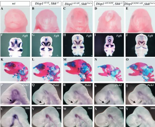
the entire embryo, the severity of the phenotype increased as Disp1 activity decreased (Fig. 2A-E). Importantly, no phenotype was observed in Disp1∆2/∆2C, Shh+/– embryos (data not shown). The midline facial defects were clearly visible at E10.5 by comparing the ventral disposition of the medial and lateral nasal process and proximal mandibular and maxillary arches that are highlighted by the expression of Fgf8 (Fig. 2F-J). In Disp1∆2/∆2C, ShhCre/+ embryos, the two nasal pits were
positioned closer to the midline, indicating a loss of facial
midline structure. A more severe phenotype was seen in
Disp1C829F/∆2C, ShhCre/+embryos as the two nasal pits start to
fuse where Shh-dependent medial nasal cells were also affected (Fig. 2J). The severity of the midline loss in conditional mutants was comparable with Disp1hypomorphic mutants Disp1∆2/∆2, Shh+/–and Disp1C829F/∆2; Shh+/–(compare
Fig. 2G and I with 2H and J). Skeleton preparations of newborn pups revealed that the premaxilla was missing from
Disp1∆2/∆2C, ShhCre/+ and Disp1C829F/∆2C, ShhCre/+ mutant embryos, just as in Disp1C829F/∆2, Shh+/–and Disp1∆2/∆2, Shh+/–
[image:6.612.48.561.215.636.2]embryos (Fig. 2K-O). Although the parietal bone was also lost
Fig. 2.Attenuating Disp1 activity specifically in Shh-producing cell phenocopies Disp1 hypomorphic mutants. (A-E) External facial morphology of E18.5 embryos. Disp1conditional mutants (C,E) display a spectrum of midline facial defects that result in a pointed face and nose that are similar but slightly milder than Disp1∆2/∆2, Shh+/–and Disp1C829F/∆2, Shh+/–embryos (B,D). (F-J) Fgf8in situ to demarcate the epithelium of the nasal pit. Two nasal pits, which are positioned well apart in wild type (F), are brought closer to the midline in Disp1∆2/∆2,
Shh+/–and Disp1∆2/∆2C, ShhCre/+(G,H), and are fused at the midline in Disp1∆2/C829F, Shh+/–(I). The fusion of the nasal pits occurs at a more medial position in Disp1C829F/∆2C, ShhCre/+ embryos (J). (K-O) Alcian Blue (non-mineralized cartilage) and Alizarin Red (mineralized cartilage and bone) stained skeletons of E18.5 embryos. The premaxilla and upper incisor are missing from all mutants. The premaxilla, upper incisor and parietal bone are missing from Disp1∆2/C829F, Shh+/–(N) but not in the conditional mutant Disp1C829F/∆2C, ShhCre/+(O). Midline facial defects are due to attenuation of Shh induction and signaling in the ventral forebrain. (P-Y) Whole-mount in situ of Ptch1(P-T) and Shh(U-Y) at E9.5 show an absence of Ptch1upregulation in the frontal nasal process of the mutants and a failure of Shh induction in the ventral forebrain.
De
137 Disp1 activity in Shh producing cell specific for N-Shhp
in Disp1C829F/∆2, Shh+/–embryos, we only observed a delay in ossification of parietal bones in Disp1C829F/∆2C, ShhCre/+mutant
(compare Fig. 2N,O). As expected from the analysis of
Disp1∆2/∆2, Shh+/– and Disp1∆2/C829F, Shh+/– mutants,
expression of Ptch1, one of the principle transcriptional targets of Hh signaling, was greatly downregulated in the frontal nasal process (FNP) of the conditional mutants at E9.5 (Fig. 2P-T, arrows). This confirms that the loss of midline structures of the frontal nasal process in the conditional mutants Disp1∆2/∆2C, ShhCre/+and Disp1C829F/∆2C, ShhCre/+was due to the attenuation
of Shh signaling in this region. Furthermore, Shhexpression within the ventral forebrain, which is itself a target of a mesendodermal derived Shh signal, was lost in all allelic combinations (Fig. 2U-Y). This most probably explains the
initial defect in FNP development in the Disp1 mutant background.
Spinal cord patterning defects in Disp1∆2/∆2C, ShhCre/+and Disp1C829F/∆2C, ShhCre/+mutant
As previously reported (Tian et al., 2004), in the Disp1∆2/∆2,
Shh+/– mutant, Shh signaling is greatly compromised in the ventral neural tube: the floorplate was absent (Foxa2–, and
Shh–) and the ventral mid-line was occupied by greatly reduced numbers of the ventral-most neural progenitor, pV3 (Nkx2.2+)
(10% of wild-type numbers; wild type, 60±6; Disp1∆2/∆2, Shh+/–,7±3, n=3, P<0.01) (Fig. 3A,B,F,G). The next
[image:7.612.77.544.247.645.2]ventral-most progenitor cells, pMN (Olig2+), were reduced to 15% of the wild type number (wild type, 71±6; Disp1∆2/∆2, Shh+/–,
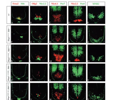
Fig. 3.Attenuation of Disp1 activity in the Shh-producing notochord leads to a similar disruption of ventral neural tube patterning, as observed in Disp1hypomorphic mutants. Sections through the neural tube of wild-type (A-E), Disp1hypomorphic mutants (Disp1∆2/∆2, Shh+/–) (F-J),
Disp1C829F/∆2, Shh+/–(P-T), and Disp1 conditional mutants [Disp1∆2/∆2C, ShhCre(K-O) and Disp1C829F/∆2C, ShhCre/+(U-Y)]. In Disp1∆2/∆2, Shh+/– mutant (F-J): the floorplate is absent (F); Nkx2.2+and Olig2+cells are greatly reduced in number (G); Nkx2.2+cells occupy the ventral midline (G); Nkx6.1+-positive cells are also affected (H); and the dorsal marker Pax7 is restricted to the dorsal domain (H). The conditional mutant
Disp1∆2/∆2C, ShhCre/+maintains the early floorplate marker Foxa2 but no Shh expression is observed in the floorplate (K). Nkx2.2+and Olig2+ cells are reduced in number to about 50% of the wild-type control levels and Nkx2.2+cells occupy the ventral midline (L). In Disp1∆2/C829F,
Shh+/–mutant (P-T), ventral progenitor cell numbers are further reduced compared with Disp1∆2/∆2, Shh+/–.No Nkx2.2+cells are present (S) and the Pax7 and Pax6 domains move ventrally (R,S). Nkx2.2+cells are still present in Disp1C829F/∆2C, ShhCre/+mutants (X).
De
10±3, n=3, P<0.01 Fig. 3B,G). The domain occupied by Nkx6.1+ cells, which demarcate pV3, pMN and pV2 progenitors, was also significantly reduced in the mutant (wild type, 175±15; Disp1∆2/∆2, Shh+/–,60±5, n=3, P<0.01) (Fig. 3C,H). The loss of these ventral cell identities was accompanied by a ventral upregulation of Pax6 and Pax7, two factors whose expression is generally repressed by Shh signaling in the ventral neural tube (Fig. 3C,D,H,I). The severe reduction of the pMN neural progenitor population translated to a significant decrease in MNR2+ motoneuron precursors that were also abnormally positioned at the ventral mid-line (wild type, 145±15,
Disp1∆2/∆2, Shh+/–, 80±10, n=3, P<0.01 Fig. 3E,J).
When Disp1∆2/∆2C, ShhCre/+ mutants were examined at E10.5, floorplate induction appears to have initiated as marked by sporadic Foxa2+ cells in the ventral midline (Fig. 3K). However, Shh, which is activated later than Foxa2 and requires Foxa2 activity (Epstein et al., 1999), failed to be induced in the floorplate (Fig. 3K). Furthermore, this apparent floorplate co-expressed Foxa2 and Nkx2.2+ (Fig. 3B,L; data not shown). Ordinarily, these are only transiently co-expressed but are rapidly restricted to their respective floorplate and pV3 progenitor domains (Fig. 3A,B; data not shown). The number of Nkx2.2+ pV3 cells was also reduced in the mutant (wild type, 60±6, Disp1∆2/∆2C, ShhCre/+, 25±4, n=3, P<0.01 Fig. 3B,L). Thus, the ventral midline cells represent some intermediate state between pV3 and floorplate identity. Other ventral progenitors marked by Olig2+ (Disp1∆2/∆2C, ShhCre/+, 35±5, Fig. 3L) and Nkx6.1+(Disp1∆2/∆2C, ShhCre/+, 125±8, Fig. 3M) were reduced to similar levels to those observed in
Disp1∆2/∆2 homozygous mutants (Tian et al., 2004). As with the face, the overall phenotype observed in conditional
Disp1∆2/∆2C, ShhCre/+ mutant was weaker than that in
Disp1∆2/∆2, Shh+/– mutants, but comparable with that of
Disp1∆2/∆2mutants. This phenotype was significantly enhanced when Disp1 levels were further reduced in Shh-expressing cells in Disp1∆2C/C829F, ShhCre/+mutants, where after recombination all Disp1 activity derives from a single hypomorphic Disp1∆2
allele. In these embryos, there was a complete failure of floorplate development (Fig. 3U); pV3 and pMN cells move to the midline and are reduced to less than 10% of the wild-type numbers, a similar reduction was also observed for Nkx6.1+ and MNR2+ cells (Fig. 3V-X). Although the severity of the phenotype was enhanced, it was still slightly less severe than that of Disp1∆2/C829F, Shh+/– mutants (Fig. 3P-T), where pV3 Nkx2.2+progenitors were completely absent (compare Fig. 3S with 3X).
Signaling by N-Shh is independent of Disp1 activity
Next, we addressed the specific requirement for Disp1 for cholesterol modified Shh ligand. We have previously reported on an N-Shh allele generated by inserting a stop-codon into the endogenous Shh gene at the position where normal cleavage and cholesterol addition occurs (Lewis et al., 2001). The protein produced is identical to that of the normal Shh signal and is expected to undergo N-terminal palmitoylation (Chen et al., 2004). Unfortunately, this allele is dominant lethal,
highlighting the importance of cholesterol modification to normal Hh regulation (Lewis et al., 2001). To overcome this dominant lethality and to enable us to address N-Shh activity in a Disp1C829F/C829F mutant background that lacks all Disp1 activity, we created a conditional N-Shh (N-ShhC) allele (J.J.
and A.P.M., unpublished). Mice heterozygous for this allele are viable and fertile permitting genetic intercrosses with the
Disp1C829F allele. Cre-mediated recombination leads to exclusive production of N-Shh from the endogenous Shhlocus, an outcome that is essentially identical to the original non-conditional N-Shhallele. To initiate recombination throughout the embryo, we used a Sox2Cre transgene to induce recombination in the entire embryo (Hayashi et al., 2002). As the resulting N-Shh allele is under identical cis-regulatory control to the wild-type Shh allele, N-Shh expression was restricted to Shh-expressing cells (data not shown). Unlike
Disp1C829F/C829Fmutants, which do not survive beyond 9.5 dpc
and show gross defects in neural, somite, cardiac, vascular, facial and limb development (Fig. 4A,B), Disp1C829F/C829F,
N-ShhC/Shhn, Sox2Creembryos were alive at E10.5 but die at or around birth. Morphologically, these embryos were indistinguishable from theN-ShhC/Shhn, Sox2Cremutants (Fig. 4C,D), which also employ N-Shh as the only available Shh ligand. At this stage, both Disp1C829F/C829F, N-ShhC/Shhn, Sox2Cre and N-ShhC/Shhn, Sox2Cremutant embryos are very
similar to the wild type, except for midline defects in the frontal nasal process. Thus, Disp1 does not appear to be required for N-Shh activity.
To confirm that the rescue of Disp1C829F/C829Fmutant by
N-Shh was due to active Hh signaling, we examined the expression of the Hh target Ptch1. In the wild-type limb bud,
Ptch1has a graded expression, occupying most of the posterior half of the distal limb bud mesenchyme, including the zone of polarizing activity (ZPA), the domain where Shh is produced in the limb (Fig. 5A,D). In Disp1∆8/∆8 mutants at 9.5 dpc, Shh
was expressed in the posterior mesenchyme as in the wild type. However, Ptch1expression was greatly reduced and restricted to the distal posterior margin of the forelimb, the Shh
expression domain (Kawakami et al., 2002). When
Disp1C829F/C829F mutants expressing N-Shh were examined,
Shh expression was slightly downregulated in the posterior mesenchyme compared with the wild type, but to a similar degree to Shh expression in N-ShhC/Shhn, Sox2Cre mutant
[image:8.612.261.562.74.193.2]embryos (Fig. 5B,C). The Ptch1expression domain was also reduced and restricted to the posterior mesenchyme in both of
Fig. 4.N-Shh rescues Disp1C829F/C829Fmutant at E10.5. Gross morphology of the (A) wild-type, (B) Disp1C829F/C829F, (C) N-ShhC/Shhn; Sox2Cre, (D) N-ShhC/Shhn;
Sox2Cre; Disp1C829F/C829Fembryos at E10.5.
De
139 Disp1 activity in Shh producing cell specific for N-Shhp
these embryos (Fig. 5E,F), as in N-Shh/Shhnlimb buds (Lewis
et al., 2001). High levels of Ptch1expressions were observed (Fig. 5D-F), with no gradient of expression across the AP axis was evident (Fig. 5E,F). However, when the two limb buds from the same embryos were stained for Shh and Ptch1
expression separately and compared, the Ptch1 expression domain clearly extended anterior to that of Shh in both
Disp1C829F/C829F, N-ShhC/Shhn, Sox2Cre and N-ShhC/Shhn, Sox2Creembryos (compare the outlined area in Fig. 5B,C with those in E,F, respectively). This result indicates that Hh signaling expanded beyond the Shh expression domain in
Disp1C829F/C829Fmutant when non-cholesterol modified N-Shh serves as the ligand. However, recent cell fate studies that demonstrate an anterior expansion of former Shh-expressing cells complicate the interpretation as the observed upregulation of Ptch1in non-Shh-expressing cells could reflect ‘historical’ signaling by cells which a few hours earlier were localized within the Shh-expressing ZPA (Harfe et al., 2004).
In the spinal cord, the movement of cells that expressed Shh do not complicate this analysis. All ventral neural progenitor types can be specified in the absence of a floorplate by notochordal Shh signaling (Matise et al., 1998; Ding et al., 1998). Furthermore, cell fate studies using the ShhCre allele
indicate that Shh-expressing cells at the midline only contribute to the floorplate (data not shown). Consistent with previous reports (Caspary et al., 2002; Kawakami et al., 2002; Ma et al., 2002), a complete loss of Disp1 activity abolishes floorplate (Foxa2–, Shh–), pV3 (Nkx2.2–) and pMN (Olig2–) induction (Fig. 6A,B,G,H). When N-Shh was introduced into
a Disp1C829F/C829Fmutant background, we observed floorplate (Foxa2+, Shh is not observed, see Discussion), pV3 (Nkx2.2+)
and pMN (Olig2+) populations at correct positions and in comparable numbers to N-ShhC/Shhn, Sox2Creembryos (Fig.
6C,D,I,J). N-Shh also rescues the Disp1hypomorphic mutant phenotype (compare Fig. 6E,K with Fig. 6C,F,L). When N-Shh was introduced into Disp1∆2/C829F mutant background (Fig. 6F,L), the phenotype was indistinguishable from that of the N-ShhC/Shhn, Sox2Crecontrol (Fig. 6C,I).
The graded expression of Hh receptor Ptch1in the ventral neural tube at E9.5 was absent in Disp1C829F/C829Fmutant (Fig. 6M,N) (Caspary et al., 2002) and greatly reduced in
Disp1∆2/C829Fmutants (Fig. 6Q). By contrast, signaling by N-Shh from the notochord and floorplate positively regulates
Ptch1expression in the ventral neural tube in Disp1C829F/C829F
or Disp1∆2/C829F backgrounds (Fig. 6P,R) as in a wild-type
Disp1 background (Fig. 6O). Thus, the rescue of Disp1 mutants by N-Shh is achieved by restoring Shh signaling to the ventral neural tube. It is noteworthy that in all samples where we detect Foxa2, but not N-Shh, protein in putative floorplate cells, Shhexpression in the floorplate was observed by in situ hybridization analysis (Fig. 6S-X) (see Discussion).
Discussion
Disp1 functions in production of a paracrine Shh signal
We have taken advantage of distinct alleles of Disp1 to examine the requirement for Disp1 in Hh signaling in the mouse. Previous reports have demonstrated that Disp1 function is essential for Shh and Ihh signaling (Caspary et al., 2002; Kawakami et al., 2002; Ma et al., 2002). Furthermore, a comparative analysis of autocrine and paracrine functions suggested that Disp1 is not required for autocrine signaling where Hh-producing cells are themselves targets of Hh signaling (e.g. Shh in the notochord), but Disp1 is essential for paracrine signaling to responsive cell populations adjacent to the sources of ligand (Caspary et al., 2002; Ma et al., 2002). These data suggest that Disp1 may function in release of active ligand from Hh-producing cells to adjacent target populations as is the case for Disp in Drosophila(Burke et al., 1999).
In this study we have used a variety of genetic strategies to examine Disp1 action. In the previous studies, the precise cellular requirements for Disp1 were unclear. Here, we specifically attenuated Disp1 activity in Shh-expressing cells; strikingly, the phenotypes closely resemble those observed when Disp1 activity is reduced in the entire embryo (Tian et al., 2004). That these phenotypes are not identical is most likely a technical limitation of our approach. In this, we used a conditional hypomorphic allele of Disp1 where essential sequence encoded within exon 2 was flanked by loxP sites and combined this allele with a ShhCreallele to ‘knock down’ Disp1
levels exclusively within Shh-expressing cells. In this genotype, Cre-dependent recombination at the Disp1∆2Clocus
can occur only after Shh transcription is initiated; hence, it is likely some Shh signaling occurs while there is a sufficient level of Disp1 to lead to normal signaling (embryos heterozygous for Disp1null/+ alleles are phenotypically
[image:9.612.43.288.74.234.2]wild-type). Alternatively, as this approach is only expected to modify Ihh signaling where Shhand Ihh expression overlap, wild-type Ihh activity from non-Shh expressing cells may
Fig. 5.Shh signaling is restored in the limb bud of N-Shh rescued
Disp1-null mutants. Whole-mount in situ hybridizations with antisense riboprobes for Shh(A-C) and Ptch1(D-F) in limb buds of wild-type (A,D), N-ShhC/Shhn; Sox2Cre(B,E) and N-ShhC/Shhn; Sox2Cre; Disp1C829F/C829F(C,F) embryos at 10.5 dpc as indicated. Shh gene expression is restricted to the posterior mesenchyme in the wild type (A). N-Shhp from this domain diffuses anteriorly, which then leads to a graded Ptch1 expression across the entire posterior half of the limb field (D). The expression of N-Shh is induced in the posterior mesenchyme in N-Shhmutant (B). Ptch1is reduced and restricted to the posterior mesenchyme (E). High levels of Ptch1
expression are restricted to Shh-producing cells and to cells immediately anterior to this domain. No gradient of expression is apparent across the AP axis. Similar levels of Shhand Ptch1
expression are observed in limb buds of N-Shh rescued Disp1mutant embryos (C,F).
De
contribute to the weaker phenotype in the conditional allele. The presence of a few Nkx6.1+ cells in ShhCre/C embryos is consistent with the former explanation.
With these provisos in mind, it is striking that the phenotypes of attenuated Disp1 activity in the Shhexpression domain and in the whole embryo are so similar. Clearly, most if not all, paracrine Shh signaling within the face, neural tube and limb is dependent on Disp1 function in the ligand-producing cell, supporting a model in which Disp1 acts in signal production and not target cell response. How Disp1 acts at the molecular and cellular level is not clear. Studies in Drosophila report the accumulation of Hh ligand in Disp1 mutant cells in the posterior compartment of the imaginal disc (Burke et al., 1999), suggesting that Disp1 may regulate the release of bulk ligand from Hh-producing cells. Further work implicates Disp in apical trafficking (Gallet et al., 2003) within Hh-producing epithelia, suggesting that the defective release and ligand accumulation in Hh-producing cells may be secondary to altered membrane trafficking. However, we failed to observe any obvious accumulation of Shh in Shh-producing cells in Disp1-null mutant embryos (data now shown). We also failed to observe any differences in the release of bulk Shh protein into the medium when Shh was expressed in Disp1C829F/C829F
or wild-type fibroblasts (Tian et al., 2004). Whether this represents a difference between polarized epithelia and fibroblasts is unclear. Interestingly, reports of a highly active multimeric complex of lipid modified Hh ligands (Zeng et al., 2001) raises the possibility that Disp1 may function not in general release of ligand, but rather in the formation and/or release of an active fraction that is composed of Hh oligomers. Clearly, there is enhanced bioactivity in media conditioned by
Shh-expressing fibroblast when Disp1 is active in these cells (Ma et al., 2002).
Disp1 is only required for paracrine signaling activity of cholesterol modified forms of Shh
Cholesterol-modification of Hh ligands plays a key role in Hh signaling. In Drosophila, removal of cholesterol from Hh increases the range of ligand action, at least in part through a disruption of normal Ptch-dependent feedback control that normally sequesters ligand (Chen and Struhl, 1996). Whereas Ptch1 mediated sequestration of N-Shh is also defective in the mouse, cholesterol modification of N-Shhp is essential for long-range action in the limb (Lewis et al., 2001). Thus, although there appears to be species or context-dependent differences in the role of cholesterol in Hh signaling, in both flies and mice cholesterol-modification of Hh ligands is essential for normal signaling within a multicellular target field. In Drosophila, Disp is required only for signaling by cholesterol-modified forms of Hh (Burke et al., 1999). Our work in the mouse indicates a similar requirement for Disp1. Whereas, Disp1-null embryos arrest at E9.5 dpc with multiple defects, including an absence of ventral cell identities in the neural tube, expression of a single allele of N-Shh is sufficient to rescue many of these deficiencies. In the resulting neural tube, all Shh-dependent ventral cell identities are represented leading to a phenotype identical to that of N-Shh/Shhnembryos on a wild-type Disp1background.
Interestingly, when we examined N-Shh localization in the floorplate and notochord in N-Shh/Shhn embryos, we were
[image:10.612.41.564.77.293.2]unable to detect any immunoreactivity (in contrast to embryos carrying a single wild-type alleles of Shh). Thus, N-Shh
Fig. 6.Ventral spinal cord patterning defects in Disp mutants are rescued by N-Shh. Floorplate and ventral progenitors see in wild type (A,G) are not induced in Disp1C829F/C829Fmutants (B,H). Similarly, floorplate and pV3 progenitors (Nkx2.2+) cells are not induced in Disp1C829F/∆2,
Shh+/–mutant (E,K). N-Shh signaling induces floorplate and ventral neural progenitors in the absence of Disp1activity in Disp1C829F/C829F; N-ShhC/Shhn; Sox2Cremutant (D,J) and in Disp1C829F/∆2, N-ShhC/Shhn, Sox2Cre embryos (F,L), similar to the pattern observed in N-ShhC/Shhn,
Sox2Cre(C,I) embryos. Shh signaling is restored in the ventral neural tube as shown by Ptch1expression (M-R). Although N-Shh is not detected by immunofluorescence in the floorplate of N-ShhC/Shhn, Sox2Cre embryos (C,D,F, compare with A), Shhis expressed at the ventral midline, indicating that floorplate induction has occurred (S-X) in the floorplate of N-ShhC/Shhn, Sox2Creembryos independent of Disp1 activity.
De
141 Disp1 activity in Shh producing cell specific for N-Shhp
appears to be rapidly lost from Shh secreting cells, whereas N-Shhp is retained and accumulates within the cell, a finding supported by earlier cell culture analyses (Bumcrot et al., 1995; Porter et al., 1996b). These results highlight the challenge faced in moving a cholesterol-tethered Shh ligand from the initial Shh-producing cell into the target field and the vital role Disp1 plays in this process. Recent studies have demonstrated that N-Shh fails to generate a soluble multimeric protein complex, lending further support to a link between Disp1 and oligomeric forms of Shh ligand (Chen et al., 2004; Zeng et al., 2001). That Disp1 shares a sterol sensing domain with Ptch1 and several proteins that regulate cholesterol biosynthesis or trafficking (Carstea et al., 1997; Hua et al., 1996) suggests cholesterol sensing by Disp1 plays some role in regulation of N-Shhp export.
Finally, given that Hh ligands undergo a second lipid modification, an N-terminal palmitoylation, how does this relate to Disp1 function? Early studies first noted that when N-Shh was highly expressed in tissue cultures, a reduced fraction of secreted ligand was palmitoylated compared with cells expressing wild-type N-Shhp (Pepinsky et al., 1998). However, more recent studies suggest that palmitoylation is largely independent of cholesterol addition (Chen et al., 2004; Gallet et al., 2003). Furthermore, loss of palmitoylation, but not cholesterol, results in a dramatic reduction in Hh and Shh activity (Chamoun et al., 2001; Chen et al., 2004; Lee et al., 2001). However, N-Shh retains biological activity in the neural tube (data herein) and limb (Lewis et al., 2001). Thus, it is likely that the N-Shh allele we have generated gives rise to an N-terminal palmitoylated ligand. If so, this lipid modification does not appear to be sufficient for retention of Shh ligand in Shh-producing cells from our data. Although both non-palmitoylated and non-cholesterol tethered Shh (N-Shh) ligands both fail to form oligomers (Chen et al., 2004), N-Shh retains bioactivity while bioactivity is lost in non-palmitoylated Shh. One possible explanation for these results is that the ready secretion of N-Shh ligand may counteract the failure of oligomerization in paracrine signaling in the embryo. By contrast, continued membrane retention and an absence of oligomerization of non-palmitoylated cholesterol-modified Shh ligand may lead to an absence of sufficient active signal within the target field to mediate any paracrine signaling in the mouse embryo.
We thank Drs Kathryn Anderson and Tamara Caspary for allowing us to use Disp1C829F/+ before the publication of this allele and for helpful discussion; Jill McMahon for ES cell injection; and Diane Faria for histology. For the gift of antibodies, we thank Jen Jensen (Nkx6.1), Ariel Ruiz i Altaba (Foxa2), Hirohide Takebayashi (Olig2) and Tom Jessell (Nkx2.2). B.H. was supported by a NRSA postdoctoral grant from NIH (NRSA AR08642). Work in C.J.T.’s laboratory was supported by a grant from the NIH (HD32443) and work in A.P.M.’s laboratory was supported by a grant from the NIH (NS33642).
References
Briscoe, J. and Ericson, J. (1999). The specification of neuronal identity by graded Sonic Hedgehog signalling. Semin. Cell Dev. Biol.10, 353-362. Briscoe, J. and Ericson, J. (2001). Specification of neuronal fates in the
ventral neural tube. Curr. Opin. Neurobiol.11, 43-49.
Briscoe, J., Sussel, L., Serup, P., Hartigan-O’Connor, D., Jessell, T. M., Rubenstein, J. L. and Ericson, J. (1999). Homeobox gene Nkx2.2 and
specification of neuronal identity by graded Sonic hedgehog signalling.
Nature398, 622-627.
Briscoe, J., Chen, Y., Jessell, T. M. and Struhl, G. (2001). A hedgehog-insensitive form of patched provides evidence for direct long-range morphogen activity of sonic hedgehog in the neural tube. Mol. Cell7, 1279-1291.
Bumcrot, D. A., Takada, R. and McMahon, A. P. (1995). Proteolytic processing yields two secreted forms of sonic hedgehog. Mol. Cell. Biol.15, 2294-2303.
Burke, R., Nellen, D., Bellotto, M., Hafen, E., Senti, K. A., Dickson, B. J. and Basler, K. (1999). Dispatched, a novel sterol-sensing domain protein dedicated to the release of cholesterol-modified hedgehog from signaling cells. Cell99, 803-815.
Cai, J., Xu, X., Yin, H., Wu, R., Modderman, G., Chen, Y., Jensen, J., Hui, C. C. and Qiu, M. (2000). Evidence for the differential regulation of Nkx-6.1 expression in the ventral spinal cord and foregut by Shh-dependent and –independent mechanisms. Genesis27, 6-11.
Carstea, E. D., Morris, J. A., Coleman, K. G., Loftus, S. K., Zhang, D., Cummings, C., Gu, J., Rosenfeld, M. A., Pavan, W. J., Krizman, D. B. et al. (1997). Niemann-Pick C1 disease gene: homology to mediators of cholesterol homeostasis. Science277, 228-231.
Caspary, T., Garcia-Garcia, M. J., Huangfu, D., Eggenschwiler, J. T., Wyler, M. R., Rakeman, A. S., Alcorn, H. L. and Anderson, K. V. (2002). Mouse dispatched homolog1 is required for long-range, but not juxtacrine, Hh signaling. Curr. Biol.12, 1628-1632.
Chamoun, Z., Mann, R. K., Nellen, D., von Kessler, D. P., Bellotto, M., Beachy, P. A. and Basler, K. (2001). Skinny hedgehog, an acyltransferase required for palmitoylation and activity of the hedgehog signal. Science293, 2080-2084.
Chen, Y. and Struhl, G. (1996). Dual roles for patched in sequestering and transducing Hedgehog. Cell87, 553-563.
Chen, M. H., Li, Y. J., Kawakami, T., Xu, S. M. and Chuang, P. T. (2004). Palmitoylation is required for the production of a soluble multimeric Hedgehog protein complex and long-range signaling in vertebrates. Genes Dev. 18, 641-659.
Chiang, C., Litingtung, Y., Lee, E., Young, K. E., Corden, J. L., Westphal, H. and Beachy, P. A. (1996). Cyclopia and defective axial patterning in mice lacking Sonic hedgehog gene function. Nature383, 407-413. Ding, Q., Motoyama, J., Gasca, S., Mo, R., Sasaki, H., Rossant, J. and Hui,
C. C. (1998). Diminished Sonic hedgehog signaling and lack of floor plate differentiation in Gli2 mutant mice.Development 125, 2533-2543. Echelard, Y., Epstein, D. J., St-Jacques, B., Shen, L., Mohler, J.,
McMahon, J. A. and McMahon, A. P. (1993). Sonic hedgehog, a member of a family of putative signaling molecules, is implicated in the regulation of CNS polarity. Cell75, 1417-1430.
Epstein, D. J., McMahon, A. P. and Joyner, A. L. (1999). Regionalization of Sonic hedgehog transcription along the anteroposterior axis of the mouse central nervous system is regulated by Hnf3-dependent and -independent mechanisms. Development126, 281-292.
Ericson, J., Morton, S., Kawakami, A., Roelink, H. and Jessell, T. M. (1996). Two critical periods of Sonic Hedgehog signaling required for the specification of motor neuron identity. Cell87, 661-673.
Ericson, J., Briscoe, J., Rashbass, P., van Heyningen, V. and Jessell, T. M. (1997a). Graded sonic hedgehog signaling and the specification of cell fate in the ventral neural tube. Cold Spring Harb. Symp. Quant. Biol.62, 451-466.
Ericson, J., Rashbass, P., Schedl, A., Brenner-Morton, S., Kawakami, A., van Heyningen, V., Jessell, T. M. and Briscoe, J. (1997b). Pax6 controls progenitor cell identity and neuronal fate in response to graded Shh signaling. Cell90, 169-180.
Gallet, A., Rodriguez, R., Ruel, L. and Therond, P. (2003). Cholesterol modification of hedgehog is required for trafficking and movement, revealing an asymmetric cellular response to hedgehog. Dev. Cell4, 191-204.
Gritli-Linde, A., Lewis, P., McMahon, A. P. and Linde, A. (2001). The whereabouts of a morphogen: direct evidence for short- and graded long-range activity of hedgehog signaling peptides. Dev. Biol.236, 364-386. Harfe, B. D., Scherz, P. J., Tian, H., McMahon, A. P. and Tabin, C. (2004).
Evidence for an expansion-based temporal Shh gradient in specifying mammalian digit identities. Cell118, 517-528.
Hayashi, S., Lewis, P., Pevny, L. and McMahon, A. P. (2002). Efficient gene modulation in mouse epiblast using a Sox2Cre transgenic mouse strain.
Gene Expr. Patterns2, 93-97.
Hua, X., Nohturfft, A., Goldsein, J. L. and Brown, M. S. (1996). Sterol
De
resistance in CHO cells traced to point mutation in SREBP cleavage-activating protein. Cell87, 415-426.
Jessell, T. M. (2000). Neuronal specification in the spinal cord: inductive signals and transcriptional codes. Nat. Rev. Genet.1, 20-29.
Karp, S. J., Schipani, E., St-Jacques, B., Hunzelman, J., Kronenberg, H. and McMahon, A. P. (2000). Indian hedgehog coordinates endochondral bone growth and morphogenesis via parathyroid hormone related-protein-dependent and -inrelated-protein-dependent pathways. Development127, 543-548. Kawakami, T., Kawcak, T., Li, Y.-J., Zhang, W., Hu, Y. and Chuang, P. T.
(2002). Mouse Dispatched mutants fail to distribute hedgehog proteins and are defective in hedgehog signaling. Development129, 5753-5765. Kohtz, J. D., Lee, H. Y., Gaiano, N., Segal, J., Ng, E., Larson, T., Baker,
D. P., Garber, E. A., Williams, K. P. and Fishell, G. (2001). N-terminal fatty-acylation of sonic hedgehog enhances the induction of rodent ventral forebrain neurons. Development128, 2351-2363.
Lee, J. D., Kraus, P., Gaiano, N., Nery, S., Kohtz, J., Fishell, G., Loomis, C. A. and Treisman, J. E. (2001). An acylatable residue of Hedgehog is differentially required in Drosophila and mouse limb development. Dev. Biol.233, 122-136.
Lewis, P. M., Dunn, M. P., McMahon, J. A., Logan, M., Martin, J. F., St-Jacques, B. and McMahon, A. P. (2001). Cholesterol modification of sonic hedgehog is required for long-range signaling activity and effective modulation of signaling by Ptc1. Cell105, 599-612.
Ma, Y., Erkner, A., Gong, R., Yao, S., Taipale, J., Basler, K. and Beachy, P. A. (2002). Hedgehog-Mediated patterning of the mammalian embryo requires transporter-like function of Dispatched. Cell111, 63-75. Matise, M. P., Epstein, D. J., Park, H. L., Platt, K. A. and Joyner, A. L.
(1998). Gli2 is required for induction of floor plate and adjacent cells, but not most ventral neurons in the mouse central nervous system. Development
125, 2759-2770.
McMahon, A. P., Ingham, P. W. and Tabin, C. J. (2003). Developmental roles and clinical significance of hedgehog signaling. Curr. Top. Dev. Biol.
53, 1-114.
Nakano, Y., Kim, H. R., Kawakami, A., Roy, S., Schier, A. F. and Ingham, P. W. (2004). Inactivation of dispatched 1 by the chameleon mutation disrupts Hedgehog signaling in the zebrafish embryo. Dev. Biol.269, 381-392.
Pepinsky, R. B., Zeng, C., Wen, D., Rayhorn, P., Baker, D. P., Williams, K. P., Bixler, S. A., Ambrose, C. M., Garber, E. A., Miatkowski, K. et al. (1998). Identification of a palmitic acid-modified form of human Sonic hedgehog. J. Biol. Chem.273, 14037-14045.
Porter, J. A., Ekker, S. C., Park, W. J., von Kessler, D. P., Young, K. E., Chen, C. H., Ma, Y., Woods, A. S., Cotter, R. J., Koonin, E. V. et al. (1996a). Hedgehog patterning activity: role of a lipophilic modification mediated by the carboxy-terminal autoprocessing domain. Cell86, 21-34. Porter, J. A., Young, K. E. and Beachy, P. A. (1996b). Cholesterol
modification of hedgehog signaling proteins in animal development. Science
274, 255-259.
Roelink, H., Porter, J. A., Chiang, C., Tanabe, Y., Chang, D. T., Beachy, P. A. and Jessell, T. M. (1995). Floor plate and motor neuron induction by different concentrations of the amino-terminal cleavage product of sonic hedgehog autoproteolysis. Cell81, 445-455.
Ruiz i Altaba, A., Jessell, T. M. and Roelink, H. (1995). Restrictions to floor plate induction by hedgehog and winged-helix genes in the neural tube of frog embryos. Mol. Cell. Neurosci.6, 106-121.
Schaeren-Wiemers, N. and Gerfin-Moser, A. (1993). A single protocol to detect transcripts of various types and expression levels in neural tissue and cultured cells: in situ hybridization using digoxigenin-labelled cRNA probes. Histochemistry100, 431-440.
Soriano, P. (1999). Generalized lacZ expression with the ROSA26 Cre reporter strain. Nat. Genet.21, 70-71.
St-Jacques, B., Dassule, H. R., Karavanova, I., Botchkarev, V. A., Li, J., Danielian, P. S., McMahon, J. A., Lewis, P. M., Paus, R. and McMahon, A. P. (1998). Sonic hedgehog signaling is essential for hair development.
Curr. Biol.8, 1058-1068.
Takebayashi, H., Yoshida, S., Sugimori, M., Kosako, H., Kominami, R., Nakafuku, M. and Nabeshima, Y. (2000). Dynamic expression of basic helix-loop-helix Olig family members: implication of Olig2 in neuron and oligodendrocyte differentiation and identification of a new member, Olig3.
Mech. Dev.99, 143-148.
Tian, H., Tenzen, T. and McMahon, A. P. (2004). Dose-dependency of Disp1 and genetic interaction between Disp1 and other Hedgehog signaling components in the mouse. Development131, 4021-4023.
Wallis, D. E. and Muenke, M. (1999). Molecular mechanisms of holoprosencephaly. Mol. Genet. Metab.68, 126-138.
Wijgerde, M., McMahon, J. A., Rule, M. and McMahon, A. P. (2002). A direct requirement for Hedgehog signaling for normal specification of all ventral progenitor domains in the presumptive mammalian spinal cord.
Genes Dev.16, 2849-2864.
Wilkinson, D. G. (1992). Whole mount in situ hybridization to vertebrate embryos. In In Situ Hybridization: A Pratical Approach, pp. 75-83. Oxford, UK: IRL Press.
Yamada, T., Placzek, M., Tanaka, H., Dodd, J. and Jessell, T. M. (1991). Control of cell pattern in the developing nervous system: polarizing activity of the floor plate and notochord. Cell64, 635-647.
Zeng, X., Goetz, J. A., Suber, L. M., Scott, W. J., Jr, Schreiner, C. M. and Robbins, D. J. (2001). A freely diffusible form of Sonic hedgehog mediates long-range signalling. Nature411, 716-720.
Zhang, X. M., Ramalho-Santos, M. and McMahon, A. P. (2001). Smoothened mutants reveal redundant roles for Shh and Ihh signaling including regulation of L/R symmetry by the mouse node. Cell106, 781-792.