S T U D Y P R O T O C O L
Open Access
Low-level laser therapy for treatment of
venous ulcers evaluated with the Nursing
Outcome Classification: study protocol for a
randomized controlled trial
Taline Bavaresco
1,4*, Ananda Ughini Bertoldo Pires
1, Vítor Monteiro Moraes
1, Viviane Maria Osmarin
1,
Denise Tolfo Silveira
2and Amália de Fátima Lucena
2,3Abstract
Background:Different methods are available for the treatment of venous ulcers. Most current approaches focus on a combination of topical and compressive therapy. Adjuvant low-level laser therapy may be helpful in lesions with a protracted healing course, but evidence for its use is still limited. This paper describes the protocol of a randomized controlled trial designed to compare the effect of adjuvant low-level laser therapy versus conventional venous ulcer tissue repair, evaluated by a nurse using clinical indicators from the Nursing Outcomes Classification (NOC).
Methods/design:For this prospective randomized controlled trial, 40 adult patients of both sexes with active venous ulcers will be recruited. Subjects will be selected by the sealed-envelope method without any
annotation or external identification that might refer to the type of study group. At the time of unblinding, a label with the description of the group to which the patient belongs (that is, control or intervention) will be found inside the envelope. Conventional treatment (topical medication and compressive therapy) will be offered to both groups. Additionally, the intervention group will receive adjuvant low-level laser therapy. All patients will be followed weekly until ulcer healing or for a maximum of 16 weeks. Evaluation of tissue repair will be based on 14 clinical indicators drawn from NOC for wound healing (secondary intention) and tissue integrity (skin and mucous membranes). The primary endpoint will be decreased wound size and scar formation. This laser therapy is expected to enhance the quality, speed, and effectiveness of the treatment of venous ulcers, a chronic condition. This should reduce associated costs to the health service and allow patients to resume their daily activities sooner.
Discussion: This randomized clinical trial will use a validated method to investigate the effect of a novel intervention for the treatment of venous ulcers.
Trial registration: ClinicalTrials.gov, NCT03229330. Registered on July 2017.
Keywords: Low-level light therapy, Venous ulcer, Outcome assessment (health care), Randomized controlled trial, Nursing care, Nursing assessment, Laser therapy
* Correspondence:[email protected]
1Nursing School at Universidade Federal do Rio Grande do Sul, São Manoel, 963, Rio Branco, Porto Alegre 90620-110, Brazil
4Caxias do Sul, Brazil
Full list of author information is available at the end of the article
Background
Chronic venous insufficiency can be defined as the set of clinical manifestations caused by reflux and/or obstruc-tion of the peripheral venous system (superficial, deep, or both), usually affecting the lower limbs [1]. Clinical examination and a taking thorough history, which usu-ally reveal clinical manifestations attributed to venous involvement (such as tingling, pain, burning, muscle cramps, edema, pruritus, restless legs, and fatigue), are the first steps in diagnosis. Diagnostic accuracy can be enhanced with Doppler ultrasonography, a noninvasive imaging modality that evaluates the anatomy of the ven-ous system and its physiology by a hemodynamic evalu-ation of blood flow [2]. The consequences of chronic venous insufficiency include edema, hyperpigmentation of the skin, and, often, development of superficial, ir-regularly shaped venous ulcers.
Venous ulcers represent about 70–90% of all leg
ulcers, with a lifetime prevalence of 0.1% to 2% in the world population. Incidence is higher in individuals over 65 years of age, and women are proportionally more affected, due to their higher survival rates com-pared to men [3, 4]. The natural history of a venous ulcer is a continuous cycle of healing and tissue de-rangement that can persist for a long time, with sub-stantial morbidity and recurrence in approximately
70% of cases [3, 4]. The negative impact of venous
ulcers on quality of life and the high costs associated with their treatment mean there is a pressing need for new therapeutic options. Conventional venous ulcer treatment is currently based on a combination of topical care (wound dressing) and compressive therapy, as well as educating patients on self-care, which includes wound dressing, hygiene, diet, and ex-ercise [5, 6].
Low-level laser therapy (LLLT) has been used as an adjuvant to conventional therapy with promising re-sults, especially in patients with acute and bloody ul-cers [7–9].
Low-level laser therapy
Proper wound care requires dressings that provide an ideal environment for the wound bed and the healing process, with properties such as hydration, thermal insulation, elimination of necrotic tissue, bacterial
control, and adequate pH level [7–9]. Thus, the
dressing and medication need to be appropriate to the type of tissue injured, so that they can have the intended healing effect. When using a conventional topical medication, numerous factors may interfere with its action on the treated tissue. In this context, conventional care can be augmented with new tech-nologies in an attempt to minimize this interference and potentiate wound healing. LLLT is a promising
adjuvant wound-care technology, which may produce additional beneficial effects in tissue regeneration.
LLLT is a form of phototherapy that employs elec-tromagnetic radiation capable of generating enough energy to interact with living tissues. It produces photochemical and photophysical effects without gen-erating heat, with the intention of reestablishing cell homeostasis. Essentially, light energy is delivered top-ically in a controlled, safe manner and it is absorbed by photo-absorbers (chromophores) that transform it
into chemical energy [8]. Positive effects include
acceleration of tissue repair, increased formation of granulation tissue, wound contraction, inflammation modulation, and pain reduction [7, 8]. The biochem-ical effects of LLLT are associated with the release of
preformed substances (histamine, serotonin, and
bradykinin), which stimulate the production of ATP and inhibit the production of prostaglandins. More-over, the bioelectric effects of laser light improve the
functioning of the sodium–potassium pump (Na+/
K+-ATPase, which is responsible for maintenance of
the cell membrane potential), again increasing the production of ATP. The resulting energy is used to normalize cellular and tissue functions according to normal tissue genetics and physiology, thus promoting more consistent tissue repair [7–10].
Although several studies have demonstrated positive outcomes with LLLT, little is known about its benefi-cial effects on the treatment of chronic wounds such as venous ulcers. A systematic review of four ran-domized clinical trials in which laser therapy was ad-ministered to patients with a diabetic foot ulcer over 2 to 16 weeks found favorable outcomes regarding
ulcer size and time to healing [7]. Another
random-ized clinical study of 51 patients with leprosy wounds reported a clinical improvement with LLLT, although there was no statistically significant difference be-tween the control group (CG) and intervention group (IG) [11].
These trials notwithstanding, evidence is still lim-ited, with most coming from observational studies. The authors of these studies note that more robust research is still needed to enable standardization of parameters and expansion of LLLT to other clinical scenarios, such as venous ulcers. Additional random-ized clinical trials are particularly necessary to bridge this knowledge gap [12–16].
Evaluation of tissue regeneration
to evaluator error, because there are no standardized clinical indicators.
The evaluation of tissue regeneration can be en-hanced using classification systems. Among such sys-tems, the Nursing Outcomes Classification (NOC) is particularly interesting as it provides a list of clinical indicators for evaluating a patient’s condition and the clinical course. These indicators are scored on 5-point Likert scales (range 1 to 5), in which the lowest score represents the worst possible state and the highest score represents the most desirable state after imple-mentation of interventions [9–17]. However, research into the clinical applicability of the NOC in wound evaluation is still incipient, which makes it impossible for nurses to know which clinical indicators to meas-ure during the clinical follow-up of patients with ven-ous ulcers. This randomized clinical trial should provide clinical evidence of the effect of LLLT as an adjuvant treatment for venous ulcers when compared to conventional topical treatment. Intervention effects and clinical evolution will be assessed through a reli-able, validated method, namely, the clinical indicators of the NOC.
The aim of this article is to describe the protocol of a randomized controlled trial designed to compare the effect of conventional venous ulcer treatment with adjunctive LLLT versus conventional treatment alone.
Throughout, patients will be evaluated with the clin-ical indicators described in the NOC.
Methods/design
Study design and centers
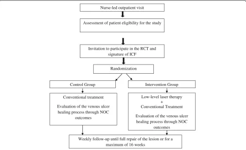
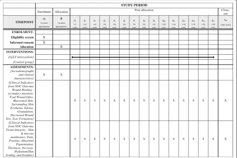
This randomized clinical trial will be conducted on a sample of participants recruited from the outpatient clinics of Hospital de Clínicas de Porto Alegre (HCPA), a tertiary-care center affiliated with the Federal University of Rio Grande do Sul, Brazil. At HCPA, patients with venous ulcers receive care from nurses, who perform nursing consultations with a focus on wound dressing and patient education. This study is conducted in accordance with the Standard Protocol Items: Recommendations for Interventional Trials (SPIRIT) Checklist (Additional file 1 and Fig. 1).
Inclusion and exclusion criteria
The study will include adult subjects (age≥18 years) of both sexes with an established diagnosis of chronic ven-ous insufficiency and an active venven-ous ulcer. All must also be available to attend the outpatient clinic weekly.
Exclusion criteria include a body mass index consistent with grade 3 obesity, current treatment for cancer, erysipelas, cellulitis, lymphangitis, chronic lymphedema, immunosup-pressant and/or corticosteroid therapy, a circular venous
Assessment of patient eligibility for the study
Invitation to participate in the RCT and signature of ICF
Nurse-led outpatient visit
Randomization
Control Group Intervention Group
Low-level laser therapy +
Conventional Treatment
Evaluation of the venous ulcer healing process through NOC
outcomes Conventional treatment
Evaluation of the venous ulcer healing process through NOC
outcomes
Weekly follow-up until full repair of the lesion or for a maximum of 16 weeks
[image:3.595.59.538.423.716.2]ulcer, and coagulation necrosis covering more than 25% of the wound bed.
The following exclusion criteria were adopted because the conditions increase the average time to chronic wound healing even further, and would, thus, require a longer follow-up period than specified for the study: cir-cumferential ulcers, due to their large extent; lymphan-gitis, erysipelas, and cellulitis, because they prolong the inflammatory phase of the wound and, consequently, hinder the tissue repair process; morbid obesity, because it hinders cellular nutrition and can make it difficult for patients to complete the proposed exercises and change their own dressings; and finally, active treatment for can-cer because it is an established contraindication to laser therapy.
Ethical considerations
Participants will be given information about the study and will read and sign an informed consent form before enter-ing the study. Continuity of conventional treatment after completion of the study will be ensured for both groups. After 6 months, all patients will meet with the study team, who will evaluate tissue repair and their lifestyle, and they will receive specific care to prevent recurrent or new ul-cers. The study protocol (15–0634) was approved by the institutional review boards of the hospital and the univer-sity. The study will be conducted in accordance with the principles of the Declaration of Helsinki and in accord-ance with Brazilian Ministry of Health guidelines and le-gislation for research on human subjects.
Sample size
The WINPEPI program, version 11.43, was used to cal-culate the sample size. A sample of 34 subjects (n= 17 per group) would be able to detect a 1-point difference on a NOC indicator score between group means (laser intervention versus conventional control) as significant. The indicators are scored on 5-point Likert scales (range 1 to 5), in which the lowest score represents the worst possible state and the highest score represents the most desirable state after implementation of interventions. A change in one level, i.e., one point on the Likert scale, characterizes a positive effect of the intervention imple-mented throughout the treatment study, according to mixed linear models and generalized estimating equations. The standard deviation common to the groups [18], with a statistical power of 80% and significance level of 5% were defined for the study. To account for possible refusals and losses to follow-up, the sample was oversized by 20%. Thus, 40 subjects (n= 20 per group) will be recruited.
Interventions
The IG will receive LLLT using an Inbramed® system, which emits laser light in the red spectrum (wavelength
660 nm and power 30 mW). The application will occur over the center of the injured area in sweep mode, with the laser tip at least 1 cm away from the bleeding area. At the wound edges and at a distance from the lesion, application will be performed in spot mode, with the laser tip leaning against the skin. The laser tip will be covered with a clear, disposable polyvinyl chloride lens to prevent infection and contamination as a result of dir-ect contact with the lesion. The nurse and patient will wear personal protective eyewear rated for laser use. After laser therapy, a conventional dressing will be ap-plied, and self-care guidelines will be given to the patient as clinically indicated.
The CG will receive conventional treatment alone, which constitutes conventional wound dressing and application of topical medication as indicated by the characteristics of the ulcer and perilesional area (includ-ing silver or calcium alginate, petrolatum-impregnated gauze, medium-chain triglycerides, papain, hydrogel, or solid petroleum jelly), followed by compressive therapy and patient self-care guidance as clinically indicated.
Study protocol
Patients eligible for the study will be divided into two groups (IG and CG) (Fig. 2). IG patients will receive LLLT as an adjunct to conventional treatment. CG pa-tients will receive conventional treatment alone. After the interventions, patients from both groups will receive guidance on self-care at home.
Conventional treatment in CG participants will be provided by a staff nurse in the outpatient wound-care clinic who has clinical experience in the care of patients with venous ulcers. IG participants will be treated by a nurse who has similar clinical experience in wound care and is also a trained laser therapist. All nurses have been trained in the standard procedures for venous ulcer care and the clinical indicators of NOC outcomes to evaluate these lesions.
In both groups, an evaluation of healing will be car-ried out by assessing the 14 constituent clinical indica-tors of the NOC outcomes for wound healing (secondary intention; 1103) and tissue integrity (skin and mucous membranes; 1101). The 14 clinical indica-tors are foul wound odor, macerated skin, surrounding skin erythema, edema, granulation, decreased wound size, scar formation, pain, pruritus, abnormal pigmen-tation, thickness, necrosis, hydration/skin scaling, and exudate. Each clinical indicator will be evaluated using a 5-point Likert scale, where the lowest score denotes the worst possible outcome, and the highest score, the best possible outcome.
protocol was based on previous studies of phototherapy for chronic wounds [12–16]. The duration of treatment was determined based on the average healing period of a chronic wound [7–12].
Randomization
During nursing consultations at the outpatient clinic, in-vestigators will assess each patient’s eligibility for the study, according to the previously established inclusion and exclusion criteria. Once deemed eligible, the patient will be invited to participate in the study. Those who accept will be asked to sign an informed consent form.
Subsequently, an additional meeting with the research assistant, the patient will be prompted to draw a sealed brown envelope with no external notations or identifica-tion that might refer to their group allocaidentifica-tion. Inside the envelope, there is a label indicating the group to which the patient belongs (that is, control or intervention). The envelope will then be opened by the patient himself. A research assistant putting the labels inside the envelopes before the study began. It organizes all the instruments that will be necesssary for the application of the re-search, confers and organizes the consultations agendas.
Demographic and clinical variables
A structured questionnaire will be administered to all study participants to obtain sociodemographic and clinical infor-mation (age, sex, educational attainment, current medica-tions, comorbidities, smoking, alcoholism, chronic venous insufficiency grade, ulcer duration, wound dressing regi-men, nutrition, and exercise). Evaluation of the ulcer itself will be performed using the 14 NOC clinical indicators de-scribed above. Patients will be instructed to perform daily plantar flexion and extension and calf-strengthening exer-cises, resting in between, and taught how to dress their wounds as appropriate to their individual needs.
Outcomes
Primary outcome
The primary outcome is decreased wound size and scar formation based on NOC outcome wound healing (sec-ondary intention; 1103).
1. Wound area in cm2(measured as the product of the longest dimension in the cephalocaudal direction by the widest dimension), evaluated by a Likert scale, with 1 being the worst possible score and 5 the best possible score. Each Likert scale STUDY PERIOD
Enrolment Allocation Post-allocation Close-out
TIMEPOINT (at nurse appointment)
0 (at nurse appointment )
(1st week)
(2nd week)
(3rd week)
(4th week)
(5th week)
(6th week)
(7th week)
(8th week)
(9th week)
(10th week)
(11th week)
(12th week)
(13th week)
(14th week)
(15th week)
(16th week)
ENROLMENT:
Eligibility screen X
Informed consent X
Allocation X
INTERVENTIONS: [LLLT intervention] [Control group]
ASSESSMENTS: [Sociodemographi and clinical characteristics]
X X
[Clinical Indicators from NOC Outcome
Wound Healing -secondary intention; Foul Wound Odor, Macerated Skin, Surrounding Skin Erythema, Edema, Granulation, Decreased Wound Size, Scar Formation]
X X X X X X X X X X X X X X X X
[Clinical Indicators from NOC Outcome Tissue Integrity - Skin & mucous membranes: Pain, Pruritus, Abnormal Pigmentation, Thickness, Necrosis, Hydration/Skin Scaling, and Exudate]
X X X X X X X X X X X X X X X X
[image:5.595.58.539.89.409.2]score of the Decreased wound size indicator has an operational definition that corresponds to the size in cm2.
2. Wound covered with epithelial tissue (new pink or bright tissue that develops from the wound edges or as islands on the surface of the wound), evaluated by a Likert scale, with 1 being the worst possible score and 5 the best possible score.
Secondary outcomes
1. Thickness
NOC tissue integrity (skin and mucous membranes; 1101) –depth reached: The layers and structures of the skin altered by loss of tissue integrity (ulcerated area) will be evaluated by a Likert scale, with 1 being the worst possible score and 5 the best possible score.
2. Pain
NOC tissue integrity (skin and mucous membranes; 1101)–Unpleasant sensory and emotional experience aris-ing from actual or potential tissue damage or described in terms of such damage, with sudden or slow onset of mild to severe intensity, constant or recurrent, without an antici-pated or predictable termination: The frequency, condition, and intensity will be evaluated by a Likert scale, with 1 being the worst possible score and 5 the best possible score.
3. Overall improvement of other correlated NOC indicators
Overall improvement of the indicators for the NOC outcomes for wound healing (secondary intention; 1103) and tissue integrity (skin and mucous membranes; 1101) will also be assessed.
Independent/exposure and confounding variables
The main exposure variable will be the tissue repair process in the CG and IG. The impact of each man-agement strategy on the outcomes will be controlled by monitoring the following confounding variables: pharmacotherapy or co-intervention at another health facility or by another professional.
Statistical analyses
Continuous variables will be expressed as mean and standard deviation or median and interquartile range ac-cording to the data distribution. Categorical variables will be expressed as absolute and relative frequencies. Generalized estimating equations will be used for com-parisons between the weekly indicators, and the
least-significance-difference post-hoc test will be used to assess the difference between weeks.
For quantitative variables with a normal distribution, the difference between the two groups will be compared by Student’st-test. This test will also be used to analyze the means of the NOC results in relation to the de-creased wound size and scar formation outcomes. The Pearson correlation coefficient will be used to evaluate the linear association between NOC results and primary
outcomes. A nonparametric Mann–Whitney test will be
used for comparison between the two groups regarding the characterization of ulcers.
To assess the effect size of the intervention, relative risks with 95% confidence intervals will be calculated. A two-tailed P< 0.05 will be considered statistically signifi-cant. All analyses will be performed in PASW Statistics, Version 18.0.
Discussion
The proposed randomized controlled trial will evaluate the efficacy of venous ulcer treatment with low-power laser therapy. Our study is innovative in many ways. We have developed an outpatient wound-care protocol de-signed to promote faster tissue regeneration with less costly care, combining technology and direct supervision.
As in all studies, we anticipate that there will be potential problems. Recruitment for clinical trials is challenging, as changes in infrastructure and supplies may change during the study. Another point to be dis-cussed is the long duration of follow-up, in this case up to 16 weeks, which may be influenced by other compo-nents. The 16-week period being proposed is understood to be adequate, considering the average healing time of a venous ulcer. One limitation is that more complex pa-tients were excluded, due to factors that could prolong healing time. However, our team’s experience in research projects and its ability to measure the outcomes of this intervention are some of our key advantages.
Trial status
Enrollment is ongoing. Recruitment started in March 2017 and is expected to conclude in December 2018. The first block of randomized patients is already receiving the study interventions, and more participants are being re-cruited. Target enrollment for the study is 40 subjects.
Additional file
Additional file 1:SPIRIT 2013 checklist: recommended items to address in a clinical trial protocol and related documents. (DOC 121 kb)
Abbreviations
Acknowledgements
We are grateful to Conselho Nacional de Desenvolvimento Científico e Tecnológico (CNPq) of the Brazilian Ministry of Science, Technology and Innovation and Fundo de Incentivo a Pesquisa e Eventos of Clinicas Hospital of Porto Alegre (FIPE/HCPA) for financial support.
Funding
This study was funded by Conselho Nacional de Desenvolvimento Científico e Tecnológico (CNPq) of the Brazilian Ministry of Science, Technology and Innovation (universal tender 01/2016 and process 405997/2016–7) and the Research and Event Incentive Fund of Hospital de Clínicas de Porto Alegre (grant 15.0634).
Availability of data and materials
Data sharing is not applicable to this article as no datasets were generated or analyzed during the current study.
Authors’contributions
TB Conceived the study, conducted the LLLT intervention, and drafted the manuscript. AP, VMM, VMO, and DTS assisted in data collection and patient randomization, and drafted the manuscript. AFL Conceived the study and drafted the manuscript. All authors read and approved the final manuscript.
Ethics approval and consent to participate
The study was approved by the Ethics and Research Committee of the Porto Alegre Hospital de Clínicas - HCPA in accordance with Brazilian Resolution No. 466 of December 2012. The consent is appropriate and satisfies the criteria required by HCPA. All patients with a venous ulcer will sign informed consent, which includes the ethical aspects of confidentiality and voluntary participation.
Consent for publication
Not applicable.
Competing interests
The authors declare that they have no competing interests.
Publisher’s Note
Springer Nature remains neutral with regard to jurisdictional claims in published maps and institutional affiliations.
Author details 1
Nursing School at Universidade Federal do Rio Grande do Sul, São Manoel, 963, Rio Branco, Porto Alegre 90620-110, Brazil.2Nursing School at Universidade Federal do Rio Grande do Sul, Hospital de Clínicas de Porto Alegre, São Manoel, 963, Rio Branco, Porto Alegre 90620-110, Brazil.3Hospital de Clínicas de Porto Alegre, Ramiro Barcelos, 2350, Santa Cecilia, Porto Alegre, RS 90035-903, Brazil.4Caxias do Sul, Brazil.
Received: 26 February 2018 Accepted: 7 June 2018
References
1. Santler B, Goerge T. Chronic venous insufficiency–a review of
pathophysiology, diagnosis, and treatment. J Dtsch Dermatol Gesellschaft. 2017;15(5):538–56.https://doi.org/10.1111/ddg.13242.
2. Spiridon M, Corduneanu D. Chronic Venous Insufficiency: a Frequently Underdiagnosed and Undertreated Pathology. Mædica. 2017;12(1):59–61. 3. Rabe E, Guex JJ, Puskas A, Scuderi A, Fernandez QF. Epidemiology of
chronic venous disorders in geographically diverse populations: results from the Vein Consult Program. Int Angiol. 2012;31(2):105–15.
4. Agale SV. Chronic leg ulcers: epidemiology, aetiopathogenesis, and management. Ulcers. 2013;1:9.https://doi.org/10.1155/2013/413604. 5. Borges EL, Caliri MHL, Haas VJ. Systematic review of topic treatment for
venous ulcers. Rev Latino-Americana Enfermagem. 2007;15(6):1163–70.
https://doi.org/10.1590/S0104-11692007000600017.
6. O’Donnell TF, Passman MA, Ennis WJ, Dalsing M, Kistner RL, Lurie F, Gloviczki P. Management of venous leg ulcers: clinical practice guidelines of the Society for Vascular Surgery® and the American venous forum. J Vasc Surg. 2014;60(2):3S–59S.https://doi.org/10.1016/j.jvs.2014.04.049.
7. Tchanque-Fossuo CN, Ho D, Dahle SE, Koo E, Li CS, Isseroff RR, Jagdeo J. A systematic review of low-level light therapy for treatment of diabetic foot ulcer. Wound Repair Regen. 2016;24(2):418–26.https://doi.org/10.1111/wrr.12399. 8. Andrade FSSD, Clark RMO, Ferreira ML. Effects of low-level laser therapy on
wound healing. Rev Colégio Brasileiro Cirurgiões. 2014;41(2):129–33.
https://doi.org/10.1590/S0100-69912014000200010.
9. Palagi S, Severo IM, Menegon DB, Lucena AF. Laserterapia em úlcera por pressão: avaliação pelas Pressure Ulcer Scale for Healing e Nursing Outcomes Classification. Rev Escola Enfermagem USP. 2015;49(5):826–33.
https://doi.org/10.1590/S0080-623420150000500017.
10. Meneguzzo DT, Lopes LA, Ribeiro MS. Terapia laser de baixa potência na reparação tecidual. In: Garcez, Ribeiro, Nunez, editors. Laser de baixa potência: princípios básicos e aplicações clínicos na Odontologia. Rio de Janeiro: Elsevier; 2012. p. 68–78.
11. Barreto JG, Salgado CG. Clinic-epidemiological evaluation of ulcers in patients with leprosy sequelae and the effect of low level laser therapy on wound healing: a randomized clinical trial. BMC Infect Dis. 2010;10(1):237– 45.https://doi.org/10.1186/1471-2334-10-237.
12. Siqueira CP, De Paula Ramos S, Gobbi CA, Kashimoto RK, Venâncio EJ, de Oliveira Toginho Filho D, Castaldin AG, Felinto AS, Silva RB, Dias IF. Effects of weekly LED therapy at 625 nm on the treatment of chronic lower ulcers. Lasers Med Sci. 2015;30(1):367–73.https://doi.org/10.1007/s10103-014-1666-5. 13. Baffoni M, Bessa LJ, Grande R, Di Giulio M, Mongelli M, Ciarelli A, Cellini L.
Laser irradiation effect on Staphylococcus aureus and Pseudomonas aeruginosa biofilms isolated from venous leg ulcer. Int Wound J. 2012;9(5): 517–24.https://doi.org/10.1111/j.1742-481X.2011.00910.x.
14. Machado RS, Viana S, Sbruzzi G. Low-level laser therapy in the treatment of pressure ulcers: systematic review. Lasers Med Sci. 2017;32(4):937–44.
https://doi.org/10.1007/s10103-017-2150-9.
15. Salvi M, Rimini D, Molinari F, Bestente G, Bruno A. Effect of low-level light therapy on diabetic foot ulcers: a near-infrared spectroscopy study. J Biomed Opt. 2017;22(3)https://doi.org/10.1117/1.JBO.22.3.038001. 16. Vitse J, Bekara F, Byun S, Herlin C, Teot L. A double-blind,
placebo-controlled randomized evaluation of the effect of low-level laser therapy on venous leg ulcers. Int J Lower Extrem Wounds. 2017;16(1): 29–35.https://doi.org/10.1177/1534734617690948.
17. Moorhead S, Johnson M, Maas M, Swanson E. Nursing outcomes classification (NOC): measurement of health outcomes. 5th ed. Philadelphia: Elsevier; 2013.