INTRODUCTION
Just over three decades ago, the discovery of splicing brought a revolution to molecular biology (Gilbert, 1978). It was then realized that alternative splicing could provide a higher coding capacity to a single gene by generating distinct proteins (Black, 2003). Although it is known that at least 43% of the genes of Drosophila melanogaster, and about two-thirds of mouse and human genes, encode alternatively spliced mRNA sequences (Johnson et al., 2003; Stolc et al., 2004; Park and Gravely, 2007; Ben-Dov et al., 2008), the functional relevance of the majority of alternative splicing events remains to be established.
In Drosophila, an extreme case of diversity obtained through alternative splicing is observed in the Dscamgene, which may potentially code for more than 38,000 splicing variants (Graveley, 2005). Alternative splicing in the double sexgene provides a clear example of functional divergence of protein isoforms, as it determines the specific development of males or females (Schütt and Nöthiger, 2000). Within the Hox genes, which encode homeodomain-containing transcription factors that specify structures along the anteroposterior axis of bilaterians (Pearson et al., 2005; Foronda et al., 2009) there are several examples of alternative splicing in mouse, human and Drosophila(Mlodzik et
al., 1988; Bermingham and Scott, 1988; Celniker et al., 1989; Zavortink and Sakonju, 1989; deLorenzi and Bienz, 1990; Shen et al., 1991; Cribbs et al., 1992; Sham et al., 1992; Fujimoto et al., 1998; Patel et al., 1999). However, the relevance of alternative splicing for Hox gene function has been little explored – except, perhaps, in the Drosophila Hox gene Ultrabithorax(Ubx).
The Drosophila Ubx gene specifies the development of adult derivatives such as the third leg, the haltere and the first abdominal segment (Lewis, 1963; Morata and Kerridge, 1981). Wings and halteres are serially homologous, and different, thoracic dorsal appendages. Ubxis expressed in the haltere discs but not in the wing discs – the primordia of halteres and wings, respectively (White and Wilcox, 1984; Beachy et al., 1985). Mutations that eliminate Ubx expression transform halteres into wings, whereas ectopic expression of Ubxtransforms wings into halteres (Lewis 1963; Lewis, 1978; Cabrera et al., 1985; White and Wilcox, 1985; White and Akam, 1985). Ubxmutations that slightly change either Ubxexpression pattern or levels in the haltere disc lead to milder, yet easily visible, transformations of haltere, revealing a highly sensitive response to the levels of Ubxinput. Several specific targets regulated by Ubxin the haltere imaginal disc have been identified (Weatherbee et al., 1998; Mohit et al., 2006; Hersh et al., 2007) (T. Pavlopoulos and M.A., unpublished). Thus, the haltere system provides an excellent framework for the qualitative and quantitative assessment of Ubx function at both developmental and molecular levels.
Ubxalternative splicing leads to the formation of six different Ubx mRNAs according to the use of alternative splice donor sites at the start of the first exon added to the optional inclusion of two 51 bp microexons located between the 5⬘and 3⬘common exons (Fig. 1A). The resulting Ubx proteins are named I, II or IV depending on whether they are encoded by: all the exons (I); the 5⬘exon, the second microexon, and the 3⬘exon (II); or just the 5⬘and 3⬘exons Development 138, 107-116 (2011) doi:10.1242/dev.051409
© 2011. Published by The Company of Biologists Ltd
1Centro de Biología Molecular Severo Ochoa (C.S.I.C.-U.A.M.), Universidad Autónoma de Madrid, Nicolás Cabrera 1, Cantoblanco, 28049 Madrid, Spain. 2Department of Zoology, University of Cambridge, Downing Street, Cambridge CB2 3EJ, UK. 3Center for Cooperative Research in Biosciences CIC Biogune, Bizkaia Technology Park, Building 801-A, Derio 48160, Spain. 4John Maynard Smith Building, School of Life Sciences, University of Sussex, Brighton BN1 9QG, UK.
*Author for correspondence ([email protected])
Accepted 19 October 2010
SUMMARY
Although most metazoan genes undergo alternative splicing, the functional relevance of the majority of alternative splicing products is still unknown. Here we explore this problem in the DrosophilaHox gene Ultrabithorax(Ubx). Ubxproduces a family of six protein isoforms through alternative splicing. To investigate the functional specificity of the Ubx isoforms, we studied their role during the formation of the Drosophilahalteres, small dorsal appendages that are essential for normal flight. Our work shows that isoform Ia, which is encoded by all Ubxexons, is more efficient than isoform IVa, which lacks the amino acids coded by two small exons, in controlling haltere development and regulating Ubxdownstream targets. However, our experiments also demonstrate that the functional differences among the Ubx isoforms can be compensated for by increasing the expression levels of the less efficient form. The analysis of the DNA-binding profiles of Ubx isoforms to a natural Ubxtarget, spalt, shows no major differences in isoform DNA-binding activities, suggesting that alternative splicing might primarily affect the regulatory capacity of the isoforms rather than their DNA-binding patterns. Our results suggest that to obtain distinct functional outputs during normal development genes must integrate the generation of qualitative differences by alternative splicing to quantitative processes affecting isoform protein expression levels.
KEY WORDS: Drosophila,Ultrabithorax, Hox, Alternative splicing, spalt
Integration of RNA processing and expression level control
modulates the function of the
Drosophila
Hox gene
Ultrabithorax
during adult development
Luis F. de Navas1, Hilary Reed2, Michael Akam2, Rosa Barrio3, Claudio R. Alonso4and
Ernesto Sánchez-Herrero1,*
D
E
V
E
LO
P
M
E
N
(IV). Each Ubx protein variant (i.e. I, II or IV) is named as ‘a’ or ‘b’ depending on: (a) the lack of, or (b) the inclusion of a small nine amino acid segment encoded between the two donor splicing sites at the 3⬘end of the first exon (O’Connor et al., 1988; Kornfeld et al., 1989) (Fig. 1A). Previous studies have revealed that each isoform has distinct expression in the embryo, that class ‘b’ isoforms are much less abundant than class ‘a’ proteins, and that isoform Ia is the predominant one in most stages (O’Connor et al., 1988; Kornfeld et al., 1989; López and Hogness, 1991; Artero et al., 1992; Bomze and López, 1994; López et al., 1996).
The extent to which the different isoforms possess different functions remains unclear. A pioneer analysis was done with the UbxMX17mutation, an inversion with breakpoints to the left and right of the second microexon, and which forms only isoforms IVa and IVb (Busturia et al., 1990; Subramaniam et al., 1994). Homozygous UbxMX17adults present small changes in phenotype, most obviously a partial transformation of haltere to wing. This effect, however, was attributed to a reduction in Ubx protein expression in the haltere disc and not to changes in the distribution of the different Ubx proteins, thus suggesting all Ubx isoforms are functionally equivalent (Busturia et al., 1990).
Other experiments, however, showed that changes in the activities of the Ia and IVa isoforms affected the peripheral nervous system (Mann and Hogness, 1990; Subramaniam et al., 1994) or the Keilin’s organs, particular sensory structures (Gebelein et al., 2002), differently. Further work on the UbxMX17mutants showed defects in flight and behaviour (Subramaniam et al., 1994). Besides, no reduction in expression was observed in haltere discs of the mutant larvae, suggesting functional differences between Ubx isoforms (Subramaniam et al., 1994). Regardless of this evidence, and perhaps because of the discrepancies between the different studies, most recent work has assumed that alternative splicing is irrelevant to Ubx function, and accordingly, a single Ubx isoform (i.e. UbxIa) is normally considered to be representative of the function of all Ubx proteins.
Here we re-examine the role of alternative splicing on Ubx function, focusing on haltere development. Our experimental design has allowed us to assess, with high sensitivity, the abilities of the different Ubx isoforms to control haltere development or the regulation of target genes in haltere discs, and to compare these results with those obtained in UbxMX17mutants. Finally, we test the DNA-binding profiles of different Ubx isoforms to one of these molecular targets, the gene spalt. We find that Ubx isoform IVa is not able to specify the normal development of halteres or to regulate Ubx target genes as efficiently as isoform Ia when expressed at comparable levels in the haltere disc. However, higher levels of isoform IVa can compensate for its lower activity and form normal halteres. The differential activity of Ia and IVa proteins does not seem to depend on major differential DNA-binding profiles. Our results indicate that alternative splicing significantly modulates Ubx function during Drosophila adult development, and also reveal that low-performance splicing isoforms can improve their function if levels of expression are increased. We suggest that developmental genes must coordinate the formation of different proteins by alternative splicing with the regulation of their levels of expression in different tissues.
MATERIALS AND METHODS Genetics
We used the following stocks: Ubx-Gal4LDN (de Navas et al., 2006), scalloped-Gal4 (Garaulet et al., 2008), C-765-Gal4 (Guillén et al., 1995), MS1096-Gal4 (Capdevila and Guerrero, 1994), UAS-UbxIa (Castelli-Gair
et al., 1994), UAS-GFP (Drysdale et al., 2005), abx bx3 pbxand Ubx1 (Lewis, 1982). In most experiments we have used a recombinant Ubx-Gal4LDNUAS-y+chromosome, and grown the larvae at 17°C (except those with the UAS-IVa2 line, grown at 25°C or 29°C), to reduce the amount of Gal4 protein (normally in excess) that activates each UAS construct. The salE/Pvenhancer was described previously (Barrio and de Celis, 2004).
Immunostaining
Immunostaining was performed according to standard protocols (Wolff, 2000). The antibodies used were mouse and rabbit anti--galactosidase (Cappel), mouse anti-Ubx (White and Wilcox, 1984), rabbit and rat anti-Sal (Barrio et al., 1999), mouse anti-Wg (Brook and Cohen, 1996), rat anti-Ara (Díez del Corral et al., 1999) and rat anti-Tub (YL/2) (Serelab). The intensity of the fluorescence signal in the haltere pouch was measured with the Measure tool of the MetaMorph program (Universal Imaging Corporation) or the Measure tool of ImageJ. Larvae of the different genotypes were put in tiny baskets, and all the baskets placed together in a wide vial in which all the fixation, incubation with antibodies and staining were carried out, allowing the free movement of the liquids, so that discs of all the genotypes received the same conditions. We took three measurements in each of five different discs and measured the average fluorescence.
RT-PCR studies
RNA was isolated using illustra RNAspin Mini (GE Healthcare) from 50 haltere discs of either wild-type or UbxMX17homozygous larvae. cDNA synthesis was performed with the M-MLV reverse transcriptase (Invitrogen) using the Ubx.3A1 primer (5⬘GCGGGTCAG -ATAATGATTCGT-3⬘), which hybridizes to nucleotides 78 to 98 bp downstream to the 3⬘splice site of the 3⬘exon. The cDNA obtained was amplified by PCR using the Ubx.5S1 (5⬘TGGAATGC -CAATTGCACCATC-3⬘), which hybridizes to the Ubx 5⬘ exon at nucleotides –133 to –113 bp upstream to the ‘b’ 5⬘splice site, and Ubx.3A1 primers. The analysis of the PCR product was made in 2% agarose gels.
Construction of UAS-Ubxtransgenic lines This has been previously described (Reed et al., 2010).
Western blots
Western blotting was performed as described (Ausubel et al., 2004). We used biotinilated anti-rat (1:10,000) and anti-mouse antibodies (1:200). Measurement of the signal was carried out with a GS-710 Calibrated Image Densitometer (BioRad) for the digitalization of the film and the Quantity One (BioRad) software for the analysis of the data.
DNA-binding experiments
A series of DNA elements derived from the salcis-regulatory region were analysed by electrophoretic mobility shift assay (EMSA) (the sequence of each element is provided on request). In brief, double-stranded oligonucleotides (33-38mer) centred on Ubx binding sites sal3, sal4and sal5/6(Galant et al., 2002; Walsh and Carroll, 2007) bearing 5⬘protruding T-overhangs (2/3x) were radioactively labelled with 32P by end-filling with the Klenow fragment of the DNA polymerase I (New England Biolabs). Proteins were produced by in vitro translation using the TnT system (Promega, Madison, USA) in the presence of small amounts of 35 S-methionine (MP Biomedicals). Protein quality and size were examined in 12% PAGE gels, protein bands were quantified in a Typhoon Trio Scanner Unit (Amersham Biosciences), and values normalized according to the number of methionines in each isoform. EMSA reactions were carried out as previously described (Galant et al., 2002; Walsh and Carroll, 2007); matching amounts of Ubx proteins were incubated with 32P-labelled DNA elements for 30 minutes at room temperature in a buffer system (4% Ficoll, 40 mM Hepes pH 7.8, 100 mM KCl, 0.5 mg/ml BSA, 2 mM DTT) in the presence of non-specific competitor p[dIdC] (Sigma). Total amount of lysate volume was kept constant across all reactions using un-programmed TnT reactions (without any template DNA). Protein-DNA binary complexes were resolved at 4°C in pre-run 5% native polyacrylamide gels; after electrophoresis, gels were fixed, dried and exposed to imaging plates (Molecular Dynamics) overnight. Images were collected as tiff files using
ImageQuant software (Amersham Biosciences).
D
E
V
E
LO
P
M
E
N
Adult cuticle analysis
This was done as previously described (de Navas et al., 2006).
RESULTS
We analysed the abilities of the Ubx splicing isoforms (Fig. 1A) to specify haltere development by two methods: first, we combined the use of Ubx mutants and a specific Ubx-Gal4 driver line to examine Ubxfunctions during haltere development; second, we re-examined the phenotype and Ubx protein expression in UbxMX17 mutant animals.
Rescue of the Ubxmutant phenotype by different Ubx isoforms
We first studied which Ubx isoforms are present in the wild-type haltere disc during late larval stages by RT-PCR. As shown in Fig. 1B, isoforms I and II are abundant in these discs, and a small amount of isoform IV can also be detected. This agrees with results showing that cDNAs encoding isoforms IVa and IVb are much less abundant than other Ubx cDNAs in third instar larvae (O’Connor et al., 1988; Kornfeld et al., 1989). We cannot resolve whether isoforms a and b (Ia and Ib, IIa and IIb) are equally represented, but previous data indicate that the ‘a’ isoforms are much more abundant than the ‘b’ ones (O’Connor et al., 1988; Kornfeld et al., 1989; Subramaniam et al., 1994; Bomze et al., 1994).
To carry out a functional analysis of the different isoforms in forming a haltere we used the GAL4/UAS method (Brand and Perrimon, 1993) with the Ubx-Gal4LDNinsertion. This Gal4 line is inserted in the Ubxgene, drives expression mainly in the haltere pouch and is mutant for Ubx(de Navas et al., 2006). Ubx-Gal4LDN
[image:3.612.54.507.62.287.2]UAS-y+/abx bx3pbxadults (we refer to this mutant combination as the Ubxmutant background, or UMB), show a transformation of the distal part of the haltere (the capitellum) into the wing (de Navas et al., 2006); halteres are bigger and bear bristles in their margins and veins in their surface (Fig. 1E, the wild-type haltere and wing are shown in Fig. 1D and 1C, respectively). As shown in Fig. 1B, this mutant combination does not alter the isoform distribution in haltere discs. We introduced different Ubx isoforms (UAS-Ubx constructs) into this mutant background and checked whether the mutant phenotype was rescued. Because the ‘b’ forms are much less abundant than the ‘a’ ones throughout development, we concentrated on studying the ‘a’ variants. As shown in Fig. 1F,G, the expression of isoforms Ia and IIa in the mutant background rescues much of the mutant phenotype, reducing the size of the haltere and suppressing the development of margin bristles and veins. We found, however, that a line that expresses the IVa protein (line UAS-IVa2) only partially rescued the mutant phenotype (Fig. 1H): the size of the transformed haltere was much bigger than that of the wild type and there were marginal bristles and vein tissue. Although the initial experiments with this line were done at 25°C, the rescue was not much better at 29°C (not shown). Similar results were observed when expressing these lines in the wing disc with scalloped-Gal4, MS1096-Gal4 or C-765 -Gal4 lines: the flies expressing the Ia protein variably transformed wings into halteres, even though the crosses are made at 17°C (Fig. 1J,L; data not shown), whereas adults expressing the IVa2 line only showed mild effects in wing development, even at 25°C (Fig. 1I,K; data not shown). We decided to study this line further and compare it with our standard Ia line (encoding the most abundant Ubx isoform).
Fig. 1.Ubxphenotypes produced by different Ubx isoforms.(A)Scheme showing the different Ubx proteins (to the right) formed by
alternative splicing. mI and mII stand for the first and second microexons, respectively. (B)RT-PCR of third instar haltere imaginal discs of wild-type
and abx bx3pbx/Ubx-Gal4LDNUAS-y+(UMB) larvae showing that the mRNAs encoding proteins I, II and (at very low levels) IV are present. (C)
Wild-type thorax, showing the wing (w) and haltere (h). (D)Wild-type haltere. (E-H)Rescue of the abx bx3pbx/Ubx-Gal4LDNUAS-y+mutant phenotype
(E, UMB), that partially transforms halteres into wings (compared with the wild-type haltere in D), by the Ia (line SG1, F), IIa (G) or IVa (line IVa2; H)
proteins. Note that the rescue in the latter is only partial. Pictures in D-H are at the same magnification. (I-L). Female adults expressing the IVa2 (I,K)
or Ia (J,L) constructs under the control of the sd-Gal4 (I,J) or MS1096-Gal4 (K,L) lines. See the very mild effects when expressing the IVa isoform (the
arrow in I indicates the absence of costal bristles) and the strong effects when expressing the Ia protein (arrowhead in J indicates the wing transformed into a haltere). SM, size marker; wt, wild type.
D
E
V
E
LO
P
M
E
N
The UbxIVa isoform is not as efficient as the UbxIa isoform in promoting a wild-type haltere
The partial rescue of the haltere mutant phenotype obtained with the IVa2 line could be due to its expressing very low Ubx levels. To check this possibility we compared, by measuring fluorescent staining and in western blots, the levels of Ubx products in the haltere discs of the wild-type and Ubx mutant background larvae with those observed when different UAS-Ubx constructs were expressed in the same mutant background. In all the cases we used an anti-Ubx antibody that recognizes all Ubx proteins (White and Wilcox, 1984).
We examined the fluorescent staining levels of haltere pouches of the following genotypes: wild type, Ubx1/+, Ubx-Gal4LDN
UAS-y+/abx bx3pbx(UMB), Ubx-Gal4LDNUAS-y+/abx bx3pbx UAS-UbxIa (SG1 line) (UAS-UAS-UbxIa/+; UMB) and UAS-UbxIVa2/+; Ubx-Gal4LDNUAS-y+/abx bx3pbx(UAS-UbxIVa2/+; UMB; this cross was made at 29°C) (Fig. 2A-E; the pattern driven by the Ubx-Gal4LDNline is shown in Fig. 2F). Data showing fluorescence
intensities in the pouches are summarized in Table 1. We found that the average intensity in the haltere pouch of UAS-UbxIVa2/+; UMB larvae is 60.2±2.4% of the wild type, a figure not statistically significantly different (P<0.05) from that observed in Ubx1/+ larvae (66.6±2.3%); the latter percentage is higher than 50% due to Ubx negatively regulating its own transcription (Irvine et al., 1993; Garaulet et al., 2008; Crickmore et al., 2009). Measurements in western blots of haltere discs gave comparable results: 62±6.7% for the UAS-UbxIVa2/+; UMB discs and 65.6±2.3% for the Ubx/+ genotype (Fig. 2I and Table 1), the two measurements not being statistically significantly different (P<0.05). Therefore, the total amount of Ubx protein in the haltere pouch of UAS-UbxIVa2/+; UMB or Ubx1/+ larvae did not differ much. However, the phenotype in the halteres of the corresponding adult flies was different: Ubx1/+ heterozygous flies only showed a slight
enlargement of the capitellum and, occasionally, one or two bristles, whereas UAS-UbxIVa2/+; UMB adults showed a partial transformation into wing (compare Fig. 2G,H). We note that in the western blots we compared the amount of protein in the whole disc, whereas our immunofluorescence measurements were concentrated in the pouch. However, the similar results obtained with the two methods support the conclusion that isoform IVa is not as efficient as isoform Ia in making a wild-type haltere when similar levels of expression are achieved.
Two results indicate, nevertheless, that isoform IVa is not completely unable to direct haltere development. First, when we expressed this protein at higher levels in the mutant background, by using the UAS-UbxIVa1 or UAS-UbxIVa33 lines (Fig. 3B, compare with a UAS-UbxIVa2/+; UMB disc in Fig. 3A), the halteres were smaller than those of wild-type flies and slightly malformed, but showed no wing characteristics (Fig. 3D, arrow, compare with a UAS-UbxIVa2/+; UMB haltere in Fig. 3C; data not shown); second, when we introduced two UAS-UbxIVa2 insertions in our mutant background we obtained an almost complete rescue of the mutant phenotype (Fig. 3E). Our conclusion is supported by the results obtained when we used the UbxIVa1 and UAS-UbxIVa33 constructs to express the UbxIVa protein in the wing pouch with the MS1096-Gal4 or the scalloped-Gal4 lines at 17°C: in these cases we obtained a transformation of wings into halteres (Fig. 3F,G, compare with Fig. 1K and 1I, respectively; data not shown).
[image:4.612.53.364.61.220.2]In our experiments the Ia isoform rescued the Ubx mutant phenotype but at levels higher than those of the wild type or those obtained with the IVa2 line (see Table 1). To ascertain if lower levels of this protein in the mutant background were sufficient to make a normal haltere, we reduced the amount of UbxIa protein by increasing the number of UAS ‘neutral’ constructs to titrate the Gal4 protein. In UAS-GFP/UAS-GFP; UAS-UbxIa/UMB haltere Fig. 2. Levels of expression of different mutant combinations.(A-E). Anti-Ubx staining in haltere
discs of wild-type (A), Ubx1/+(B), UMB (C),
UAS-UbxIVa2/+; UMB (D) and UAS-UbxIa/+; UMB (E)
larvae. (F)Haltere of the genotype Ubx-Gal4LDN
/UAS-GFP showing the expression driven by the Gal4 line.
(G,H)Halteres of Ubx1/+(G) and UAS-UbxIVa2/+;
UMB (H) adults. Note the different size and
morphology. (I)Western blot showing the Ubx
protein signal in haltere discs of the following
genotypes: (1) wild type, (2) Ubx1/+,(3) UMB, (4)
UAS-UbxIVa2/+; UMB, (5) UAS-UbxIa UMB and (6)
UbxMX17. The expression of tubulin (tub) was used as
an internal control.
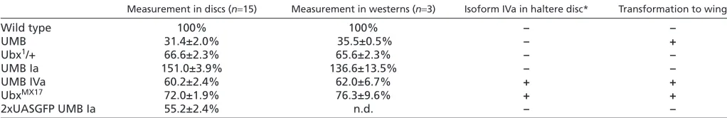
Table 1. Relative abundance of Ubx protein in the haltere disc in different mutant backgrounds when compared with the wild type (100%)
Measurement in discs (n15) Measurement in westerns (n3) Isoform IVa in haltere disc* Transformation to wing
Wild type 100% 100% – –
UMB 31.4±2.0% 35.5±0.5% – +
Ubx1/+ 66.6±2.3% 65.6±2.3% – –
UMB Ia 151.0±3.9% 136.6±13.5% – –
UMB IVa 60.2±2.4% 62.0±6.7% + +
UbxMX17 72.0±1.9% 76.3±9.6% + +
2xUASGFP UMB Ia 55.2±2.4% n.d. – –
*Only high levels considered.
n.d., not determined.
D
E
V
E
LO
P
M
E
N
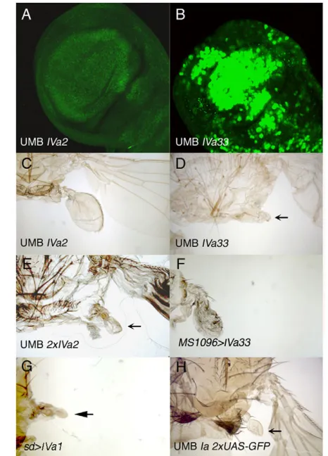
[image:4.612.49.560.637.720.2]discs, the amount of Ubx protein was about 55.2±2% that of the wild-type discs (Table 1), but the halteres were similar to those of Ubx1/+flies (Fig. 3H); by contrast, the halteres of UAS-GFP/UAS-GFP; UAS-UbxIa/UMB flies were different from those of the UAS-UbxIVa2/+; UMB genotype (Fig. 3C), although levels of Ubx expression were slightly higher in the latter mutant combination. These results strongly suggest that the Ia protein is able to make an almost normal haltere at levels that the IVa variant is unable to.
Isoform IVa does not efficiently suppress Ubx targets
To explore in more detail the different ability of the IVa2 and Ia lines in rescuing the Ubxmutant phenotype, we compared the expression of Ubxtargets in the haltere pouches of wild type and different mutant combinations.
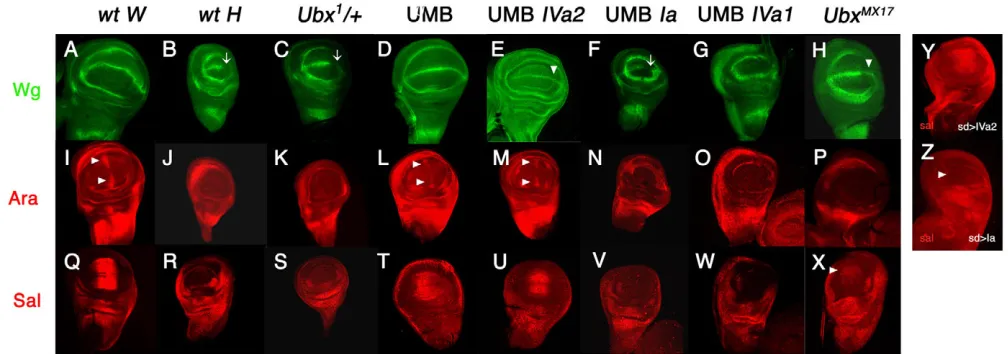
The gene wingless(wg) is required for the formation of the wing margin and is expressed at the dorsoventral boundary of wing discs (Phillips and Whittle, 1993; Couso et al., 1994) (Fig. 4A). By contrast, it is only present in the anterior compartment of the haltere disc dorsoventral boundary (Weatherbee et al., 1998; Shashidara et al., 1999) (Fig. 4B, arrow). The araucan(ara) gene is required for the development of vein L3 and associated sensilla campaniformia (Gomez-Skarmeta et al., 1996a). arais expressed in two patches of anterior cells close to the anteroposterior boundary of the wing pouch (Fig. 4I, arrows) (Gómez-Skarmeta et al., 1996a; Gómez-Skarmeta et al., 1996b), but it is not present in the haltere pouch (Fig. 4J). Finally, the gene spalt(sal) is needed for the positioning of veins L2 and L5 (reviewed in de Celis and Barrio, 2009) and is expressed in the central domain of the wing pouch (de Celis et al., 1996; Lecuit et al., 1996; Nellen et al., 1996) (Fig. 4Q) but not in the haltere pouch (Weatherbee et al., 1998; Barrio et al., 1999) (Fig. 4R).
In Ubx1/+haltere discs, the wg, araor salexpression patterns remain as in the wild type (Fig. 4C,K,S), but they change to the wing pattern in the UMB haltere discs (Fig. 4D,L,T). The wild-type haltere pattern for any of these three genes is restored if the constructs expressing the Ia (Fig. 4F,N,V) or IVa (line IVa1) (Fig. 4G,O,W) proteins are introduced in the mutant background (except that the IVa1 construct also eliminates Wg expression in the anterior pouch), but not if we introduce the IVa2 line in the same mutant background (Fig. 4E,M,U). These results indicate that: (1) wg, araand salare repressed by just one dose of Ubx in the haltere pouch; (2) isoform Ia is able to repress the expression of these targets in the mutant background that transform halteres into wings; and (3) isoform IVa is able to achieve a similar repression only when expressed at high levels but not when present at comparable levels to those of Ubx1/+haltere pouchs.
These results are paralleled by those obtained when expressing different isoforms in the wing disc, where no endogenous protein is present. In sd-Gal4/+; UAS-UbxIVa2/+ wing discs, the expression of Sal was only barely changed (Fig. 4Y), whereas expression of the Ia isoform under the same driver eliminated Sal expression (Fig. 4Z).
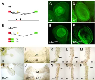
A functional analysis of the UbxMX17mutation in haltere development
The UbxMX17mutation is an inversion between the second and third introns of the Ubxgene that prevents the correct splicing between microexons I and II so that only isoforms IVa and IVb are made in UbxMX17 homozygotes (Fig. 5A,B) (Busturia et al., 1990; Subramaniam et al., 1994). The distribution of the IVa (and IVb) proteins in UbxMX17embryos follows that of the compound wild-type Ubx pattern, and not the particular one of the IV isoforms in the wild type (Busturia et al., 1990; Subramaniam et al., 1994). Similarly, the expression of the IV variants in UbxMX17third leg and haltere discs (Fig. 5E,F) closely resembled the Ubx wild-type pattern (Fig. 5C,D), although the levels of expression were slightly reduced (Table 1).
[image:5.612.54.287.60.384.2]UbxMX17homozygous halteres are partially transformed into wings (Busturia et al., 1990) (Fig. 5H, compare with the wild type in Fig. 5G) and the number of bristles in the first abdominal segment is reduced compared with that of the wild type (Fig. 5J, compare with Fig. 5I) (Busturia et al., 1990). We have also found small changes in the pattern of the metathoracic legs: in 83% of the mutant legs (n24) there was a small apical bristle in the anterior compartment (Fig. 5K, arrow), similar to that present in the wild-type mesothoracic leg (Fig. 5L, arrow) but absent in the wild-wild-type Fig. 3. Correlation between the amount of UbxIVa and UbxIa
proteins and phenotypic rescue.(A,C)Haltere imaginal disc (A) and haltere (C) of a UAS-UbxIVa2/+; UMB larva (A) or adult (C).
(B,D)Haltere imaginal disc (B) and haltere (D) of a UAS-UbxIVa33/+;
UMB larva (B) or adult (D). Note the reduced size of the haltere in D (arrow; the haltere is also of abnormal shape) compared with that of C, which correlates with the much higher Ubx protein expression (B).
(E)Halteres of a UAS-UbxIVa2/UAS-UbxIVa2; UMB fly, showing an
almost complete rescue of the mutant phenotype (arrow). (F) MS1096
-Gal4/+; UAS-UbxIVa33/+ female, showing a partial transformation of wing into haltere. A similar result is seen when expressing the IVa1 line
(not shown). (G)sd-Gal4/+; UAS-UbxIVa1/+ female, in which the wing
(arrow) is transformed into a haltere. A similar result is observed with
the IVa33 line (not shown). (H)UAS-GFP/UAS-GFP; UAS-UbxIa UMB fly.
An almost wild-type haltere develops (arrow).
D
E
V
E
LO
P
M
E
N
anterior metathoracic leg (Fig. 5M; n15). There is also a reduction in the number of big bristles present in the posterior compartment of the metathoracic basitarsus of UbxMX17flies (1.2 bristles; Fig. 5N, arrow) compared with the wild type (2.6 bristles; Fig. 5P, arrow), indicating a partial transformation into the basitarsus of the wild-type mesothoracic leg (which bears no bristles; Fig. 5O). All these characteristics point to a partial anteriorwards transformation of the third thoracic and first abdominal segments; that is, a partial reduction of Ubx activity.
In accordance with these transformations, in UbxMX17haltere pouches there was weak expression of wg in the posterior compartment (Fig. 4H, compare with the wild-type wing and haltere disc expression in Fig. 4A,B, respectively) and of salin the central region of the pouch (Fig. 4X; the wild-type expression in wing and haltere discs is shown in Fig. 4Q,R, respectively). These results are similar to those obtained when the IVa2 line is expressed in the UMB and support the conclusion that the IVa isoform is not able to repress Ubxtargets as efficiently as does the Ia isoform. By contrast, there is no araexpression in UbxMX17haltere discs (Fig. 4P), as in the wild-type haltere disc (Fig. 4J; wild type wing disc signal in Fig. 4I), perhaps owing to the higher levels of Ubx products in UbxMX17 compared with UAS-UbxIVa2/+; UMB haltere discs.
To characterize in more detail the different activities of the Ia and IVa proteins, we selected the salgene. Ubx directly regulates salin the haltere disc, as functional Ubx-binding sites are present in the salregulatory region (Galant et al., 2002; Walsh and Carroll, 2007). An EcoRI/PvuII 1085 bp fragment from this region, named salE/Pv(see Fig. S1A in the supplementary material), reproduces salexpression in the wing pouch when fused to a lacZreporter gene (Barrio and de Celis, 2004) (see Fig. S1B in the supplementary material; compare with Fig. 4Q). This fragment is similar in size and location to the 1.1 kb fragment previously
characterized (Galant et al., 2002). The expression of this reporter mimicked the endogenous salexpression in the haltere pouches of wild type, Ubx1/+, UMB, UMB UbxIVa2 and UMB UAS-UbxIa larvae (see Fig. S1C-G in the supplementary material; compare with Fig. 4R-V).
Binding of UbxIa and UbxIVa proteins to sal regulatory sequences
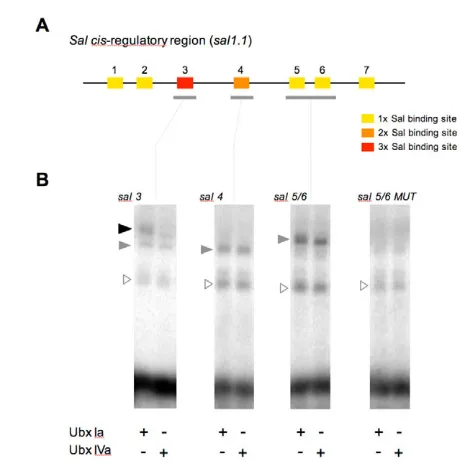
The preceding results indicate that Ubx isoforms Ia and IVa differently regulate Ubx targets and show different abilities to rescue a Ubx mutant phenotype when expressed at similar levels. In a molecular framework, these different abilities could be the result of: (1) differential binding profiles of the Ubx proteins to DNA-binding sites present in the cis-regulatory sequences of Ubx target genes; (2) distinct regulatory capabilities of the Ubx proteins independently of their binding profiles; or (3) a combination of both. To investigate this issue, we measured the abilities of Ubx isoforms Ia and IVa to form DNA-protein complexes on Ubx-binding sites present in the sal regulatory region contained in the salE/Pvfragment [named sal1.1in Galant et al. (Galant et al., 2002) and Walsh and Carroll (Walsh and Carroll, 2007)]. To this end, in vitro translated Ubx proteins were resolved by PAGE, quantified (see Materials and methods) and used in EMSA applied to the sal3, sal4and sal5/6 DNA elements (Galant et al., 2002) (Fig. 6A).
[image:6.612.53.557.61.238.2]Our results indicate that UbxIa and UbxIVa proteins possess very similar DNA-binding abilities to sal4 andsal5/6DNA elements (Fig. 6B), forming binary DNA-protein complexes in comparable proportions in the presence of excess DNA target. By contrast, isoform UbxIa seems to bind the sal3element in a slightly more stable manner than UbxIVa (assayed with proteins at equal molar proportions) (Fig. 6B). Mutation of Ubx-binding sites leads to no significant binding of either Ia or IVa proteins (Fig. 6B), showing that these proteins bind specifically to these sites (see Galant et al., 2002). Fig. 4. Expression of Ubxtargets in the wild type and in different isoform-specific mutant backgrounds. (A-X)The expression of Wg
(A-H), Ara (I-P) and Sal (Q-X) is shown in wild-type wing discs (wt W; A,I,Q), wild-type haltere discs (wt H; B,J,R), Ubx1/+ (C,K,S), UMB (D,L,T),
UAS-UbxIVa2/+; UMB (E,M,U), UAS-UbxIa/+UMB (F,N,V), UAS-UbxIVa1/+; UMB (G,O,W) and UbxMX17(H,P,X) haltere discs. Note that, differently from
the wing disc, Wg is not present in the posterior region of the haltere pouch (arrow in B) and that Sal and Ara are not expressed in the haltere
pouch (J and R). The expression in the haltere disc of the three genes does not change in Ubx1/+larvae (C, arrow, K and S), it changes to the wing
disc pattern in the mutant background, independently of whether or not there is expression of the IVa2 line (D,E,L,M,T,U; arrowheads in E, L and M indicate lack of repression), but remains as haltere expression if the Ia or IVa1 insertions are present in the mutant background (F,G,N,O,V,W; arrow
in F indicates represion of Wg; Wg disappears also from anterior compartment when using the IVa1 line). In UbxMX17mutants there is weak
de-repression of wgand sal(H and X, arrowheads) but not of ara(P). (Y,Z)In wing discs carrying the sd-Gal4 driver and the IVa2 construct (at 25°C)
the expression of saldoes not change (Y; compare with Q) whereas it disappears if the Ia protein is expressed with the same driver, even at 17°C (Z;
arrowhead marks absence of expression).
D
E
V
E
LO
P
M
E
N
DISCUSSION
Although the prevalence of alternative splicing in metazoans is indisputable, the functional relevance of most alternative splicing events is still unknown (Biencowe, 2006; Hartmann and Valcárcel, 2009). Here we examined the impact of alternative splicing on the functions of the Drosophila Hox gene Ubx.
Specificity of Ubx isoforms, levels of Ubx protein and morphology
Previous experiments resulted in conflicting views regarding specificity of Ubx isoforms: some claim that UbxIa and UbxIVa perform different roles in some contexts (Mann and Hogness, 1990; Subramaniam et al., 1994; Gebelein et al., 2002; Reed et al., 2010) whereas in the original work describing the UbxMX17
mutation it was concluded that the activity of all Ubx isoforms was basically the same (Busturia et al., 1990). However, some of these studies did not take into account the amount of Ubx protein obtained in each experiment. Furthermore it has been recently demonstrated that relatively small differences in the amount of Ubx protein cause major changes in development (Tour et al., 2005).
Our study has re-examined this controversy, focusing on the development of the haltere, and measuring the amount of Ubx protein in different mutant combinations. We have not found a major difference in Ubx protein staining in UbxMX17haltere (or
third leg) discs, which would account for the mutant phenotype (Busturia et al., 1990). By contrast, the analysis of UbxMX17
animals, and of flies expressing different isoforms in a Ubxmutant background, suggests that isoform UbxIVa is not able to form a normal haltere at the levels that the Ubx isoform Ia does, suggesting different activity and helping to explain the UbxMX17
mutant phenotype. Although we have based some of these conclusions on experiments expressing different isoforms in a non-null Ubx mutant background, which allowed the precise comparison of Ubx protein levels in the haltere disc, we have also
obtained similar results when expressing different isoforms in the wing disc, where no Ubx is present, except in the peripodial membrane.
We have also found that if a higher amount of the IVa protein is achieved (using the IVa33 or IVa1 insertions, or with two doses of the IVa2 construct) the transformation of haltere into wing in the mutant background is suppressed. Accordingly, some Ubxtargets (wg, sal, ara), silenced in the wild-type haltere pouch, are de-repressed when the UbxIVa isoform is expressed at low levels in the mutant background, but suppressed if the levels are high. Therefore, higher levels of expression can compensate for the functional differences of different Ubx isoforms caused by alternative splicing. During normal development, genes must integrate the generation of qualitative differences by alternative splicing with the regulation of the levels of expression of each isoform; the molecular coupling of transcriptional regulation to alternative splicing via modulations in transcriptional elongation rate may provide a mechanism to achieve this molecular coordination during gene expression (Cramer et al., 1997; de la Mata et al., 2003).
[image:7.612.51.371.63.335.2]Binding efficiency and activity of Ubx proteins Most Hox proteins include a hexapeptide motif that is required to interact with the extradenticle protein, the main Hox protein cofactor in Drosophila(Mann and Chan, 1996). The Ia and IVa proteins differ in the length of a region located between the hexapeptide motif and the homeodomain (the linker region, longer in the Ia variant). It has been shown that this linker region is necessary for Ubx repression of embryonic Dllbut not for the binding of Ubx to the relevant Dllbinding region, and that the UbxIa and UbxIVa proteins have different transcriptional regulatory properties (Gebelein et al., 2002). Similarly, we have shown that the Ia and IVa proteins do not show major differences in binding in vitro to a relevant region of the salregulatory domain (Galant et al., 2002) but do not repress the salgene with equal
Fig. 5. Phenotype and Ubxexpression in
UbxMX17mutants.(A,B)Scheme of the UbxMX17 mutation (B), compared with the wild type (A). The inversion in the DNA (arrows indicate the inversion points and the DNA inverted is in red) allows the production of only the IVa and IVb splicing
variants. (C-F). Haltere (C,E) and third leg (D,F)
discs of wild-type (C,D) and UbxMX17homozygous
larvae (E,F) showing a similar distribution and levels of expression of Ubx proteins (I and II in the wild
type, IV in the mutant). (G-P). The UbxMX17adults
show typical defects of reduction of Ubxfunction
in halteres (H, compare with the wild type in G), first abdominal segment (A1), which is reduced and with a lower number of bristles (J, compare with the wild type in I), and metathoracic leg (K, N), which show a reduced apical bristle (arrow in K) and few posterior bristles in the basitarsus (arrow in N); a (bigger) apical bristle appears in the wild-type mesothoracic leg (L, arrow) but not in the wild-type metathoracic one (M), and the posterior bristles in the basitarsus are present in the wild-type metathoracic leg (P, arrow) but not in the mesothoracic one (O).
D
E
V
E
LO
P
M
E
N
efficiency. The repression of Dllby Ubx, however, requires the co-factors Extradenticle (Exd) and Homothorax (Hth) (Gebelein et al., 2002). By contrast, Exd is cytoplasmic and inactive in the haltere pouch (González-Crespo and Morata, 1995; Aspland and White, 1997) and Hth is not expressed and probably not required in the haltere pouch (as it is not in the wing pouch) (Pai et al., 1998; Casares and Mann, 2000; Azpiazu and Morata, 2000). Therefore, a different interaction with Exd or Hth cannot account for the different activity of the two splicing variants in the distal haltere disc. It may be that other, yet undiscovered, co-factors may differently interact with Ubx Ia and IVa proteins in the haltere disc. Alternatively, the different activity may rely on slight differences in binding affinity, or in the transcription-regulating properties of the bound proteins. We have found slight differences between the two isoforms of Ubx in binding to one element containing three Ubx core consensus binding sites within the salregulatory domain, but no differences in Ubx binding to other sites within this same region. The Ubx-binding sites within the salregulatory domain contribute additively to the repression of salby Ubx(Galant et al., 2002). It is possible, therefore, that even small differences in the binding of Ubx to each of these sites may accumulate and result in appreciable differences in gene expression. We speculate that
higher amount of isoform IV may compensate for its lower binding or activity by increasing the probability of physical interactions with the basal transcription machinery. Perhaps the functional differences observed among Ubx isoforms may explain, to some degree, the evolutionary conservation in the spatial distribution of Ubx isoforms within distantly related Drosophila species.
Regulation of Ubx targets by different Ubx isoforms
Based on our data and on previous reports we think that there may be three different responses when either the Ia or IVa proteins are expressed: (1) some Ubxtargets, such as decapentaplegicin the visceral mesoderm (at least as to its anterior repression) and those needed to specify the embryonic cuticle, probably respond similarly to the different Ubx proteins (Busturia et al., 1990; Mann and Hogness, 1990; Subramaniam et al., 1994; Gebelein et al., 2002; Reed et al., 2010); (2) other targets, such as sal, wgand ara in the haltere disc, as well as others needed to repress wing development and promote haltere development, are more efficiently regulated by isoform Ia than IVa, but if the levels of the latter are increased, a similar regulation is achieved; and (3) the expression of dppin the posterior visceral mesoderm (Reed et al., 2010) or of targets needed to specify the segmental pattern in the embryonic peripheral nervous system (Mann and Hogness, 1990; Subramaniam et al., 1994), are differently controlled by proteins Ia and IVa, independently of their levels of expression.
Other Ubxtargets can be assigned to one of these three classes with less certainty. Thus, Dllis repressed in the embryo by the Ia protein, and not by the IVa isoform (Gebelein et al., 2002), and may be included in the second group. Although Dllexpression in UbxMX17 animals, or in embryos with high levels of the IVa isoform, has not been detailed, high levels of this protein repress the formation of Keilin’s organs, which is driven by Dllexpression (Mann and Hogness, 1990). We note that UbxMX17adults have abnormalities in metathoracic legs and the A1 (Busturia et al., 1990; and this report), in the development of A1 and A2 larval abdominal muscles (Reed et al., 2010) and are not as healthy as wild-type flies (Subramaniam et al., 1994), suggesting that isoform IVa may be less efficient than isoform Ia in controlling many Ubx targets, not just those making a haltere. In conclusion, we think that the particular architecture of cis-regulatory regions in each Ubx target may account for the inclusion of each gene in one of these three categories, so a specific research may be needed for each case.
Our studies also point to the importance of measuring protein levels when trying to correlate isoform activity and morphology. Several studies in different systems have also shown that the different levels of Ubx (Tour et al., 2005) or other Hox proteins may also end up in distinct phenotypes (Cribbs et al., 1995; Greer et al., 2000; Trvdik and Capecchi, 2006). In sum, we conclude that alternative splicing modulates Ubx function during Drosophila adult development, providing a good example of the functional relevance of alternative splicing events and protein level control within the physiological context of animal development.
Acknowledgements
[image:8.612.51.289.56.288.2]We thank G. Morata and A. Busturia for critically reading the manuscript; B. Hersh, J. Romero and E. Turiégano for their help in the DNA-binding experiments, westerns blots and statistical analysis, respectively; M. Calleja, S. Campuzano, S. Carroll, T. Kaufman, J. López, W. McGinnis, A. Michelson, G. Morata, R. White and the Bloomington Stock Center for stocks, probes or antibodies; and R. González for technical assistance. This work has been supported by a New Investigator Grant from the UK’s BBSRC to C.R.A. (Refs: BB/E01173X/1, BB/E01173X/2), grants from the Ministerio de Ciencia y
Fig. 6. DNA binding of UbxIa and UbxIVa isoforms to elements within the salregulatory region.(A)Diagram of the sal
cis-regulatory region contained within the sal1.1fragment as described in
Galant et al. (Galant et al., 2002) (the same fragment as the salE/Pv
fragment). The regulatory elements, termed sal1-7, include sequences
containing one, two or three Ubx core consensus binding sites
(indicated in yellow, orange and red, respectively). (B)EMSA comparing
the binding abilities of Ubx proteins UbxIa and UbxIVa to the wild-type
salelements sal3, sal4and sal5/6, as well as to the mutated version of
sal5/6sites. In the conditions of this experiment, all three labelled probes are shifted in the presence of Ubx proteins producing protein-DNA complexes; of these, some appear to form in a similar manner with either UbxIa or UbxIVa proteins (grey arrowheads), whereas others
seem to be more stably formed with UbxIa (black arrowhead, sal3
element). We also detect a series of nonspecific products also present in control reactions lacking Ubx proteins (empty triangles). When binding sites are mutated, neither UbxIa nor UbxIVa proteins bind significantly to the DNA (see also Galant et al., 2002).
D
E
V
E
LO
P
M
E
N
Tecnología (no. BFU2005-04342, BFU2008-00632 to E.S. and BFU2008-01884 to R.B., and Consolider CSD2007-00008 to E.S. and R.B.), and an Institutional Grant from the Fundación Ramón Areces. L. de N. was supported by a fellowship from the Spanish Ministerio de Educación y Ciencia. R. B.
acknowledges the Government of the Autonomous Community of the Basque Country (PI2009-16 and Etortek Research Programs 2008/2009) and the Bizkaia County.
Competing interests statement
The authors declare no competing financial interests.
Supplementary material
Supplementary material for this article is available at
http://dev.biologists.org/lookup/suppl/doi:10.1242/dev.051409/-/DC1
References
Artero, R. D., Akam, M. and Pérez-Alonso, M.(1992). Oligonucleotide probes detect splicing variants in situin Drosophilaembryos. Nucleic Acids Res. 20, 5687-5690.
Aspland, S. E. and White, R. A.(1997). Nucleocytoplasmic localisation of extradenticle protein is spatially regulated throughout development in Drosophila. Development124, 741-747.
Ausubel, M., Brent, R., Kingston, R. E., Moore, D. D., Seidman, J. G., Smith, J. A. and Struhl, K.(2004). Current Protocols in Molecular Biology.John Wiley and Sons.
Azpiazu, N. and Morata, G.(2000). Function and regulation of homothorax in the wing imaginal disc of Drosophila. Development127, 2685-2693.
Barrio, R. and de Celis, J. F.(2004). Regulation of spaltexpression in the Drosophilawing blade in response to the Decapentaplegic signaling pathway. Proc. Natl. Acad. Sci. USA 101, 6021-6026.
Barrio, R., de Celis, J. F., Bolshakov, S. and Kafatos, F. C.(1999). Identification of regulatory regions driving the expression of the Drosophila spaltcomplex at different developmental stages. Dev. Biol. 215, 33-47.
Beachy, P. A., Helfand, S. L. and Hogness, D. S.(1985). Segmental distribution of bithorax complex proteins during Drosophiladevelopment. Nature313, 545-551.
Ben-Dov, C., Hartmann, B., Lundgren, J. and Valcárcel, J.(2008). Genome-wide analysis of alternative pre-mRNA splicing. J. Biol. Chem. 283, 1229-1233.
Bermingham, J. R. and Scott, M. P.(1988). Developmentally regulated alternative splicing of transcripts from the Drosophilahomeotic gene Antennapedia can produce four different proteins. EMBO J. 7, 3211-3222.
Biencowe, B. J.(2006). Alternative splicing, new insights from global analyses. Cell126, 37-47.
Black, D. L.(2003). Mechanisms of alternative pre-messenger RNA splicing. Annu. Rev. Biochem. 72, 291-336.
Bomze, H. M. and López, A. J.(1994). Evolutionary conservation of the structure and expression of alternatively spliced Ultrabithoraxisoforms from Drosophila. Genetics136, 965-977.
Brand, A. and Perrimon, N. (1993). Targeted gene expression as a means of altering cell fates and generating dominant phenotypes. Development118, 401-415.
Brook, W. J. and Cohen, S. M.(1996). Antagonistic interactions between wingless and decapentaplegic responsible for dorsal-ventral pattern in the Drosophilaleg. Science273, 1373-1377.
Busturia, A., Vernós, I., Macías, A., Casanova, J. and Morata, G.(1990). Different forms of Ultrabithoraxproteins generated by alternative splicing are functionally equivalent. EMBO J. 9, 3551-3556.
Cabrera, C., Botas, J. and García-Bellido, A.(1985). Distribution of Ultrabithorax proteins in mutants of Drosophilabithorax complex and its transregulatory genes. Nature318, 569-572.
Capdevila, J. and Guerrero, I.(1994). Targeted expression of the signaling molecule decapentaplegic induces pattern duplications and growth alterations in Drosophilawings. EMBO J. 13, 4459-4468.
Casares, F. and Mann, R. S.(2000). A dual role for homothorax in inhibiting wing blade development and specifying proximal wing identities. Development127, 1489-1498.
Castelli-Gair, J., Greig, S., Micklem, G. and Akam, M.(1994). Dissecting the temporal requirements for homeotic gene function. Development120, 1983-1995.
Celniker, S., Keelan, D. J. and Lewis, E. B.(1989). The molecular genetics of the bithorax complex of Drosophila, characterization of the products of the Abdominal-B domain. Genes Dev. 3, 1424-1436.
Couso, J. P., Bishop, S. A. and Martínez-Arias, A.(1994). The wingless signalling pathway and the patterning of the wing margin in Drosophila. Development120, 621-636.
Cramer, P., Pesce, C. G., Baralle, F. E. and Kornblihtt, A. R.(1997). Functional association between promoter structure and transcript alternative splicing. Proc. Natl. Acad. Sci. USA 94, 11456-11460.
Cribbs, D. L., Pultz, M. A., Johnson, D., Mazzulla, M. and Kaufman, T. C.
(1992). Structural and evolutionary conservation of the Drosophilahometic gene proboscipedia. EMBO J. 11, 1437-1449.
Cribbs, D. L., Benassayag, C., Randazzo, F. M. and Kaufman, T. C.(1995). Levels of homeotic protein function can determine developmental identity, evidence from low-level expression of the Drosophilahomeotic gene proboscipedia under Hsp70 control. EMBO J. 14, 767-778.
Crickmore, M. A., Ranade, V. and Mann, R. S.(2009). Regulation of Ubx expression by epigenetic enhancer silencing in response to Ubx levels and genetic variation. PLoS Genet.5, e1000633.
De Celis, J. F. and Barrio, R.(2009). Regulation and function of Spalt proteins during animal development. Int. J. Dev. Biol. 53, 1385-1398.
De Celis, J. F., Barrio, R. and Kafatos, F. C.(1996). A gene complex acting downstream of dpp in Drosophilawing morphogenesis. Nature381, 421-424.
De la Mata, M., Alonso, C. R., Kadener, S., Fededa, J. P., Blaustein, M., Pelisch, F., Cramer, P., Bentley, D. and Kornblihtt, A. R.(2003). A slow RNA polymerase II affects alternative splicing in vivo. Mol. Cell2, 525-532.
DeLorenzi, M. and Bienz, M.(1990). Expression of Abdominal-B homeoproteins in Drosophilaembryos. Development108, 323-329.
de Navas, L. F., Foronda, D., Suzanne, M. and Sánchez-Herrero, E.(2006). A simple and efficient method to identify replacements of P-lacZby P-Gal4 lines allows obtaining Gal4 insertions in the bithorax complex of Drosophila. Mech. Dev. 123, 860-867.
Díez del Corral, R., Aroca, P., Gómez-Skarmeta, J.-L., Cavodeassi, F. and Modolell, J.(1999). The Iroquois homeodomain proteins are required to specify body wall identity in Drosophila. Genes Dev. 13, 1754-1761.
Drysdale, R. A., Crosby, M. A. and the FlyBase Consortium(2005). FlyBase, genes and gene models. Nucleic Acids Res. 33, D390-D395.
Foronda, D., de Navas, L. F., Garaulet, D. L. and Sánchez-Herrero, E.(2009). Function and specificity of Hox genes. Int. J. Dev. Biol. 53, 1409-1419.
Fujimoto, S., Araki, K., Chisaka, O., Araki, M., Takagi, K. and Yamamura, K.
(1998). Analysis of the murine Hoxa-9 cDNA, an alternatively spliced transcript encodes a truncated protein lacking the homeodomain. Gene209, 77-85.
Galant, R., Walsh, C. M. and Carroll, S. B.(2002). Hox repression of a target gene, extradenticle-independent, additive action through multiple monomer binding sites. Development129, 3115-3126.
Garaulet, D. L., Foronda, D., Calleja, M. and Sánchez-Herrero(2008). Polycomb-dependent Ultrabithorax Hox gene silencing induced by high Ultrabithorax levels in Drosophila. Development135, 3219-3228.
Gebelein, B., Culi, J., Ryoo, H.-D., Zhang, W. and Mann, R. S.(2002). Specificity of Distalless repression and limb primordia development by Abdominal Hox proteins. Dev. Cell3, 487-498.
Gilbert, W.(1978). Why genes in pieces? Nature271, 501.
Gómez-Skarmeta, J. L. and Modolell, J.(1996b). Araucanand caupolican provide a link between compartment subdivisions and patterning of sensory organs and veins in the Drosophilawing. Genes Dev. 10, 2935-2945.
Gómez-Skarmeta, J. L., Díez del Corral, R., de la Calle Mustienes, E., Ferrés-Marco, D. and Modolell, J.(1996a). Araucanand caupolican, two members of the novel iroquois complex, encode homeoproteins that control proneural and vein-forming genes. Cell85, 95-105.
González-Crespo, S. and Morata, G.(1995). Control of Drosophilaadult pattern by extradenticle. Development121, 2117-2125.
Graveley, B. R.(2005). Mutually exclusive splicing of the insect Dscam pre-mRNA directed by competing intronic RNA secondary structures. Cell123, 65-73.
Greer, J. M., Puetz, J., Thomas, K. R. and Capecchi, M. R.(2000). Maintenance of functional equivalence during paralogous Hox gene evolution. Nature403, 661-665.
Guillén, I., Mullor, J. L., Capdevila, J., Sánchez-Herrero, E., Morata, G. and Guerrero, I.(1995). The function of engrailedand the specification of Drosophilawing pattern. Development121, 3447-3456.
Hartmann, B. and Valcárcel, J.(2009). Decrypting the genome’s alternative messages.Curr. Opin. Cell Biol. 21, 377-386.
Hersh, B. M., Nelson, C. E., Stoll, S. J., Norton, J. E., Albert, T. J. and Carroll, S. B.(2007). The UBX-regulated network in the haltere imaginal disc of D. melanogaster. Dev. Biol. 302, 717-727.
Irvine, K. D., Botas, J., Jha, R. S., Mann, R. S. and Hogness, D.(1993). Negative autoregulation by Ultrabithorax controls the level and pattern of its expression. Development117, 387-399.
Johnson, J. M., Castle, J., Garrett-Engele, P., Kan, Z., Loerch, P. M., Armour, C. D., Santos, R., Schadt, E. E., Stoughton, R. and Shoemaker, D. D.(2003). Genome-wide survey of human alternative pre-mRNA splicing with exon junction microarrays. Science302, 2141-2144.
Kornfeld, K., Saint, R. B., Beachy, P. A., Harte, P. J., Peattie, D. A. and Hogness, D. S.(1989). Structure and expresión of a family of Ultrabithorax mRNAs generated by alternative splicing and polyadenilation in Drosophila. Genes Dev.3, 243-258.
Lecuit, T., Brook, W. J., Ng, M., Calleja, M., Sun, H. and Cohen, S. M.(1996). Two distinct mechanisms for long-range patterning by Decapentaplegic in the Drosophilawing. Nature381, 387-393.
Lewis, E. B.(1963). Genes and developmental pathways. Am. Zool.3, 33-56.
D
Lewis, E. B.(1978). A gene complex controlling segmentation in Drosophila. Nature 276, 565-570.
Lewis, E. B.(1982). Control of body segment differentiation in Drosophila by the bithorax gene complex. Prog. Clin. Biol. Res. 85, 269-288.
López, A. J. and Hogness, D. S.(1991). Immunochemical dissection of the Ultrabithoraxhomeoprotein family in Drosophila melanogaster. Proc. Natl. Acad. Sci. USA 88, 9924-9928.
López, A. J., Artero, R. D. and Pérez-Alonso, M.(1996). Stage, tissue, and cell specific distribution of alternative Ultrabithorax mRNAs and protein isoforms in the Drosophilaembryo. Roux’s Arch. Dev. Biol. 205, 450-459.
Mann, R. S. and Hogness, D. S.(1990). Functional dissection of Ultrabithorax proteins in D. melanogaster. Cell60, 597-610.
Mann, R. S. and Chan, S. K.(1996). Extra specificity from extradenticle, the partnership between HOX and PBX/EXD homeodomain proteins. Trends Genet.
12, 258-262.
Mlodzik, M., Fjose, A. and Gehring, W. J.(1988). Molecular structure and spatial expression of a homeobox gene from the labial region of the Antennapedia complex. EMBO J. 7, 2569-2578.
Mohit, P., Makhijani, K., Madhavi, M. B., Bharathi, V., Lal, A., Sirdesai, G., Reddy, V. R., Ramesh, P., Kannan, R., Dhawan, J. et al.(2006). Modulation of AP and DV signaling pathways by the homeotic gene Ultrabithorax during haltere development in Drosophila. Dev. Biol. 291, 356-367.
Morata, G. and Kerridge, S.(1981). Sequential functions of the bithorax complex of Drosophila. Nature290, 778-781.
Nellen, D., Burke, R., Struhl, G. and Basler, K.(1996). Direct and long-range action of a Dpp morphogen gradient. Cell85, 357-368.
O’Connor, M. B., Binari, R., Perkins, L. A. and Bender, W.(1988). Alternative products from the Ultrabithorax domain of the bithorax complex. EMBO J. 7, 435-445.
Pai, C. Y., Kuo, T. S., Jaw, T. J., Kurant, E., Chen, C. T., Bessarah, D. A., Salzberg, A. and Sun, Y. H.(1998). The Homothorax homeoprotein activates the nuclear localization of another homeoprotein, extradenticle, and suppresses eye development in Drosophila. Genes Dev. 12, 435-446.
Park, J. W. and Graveley, B. R.(2007). Complex alternative splicing. Adv. Exp. Med. Biol. 623, 50-63.
Patel, C. V., Sharangpani, R., Bandyopadhyay, S. and DiCorleto, P. E.(1999). Endothelial cells express a novel, tumor necrosis factor-alpha-regulated variant of HOXA9. J. Biol. Chem. 274, 1415-1422.
Pearson, J. C., Lemons, D. and McGinnis, W.(2005). Modulating Hox gene functions during animal body development. Nat. Rev. Genet. 6, 893-903.
Phillips, R. G. and Whittle, J. R.(1993). wingless expression mediates determination of peripheral nervous system elements in late stages of Drosophilawing disc development. Development118, 427-438.
Reed, H. C., Hoare, T., Weaver, T. A., White, R. A. H., Akam, M. and Alonso, C. R.(2010). Alternative splicing modulates Ubx protein function in Drosophila melanogaster. Genetics184, 745-758.
Schütt, C. and Nöthiger, R.(2000). Structure, function and evolution of sex-determining systems in Dipteran insects. Development127, 667-677.
Sham, M. H., Hunt, P., Nonchev, S., Papalopulu, N., Graham, A., Boncinelli, E. and Krumlauf, R.(1992). Analysis of the murine Hox-2.7 gene, conserved alternative transcripts with differential distributions in the nervous system and the potential for shared regulatory regions. EMBO J. 11, 1825-1836.
Shashidhara, L. S., Agrawal, N., Bharathi, V. and Sinha, P.(1999). Negative regulation of dorsoventral signaling by the homeotic gene Ultrabithorax during haltere development in Drosophila. Dev. Biol. 212, 491-502.
Shen, W. F., Detmer, K., Simonitch-Eason, T. A., Lawrence, H. J. and Largman, C.(1991). Alternative splicing of the HOX 2.2 homeobox gene in human hematopoietic cells and murine embryonic and adult tissues. Nucleic Acids Res. 11, 539-545.
Stolc, V., Gauhar, Z., Mason, C., Halasz, G., van Batenburg, M. F., Rifkin, S. A., Hua, S., Herreman, T., Tongprasit, W., Barbano, P. E. et al.(2004). A gene expresión map for the euchromatic genome of Drosophila melanogaster. Science306, 655-660.
Subramaniam, V., Bonze, H. M. and López, A. J.(1994). Functional differences between Ultrabithorax protein isoforms in Drosophila melanogaster, evidence from elimination, substitution and ectopic expresión of specific isoforms. Genetics136, 979-991.
Tour, E., Hittinger, C. T. and McGinnis, W.(2005). Evolutionary conserved domains required for activation and repression functions of the DrosophilaHox protein Ultrabithorax. Development132, 5271-5281.
Tvrdik, P. and Capecchi, M. R.(2006). Reversal of Hox1 gene subfunctionalization in the mouse. Dev. Cell11, 239-250.
Walsh, C. M. and Carroll, S. B.(2007). Collaboration between Smads and a Hox protein in target gene repression. Development134, 3585-3592.
Weatherbee, S. D., Halder, G., Kim, J., Hudson, A. and Carroll, S.(1998). Ultrabithoraxregulates genes at several levels of the wing-patterning hierarchy to shape the development of the Drosophilahaltere. Genes Dev. 12, 1474-1482.
White, R. A. H. and Wilcox, M.(1984). Protein products of the bithorax complex in Drosophila. Cell39, 163-171.
White, R. A. H. and Akam, M. E.(1985). Contrabithoraxmutations cause inappropriate expression of Ultrabithorax products in Drosophila. Nature 318, 567-569.
White, R. A. H. and Wilcox, M.(1985). Distribution of Ultrabithorax proteins in Drosophila. EMBO J.4, 2035-2043.
Wolff, T.(2000). Histological techniques or the Drosophilaeye. Part I, larva and pupa. In Drosophila Protocols (ed. W. Sullivan, M. Ashburner and R. S. Hawley), pp. 201-227. Cold Spring Harbor, NY: Cold Spring Harbor Laboratory Press.
Zavortink, M. and Sakonju, S.(1989). The morphogenetic and regulatory functions of the DrosophilaAbdominal-B gene are encoded in overlapping RNAs transcribed from separate promoters. Genes Dev. 3, 1969-1981.