RESEARCH ARTICLE
A novel role for
sox7
in
Xenopus
early primordial germ cell
development: mining the PGC transcriptome
Amanda M. Butler1, Dawn A. Owens1, Lingyu Wang2,* and Mary Lou King1,‡ABSTRACT
Xenopus primordial germ cells (PGCs) are determined by the presence of maternally derived germ plasm. Germ plasm components both protect PGCs from somatic differentiation and begin a unique gene expression program. Segregation of the germline from the endodermal lineage occurs during gastrulation, and PGCs subsequently initiate zygotic transcription. However, the gene network(s) that operate to both preserve and promote germline differentiation are poorly understood. Here, we utilized RNA-sequencing analysis to comprehensively interrogate PGC and neighboring endoderm cell mRNAs after lineage segregation. We identified 1865 transcripts enriched in PGCs compared with endoderm cells. We next compared the PGC-enriched transcripts with previously identified maternal, vegetally enriched transcripts and found that ∼38% of maternal transcripts were enriched in PGCs, includingsox7. PGC-directedsox7knockdown and overexpression studies revealed an early requirement for sox7 in germ plasm localization, zygotic transcription and PGC number. We identified pou5f3.3 as the most highly expressed and enriched POU5F1 homolog in PGCs. We compared theXenopusPGC transcriptome with human PGC transcripts and showed that 80% of genes are conserved, underscoring the potential usefulness of Xenopus for understanding human germline specification.
KEY WORDS:Xenopus, RNA-seq, PGCs,sox7,pou5f3.3(oct60), Germ plasm
INTRODUCTION
The exclusion of germ cells from somatic cell fates in early development is an essential process in metazoans that ensures continuation of the species. Germline specification in Xenopus is established by inheritance of germ plasm, a subcellular matrix containing maternally derived RNAs and proteins. Germ plasm contains all the genetic information that protects primordial germ cells (PGCs) from somatic differentiation and initiates a unique gene expression program that preserves their potential for totipotency and differentiation. Furthermore, germ plasm has been shown to be both required and sufficient to determine germ cell fate inXenopus(Tada et al., 2012). Germ plasm components are localized, along with somatic determinants, to the vegetal pole during oogenesis (Forristall et al., 1995; Heasman et al., 1984; Kloc and Etkin, 1995; Zhang et al.,
1998). During cleavage stages, cells containing germ plasm undergo asymmetric division so that the germ plasm is only inherited by one daughter cell termed the presumptive PGC ( pPGC). Although somatic determinants are partitioned into pPGCs during cleavage stages, the genetic programs for somatic fate are not activated there because of translational repression and transient suppression of RNA polymerase II-regulated transcription (Lai and King, 2013; Venkatarama et al., 2010). Segregation of the germline occurs at gastrulation when the germ plasm moves to a perinuclear location and subsequent divisions result in both daughter cells, now termed PGCs, receiving germ plasm. PGCs then initiate their zygotic transcription program driven by unknown maternal transcription factors. However, the activated gene network necessary for proper PGC specification and development has not been characterized inXenopus.
To begin to understand the gene network required for proper PGC development, we recently completed RNA-sequencing (RNA-seq) analysis of both animal and vegetal pole tips isolated from stage VI oocytes (Owens et al., 2017). This work defined 198 annotated RNAs highly enriched at the vegetal pole including several known germline and somatic determinants. Further analysis confirmed known germline components, such aspgat(also known asxpat),sybuand otx1, and identified novel components including the transcription factor (TF)sox7(Owens et al., 2017). The F-sox family membersox7 has previously been shown to be an early downstream target of VegT and to induce expression ofnodalgenes necessary for somatic fates (Zhang, et al., 2005). Similar toXenopusSox7 (Hudson et al., 1997; Zhang et al., 2005), human SOX17, another F-sox family member, has also historically been reported as an essential transcription factor required for endoderm specification (Charney et al., 2017; Hudson et al., 1997; Irie et al., 2015). Interestingly, Irie and co-workers generated human primordial germ cell-like cells (hPGCLCs) from embryonic stem cells and identifiedSOX17as the primary regulator of human primordial germ cell-like fate (Irie et al., 2015).
In the present study, we utilized RNA-seq analysis to determine the zygotic PGC transcriptome in Xenopus by comprehensive interrogation of PGC and neighboring endoderm cell RNAs just after lineage segregation. We identified 1865 transcripts enriched in PGCs, and over a third of the 198 annotated, vegetally enriched transcripts (Owens et al., 2017) were among them, includingsox7. To elucidate the specific role ofsox7in PGCs, we directed sox7 knockdown and overexpression constructs to the germline. Our results indicate that, prior to neurula,sox7is necessary for proper germ plasm localization, timely zygotic transcription and correct PGC number. These data provide further evidence that sox7is a crucial TF required for PGC development.
In addition tosox7,pou5f3.3(also known asoct60), which is in the same POU subclass as the human pluripotency factorPOU5F1 (also known asOCT3/4) (Frankenberg and Renfree, 2013; Hellsten et al., 2010; Hinkley et al., 1992), is enriched in PGCs. Aspou5f3.3 is not enriched at the vegetal pole of stage VI oocytes (Owens et al., 2017), along with other known germ plasm transcripts, it might Received 13 June 2017; Accepted 6 November 2017
1Department of Cell Biology, University of Miami Miller School of Medicine, 1011
NW 15th St, Miami, FL 33136, USA.2Department of Biology, University of Miami,
Coral Gables, FL 33124, USA.
*Present address: Department of Biology, Duke University, 125 Science Drive, Box 90338, Durham, NC 27708, USA.
‡Author for correspondence ([email protected])
M.L.K., 0000-0002-1923-4283
DEVEL
O
represent a zygotic germ plasm transcript required for proper PGC specification. In fact,POU5F1is considered a key gene necessary for human PGC (hPGC) specification (Tang et al., 2016), and POU5F1 acts as a functional homolog for pou5f3.3 in rescue experiments (Frankenberg and Renfree, 2013; Hellsten et al., 2010; Hinkley et al., 1992). In the present study, we show for the first time thatpou5f3.3 plays a crucial role in early development ofXenopus PGCs. Furthermore, we compared theXenopus PGC transcriptome with the human PGC and hPGCLC transcriptomes (see supplementary information in Irie et al., 2015), and show that 80% of genes are conserved. Taken together, these data indicate that Xenopus is a genetically relevant system for modeling the gene networks necessary for human germline specification and development.
RESULTS
RNA-sequence analysis of PGCs after segregation from the endoderm
PGCs initiate zygotic transcription after they segregate from the endoderm at gastrulation (Venkatarama et al., 2010). To identify transcripts involved in PGC development, we took advantage of the large abundance of mitochondria in germ plasm to identify these cells. Four-cell embryos were stained briefly with the mitochondrial lipophilic dye DiOC6, and when they reached stages (st.) 12.5-14, PGCs and embryo-matched endoderm cells were collected as described previously (Butler et al., 2017). In total, RNA was extracted from∼1040 PGCs and∼2100 paired endoderm cells from embryos generated by 28 female frogs. The respective RNA samples were split into three technical replicates each and then processed and sequenced as shown in Fig. S1 and described in Materials and Methods. Although version 9.1 of the Xenopus laevis genome (Xenbase.org, September, 2015) contained a higher number of transcripts and was better annotated, it did not contain all the transcripts in the previous version, 7.1. Therefore, the raw reads of
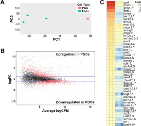
each sample were aligned to both v9.1 and v7.1 of theXenopus laevis genome (Xenbase.org, September, 2015). The aligned reads for each genome version were sorted and counted, then merged together before normalization and data analysis. For transcripts identified in both v9.1 and v7.1, read counts from v9.1 were used. In total, 13,469 transcripts were identified (Table S1, transcripts unique to v7.1 are highlighted in yellow). Two-dimensional principal component analysis (PCA) showed that all three PGC samples cluster together and away from the three endoderm samples, which also cluster together (Fig. 1A). Differential expression analysis was performed and results are shown as a MA plot, in which M represents the log ratio and A represents mean average (Fig. 1B).
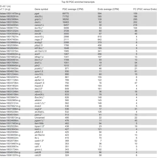
Transcripts were considered significantly enriched in PGCs or endoderm cells if their expression was increased at least threefold compared with endoderm cells or PGCs, respectively. Using these criteria, 1865 transcripts were enriched in PGCs (red) and 791 transcripts were enriched in endoderm cells (blue) (Table S1). A heat map was generated with the top 50 PGC-enriched transcripts (Fig. 1C). Several genes known to be expressed in PGCs, including pgat,ddx25(also known asdeadsouth),dazl,nanos1,grip2,velo1 and dnd1 (Colozza and De Robertis, 2014; King et al., 2005; Tarbashevich et al., 2007), are among the top 50, confirming our sample identity. In addition to known germline RNAs, six of the top 50 PGC-enriched transcripts, namely impad1, dsc3, fbxo34, fam168b,tspan1and cab39, have not previously been shown to play a role in germ cell development (Fig. 1C).
Biological processes and network analysis of PGC-enriched transcripts
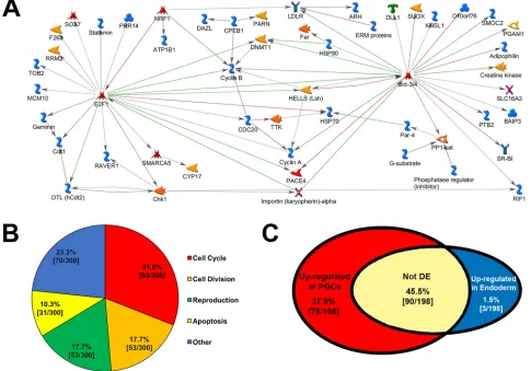
[image:2.612.48.338.438.697.2]MetaCore (GeneGo) was used to interrogate potential gene networks operating in PGCs. Network analysis revealed five hubs (five or more connections) linking, in a direct-interaction network, 53 out of the top 150 PGC-enriched transcripts. Hubs included the
Fig. 1. RNA-seq analysis of PGC versus endoderm cell transcripts inX. laevisembryos.(A) Two-dimensional principal component analysis (PCA) of PGC and Endo transcripts from st. 12.5-14 embryos. Variance was 73.5% and 11.3% in PC1 and PC2, respectively. (B) Differential expression analysis of PGC versus Endo transcripts; red dots indicate differentially expressed transcripts. (C) Heat map of the top 50 PGC-enriched transcripts; novel germline transcripts are underlined. Endo, endoderm cell sample. Asterisks indicate transcripts that express more than one copy.‘.L’and‘.S’indicate long and short chromosomes, respectively. Note that only the more highly expressed copy in PGCs is shown.
DEVEL
O
TFs e2f1, pou5f3.3/pou5f3.2 (two of three POU5F1 Xenopus homologs; pou5f3.2 is also known as oct25), cyclin B2, chk1 (chek1) andhells(Fig. 2A). Interestingly, only 20 of the top 150 endoderm cell-enriched transcripts form a direct-interaction network (data not shown). This difference is probably because PGCs represent a single lineage, whereas several cell lineages are derived from endoderm cells (Charney et al., 2017).
To identify potential biological processes necessary for PGC specification, the 300 most highly expressed, PGC-enriched transcripts were subjected to gene ontology analysis using GeneGo. The top 50 GeneGo processes are shown in Table S2. These processes were further categorized into four molecular functions including: cell cycle (93/300), cell division (53/300), reproduction (53/300) and apoptosis (31/300) (Fig. 2B). Considering that PGCs undergo few cell divisions before they infiltrate the somatic gonads much later in development, it was surprising that the most highly expressed PGC-enriched transcripts were primarily involved in cell cycle regulation and cell division.
We next identified 195 TFs that were enriched in PGCs, including sox7, pou5f3.3, pou5f3.2, e2f1, mixer, otx1 and twist1, and 16 transcriptional co-factors (Table S1, tabs 3-5). Surprisingly, 89 of the identified TFs contain homeodomains. Homeodomain-containing TFs are crucial for proper developmental patterning in somatic tissues (Pearson et al., 2005; Philippidou et al., 2012), but their role in PGC development has not been characterized.
PGC transcriptome is conserved betweenXenopusand human PGCs
Irie et al. (2015) recently performed RNA-seq analysis on human gonadal PGCs from week 7 human embryos (hPGCs), and PGC-like cells (hPGCLCs), created through differentiation of human embryonic stem cells. They identifiedSOX17as an early marker and necessary regulator of hPGCLC fate. Here, we performed RNA-seq analysis onXenopusPGCs after segregation from the endoderm and showed that another F-sox family member,sox7is highly enriched in Xenopus PGCs (Table S1). To identify additional similarities between the Xenopus and human PGC transcriptomes, we performed a comparative analysis of hPGC and hPGCLC transcripts (Irie et al., 2015), versus our Xenopus PGC-enriched data set. Interestingly, we found that∼80% (1489/1865) ofXenopus PGC-enriched genes were also expressed in hPGCs (gray), hPGCLCs (green) or both (orange) (Table S3). Furthermore, of the top 50XenopusPGC-enriched transcripts, 90% are expressed in hPGCs and/or hPGCLCs (Table S3, tab 1). These results suggest thatXenopusis a relevant model system to study the gene networks required for human germline development.
Maternal, vegetally enriched transcripts are expressed in PGCs
[image:3.612.65.550.362.701.2]During Xenopus oogenesis, hundreds of RNAs are selectively localized to the vegetal pole, including germ cell and endoderm
Fig. 2. Analysis of PGC-enriched transcripts.(A) GeneGo direct interaction network connecting 53 of the 150 most highly expressed PGC-enriched transcripts (network key: https://portal.genego.com/help/MC_legend.pdf ). (B) Top 300 PGC-enriched transcripts categorized by molecular function. (C) Distribution of
zygotically expressed maternal, vegetally enriched transcripts. DE, differentially expressed.
DEVEL
O
determinants. Recently, Owens et al. (2017) identified 411 RNAs enriched at the vegetal pole (compared with the animal pole), 198 of which were annotated. Considering that the maternal, vegetally enriched transcripts are known to contain both germ- and endoderm-cell determinants, we compared those annotated transcripts with the PGC/Endoderm cell data set. Of the 198 maternal, vegetally enriched transcripts, ∼84% (167/198) were indeed expressed in either endoderm cells, PGCs, or both (Table S4). We therefore, considered those transcripts to be maternal, and the remaining transcripts were considered to be zygotically expressed. Only ∼1.5% (3/198) of maternal mRNAs were enriched in endoderm cells compared with PGCs (blue), whereas ∼45% (90/198) were expressed in both PGCs and endoderm cells, but not differentially expressed (white) (Fig. 2C; Table S4). However, over a third of the maternal transcripts (75/198) were enriched in PGCs compared with endoderm cells (red) (Fig. 2C; Table S4), which suggests that many of the annotated maternal, vegetally enriched RNAs are involved in germline specification. Whether they are essential will require further functional testing. Furthermore, although most PGC-enriched transcripts were zygotic, relative to germ plasm expression (i.e. not enriched at the vegetal pole of stage VI oocytes) (1760/1865), about half of the 50 most highly expressed PGC-enriched transcripts were maternal (vegetally enriched transcripts) (Table 1, gray).
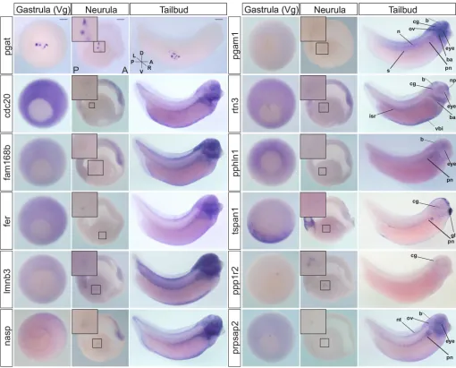
Expression of PGC-enriched mRNAs during development We next utilized whole-mount in situ hybridization (WISH) analysis to confirm mRNA expression in PGCs for a set of 15 highly expressed, PGC-enriched transcripts. Expression of selected transcripts was analyzed at gastrula (st. 11.5), neurula (st. 16) and tailbud (st. 33/34) stages.pgat, a PGC-specific transcript, was used as a positive control to reference expression in PGCs at each stage. Consistent with our RNA-seq data, all of the 15 transcripts tested (fer,pphln1,prpsap2,cdc20,fam168b,lmnb3,nasp,rtn3,pgam1, tspan1,ppp1r2,pgat,h1foo,zfyve26andimpad1), were expressed in PGCs at neurula (12/15 are shown in Fig. 3), and all but four transcripts (pphln1,cdc20,h1fooandimpad1) were also expressed at st. 11.5. However, of the 11 transcripts expressed at st. 11.5, only four (fam168b,pgam1,ppp1r2andzfyve26) were vegetally enriched, maternal transcripts (Owens et al., 2017). These data suggest that PGCs may have initiated zygotic transcription by st. 11.5. Furthermore, onlypgatwas still expressed, as detected by WISH, in PGCs at tailbud stages. At tailbud stages, all PGC-enriched transcripts tested, exceptpgat, were expressed only in somatic tissues. Interestingly, the most represented expression pattern at tailbud was in neural regions, including the eye, cranial ganglia, brain and neural tube (Fig. 3). This observation is consistent with what was previously found (Owens et al., 2017), and could suggest that these transcripts play dual roles in germline and neural specification during development.
sox7knockdown or overexpression increased PGC number in early development
Historically, mice have been used as the primary model system to study human PGC development (Hayashi et al., 2007; Ohinata et al., 2009, 2005; Saitou et al., 2002; Saitou and Yamaji, 2012). Mouse studies have revealed specific gene sets required to specify the germ cell lineage, including the essential pluripotency genes:NANOG, POU5F1andSOX2(Tang et al., 2016). However, contrary to what has been predicted for human PGC development (based on mouse studies), Irie et al. (2015) identified the F-sox family member SOX17, as a primary regulator of human primordial germ cell-like
fate, notSOX2(a SOXB1 family member). Interestingly, although severalsoxtranscripts were enriched in PGCs (namelysox2,sox3, sox4,sox7andsox8),sox7, another F-sox family member, is highly enriched (74-fold versus endoderm) and the most highly expressed [269 counts per million (CPM)]soxtranscript in XenopusPGCs (Table S1). Furthermore,sox7was recently shown to be expressed in germ plasm through gastrulation, and both morpholino (MO)-mediatedsox7knockdown and overexpression ofsox7in the whole embryo resulted in significantly reduced PGCs at tailbud stages (Owens et al., 2017). Therefore, we hypothesized that sox7 is necessary for proper PGC development.
To test our hypothesis, sox7 MO-mediated knockdown and overexpression of sox7-FL mRNA was targeted to PGCs as described in Materials and Methods (Fig. S2). Interestingly, PGC-directed sox7 knockdown and overexpression resulted in significantly more PGCs during gastrulation (st. 11.5) (Fig. 4A, B). These effects were significantly rescued by co(PGC-directed)-injection of asox7mRNA construct that cannot bind thesox7-MO (sox7-MO rescue; Fig. S3) (Fig. 4A,B), thereby confirming specificity of the observed phenotype. However, by tailbud stages (st. 33/34), normal PGC numbers were detected in PGC-directed sox7 knockdown embryos whereas PGC-directed sox7 -overexpressing embryos had significantly fewer PGCs (Fig. 4C, D). The observed increase in PGC number at gastrulation was both remarkable and unexpected as the amount of germ plasm assembled during oogenesis remains constant during development. One possible explanation for more germ plasm-bearing cells aftersox7 misexpression might be the premature migration of germ plasm to a perinuclear position, resulting in both daughter cells, rather than only one cell, receiving germ plasm.
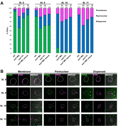
Misexpression ofsox7alters germ plasm localization DuringXenopusPGC development, germ plasm at blastula stages is localized in close apposition to the plasma membrane of pPGCs. Subsequent asymmetric divisions result in only one of the two daughter cells inheriting germ plasm (Aguero et al., 2017). At gastrula, the germ plasm in these cells moves to a perinuclear location, establishing them as PGCs. PGCs will subsequently undergo symmetric divisions at specific times during development. Although the timing for germ plasm migration has been described previously inex vivostudies (Yamaguchi et al., 2013), it is currently unknown precisely when the germ plasm becomes perinuclear in vivo. To determine whether germ plasm migration is affected when sox7 expression is altered in PGCs, we first set out to determine normal germ plasm patterningin vivofrom late blastula to gastrula (st. 8-11).
Immunodetection of the nuclear protein PCNA was used to identify cell nuclei and, when overexposed, the outline of individual cells. Co-immunodetection of Piwi protein, a specific germ plasm marker, was used to identify germ plasm location in PGCsin vivo (Lau et al., 2009). We identified three distinct germ plasm phenotypes: membrane (germ plasm located along the plasma membrane), perinuclear and dispersed (germ plasm found in various regions of the PGC). The dispersed phenotype was rare (2.8-14.4%) at all stages analyzed (Fig. 5). Similar to what Taguchi et al. (2012) reported in their ex vivo studies, germ plasm was located at the membrane in 93.4% (99/106) of pPGCs at stage 8, and 11.6% (22/189) at stage 10. However, at stage 9 Taguchi et al. (2012) reported only 55% of PGCs with membrane-localized germ plasm, whereas we observed that the majority of PGCs (81.9%, 172/ 210) still exhibited membrane-localized germ plasm at stage 9. Taken together, these results indicate that germ plasm
DEVEL
O
reorganization from membrane to perinuclear occurs late in stage 9, after the midblastula transition (MBT).
Whensox7was knocked down or overexpressed, the dispersed germ plasm patterning was significantly more common at all stages tested (Fig. 5). In addition to dispersed germ plasm, significantly more PGCs exhibited perinuclear localized germ plasm prematurely, as early as stage 8 (Fig. 5). Movement of germ plasm to a perinuclear position requires microtubules (Taguchi et al., 2012). Germes (also known as Loc779566) is a germ plasm
[image:5.612.50.565.70.589.2]component that binds to dynein light chains and plays a crucial role in germ plasm translocation from the plasma membrane to the perinuclear region in PGCs (Berekelya et al., 2007; Yamaguchi et al., 2013). In addition to Germes, Ddx25 also affects germ plasm localization, but in a distinctly different manner, as its timely movement to a perinuclear position is delayed along with cell division (Yamaguchi et al., 2013). Thus, Sox7 might play a role in germ plasm localization through transcriptional regulation of the genes necessary for this process.
Table 1. Maternal versus zygotic PGC-enriched transcripts
Top 50 PGC-enriched transcripts
ID v9.1 (m)
v7.1 (m.g) Gene symbol PGC average (CPM) Endo average (CPM) FC (PGC versus Endo)
Xelaev15011470m.g pgat 89775 564 159
Xelaev18035291m ddx25.L 71752 385 186
Xelaev18023956m grip2.L* 38292 130 295
Xelaev18031029m dazl.L 16463 82 202
Xelaev18034293m nanos1.L 6457 30 216
Xelaev18006177m tex15.L* 5499 53 104
Xelaev18031232m trank1.L 3728 80 46
Xelaev15043509m.g trim36 3005 67 45
Xelaev18017827m mcm10.L 2998 810 4
Xelaev18047493m naga.S* 2111 642 3
Xelaev15038666m.g acsbg2 2088 379 6
Xelaev18025369m ptbp2.S 1766 456 4
Xelaev18028249m kif13b.L* 1648 189 9
Xelaev18010315m slc12a3.2.S 1585 341 5
Xelaev15046920m.g acsl1 1467 294 5
Xelaev15016832m.g ldlrap1-a 1341 107 13
Xelaev18034461m bicc1.L* 1169 93 13
Xelaev18017004m dnd1.L 1051 19 54
Xelaev15069945m.g plin2 1015 47 22
Xelaev18018420m pcsk6.L* 971 46 21
Xelaev18046219m spire1.S 930 11 81
Xelaev18012344m mov10.L 890 60 15
Xelaev18032097m sulf1.L 861 155 6
Xelaev18011128m atp5a1.S 789 122 6
Xelaev18033736m impad1.S 750 16 47
Xelaev18041802m hspa5.S 690 193 4
Xelaev18033676m dsc3.S* 649 161 4
Xelaev18031248m velo1.L 639 36 18
Xelaev18040158m zfyve26.L 633 78 8
Xelaev18039847m fbxo34.L* 626 148 4
Xelaev15002965m.g alkbh4 580 145 4
Xelaev18023131m ccnb1.2.L* 562 148 4
Xelaev15070621m.g lmnb3 536 99 5
Xelaev15071996m.g vegt-b* 536 17 31
Xelaev18005239m slc30a9.L 532 126 4
Xelaev18045154m parn.L* 513 41 13
Xelaev15018513m.g Unnamed 495 22 22
Xelaev18034686m pgam1.L* 476 11 45
Xelaev15017463m.g fam168b 462 94 5
Xelaev18025228m tspan1.S* 438 23 19
Xelaev18023643m tob2.L 427 121 4
Xelaev18020909m pfkfb3.S 425 94 4
Xelaev15038059m.g slc16a3-b 423 33 13
Xelaev18008022m fer.L 415 70 6
Xelaev18020738m cpeb1.S* 368 7 52
Xelaev15019487m.g nasp 353 36 10
Xelaev18003587m cdt1.1 351 71 5
Xelaev18031734m gmnn.L 343 102 3
Xelaev15049105m.g cab39 339 96 4
Xelaev15061337m.g cdc20 324 58 6
Maternal, vegetally enriched transcripts expressed in PGCs (gray), and zygotic transcripts (white). CPM, counts per million; Endo, endoderm cell sample; FC, fold change; PGC, primordial germ cell sample. *Most highly expressed copy is shown.
DEVEL
O
Altered expression ofsox7in PGCs results in premature zygotic transcription
PGCs are transcriptionally silent until gastrulation, when they segregate from the endoderm and initiate zygotic transcription (Lai and King, 2013; Venkatarama et al., 2010). The transcription factor(s) within the germ plasm that initiate the PGC-specific gene program remain unknown, but are likely to act after the germ plasm has migrated to a perinuclear position. In this case, premature perinuclear localization would be predicted to result in premature transcriptional activity. To test whethersox7plays a role in PGC transcription, embryos with PGC-directed altered expression of sox7 were collected at st. 10 and 11 and subjected to co-immunofluorescence with antibodies recognizing Piwi (to identify PGCs; Lau et al., 2009) and RNA Pol II CTD-pSer2 (to identify transcriptionally active nuclei) (Bensaude et al., 1999; Bregman et al., 1995; Seydoux and Dunn, 1997). Consistent with our prediction, sox7 knockdown caused a significant increase in the number of transcriptionally active PGCs at both st. 10 and st. 11
compared with control (Fig. 6). This effect was significantly rescued by co(PGC-directed)-injection of sox7-MO with a sox7 mRNA construct that thesox7-MO cannot bind (sox7-FL rescue) (Fig. 6), thereby confirming specificity of the observed phenotype. Furthermore, PGC-directedsox7overexpression also caused both premature germ plasm migration and a significant increase in the number of transcriptionally active PGCs at st. 10, albeit to a lesser degree thansox7-MO; this effect did not persist to st. 11 (Fig. 6). These results show thatsox7is necessary for proper timing of PGC transcriptional activity.
PGC-directedsox7knockdown alters expression of germline genes
[image:6.612.51.561.57.468.2]Our functional studies showed that both PGC-directed sox7 knockdown and overexpression caused germ plasm to become dispersed or localize prematurely to the perinuclear region as early as st. 8 (Fig. 5), and caused PGCs to undergo premature transcription (Fig. 6). Thus, we hypothesized that Sox7 might Fig. 3. WISH analysis of PGC transcripts.Expression of selected PGC-enriched mRNAs at post-MBT st. 11.5 (gastrula), 16 (neurula) and 33/34 (tailbud).pgat expression marks germ plasm in PGCs. Transcripts detected in PGCs at neurula are shown at higher magnification in insets. WISH analysis was performed on ≥21 total embryos from at least three adult female frogs. Insets show magnifications of the boxed areas. A, anterior; b, brain; ba, brachial arches; cg, cranial ganglia; D, dorsal; gl, cement gland; isr, intersegmental region; L, left; n, notochord; np, nasal placodes; nt, neural tube; ov, otic vesicle; P, posterior; pn, pronephros; R, right; s, somites; V, ventral; vbi, ventral blood islands; Vg, vegetal view. Scale bars: 200μm.
DEVEL
O
regulate transcription of germline genes in PGCs. As there are over 1800 transcripts enriched in PGCs (Table S1), we first narrowed down possible candidates by selecting genes for which expression is both restricted to the germline and which were identified in preliminary chromatin immunoprecipitation (ChIP) experiments. The preliminary ChIP-seq data was kindly provided by Dr Ken Cho (University of California, Irvine, CA, USA; unpublished). In their study, ChIP-seq was performed on st. 8Xenopus tropicalisembryos using an anti-Sox7 antibody. In addition to genes for which expression is restricted to the germline, we also looked at the human POU5F1homologpou5f3.3, because it is a primary regulator of human PGC specification and is highly enriched inXenopusPGCs. The preliminary ChIP-seq results revealed that of our selected candidates,germes,dazl,grip2,ddx4(also known asvasa),dnd1 and pou5f3.3 all had one or more peaks, indicating that Sox7 directly binds to these genesin vivo.
Because the observed effects ofsox7misexpression we observed occurred as early as st. 8 (Fig. 5), and 99.6% ofXenopus tropicalis andlaevisorthologous genes are syntenic between the two species (Riadi et al., 2016), we investigated whether PGC-directed sox7 knockdown or overexpression alters transcription of the identified germline genes in st. 8, 9, 10 and 11 embryos by RT-qPCR. Interestingly, sox7 knockdown caused a significant increase in grip2 expression compared with scramble-MO control, and a decrease ingermesexpression as early as st. 8 (Fig. 7A). These data
indicate thatsox7might play a role in germ plasm localization by directly regulating germes expression, and negatively regulating expression ofgrip2during early PGC development. Surprisingly, we saw no significant effects on expression of the germline genes whensox7was overexpressed (Fig. 7B). These data suggest that the increased PGC number observed insox7-overexpressing embryos (Fig. 4A,B) might not be due to altered transcription of germline genes. Alternative explanations that could account for the observed phenotype are proposed in the Discussion section below.
[image:7.612.53.426.55.443.2]Unlikegermes,dazl,grip2,ddx4anddnd1,pou5f3.3expression is not restricted to the germline in st. 8, 9, 10 and 11 embryos. Therefore, although we did not observe a significant change in pou5f3.3 expression in PGC-directed sox7 knockdown or overexpressing embryos, this could be because any effect specific to PGCs was overshadowed bypou5f3.3expression in the rest of the embryo. Therefore, to determine whetherpou5f3.3plays a role in PGC development, pou5f3.3 MO-mediated knockdown and overexpression ofpou5f3.3-FL mRNA (Fig. S4) was targeted to PGCs as described in Materials and Methods (Fig. S2). Interestingly, PGC-directed pou5f3.3 knockdown resulted in significantly more PGCs, in five out of eight independent experiments, during gastrulation (st. 11.5; Fig. S5A,B); however, normal PGC numbers were recovered by tailbud stages (st. 33/34; Fig. S5C,D). These data suggest thatpou5f3.3might play a role in early PGC development, consistent with what has been shown for
Fig. 4. PGC number increased after PGC-directedsox7knockdown or overexpression in early development. (A-D) The number of PGCs per embryo was determined in control,sox7-MO,sox7-FL, and sox7-MO rescue embryos at gastrula (st. 11.5) (A,B) and tailbud (st. 33-34) (C,D) stages. Representative images are
shown (B,D). In box and whisker plots, middle bar is the median, top of the box is the third quartile, bottom of the box is the first quartile, and the whiskers represent the minimum and maximum number of PGCs observed in one embryo of the set analyzed (i.e. the range). ctrln=23,sox7-MOn=24,sox7-FLn=23, sox7-MO rescuen=25. *P<0.05, **P<0.005 (compared with uninjected control);#P<0.05
(compared withsox7-MO).pgatexpression by WISH was used to identify PGCs. Analysis shown for one of at least three independent experiments. ctrl, control; FL, full-length, flag-tagged; MO, morpholino. Scale bars: 500μm (B); 1 mm (D).
DEVEL
O
POU5F1 in humans. Further functional studies are necessary to determine the exact role ofpou5f3.3inXenopusPGC development, and whether Pou5f3.3 works in conjunction with Sox7 to promote specification of this cell type.
DISCUSSION
In the present study, we identify for the first time theXenopus laevis PGC transcriptome after its segregation from the endoderm lineage. In total, 1865 transcripts were enriched in PGCs (versus embryo-and stage-matched endoderm cells), embryo-and six of the top 50 PGC-enriched transcripts have never been shown to play a role in the germline. Cell cycle, cell division, and reproduction are the three most highly represented molecular functions in the top 300 enriched transcripts, and 53 of the 150 most highly expressed PGC-enriched transcripts form a direct interaction network. Interestingly, 84% of maternal, vegetally enriched transcripts are zygotically
expressed, and of those,∼38% were enriched in PGCs. Similar to humans, bothpou5f3.3(POU5F1homolog) and an F-sox family member (sox7inXenopusandSOX17in humans) are required for proper PGC development. Functional studies show that sox7 is necessary for proper germ plasm localization and for timing of PGC transcription during early development of theXenopusgermline. Finally, 80% ofXenopus laevisPGC-enriched genes are expressed in human PGCs and/or hPGCLCs.
Identification of the zygotic PGC transcriptome
[image:8.612.107.507.54.482.2]The majority (88%) of the 50 most highly expressedXenopus PGC-enriched transcripts are known germline components, which confirms the specificity of our PGC isolation (Fig. 1C). Additionally, we discovered six (of the top 50) PGC-enriched transcripts that have not been previously shown to play a role in the germline:impad1,cab39,dsc3,fam168b,tspan1andfbxo34. These Fig. 5. PGC-directedsox7knockdown or overexpression altered germ plasm localization in early PGC development.Control,sox7-MO,sox7-FL, and sox7-MO rescue embryos were analyzed for Piwi (germ plasm; pink) and PCNA (nuclear; green) expression by immunofluorescence at the indicated stages. (A) Germ plasm localization in each PGC was classified as membrane, nuclear or dispersed and quantified;nnumbers of PGCs analyzed are indicated. (B) Representative images of each classification are shown for all stages analyzed. Representative PGC in each panel is outlined in white. *P<0.05, **P<0.005 (compared with ctrl);#P<0.05 (compared withsox7-MO). ctrl, control; FL, full length, flag-tagged; MO, morpholino. All images were taken at 20×.
Scale bars: 100μm.
DEVEL
O
genes have, however, been shown to play roles in cellular processes necessary for PGC development. impad1 is involved in inositol phosphate and sulfur metabolism, and might be necessary for maintaining pluripotency, which is required for proper PGC development (Fagerberg et al., 2014). Furthermore, cab39 (Guo et al., 2017; Zhao et al., 2017), dsc3 (Getsios et al., 2004) and fam168b(also known asmani) (Mishra et al., 2011, 2012) have been shown to mediate cell migration, and thus may be enriched in PGCs to prime them for future migration through the endoderm to the presumptive gonads.
Members of the transmembrane 4 superfamily, which includes tspan1, have been shown to form complexes with EPCAM to mediate pluripotency reprogramming by upregulation of OCT4 (Chen et al., 2010; Chuang et al., 2012; Jiang et al., 2015; Kanatsu-Shinohara et al., 2011), a well-established pluripotency gene necessary for PGC development. Similarly, members of the F-box protein family, such as Fbxo34, regulate proteins involved in differentiation and cell cycle progression including the pluripotency factor KLF4 and members of the Wnt signaling pathway (Randle and Laman, 2016), respectively. Tight regulation of the Wnt signaling pathway is necessary for proper PGC development (Chawengsaksophak et al., 2012; Kimura et al., 2006; Laird et al., 2011). Therefore,fbxo34might be necessary to maintain pluripotency and regulate the cell cycle of PGCs. In fact,
genes involved in cell cycle and cell division are very abundant in PGCs after segregation from the endoderm (Fig. 2B). Although PGCs undergo very few cell divisions prior to arriving in the gonads, of the top 300 PGC-enriched genes that are involved in cell cycle regulation, only about half are negative regulators (www.genecards. org). One possible explanation is that, similar to oogenesis, when products are made and stored for future use in the developing embryo, PGCs may make and store the products that they will need after they enter the gonads and undergo an intense period of proliferation. Functional studies are necessary to address this hypothesis and to deduce the specific roles of novel germline transcripts in PGCs.
[image:9.612.49.400.50.468.2]About 10% of PGC-enriched genes are TFs and thus likely to be involved in PGC specification. Surprisingly, the gene encoding the maternal, vegetally enriched T-box transcription factor VegT, which is known to initiate mesoendodermal differentiation (Xanthos et al., 2001; Zhang et al., 1998; Zhang and King, 1996), is among the PGC-enriched TFs (Table S1). Heasman et al. (2001) have shown thatvegtmRNA is necessary to anchor late pathway mRNAs to the vegetal cortex of the fully grownXenopusoocyte. We have recently identifiedsox7as a late pathway mRNA (Owens et al., 2017), and in the present study show that sox7 is necessary for proper PGC development (Figs 4-6). Thus, vegt RNA may be necessary to anchorsox7to the vegetal cortex and thereby concentrate it within
Fig. 6. PGC-directedsox7knockdown or overexpression increased the number of transcriptionally active PGCs in early development.Control,sox7-MO,sox7-FL, andsox7-MO rescue embryos were analyzed for Piwi (germ plasm; pink) and pSer2 (transcriptionally active nuclei; green) expression by immunofluorescence at the indicated stages. (A) The percentage of pSer2-positive PGCs was determined; nnumbers of PGCs analyzed are indicated. (B) Representative images of PGCs from control and injected embryos are shown for each stage. Representative PGC in each panel is outlined in white; arrowheads indicate pSer2-positive PGCs; arrows indicate pSer2-pSer2-positive somatic cell nuclei. *P<0.05, **P<0.005 (compared with ctrl);#P<0.005 (compared with
sox7-MO). ctrl, control; MO, morpholino; FL, full length, flag-tagged; pSer2, RNA polymerase II carboxy-terminal-domain phosphorylated at Ser2. All images were taken at 20×. Scale bars: 50μm.
DEVEL
O
the germ plasm during oogenesis. If this is true, and maternalsox7 mRNA persists in the germ plasm of PGCs until st. 12.5-14, then, presumably, maternalvegtmRNA might also persist longer in PGCs
[image:10.612.73.533.53.640.2]than in surrounding somatic cells. To address this question, we took advantage of the fact that maternal (Zhang and King, 1996) and zygotic (Stennard et al., 1996) vegt are slightly different splice Fig. 7. PGC-directedsox7knockdown, but not overexpression, alters expression of germline genes.(A,B) RT-qPCR of expression of germline genes in PGC-directed scramble-MO control andsox7knockdown embryos (sox7-MO) relative to uninjected control embryos (dashed line) (A) and uninjected control (ctrl) and PGC-directedsox7overexpression (sox7-FL) embryos (B) at st. 8, 9, 10 and 11. *P<0.05 (compared with relative control).odc1expression was used as the reference gene. Error bars represent s.d. of at least two experiments.
DEVEL
O
isoforms (Stennard et al., 1999). We further analyzed the reads from our PGC and endoderm cell samples to determine whether the maternal, zygotic, or bothvegtisoforms were expressed. Consistent with our hypothesis, maternal and not zygoticvegtis expressed in PGCs (Fig. S6). However, we were unable to detect distinct maternal or zygotic isoforms in our endoderm samples based on alignment to the unique regions of these two isoforms (Fig. S6A). Becausevegtis not highly expressed in the endoderm samples (17 CPM, Table S1), deeper RNA sequencing would be required to decipher what vegt isoform(s) are expressed in endoderm cells. Additionally, Zhang et al. (2005) revealed that VegT directly regulatessox7 expression, which induces thenodal-related genes nodal1,nodal2,nodal,nodal5 andnodal6 (also known asxnr1, xnr2,xnr4,xnr5 andxnr6, respectively) and the pan-endodermal markera2m(also known asendodermin), to specify the endoderm. Ifsox7is zygotically re-expressed in PGCs, then perhapsvegt, in addition to its role in endoderm specification, is enriched in PGCs to also induce new zygotic germlinesox7expression.
Interestingly, almost half of the PGC-enriched TFs contain homeodomains (Table S1). Historically, homeodomain-containing TFs are known to be required for proper developmental patterning in somatic tissues (Pearson et al., 2005; Philippidou et al., 2012). Their role in PGCs is currently unknown; however, a recent report by Zheng et al. (2015) suggests a new function for a subset of homeobox-containing genes that may be relevant. Using touch receptor neurons, Zheng et al. (2015) revealed that the homeodomain-containing proteins CEH-13/Lab and EGL-5/Abd-B act as ‘guarantors’ to increase the likelihood of mec-3 transcription by a POU-homeodomain TF (UNC-86), via the same Hox/Pbx binding site. These data lead us to suggest that at least some of the homeodomain TFs enriched in PGCs might act as ‘guarantors’to ensure differentiation of the germline. It is tempting to speculate that, similar to what has been shown in touch receptor neurons, PGC-specific differentiation factors are activated by Pou5f3.3 (or other POU proteins) in conjunction with homeodomain-containing proteins to promote differentiation in the germline. This idea is also consistent with the hypothesis that, following lineage segregation from the endoderm, PGCs transcribe and store products for later use, after entry into the somatic gonads.
PGC transcriptome is conserved betweenXenopusand human PGCs
Complete analysis of the Xenopus laevis PGC transcriptome showed that∼80% of genes were also expressed in hPGC and/or hPGCLCs, includingnanos,pou5f3.3,grip2anddazl(Table S3). It is interesting that such a large percentage of genes expressed in Xenopus PGCs by late gastrula/early neurula are conserved in human PGCs considering that these cell types are generated through unique mechanisms (inheritance and induction, respectively). We therefore investigated whether transcripts enriched at the vegetal pole of fully mature Xenopus laevis oocytes (Owens et al., 2017) are also conserved in mature human oocytes (Yan et al., 2013). Just over half (60%; 119/198) of the transcripts enriched at the vegetal pole of stage VIXenopus laevis oocytes are also expressed in mature human oocytes (Table S5) indicating that there are more differences in gene expression in Xenopus and human oocytes compared with PGCs. These data suggest that, although the Xenopus and human PGC transcriptomes are well conserved, the unique mechanisms used to generate this cell type could account for differences in gene expression profiles during PGC development.
sox7is required for PGC development
Typically, manipulation of PGC-specific genes results in fewer PGCs (Horvay et al., 2006; Houston and King, 2000; Lai et al., 2012). However, PGC-directed sox7 knockdown and overexpression resulted in significantly more germ plasm-bearing cells during gastrulation (Fig. 4A,B). These results can probably be attributed to altered germ plasm localization and premature initiation of PGC transcription, which was also observed in both PGC-directed sox7 knockdown and overexpressing embryos (Figs 5 and 6). Altered germ plasm location, whether it be dispersed throughout the cell or localized prematurely to the perinuclear region, would cause symmetric (rather than asymmetric) division of pPGCs and thus account for the increased number of PGCs observed insox7-misexpressing embryos. However, except for the slight differences in the level of altered transcription insox7 -misexpressing embryos (Fig. 6), it was difficult to distinguish how both knockdown and overexpression of the same gene in PGCs generated similar phenotypes.
To begin to shed light on potential mechanisms that could explain these similarities, we investigated expression of known germline genes that Sox7 was shown (in preliminary ChIP-seq studies) to bind directly. Interestingly, no differences were observed in PGC-directed sox7-overexpressing embryos, but PGC-directed sox7 knockdown significantly increasedgrip2expression but decreased expression ofgermes(Fig. 7). These results indicate a dual role for Sox7 in PGCs as both a negative and positive regulator of transcription, both of which have been previously reported for Sox7 (Charney et al., 2017; Ko et al., 2017; Wang et al., 2014; Futaki et al., 2004; Séguin et al., 2008; Zhang et al., 2005).
As mentioned previously, germes plays an essential role in microtubule-based germ plasm translocation from the plasma membrane to a perinuclear location (Yamaguchi et al., 2013). Similarly, Grip2 has been shown to play a crucial role in microtubule alignment and bundling at the vegetal cortex in zebrafish (Ge et al., 2014). Interestingly, mutation of the zebrafish genegrip2(also known ashecate) causes defects in the asymmetric movement ofwnt8amRNA, and disrupts anchoring ofsybu, two mRNAs enriched in PGCs (Table S1). Furthermore,hecatemutants also have altered PGC number and location (Ge et al., 2014). Therefore, the effect on germ plasm location in PGC-directedsox7 knockdown embryos is likely to be due to altered expression of germesandgrip2.
As no differences in expression of PGC genes were observed when sox7was overexpressed, the effect ofsox7overexpression on PGC number might be influenced by an alternative role ofsox7. Recently, a new paradigm called coupling has emerged that links transcription with RNA stability (reviewed by Haimovich et al., 2013). One possibility is maternal Sox7 protein occupies specific promoters and facilitates the recruitment of RNA stability regulators,‘imprinting’ respective genes. Therefore,sox7overexpression could enhance the stability of germline genes in this manner (rather than directly enhancing or reducing transcription). Whether Sox7 protein can bind and stabilize germ plasm RNAs is an interesting question to be resolved.
Although we focused on the role of sox7 in early PGC development, it is worth noting that by st. 33/34, PGC numbers for PGC-directedsox7-overexpressing embryos were significantly decreased, and sox7 knockdown embryos were indistinguishable from controls (Fig. 4C,D). For PGC-directed sox7 morphant embryos this could be explained by the dispersed germ plasm pattern leading to some pPGCs not inheriting enough germ plasm to maintain PGC specification later in development. However,sox7
DEVEL
O
overexpression could be causing ectopic expression of somatic genes such assox17in PGCs (Zhang et al., 2005). Misexpression of sox17in PGCs results in apoptosis during early tailbud stages (∼st. 28) (Lai et al., 2012), which could account for the reduced number of PGCs observed at mid-tailbud stages (st. 33/34; Fig. 4C,D) in PGC-directedsox7-overexpressing embryos. Additionally, because PGC-directed injections also target the surrounding endoderm of PGCs (Fig. S2), an effect on the PGC niche cannot be overlooked.
pou5f3.3might play a role in PGC development
The TF POU5F1 has been well-established as a pluripotency gene that is crucial for proper PGC development in animals from zebrafish to humans (Lacerda et al., 2014; Tang et al., 2016). We previously isolated PGCs and, using semi-quantitative PCR, identified pou5f3.1 (also known as oct91) as a zygotic PGC transcript (Venkatarama et al., 2010). Since then, we modified our PGC isolation procedure to achieve more precise PGC selection (Butler et al., 2017). Using this modified method, we isolated over ten times more PGCs than the original study to perform highly sensitive RNA-sequence analysis. In the current study, all three POU5F1 homologs, pou5f3.3, pou5f3.2 and pou5f3.1, were expressed in PGCs; however, only pou5f3.3 and pou5f3.2 were enriched in PGCs compared with embryo- and stage-matched endoderm cells (Table S1). pou5f3.3 was the predominant POU member in PGCs and, similar tosox7knockdown, PGC-directed pou5f3.3knockdown resulted in significantly more PGCs in early development (Fig. S5). However, this increase in PGC number was observed in only five out of eight experiments, suggesting that, in contrast tosox7knockdown,pou5f3.3knockdown alone was not sufficient to cause the phenotype 100% of the time. These results suggest that in the absence ofpou5f3.3,pou5f3.2(and/orpou5f3.1) might serve a redundant role in PGCs.
Interestingly,pou5f3.3contains octamer and Sox binding motifs within thepou5f3.3regulatory ( promoter) region, which are crucial for pou5f3.3 transcription in oocytes (Morichika et al., 2014). Furthermore, althoughpou5f3.3is expressed in the oocyte, it is not enriched at the vegetal pole, where germ plasm and its maternal components are localized (Owens et al., 2017); therefore, we considerpou5f3.3to be a zygotic PGC transcript (with respect to germ plasm). Unlikepou5f3.3,sox7is enriched at the vegetal pole of stage VI oocytes along with germ plasm components, and is thus considered a maternal PGC transcript (Table S4). Aspou5f3.3 is considered a zygotic PGC transcript that contains an octamer-sox binding motif, it is reasonable to suggest thatsox7initiates zygotic pou5f3.3 expression, and together they turn on the zygotic PGC transcriptome to specify the germline. It is worth noting that, to date, the maternal TF(s) that activate zygotic transcription in PGCs remain unknown. Although we investigated sox7 in the present study,e2f1 is another maternal TF that is enriched in PGCs and represents a major hub in the PGC direct-interaction network (Table S1; Fig. 2A). Further studies are necessary to elucidate the specific roles of e2f1, and the POU5F1 homologs pou5f3.3, pou5f3.2andpou5f3.1, and to investigate their possible regulation bysox7inXenopusPGC development and specification.
MATERIALS AND METHODS
Isolation of PGC and endoderm cell samples
Primordial germ cells and stage-matched endoderm cells were isolated from
st. 12-14Xenopus laevisembryos and collected into RNA lysis buffer as
described by Butler et al. (2017). Total RNA was extracted using the
RNAqueous-Micro Total RNA Isolation Kit as per the manufacturer’s
protocol (Ambion). To acquire enough RNA for processing, RNA was
extracted from∼1040 PGCs and∼2100 endoderm cells isolated from∼245
total embryos that were generated by 28 different females and pooled. The total RNA was quantified and assessed for quality by electrophoresis on an Agilent 2100 Bioanalyzer RNA 6000 Pico Chip (Agilent Technologies).
RNA library preparation and sequencing
Ribosomal RNA (rRNA) was depleted from a total of 300 ng of each RNA sample using the RiboZero Gold (Human/Mouse/Rat) kit (Illumina, MRZG12324). The yield of rRNA-depleted RNA was 6.9 ng for the PGC sample and 10.2 ng for the endoderm cell sample. Three 1-ng aliquots of each rRNA-depleted sample were used as template for preparation of sequence-able template DNA molecules using the ScriptSeq v2 RNASeq Library Preparation Kit (Illumina, SSV21124). The quality and size distribution of the amplified libraries were determined utilizing an Agilent 2100 Bioanalyzer High Sensitivity DNA Chip. Libraries were quantified using the KAPA Library Quantification Kit (Kapa Biosystems), and equimolar concentrations were diluted prior to loading onto the flow cell of the Illumina cBot cluster station. The libraries were extended and bridge amplified to create sequence clusters using the Illumina HiSeq PE Cluster Kit v4, and sequenced on an Illumina HiSeq Flow Cell v4 with 100-bp paired-end reads plus index read using the Illumina HiSeq SBS Kit v4. Real-time image analysis and base calling were performed on the instrument using the HiSeq Sequencing Control Software version 2.2.58. Three DNA
libraries were prepared fromXenopus laevisprimordial germ cell RNA and
three from matched endoderm cell RNA were sequenced on the HiSeq2500. All samples had a minimum of 46,313,867 passed-filter paired-end reads.
RNA-sequence data analysis
Bowtie 2 v2.2.6 was used to create index reference genomes forXenopus
laevisv7.1 and v9.1 (xenbase.org, September, 2015), then the raw reads from PGC and endoderm samples were aligned to each genome version
using TopHat2 v2.1.0. Alignment toX. laevisv7.1 ranged from 45.9% to
62.5%, and from 45.9% to 62.6% for v9.1. Aligned reads were sorted and counted with Samtools v1.2 and HTSeq v0.6.0, respectively. Raw count data were filtered to remove transcripts with FDR>0.1, and CPM<3 in at least three samples. Filtered counts were TMM normalized and GLMTagwise dispersion was estimated. In total, 7902 transcripts from v7.1 and 10,364 from v9.1 were retained. Data from alignment to v7.1 and v9.1 were merged and normalized generating 13,469 total unique transcripts (Table S1). Note, for transcripts identified in both genome versions, the counts from v9.1 were used. These transcripts were then subjected to principal component analysis (PCA), dispersion, and differential expression analysis using RStudio. EdgeR v3.12.0 was used for differential gene expression analysis. Transcripts were considered to be differentially
expressed if the FDR corrected (Benjamini–Hochberg) P-value was
<0.05. Average CPM from the three PGC samples was calculated and compared with that from the three endoderm cell samples. Transcripts at least threefold more highly expressed in PGCs compared with endoderm cells were considered enriched in PGCs, and those threefold more highly expressed in endoderm cells compared with PGCs were considered enriched in endoderm cells. Using these criteria, 1865 transcripts were enriched in PGCs, and 791 in endoderm cells (Table S1).
Gene ontology, biological process, and pathway analysis Human homologs of the top 300 PGC-enriched transcripts were submitted to GeneGo for gene ontology and biological process analysis. Additionally, human homologs of the top 150 PGC-enriched transcripts were subject to pathway analysis via GeneGo (MetaCore Bioinformatics software from Thomson Reuters; https://portal.genego.com).
Whole-mountin situhybridization (WISH)
X. laevisembryos were obtained as described in Sive et al. (2000). WISH was performed exactly as described by Owens et al. (2017). Plasmids
containing full-length clones were purchased from GE Dharmacon (rtn3,
pgam1,ppp1r2, zfyve26), Transomic Technologies (fer, pphln1, cdc20,
DEVEL
O
lmnb3, nasp, tspan1, h1foo, impad1) and ThermoScientific (prpsap2,
fam168b). Inserts were verified by restriction digestion and sequencing. Inserts were PCR amplified using the following primer sets: T7/Sp6 (pphln1,cdc20,fer,h1foo,impad1,lmnb3,rtn3,fam168b,pgam1,zfyve26,
prpsap2) or T3/Sp6 (ppp1r2,tspan1,nasp).pgatclone was synthesized as described by Lai et al. (2012). Antisense probes containing digoxigenin-11-UTP were synthesized using the T7 or T3 RNA polymerase (NEB). Primer
sequences: T7, 5′-TAATACGACTCACTATAG-3′; T3, 5′-AATTAACCCT
CACTAAAG-3′.
PGC-targeted injections
To show the ability to target the germline, FITC-dextran (0.2%) was injected in the germ plasm-bearing blastomeres of 16- to 32-cell embryos as shown in Fig. S2. Injected embryos were incubated in the dark at 18°C, then collected and fixed for WISH at st. 11.5 and 33/34. Embryos were staged according to Nieuwkoop and Faber (1956). WISH was performed as
described by Owens et al. (2017). The germ plasm markerpgatwas used to
identify PGCs and FITC as a lineage tracer to identify progeny of injected blastomeres. It is important to note that although this method does restrict the injected material to the PGC region, the surrounding endoderm is also targeted to some degree (Fig. S2).
Functional analysis ofsox7andpou5f3.3in PGCs
Morpholinos targeting the following RNAs were purchased from Gene
Tools: pou5f3.3-MO (5′-ATTGGTCCATTTCCAGCACTTGGTC), and
sox7-MO (Owens et al., 2017). To test pou5f3.3-MO efficiency,
flag-tagged full-lengthpou5f3.3(pou5f3.3-FL) was synthesized and purchased
from Genewiz.sox7-MO efficiency was previously determined using
flag-tagged full-lengthsox7(sox7-FL) (Owens et al., 2017).pou5f3.3-FL-rescue
and sox7-FL-rescue were generated from pou5f3.3-FL and sox7-FL,
respectively, by introducing conservative mutations in the regions that
bind the morpholinos (pou5f3.3-FL-rescue, 5′-GAtCAgGTtCT
aGAgATGGAtCAgT;sox7-FL-rescue, 5′-TaAGtAAaTCcGTgGGcATcAT
GAC), rendering the respective morpholinos ineffective (Fig. S4C and Fig. S3, respectively). Mutations were introduced using the Q5 Site-Directed Mutagenesis Kit (New England Biosciences, E0554) as
per the manufacturer’s protocol, with the following primer sets:
pou5f3.3-FL-rescue-forward, 5′-agatggatcagTCCATATTGTACAGCC
AAAG; pou5f3.3-FL-rescue-reverse: 5′-ctagaacctgaTCAGGGAATTCG
AATCGATG; sox7-FL-rescue-forward, 5′-gtgggcatcATGACTACCC
TGATGGGATC;sox7-FL-rescue-reverse: 5′-ggatttacttATGATCGATGGG
ATCCTG.
pou5f3.3-MO was tested for knockdown efficiency, and the effectiveness
of thepou5f3.3-FL rescue andsox7-FL rescue constructs in the presence of
pou5f3.3-MO andsox7-MO, respectively, was tested using the Wheat Germ
Extract Kit (Promega, L4380) as per the manufacturer’s protocol.
Translation ofsox7-FL rescue,pou5f3.3-FL andpou5f3.3-FL rescue were
detected by Flag immunoprecipitation followed by western blot analysis for Flag (Figs S3 and S4, respectively). A histogram was generated by first comparing each band intensity to respective loading controls using ImageJ,
then calculating percentage inhibition relative to either sox7-FL rescue,
pou5f3.3-FL or pou5f3.3-FL rescue expression in the absence of the respective MO. Primary antibody was monoclonal mouse anti-Flag (Sigma, F1804); secondary antibody was HRP-conjugated anti-mouse IgG (Promega, W402B).
Synthetic capped mRNAs for microinjection were obtained byin vitro
transcription using the mMESSAGE mMACHINE SP6 Kit (Ambion). DNA templates were generated via PCR using SP6/T3 primers, then
transcribed as per the manufacturer’s protocol. PGC-targeted injections
were performed on 16- to 32-cell embryos with the following:pou5f3.3-MO
(16 ng total); pou5f3.3-FL (200 pg); pou5f3.3-MO (16 ng total) and
pou5f3.3-FL-rescue (200 pg) together; sox7-MO (16 ng total); sox7-FL
(150 pg);sox7-MO (16 ng total) andsox7-FL-rescue (150 pg) together.
Injected embryos were collected and fixed at st. 11.5 and 33/34 (staging according to Nieuwkoop and Faber, 1956), and PGCs were identified using
WISH against pgat as previously described (Owens et al., 2017). PGC
number per embryo was calculated by manually counting PGCs in each embryo. The yolk plugs along with the endodermal cores, which contain the
PGCs, were removed from st. 11.5 embryos and dissected into individual cells to ensure all PGCs were counted. Note: PGCs on the surface of the yolk plug stained purple, and internal PGCs stained bluish-purple. All results
shown are representative of at least three independent experiments. TheP
(t)-values were determined using a two-tailed unpaired Student’s t-test. P
-values <0.05 were considered significant.
Sample preparation for confocal analysis
These methods were modified from Lai et al. (2012). Staged embryos were
fixed in Dent’s fixative for at least 24 h at−20°C, then re-hydrated with
75%, 50% and 25% methanol in PBS-tween (PBST) for 5 min each at room temperature (RT) followed by 3×10 min washes with PBST at RT. Next, embryos were bleached in 0.5× standard saline citrate (SSC) solution with
5% formamide and 1% H2O2for 1-2 h under fluorescent light. Embryos
were then washed twice (10 min each wash) in PBST then twice (30 min each wash) in 0.3% Triton X-100 in PBS at RT. Embryos were blocked in blocking solution (2% bovine serum albumin, 0.15% Triton X-100 in PBS) for 15 min followed by blocking solution with 10% goat serum for 30 min at RT with rocking. Embryos were incubated with primary antibody in blocking solution overnight at 4°C. The following day, embryos were washed four times (1 h each wash) in PBST then incubated with Alexa Fluor-labeled secondary antibody in blocking solution overnight at 4°C in the dark. The following day, embryos were washed four times (1 h each
wash) in PBST. Embryos were analyzed in Murray’s Clear (2:1
benzylbenzoate: benzylalcohol) using an inverted Zeiss LSM-510 confocal laser scanning microscope equipped with argon ion, helium-neon and green-neon lasers after dehydration with ethanol.
Germ plasm analysis
Uninjected (control) embryos and embryos injected withsox7-MO (16 ng
total), sox7-FL (150 pg), or sox7-MO (16 ng total) and sox7-FL-rescue
(150 pg) together were collected at st. 8, 9, 10 and 11, then fixed and processed for confocal analysis. The following primary antibodies were used: rabbit anti-Piwi1 (1:1000; a gift from Dr Nelson Lau; Lau et al., 2009) and mouse anti-PCNA Clone 10 (1:200; Sigma, P8825). The following secondary antibodies were used: Alexa Fluor 488 goat anti-mouse IgG (H +L) (1:500; Molecular Probes, A10680) and Alexa Fluor 633 goat anti-rabbit IgG (H+L) (1:500; Molecular Probes, A21071). PCNA fluorescence was over-saturated to identify cell nuclei and to outline individual cells. Each embryo was scanned for Piwi expression to identify PGCs. Once PGCs were identified, the merged image was used to visualize the location of germ plasm (detected by Piwi immunofluorescence) in each individual PGC relative to cell membrane and nuclei (detected by PCNA immunofluorescence). Germ plasm location was classified as either membrane, perinuclear or dispersed. Over 100 PGCs were assessed in control and injected embryos at st. 8, 9, 10 and 11; embryos used were generated from four different female frogs. Germ plasm location in PGCs from control embryos was compared with that in injected embryos, and
P(t)-values were determined using a two-tailed unpaired Student’st-test.
P-values <0.05 were considered significant.
PGC transcription analysis
Uninjected (control) embryos and embryos injected withsox7-MO (16 ng
total),sox7-FL (150 pg), orsox7-MO (16 ng total) andsox7-FL-rescue
(150 pg) together were collected at st. 10 and 11, then fixed and processed for confocal analysis. The following primary antibodies were used: rabbit anti-Piwi1 (1:1000; a gift from Dr Nelson Lau; Lau et al., 2009) and mouse anti-RNA polymerase II carboxy-terminal-domain phosphorylated at Ser2 (CTD-pSer2) (1:300; Biolegend, 920204). The following secondary antibodies were used: Alexa Fluor 488 goat anti-mouse IgG (H+L) (1:500; Molecular Probes, A10680) and Alexa Fluor 633 goat anti-rabbit IgG (H +L) (1:500; Molecular Probes, A21071). Each embryo was scanned for Piwi and CTD-pSer2 expression to identify PGCs and transcriptionally active cells, respectively. Once PGCs were identified, the merged image was used to determine transcriptionally active PGCs. A total of 96-143 PGCs were assessed at st. 10, and 153-251 PGCs at st. 11, from embryos generated from three different female frogs. The percentages of
transcriptionally active PGCs from control embryos were compared with
DEVEL
O
those of injected embryos, andP(t)-values were determined using a
two-tailed unpaired Student’s t-test. P-values <0.05 were considered
significant.
RNA isolation and real-time quantitative PCR (RT-qPCR)
Total RNA was isolated using Trizol reagent exactly as described by Owens et al. (2017) from ten uninjected control, PGC-directed scramble-MO
(5′-CCTCTTACCTCAGTTACAATTTATA), sox7 knockdown (sox7-MO),
and sox7-overexpressing (sox7-FL) embryos at st. 8, 9, 10 and 11. For
each sample, total RNA was used to synthesize cDNA via the High Capacity cDNA Reverse Transcription Kit (Applied Biosystems). SsoAdvanced Universal SYBR Green Supermix (Bio-Rad) was used according to the
manufacturer’s protocol for quantitative, real-time PCR (qRT-PCR)
analysis of odc1 (also known as odc) (Xanthos, et al., 2001);germes
(forward: 5′-CAAGATGAACATCAGGAGAGG; reverse: 5′-GCACA
GCTTGATAACCAAAGG); dazl (forward: 5′-GTTCAGGCTTGCC
CATATCCAAG; reverse: 5′-TTGGATCCATATCACAGCAGTGG);
grip2(forward: GACCTTGAAACATGTGGACAGTCAG; reverse:
5′-TGTTGCTGCTGATGTGATGGCTTCC); ddx4 (forward: 5′-CATCAA
CAAGCATTCACGGTG; reverse: 5′-CCAATTCTATGGACGTACTC
ATC); dnd1 (forward: TGGTAATGCTCCAGTCAGTG; reverse:
5′-TAAGCGAACCCTCGATTCAG); and pou5f3.3(forward: 5′-GGAACT
GAAGAGGATGGAATG; reverse: 5′- CAGTTGCAGGGACTCAAAGC). qRT-PCR analyses were carried out using 50 ng of cDNA on a Bio-Rad
C1000 Thermal Cycler, CFX96 Real-Time System.germes,dazl,grip2,
ddx4, dnd1 and pou5f3.3 expression was normalized to odc1. mRNA expression values were calculated as 2-[CT(target)-CT(odc1)]. Two (Fig. 7B) or three (Fig. 7A) independent experiments were performed and the average expression of each gene was calculated relative to uninjected control embryos
(set to 100%) then plotted. Relative gene expression insox7knockdown
embryos was compared with scramble-MO control embryos, and relative
gene expression in sox7-overexpressing embryos was compared with
uninjected control embryos, andP(t)-values were determined using a
two-tailed unpaired Student’st-test.P-values <0.05 were considered significant.
vegtanalysis
The raw reads from PGC and endoderm samples were aligned to specific regions of the maternal (Zhang and King, 1996) and zygotic (Stennard
et al., 1996) isoforms ofvegt. Because the unique region of zygoticvegt
is only 32 bp, and the size of our sample reads was 100 bp, we extended
the zygoticvegtreference sequence to also include 70 bp of thevegt
region that is conserved between maternal and zygotic isoforms (Fig. S6A). The reference for the maternal isoform was generated using the 131 bp unique region plus the same 70 bp conserved region used for the
zygoticvegtisoform (Fig. S6A). The raw reads from PGC and endoderm
samples were also aligned to a vegt region that is 100% conserved
between maternal and zygotic isoforms (Fig. S6A), which served as a
positive control forvegtalignment. Reads were aligned to sea urchin
wnt8andXenopus laevis wnt8to serve as negative and positive controls, respectively, for the alignment assay.
Xenopusand human oocyte transcript comparison
Yan et al. (2013) investigated the transcriptome of fully mature human oocytes using RNA sequencing followed by alignment to two human references (see Yan et al., 2013 for specific details). Using these publicly available data (deposited in Gene Expression Omnibus under accession number GSE60138), we considered genes to be expressed in the human oocyte if at least two of their three samples had a FPKM
value ≥1 when aligned to either reference genome. After applying
these criteria, we generated a list of human oocyte transcripts. Next, we compared the list of human oocyte transcripts to the set of 198
transcripts enriched at the vegetal pole of stage VI Xenopus laevis
oocytes previously reported (Owens et al., 2017) to determine which transcripts are conserved in humans.
Acknowledgements
We thank Dr Mike Klymkowsky for sharing preliminary data showingsox7in germ plasm. We thank Dr Ken Cho for sharing preliminary Sox7 ChIP-seq data on
stage 8Xenopus tropicalisembryos. We thank Dr Nelson Lau for the Piwi antibody and Dr Karen Newman for expert technical assistance.
Competing interests
The authors declare no competing or financial interests.
Author contributions
Conceptualization: A.M.B., D.A.O., M.L.K.; Methodology: A.M.B., D.A.O., L.W., M.L.K.; Software: A.M.B., L.W.; Validation: A.M.B., D.A.O., M.L.K.; Formal analysis: A.M.B., D.A.O., L.W.; Investigation: A.M.B., D.A.O.; Resources: M.L.K.; Data curation: A.M.B., L.W.; Writing - original draft: A.M.B.; Writing - review & editing: A.M.B., D.A.O., L.W., M.L.K.; Visualization: A.M.B., D.A.O., L.W., M.L.K.; Supervision: M.L.K.; Project administration: A.M.B., M.L.K.; Funding acquisition: M.L.K.
Funding
This work was supported by the National Institutes of Health (HD072340 and GM102397 to M.L.K.). L.W. was supported by a National Science Foundation grant (IOS 1257967). Deposited in PMC for release after 12 months.
Data availability
All RNA-seq data are available at Gene Expression Omnibus with accession number GSE102047.
Supplementary information
Supplementary information available online at
http://dev.biologists.org/lookup/doi/10.1242/dev.155978.supplemental
References
Aguero, T., Kassmer, S., Alberio, R., Johnson, A. and King, M. L.(2017). Mechanisms of vertebrate germ cell determination.Adv. Exp. Med. Biol.953, 383-440.
Bensaude, O., Bonnet, F., Cassé, C., Dubois, M.-F., Nguyen, V. T. and Palancade, B.(1999). Regulated phosphorylation of the RNA polymerase II C-terminal domain (CTD).Biochem. Cell Biol.77, 249-255.
Berekelya, L. A., Mikryukov, A. A., Luchinskaya, N. N., Ponomarev, M. B., Woodland, H. R. and Belyavsky, A. V.(2007). The protein encoded by the germ plasm RNA Germes associates with dynein light chains and functions in Xenopus germline development.Differentiation75, 546-558.
Bregman, D. B., Du, L., van der Zee, S. and Warren, S. L.(1995). Transcription-dependent redistribution of the large subunit of RNA polymerase II to discrete nuclear domains.J. Cell Biol.129, 287-298.
Butler, A. M., Aguero, T., Newman, K. M. and King, M. L.(2017). Primordial germ cell isolation from Xenopus laevis embryos.Methods Mol. Biol.1463, 115-124. Charney, R. M., Forouzmand, E., Cho, J. S., Cheung, J., Paraiso, K. D.,
Yasuoka, Y., Takahashi, S., Taira, M., Blitz, I. L., Xie, X. et al.(2017). Foxh1 occupies cis-regulatory modules prior to dynamic transcription factor interactions controlling the mesendoderm gene program.Dev. Cell40, 595-607.e594. Chawengsaksophak, K., Svingen, T., Ng, E. T., Epp, T., Spiller, C. M., Clark, C.,
Cooper, H. and Koopman, P.(2012). Loss of Wnt5a disrupts primordial germ cell migration and male sexual development in mice.Biol. Reprod.86, 1-12. Chen, L., Yuan, D., Zhao, R., Li, H. and Zhu, J.(2010). Suppression of TSPAN1 by
RNA interference inhibits proliferation and invasion of colon cancer cells in vitro.
Tumori96, 744-750.
Chuang, C.-Y., Lin, K.-I., Hsiao, M., Stone, L., Chen, H.-F., Huang, Y.-H., Lin, S.-P., Ho, H.-N. and Kuo, H.-C.(2012). Meiotic competent human germ cell-like cells derived from human embryonic stem cells induced by BMP4/WNT3A signaling and OCT4/EpCAM (epithelial cell adhesion molecule) selection.J. Biol. Chem.287, 14389-14401.
Colozza, G. and De Robertis, E. M.(2014). Maternal syntabulin is required for dorsal axis formation and is a germ plasm component in Xenopus.Differentiation
88, 17-26.
Fagerberg, L., Hallström, B. M., Oksvold, P., Kampf, C., Djureinovic, D., Odeberg, J., Habuka, M., Tahmasebpoor, S., Danielsson, A., Edlund, K. et al. (2014). Analysis of the human tissue-specific expression by genome-wide integration of transcriptomics and antibody-based proteomics. Mol. Cell. Proteomics13, 397-406.
Forristall, C., Pondel, M., Chen, L. and King, M. L.(1995). Patterns of localization and cytoskeletal association of two vegetally localized RNAs, Vg1 and Xcat-2.
Development121, 201-208.
Frankenberg, S. and Renfree, M. B.(2013). On the origin of POU5F1.BMC Biol.
11, 56.
Futaki, S., Hayashi, Y., Emoto, T., Weber, C. N. and Sekiguchi, K.(2004). Sox7 plays crucial roles in parietal endoderm differentiation in F9 embryonal carcinoma cells through regulating Gata-4 and Gata-6 expression. Mol. Cell. Biol. 24, 10492-10503.
Ge, X., Grotjahn, D., Welch, E., Lyman-Gingerich, J., Holguin, C., Dimitrova, E., Abrams, E. W., Gupta, T., Marlow, F. L., Yabe, T. et al.(2014). Hecate/Grip2a