Three Methods
Dominique Hasboun, Martine Chanto
ˆme, Abderrezak Zouaoui, Mokrane Sahel, Miche`le Deladoeuille, Nader Sourour,
Michel Duyme, Michel Baulac, Claude Marsault, and Didier Dormont
PURPOSE: To determine whether measurements of the volume of the hippocampal formation
obtained from a three-dimensional acquisition not perpendicular to the hippocampus are statisti-cally different from those obtained from a perpendicular acquisition. METHODS: Both hippocampi were studied in 10 healthy volunteers with two three-dimensional acquisitions, allowing three different volume-calculation protocols: (a) on sections from a coronal 3-D acquisition not perpen-dicular to the axis of the hippocampal formation (NOPERP protocol), (b) on sections obtained with the same acquisition but reformatted perpendicular to the axis of the hippocampal formation (REFOR protocol), and (c) on sections from a coronal 3-D acquisition perpendicular to the axis of the hippocampal formation (PERP protocol) obtained with the patient’s head tilted backward. To obtain measurements of the volume of the hippocampal formations, an accurate 3-D processing technique was used to segment the hippocampus. In all subjects, two hippocampal formation right-left asymmetry indexes were calculated by using each of the three protocols. RESULTS: For the right hippocampus, the mean volume was 3.42 cm3(NOPERP protocol), 4.18 cm3(REFOR protocol), and 3.91 cm3(PERP protocol). For the left hippocampus, the mean volume was 3.29 cm3(NOPERP protocol), 4.02 cm3(REFOR protocol), and 3.74 cm3(PERP protocol). For both hippocampi, the differences of the mean volumes were significant between each protocol. How-ever, for both hippocampi, a high correlation was observed between volumes obtained with the different protocols. For the two asymmetry indexes, there were no significant differences for the means obtained with the three protocols. CONCLUSION: With the use of 3-D acquisitions in the study of hippocampal formation biometry, different procedures lead to significant variations in the absolute values of the volume of the hippocampal formation. However, there is a strong correlation between the results obtained by each method.
Index terms: Brain, magnetic resonance; Brain, measurements; Hippocampus
AJNR Am J Neuroradiol17:1091–1098, June 1996
Magnetic resonance (MR) imaging has been
used in studies of the hippocampal formation
and in amygdala biometric studies for several
years. The biometric data are useful for
evalu-ating selective hippocampal atrophy in patients
with intractable partial seizures (1– 6),
Alzhei-mer-type dementia (7), amnesic syndromes
(8), or schizophrenia (9, 10). Obvious
hip-pocampal atrophy seen on MR images
corre-sponds to severe neuronal loss (11–13). Thus,
early diagnosis depends on the ability to detect
small variations in hippocampal volume.
Differ-ent MR sequences have been used to evaluate
the volume of the hippocampal formation, and
technical progress has allowed reduction of
sec-tion thickness. At present, a technique using
three-dimensional gradient-echo acquisition
al-lows very thin contiguous sections to be
ob-tained (4, 5, 9, 12, 14 –17). In addition, the use
of an independent workstation with 3-D
soft-ware has improved data processing. However,
some problems concerning the use of 3-D
ac-quisition for measuring the volume of the
hip-pocampal formation still exist. Many authors (7,
Received August 31, 1995; accepted after revision January 18, 1996. From the Departments of Neuroradiology (D.H., A.Z., M.S., M.De., N.S., C.M., D.D.) and Neurology (D.H., M.B.), Pitie´-Salpeˆtrie`re Hospital University Paris VI, and the Department of Biological Anthropology and Genetic Epidemiology, INSERM U155 (M.C., M.Du.), Paris, France.
Address reprint requests to Didier Dormont MD, Department of Neuro-radiology, Pitie´-Salpeˆtrie`re Hospital, 83 Boulevard de L’Hoˆpital, 75651 Paris, Cedex 13 France.
AJNR 17:1091–1098, June 1996 0195-6108/96/1706 –1091
q
American Society of Neuroradiology18, 19) state that, in measurements of
hip-pocampal volume, acquisitions in the plane
perpendicular to the axis of the hippocampus is
mandatory. This is easily accomplished with the
use of two-dimensional spin-echo or
inversion-recovery sequences; but, on most MR imaging
units, 3-D oblique acquisitions are impossible.
Although this can be overcome by modifying
the patient’s position, the angle at which the
head must be tilted to obtain an acquisition
perpendicular to the axis of the hippocampal
formation is very uncomfortable and cannot be
used routinely in clinical practice.
The purpose of this study was to ascertain
whether measurements of the volume of the
hippocampal formation obtained from a 3-D
acquisition not perpendicular to the axis of the
hippocampal formation were significantly
dif-ferent from those obtained from a perpendicular
acquisition and to evaluate whether, in 3-D
non-perpendicular acquisitions, there were
discrep-ancies in the volume measurements between
nonreformatted and reformatted sections.
Subjects and Methods
Ten healthy volunteers were studied. The subjects
in-cluded five men and five women who were 22 to 47 years
old (mean, 29 years; SD, 6.7). They were neurologically
intact and had no systemic disease.
MR imaging was performed on a 1.5-T unit. We
per-formed two volumetric acquisitions for each subject, one in
a coronal plane (not perpendicular to the hippocampal
formation) and the other in a plane perpendicular to the
axis of the hippocampal formation. During the latter
ac-quisition, the subject was placed supine within the magnet,
and the head was tilted backward so that an imaginary line
joining the lips and the external auditory canal was
per-pendicular to the examination table. A 600/11/1
(repeti-tion time/echo time/excita(repeti-tions) sagittal spin-echo
se-quence was obtained to verify that the hippocampus was
perpendicular to the coronal plane. If necessary, the head
orientation was adjusted to position the hippocampus
properly. The volumetric acquisitions were obtained with
the spoiled gradient recalled acquisition in a steady state
(GRASS) sequence. Parameters of the sequence were 23/
5/1; flip angle was 35
8
; field of view was 22 cm, and matrix
size was 256
3
192. One hundred twenty-four contiguous
sections were obtained of the entire head. Section
thick-ness was 1.5 mm.
Each acquisition was transferred to a workstation.
Volu-metric measurements were performed using the 3-D
op-tion software. A 3-D model of the head was obtained from
the 124 sections by using a low threshold of 25 and a high
threshold of 400 (arbitrary units). These values limited the
range of voxel intensity used in generating the 3-D model.
The model was seen in four synchronized windows: 3-D,
coronal, axial, and sagittal. The width of the gray scale and
the level of the four windows were adjusted visually, and
images were magnified by a factor of 3.4. Processing was
performed with a 3-D mouse-driven cursor, which
ap-peared simultaneously at the same location in all the
ac-tive visualized planes. This device permits the operator to
know the exact anatomic location of the cursor and, in
particular, to differentiate the most anterior part of the
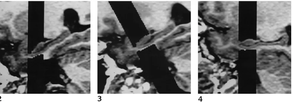
hippocampus from the amygdala (Fig 1). Three
segmen-tations of the hippocampal formation allowing three
differ-ent volumetric calculations were performed in all subjects
as follows: (
a
) volume was measured on sections of the
coronal 3-D acquisition that was not perpendicular to the
axis of the hippocampal formation (NOPERP protocol)
(Fig 2); (
b
) using the same acquisition, segmentation was
performed on sections reformatted (REFOR protocol) in
the plane perpendicular to the axis of the hippocampal
formation (Fig 3); the plane perpendicular to the
hip-pocampus was determined from the sagittal reformatted
view centered on the right hippocampus; and (
c
)
segmen-tation was performed on the coronal 3-D acquisition
ob-tained with the patient’s head positioned in such a way that
the acquisition plane was perpendicular (PERP protocol)
to the axis of the hippocampal formation (Fig 4). The
segmentations were performed by two different operators.
Total time for the segmentation of one hippocampus with
one protocol was approximately 40 minutes.
The measurements included the entire rostrocaudal
ex-tent of the hippocampus (eg, CA-1 through CA-4 sectors
of the hippocampus proper, the dentate gyrus, the alveus,
the fimbria, and a part of the subiculum). The
hippocam-pus was progressively segmented rostrocaudally. The
boundaries were outlined in the coronal plane. The
incre-ment between two segincre-mentation planes was 2 mm. This
increment was slightly greater than the section thickness,
but it could be obtained with the 3-D software (with
tions parallel to the acquisition or with reformatted
sec-tions) and it was chosen to shorten the total time needed
for segmentation. The first section, checked in the sagittal
window (Fig 5), was located just 2 mm caudal to the plane
intersecting the most anterior extension of the alveus, just
rostral to the uncus. The most accurate anterior limit was
searched with the 3-D cursor interactively in the coronal,
sagittal, and axial planes. Anatomic landmarks of the
hip-pocampal formation were defined at the level of the head,
body, and tail of the hippocampus, as described below.
Hippocampal Head
[image:3.612.63.557.86.259.2]Dorsally and laterally, the alveus provides a landmark
for the hippocampal head. It allows the examiner to
differ-entiate the hippocampus from the overlying amygdala
with the 3-D cursor. At this level, the hippocampus has a
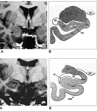
characteristic triangular shape (Fig 6). The location of the
cursor in the coronal plane was always simultaneously
Fig 2. Image processing, right hippo-campus in healthy volunteer: hippocampal segmentation on sections obtained from the acquisition not perpendicular to the hippocampus (NOPERP protocol).
Fig 3. Image processing, right hip-pocampus in healthy volunteer: hip-pocampal segmentation on reformatted coronal sections (perpendicular to the hip-pocampus) obtained from the nonperpen-dicular acquisition (REFOR protocol).
Fig 4. Image processing, right hip-pocampus in healthy volunteer: hip-pocampal segmentation on sections ob-tained from the acquisition perpendicular to the hippocampus (PERP protocol).
Fig 5. A, Spoiled GRASS image refor-matted in the sagittal plane shows the an-terior limit of the hippocampal segmenta-tion just rostral to the uncus (arrow).
[image:3.612.57.387.334.509.2]checked in the sagittal plane. More caudally, at the level of
the digitations of the pes hippocampus, the temporal horn
appears and enhances this dorsal limit. At this level, the
medial part of the hippocampal head merges with the
amygdala at the level of the amygdalohippocampal
tran-sition area (Fig 7). We traced a horizontal line along the
extent of the alveus to cut this amygdalohippocampal
area. The ventral limit was clearly defined by the gray–
white matter junction between the white matter of the
entorhinal cortex and the subiculum. Medially, the
bound-ary of the hippocampal head was limited by the uncal
sulcus and ambient fissure. The intralimbic gyrus was
out-lined in the most caudal planes of the pes hippocampus.
Hippocampal Body
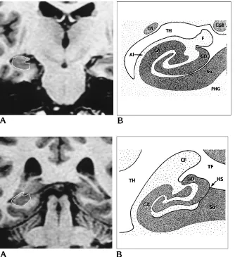
The hippocampal body was easier to outline (Fig 8):
dorsally and laterally we included the alveus overlying the
cornu Ammonis. This boundary is well defined in the floor
of the temporal horn. The fimbria was included in the
measurements. Medially, we chose an arbitrary landmark
located in the middle of the subiculum. The dentate gyrus
located between the fimbria and the hippocampal fissure
was included. Ventrally and laterally, the white matter was
well distinguished from the subiculum and from CA-1.
Hippocampal Tail
Dorsally and laterally, the alveus was outlined up to the
origin of the crus fornices medially, which was cut along
the extent of the alveus (Fig 9). The medial landmark was
an arbitrary vertical line traced at the level of the medial
limit of the hippocampal sulcus. The section showing the
entire length of the crus fornices was considered the
pos-terior limit of the hippocampal tail and it was not included
in the segmentation process.
After the segmentation process, the hippocampus was
portrayed on a 3-D–rendered image (Fig 10) and the
nu-meric values of the volume obtained by the 3-D software
were also displayed. Both hippocampi were studied in all
subjects. Using each protocol, we calculated two
hip-pocampal asymmetry indexes for each subject. The first
index (R1), was computed as follows:
R1(%)
5
100
3
(
R
2
L
)
Max
(
R, L
)
Fig 6. A, Spoiled GRASS coronal im-age (PERP protocol) shows the landmarks of the rostral part of the hippocampal head.
B, Anatomic drawing at the same level shows the close amygdalohippocampal relations. Am indicates amygdala; CA, cornu Ammonis;EA, entorhinal area;GA, gyrus ambiens;OT, optical tract;SL, semi-lunar gyrus; TH, temporal horn; and US, uncal sulcus.
Fig 7. A, Spoiled GRASS coronal im-age (PERP protocol) shows the landmarks of the hippocampal head at the level of the digitations.
[image:4.612.226.555.84.458.2]where
R
and
L
are, respectively, the values of the right and
left hippocampus volumes and
Max(R, L)
is the highest
value among the right and the left volumes (1). The
sec-ond index (R2) was computed as follows:
R 2
~
%
!
5
100
3
(
R
2
L
)
S
R
1
L
2
D
(20). The statistical analysis included the calculation of the
Pearson product-moment correlation between each
proto-col and a
t
test for dependent samples to test the difference
of means between each protocol.
Interobserver reproducibility was studied on both
hip-pocampi for the 10 subjects with the REFOR protocol and
was very elevated. Variance analysis showed that, for the
right hippocampi, 95.3% of the variance was due to the
true variance of the hippocampi; for the left, it was 92.7%.
Thus, only 4.7% of the variance for the right hippocampi
and 7.3% for the left hippocampi was due to interobserver
variability.
Results
Values of hippocampal volumes and
asym-metry indexes obtained with the three
measure-ment protocols in the 10 subjects are described
successively in the following sections.
Right Hippocampus
The mean volumes of the right hippocampus
were 3.42 cm
3(NOPERP protocol), 4.18 cm
3(REFOR protocol), and 3.91 cm
3(PERP
proto-col) (Table). The difference of the mean volume
of the right hippocampus was significant (
P
,
.001) between NOPERP and PERP protocols,
Fig 8. A, Spoiled GRASS coronal im-age (PERP protocol) shows the landmarks of the hippocampal body.
B, Anatomic drawing at the same level. Alindicates alveus;CA, cornu Ammonis; CN, caudate nucleus;F, fimbria;GD, gyrus dentatus; LGB, lateral geniculate body; PHG, parahippocampal gyrus;Su, subicu-lum; andTH, temporal horn.
Fig 9. A, Spoiled GRASS coronal image (PERP protocol) shows the landmarks of the hippocampal tail.
B, Anatomic drawing at the same level. CA indicates cornu Ammonis; CF, crus fornices; GD, gyrus dentatus;HS, hippocampal sulcus; Su, subiculum; TF, transverse fissure; andTH, temporal horn.
[image:5.612.58.392.86.451.2]between REFOR and PERP protocols, and
be-tween NOPERP and REFOR protocols. The
dif-ference between mean volumes of the right
hip-pocampus
computerized
with
PERP
and
NOPERP protocols (0.49 cm
3) was higher than
the difference of mean volumes calculated with
REFOR and PERP protocols (0.27 cm
3), but
this latter result was not statistically significant
(
P
5
.06). The volumes of the right
hippocam-pus were significantly correlated (
P
,
.001)
be-tween NOPERP and PERP protocols (
r
5
.91),
REFOR and PERP protocols (
r
5
.95), and
NOPERP and REFOR protocols (
r
5
.91).
Left Hippocampus
The mean volumes of the left hippocampus
were 3.29 cm
3(NOPERP protocol), 4.02 cm
3(REFOR protocol), and 3.74 cm
3(PERP
proto-col) (Table). The difference of the mean volume
of the left hippocampus was significant (
P
,
.001) between NOPERP and PERP protocols,
between REFOR and PERP protocols, and
be-tween NOPERP and REFOR protocols. The
dif-ference between left hippocampus mean
vol-umes computerized with PERP and NOPERP
protocols (0.45 cm
3) was higher than the
differ-ence of mean volumes calculated with REFOR
and PERP protocols (0.28 cm
3), but again this
latter result was not statistically significant (
P
5
.18). The volumes of the left hippocampus were
significantly correlated (
P
,
.001) between
NOPERP and PERP protocols (
r
5
.91), REFOR
and PERP protocols (
r
5
.98), and NOPERP and
REFOR protocols (
r
5
.95).
Asymmetry Indexes
The mean R1s were 4.3% (NOPERP
proto-col), 4.4% (REFOR protoproto-col), and 4.6% (PERP
protocol) (Table). The difference of the mean
ratio R1 was not significant between NOPERP
and PERP protocols (
P
5
.86), between REFOR
and PERP (
P
5
.80), and between NOPERP and
REFOR (
P
5
.97). The difference between
NOPERP (0.29%) and between
PERP-REFOR (0.24%) was not significant (
P
5
.80).
The correlations of the R1 asymmetry indexes
between each protocol were all significant (
P
,
.05).
The mean R2s were 4.57% (NOPERP
proto-col), 4.65% (REFOR protoproto-col), and 4.91%
(PERP protocol). The difference of the mean
ratio R2 was not significant between NOPERP
and PERP (
P
5
.84), between REFOR and PERP
(
P
5
.79), and between NOPERP and REFOR (
P
5
.96). The difference PERP-NOPERP (0.34%)
and between PERP-REFOR (0.26%) was not
significant (
P
5
.80). The correlations of the R2
asymmetry indexes between each protocol
were all significant (
P
,
.05).
Discussion
The in vivo biometry of the hippocampus has
been applied to the study of temporal lobe
epi-lepsy (1– 6, 21, 22), Alzheimer disease (7),
am-nesia (8, 23), and schizophrenia (9, 10).
Progress regarding biometry techniques in the
hippocampus has involved image acquisition
and data processing (5, 7–9, 17, 23, 24 –38).
At present, many researchers are using 3-D
acquisition sequences (5, 9, 17, 31), which can
be successively transferred on a workstation to
permit segmentation and volume calculation.
All authors (4, 5, 9, 17) report segmentation of
the hippocampal formation in the coronal plane
without the use of any 3-D control. However,
these 2-D segmentations have important
limi-tations, principally concerning the anatomic
identification of complex structures. For
exam-ple, to separate the pes hippocampus (which is
one of the most voluminous parts of the
hip-pocampal formation) from the overlying
amyg-dala is very difficult and sometimes impossible
with the use of 2-D processing software. Some
authors (8, 17, 23, 37) have preferred to
ex-clude this region despite its significant volume.
Mean values of right and left hippocampal volumes, R1 and R2 ratios obtained with the three different protocolsProtocol R Hippocampus, mean6SD L Hippocampus, mean6SD R1 (%), mean6SD R2 (%), mean6SD
NOPERP 3.42 cm360.49 3.29 cm360.63 4.366.0 4.5766.5
REFOR 4.18 cm360.53 4.02 cm360.74 4.466.5 4.6566.8
PERP 3.91 cm360.53 3.74 cm360.64 4.666.0 4.9166.4
Note.—NOPERP indicates acquisition not perpendicular to axis of hippocampal formation; REFOR, same acquisition, sections reformatted perpendicular to axis; and PERP, coronal 3-D acquisition perpendicular to axis. See “Hippocampal Tail” section for formulations of asymmetry indexes R1 (%) and R2 (%).
Our study shows that these anatomic problems
can be solved by means of 3-D processing of
the data. The major benefit of 3-D processing is
the 3-D cursor, which enables simultaneous
identification of an anatomic structure in three
orthogonal reformatted planes. With the use of
3-D processing, the major problem related to
the complex anatomy of the pes hippocampus
is easily solved. This is useful for the small
an-atomic structures (such as the alveus and the
fimbria), because each point of the structure,
located in the coronal plane, can be checked
and simultaneously displayed in the sagittal and
axial planes. Thus, the rostrocaudal and
medio-lateral extents of the hippocampal formation are
respectively displayed in the sagittal and axial
planes. The major benefit of 3-D processing is
in resolving anatomic ambiguities; for example,
when a boundary is not clear in the coronal
plane, it can be examined by looking at it
simul-taneously in the other planes.
Using this method of hippocampal
segmen-tation, we made three measurements of the
vol-ume of the hippocampal formation in each
sub-ject in order to evaluate and compare the
different results. It is commonly accepted that
all volume measurements of the hippocampal
formation should be performed in a plane
per-pendicular to its axis because of problems
re-lated to partial volume effects (8). However,
with most MR units, 3-D oblique acquisitions
are not available. Some authors have used one
of the three protocols of measuring the volume
of the hippocampal formation that we used (5,
9, 14 –17, 38). However, a study comparing the
results of the different protocols is not available.
The aim of our study was to compare the results
of these three protocols. In our series,
acquisi-tion and measurements performed directly
per-pendicular to the hippocampal formation (PERP
protocol) were considered as the reference
be-cause it avoided the possibility of introducing
errors caused by partial volume effects (as with
the NOPERP protocol) or by interpolated voxel
values (as with the REFOR protocol). Our study
shows that, even with a small group of 10
sub-jects, there was a significant difference between
the results obtained with each protocol. The
smallest values of the volume of the
hippocam-pal formation were obtained with the NOPERP
protocol; the highest values with the REFOR
protocol. In all subjects, volumes obtained with
the reference protocol (PERP protocol) were
al-ways between those obtained with the NOPERP
and REFOR protocols. Our results show that the
mean error obtained with the NOPERP protocol
was higher than the mean error obtained with
the REFOR protocol (in reference to the values
obtained with the PERP protocol). However,
probably because of the small size of our
sam-ple, this difference was not significant. Despite
the different values obtained with the three
pro-tocols, there was a strong correlation between
the volumes obtained by each method. This
correlation between the three protocols explains
the fact that the asymmetry indexes are not
significantly modified by the protocol used.
In conclusion, the use of different protocols
for measuring the volume of the hippocampal
formation led to important variations in the
ab-solute values of the calculated volumes.
How-ever, there was a strong correlation between the
results obtained by each method.
Conse-quently, any of the methods may be used to
study a group of patients, but it is important to
apply the same protocol throughout a given
patient population. Moreover, if a preliminary
study of control subjects is performed, it must
also be done with the same protocol.
Acknowledgments
We thank Alessandra Biondi, MD, for helpful
discus-sions in the revision of the manuscript, and Ste´phan
Bla-trix, Carine Brunello, Cyrille Martinet, and Claire Savourat,
the medical illustrators who created the original drawings.
References
1. Adam C, Baulac M, Saint-Hilaire JM, Landau J, Granat O, Laplane D. Value of magnetic resonance imaging-based measurements of hippocampal formations in patients with partial epilepsy.Arch Neurol1994;51:130 –138
2. Bronen RA. Epilepsy: the role of MR imaging.AJR Am J Roent-genol1992;159:1165–1174
3. Cascino GD, Jack C Jr, Sharbrough FW, Kelly PJ, Marsh WR. MRI assessments of hippocampal pathology in extratemporal lesional epilepsy.Neurology1993;43:2380 –2382
4. Cendes F, Andermann F, Gloor P, et al. MRI volumetric measure-ment of amygdala and hippocampus in temporal lobe epilepsy. Neurology1993;43:719 –725
5. Cook MJ, Fish DR, Shorvon SD, Straughan K, Stevens JM. Hip-pocampal volumetric and morphometric studies in frontal and temporal lobe epilepsy.Brain1992;115:1001–1015
6. Jack C Jr, Bentley MD, Twomey CK, Zinsmeister AR. MR imag-ing-based volume measurements of the hippocampal formation and anterior temporal lobe: validation studies.Radiology1990; 176:205–209
8. Press GA, Amaral DG, Squire LR. Hippocampal abnormalities in amnesic patients revealed by high-resolution magnetic resonance imaging.Nature1989;341:54 –57
9. Shenton ME, Kikinis R, Jolesz FA, et al. Abnormalities of the left temporal lobe and thought disorder in schizophrenia: a quantita-tive magnetic resonance imaging study.N Engl J Med1992;327: 604 – 612
10. Suddath RL, Christison GW, Torrey EF, Casanova MF, Wein-berger DR. Anatomical abnormalities in the brains of monozygotic twins discordant for schizophrenia.N Engl J Med1990;322:789 – 794
11. Cascino GD, Jack C Jr, Parisi JE, et al. Magnetic resonance imaging-based volume studies in temporal lobe epilepsy: patho-logical correlations.Ann Neurol1991;30:31–36
12. Cendes F, Andermann F, Dubeau F, et al. Early childhood pro-longed febrile convulsions, atrophy and sclerosis of mesial struc-tures, and temporal lobe epilepsy: an MRI volumetric study. Neu-rology1993;43:1083–1087
13. Lencz T, McCarthy G, Bronen RA, et al. Quantitative magnetic resonance imaging in temporal lobe epilepsy: relationship to neu-ropathology and neuropsychological function.Ann Neurol1992; 31:629 – 637
14. Jack CR. MRI-based hippocampal volume measurements in epi-lepsy.Epilepsia1994;35:S21–S26
15. Kuks JB, Cook MJ, Fish DR, Stevens JM, Shorvon SD. Hip-pocampal sclerosis in epilepsy and childhood febrile seizures. Lancet1993;342:1391–1394
16. Soininen HS, Partanen K, Pitkanen A, et al. Volumetric MRI anal-ysis of the amygdala and the hippocampus in subjects with age-associated memory impairment: correlation to visual and verbal memory.Neurology1994;44:1660 –1668
17. Spencer SS, McCarthy G, Spencer DD. Diagnosis of medial tem-poral lobe seizure onset: relative specificity and sensitivity of quantitative MRI.Neurology1993;43:2117–2124
18. Jack C Jr, Gehring DG, Sharbrough FW, Felmlee JP, Forbes G, Hench VS. Temporal lobe volume measurement from MR images: accuracy and left-right asymmetry in normal persons.J Comput Assist Tomogr1988;12:21–29
19. Conlon P, Trimble MR, Rogers D, Callicott C. Magnetic resonance imaging in epilepsy: a controlled study. Epilepsy Res1988;2: 37– 43
20. White LE, Lucas G, Richards A, Purues D. Cerebral asymmetry and handedness.Nature1994;368:197–198
21. Kuzniecky R, Burgard S, Faught E, Morawetz R, Bartolucci A. Predictive value of magnetic resonance imaging in temporal lobe epilepsy surgery.Arch Neurol1993;50:65– 69
22. Jackson GD, Berkovic SF, Duncan JS, Connelly A. Optimizing the diagnosis of hippocampal sclerosis using MR imaging.AJNR Am J Neuroradiol1993;14:753–762
23. Squire LR, Amaral DG, Press GA. Magnetic resonance imaging of the hippocampal formation and mammillary nuclei distinguish medial temporal lobe and diencephalic amnesia.J Neurosci1990; 10:3106 –3117
24. Kesslak JP, Nalcioglu O, Cotman CW. Quantification of magnetic resonance scans for hippocampal and parahippocampal atrophy in Alzheimer’s disease.Neurology1991;41:51–54
25. Jack C Jr, Twomey CK, Zinsmeister AR, Sharbrough FW, Pe-tersen RC, Cascino GD. Anterior temporal lobes and hippocampal formations: normative volumetric measurements from MR images in young adults.Radiology1989;172:549 –554
26. Ashtari M, Barr WB, Schaul N, Bogerts B. Three-dimensional fast low-angle shot imaging and computerized volume measurement of the hippocampus in patients with chronic epilepsy of the tem-poral lobe.AJNR Am J Neuroradiol1991;12:941–947
27. Bhatia S, Bookheimer SY, Gaillard WD, Theodore WH. Measure-ment of whole temporal lobe and hippocampus for MR volumetry: normative data.Neurology1993;43:2006 –2010
28. Murro AM, Park YD, King DW, et al. Seizure localization in tem-poral lobe epilepsy: a comparison of scalp-sphenoidal EEG and volumetric MRI.Neurology1993;43:2531–2533
29. Yoneda Y, Mori E, Yamashita H, Yamadori A. MRI volumetry of medial temporal lobe structures in amnesia following herpes sim-plex encephalitis.Eur Neurol1994;34:243–252
30. Cendes F, Andermann F, Gloor P, et al. Atrophy of mesial struc-tures in patients with temporal lobe epilepsy: cause or conse-quence of repeated seizures?Ann Neurol1993;34:795– 801 31. Jack CR Jr, Mullan DP, Sharbrough FW, et al. Intractable non
lesional epilepsy of temporal lobe origin: lateralization by interic-tal SPECT versus MRI.Neurology1994;44:829 – 836
32. Jack CR Jr, Krecke KN, Luetmer PH, et al. Diagnosis of mesial temporal sclerosis with conventional versus fast spin-echo MR imaging.Radiology1994;192:123–127
33. Kim JH, Tien RD, Felsberg GJ, Osumi AK, Lee N. MR measure-ments of the hippocampus for lateralization of temporal lobe epilepsy: value of measurements of the body vs the whole struc-ture.AJR Am J Roentgenol1994;163:1453–1457
34. Tien RD, Felsberg GJ, Crain B. Normal anatomy of the hippocam-pus and adjacent temporal lobe: high-resolution fast spin-echo MR images in volunteers correlated with cadaveric histologic sec-tions.AJR Am J Roentgenol1992;159:1309 –1313
35. Tien RD, Felsberg GJ, Campi de Castro C, et al. Complex partial seizures and mesial temporal sclerosis: evaluation with fast spin-echo MR imaging.Radiology1993;189:835– 842
36. Bathia S, Bookheimer SY, Gaillard WD, Theodore WH. Measure-ment of whole temporal lobe and hippocampus for MR volumetry: normative data.Neurology1993;43:2006 –2010
37. Ikeda M, Tanabe H, Nakagawa Y, et al. MRI-based quantitative assessment of the hippocampal region in very mild to moderate Alzheimer’s disease.Neuroradiology1994;36:7–10