J. Exp. Biol. (1966), 45, 83-99 8 3 With I I text-figures
Printed in Great Britain
ACTIVITY IN THE OPTIC NERVE OF PECTEN MAXIMUS
IN RESPONSE TO CHANGES IN LIGHT INTENSITY, AND
TO PATTERN AND MOVEMENT IN THE
OPTICAL ENVIRONMENT
BY M. F. LAND
Department of Physiology, University College London [Received 7 February 1966)
In order to find out what sort of information a simple eye is capable of transmitting about the nature of the optical environment, it is necessary to answer two questions. First, to what conditions of illumination do the individual receptor cells respond? Secondly, to what extent does the functional organization of the eye—the optical system and the retina—convey additional powers, such as spatial resolution, to the system as a whole?
(0)
I
•iOO/i Nerve fromdistal retina
[image:1.451.60.399.307.520.2]Nerve from proximal retina
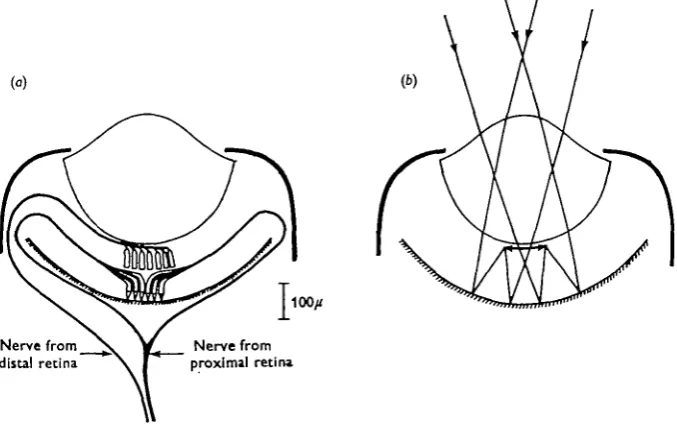
Fig. 1. Diagrams of (a) innervation and (6) optical system of the eye of Pecten.
In the eye of the scallop, Pecten, the first question has already been investigated. Hartline (1938) found that receptor axons in the optic nerve responded either to onset of illumination, or to darkening, but not to both (Hartline was illuminating the whole eye). He found that cutting the distal nerve (Fig. 1 a) abolished only the' off' responses, which, he concluded, were initiated in the cells of the distal retina, the ' on' responses originating in the proximal cells. Hartline was undecided as to whether the distal cells were primary 'off' receptors, or ganglion cells, functionally dependent on the
activity of the proximal retina. However, Miller (1958) found that the distal cells contained photoreceptor-like organelles of ciliary origin, and concluded that these were, anatomically, primary sense cells.
In this eye an image is formed not by the lens but by reflexion at the 'argentea' which lines the back of the eye (Land, 1965). The image is of good quality, and falls on the distal cells in the region containing Miller's organelles (Fig. 1 b). There is much behavioural evidence (e.g. Buddenbrock & Moller-Racke, 1953) which indicates that scallops do react, by closing, to the presence of moving objects which cast no direct shadow on the eyes. From the optical system, it seems logical to infer that the way
Pecten detects moving objects is by the production of a series of 'off' responses, as the
image of a dark object crosses successive cells of the distal retina. The aim of this paper is:
(a) to re-examine the responses of the proximal and distal retinae to
non-direc-tional stimuli,
(b) to investigate the nature of the activity in the optic nerve in response to
pat-terned stimuli, and to try to rationalize this in terms of the effect of the image on single retinal cells.
METHODS
Pecten maximus were obtained from Plymouth, and kept in a sea-water aquarium.
Experiments were usually performed within the first week, although the animals often survived for months in good condition.
A small piece of mantle, containing one of the larger eyes (1 mm.), was removed from the upper, flat, valve, and pinned to a cork block. The tentacles were removed from the area immediately around the eye, to prevent them entering the field of vision. The common optic nerve was located in the radial pallial musculature, and dissected free for a few mm. Recordings were made either between two platinum elec-trodes, or between one electrode and the bath, the nerve being raised into a paraffin layer above the sea water. The responses were recorded via an a.c. amplifier, and dis-played on the upper trace of a Tektronix 502 oscilloscope. Spike heights were between 30 and 100/tV.
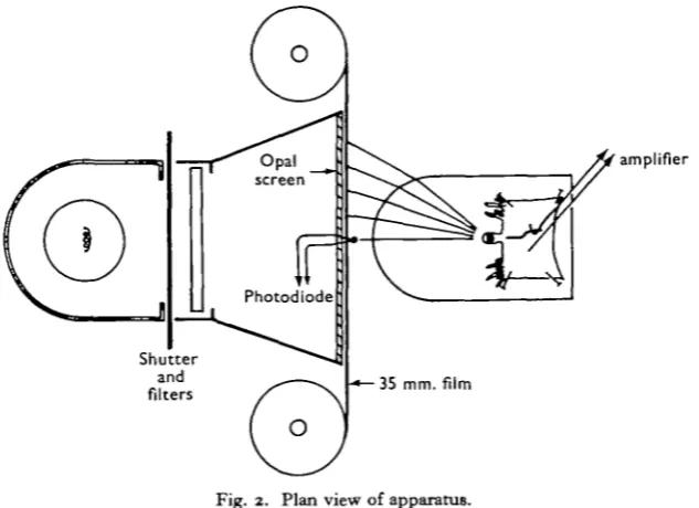
The light source of the stimulator, a 12 V., 36 W. car bulb, illuminated an opal Perspex screen, 8 x 2-4 cm. Between die lamp and the screen was a manually operated shutter, which could be closed in about 5 msec, and a chamber to hold neutral density filters. 35 mm. film, used for displaying light and dark stripes, could be moved in front of this screen, either by hand or using a kymograph motor. Changes in illu-mination at the centre of the screen were monitored by a photodiode, and displayed on the lower oscilloscope trace.
The curvature of the front face of the bath containing the eye was arranged so that equal distances across the screen subtended approximately equal angles at the eye. A strip 8-2 mm. wide subtended io° ± 1 ° at the eye, irrespective of its position on the screen -(Fig. 2). The 'field of view' of the eye was thus about 950 by 250. With a maximum screen brightness of 500 cd./m.2, the illumination at the eye from the whole field was about 700 lm./m2. (calculated, and checked with a photocell in position in the bath). A sloping platform on which the eye was mounted brought the eye's optical axis horizontal.
Optic nerve activity in Pecten
RESULTS
Preliminary experiments
A number of experiments were performed to check Hartline's (1938) demonstration of the separate origin of the 'on' and 'off' responses.
(i) Action potentials were recorded from the common optic nerve only during the periods immediately following illumination increase (on) and decrease (off). There was usually no activity during either constant illumination or darkness. P. maximus appears to differ in this respect from P. irradians where Hartline recorded a per-sistent discharge at a reduced frequency throughout illumination. This was only occasionally seen in P. maximus.
[image:3.451.70.387.216.446.2]amplifier
Fig. 2. Plan view of apparatus.
(ii) Spike heights during 'off' responses were greater than those recorded during ' on' responses. An estimate of the conduction velocities was obtained during the two types of response, by recording diphasically with electrodes 1 cm. apart, and measuring the time between the positive and negative peaks of individual action potentials. During 'off' responses the mean conduction velocity was about I-I m./sec, and 0-7 during 'on' responses, suggesting that the two responses are carried in different classes of fibre.
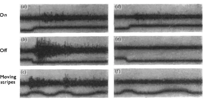
(iii) Cutting the distal nerve at the side of the eye, before it joined with the proximal nerve, abolished only the 'off' responses; the 'on' responses were unimpaired (Fig. 3) (iv) When recordings were made from the stump of the distal nerve as it emerged from the eye, only 'off responses were observed.
These experiments confirm that the 'on' and 'off' responses originate in the proximal and distal cells respectively.
expected that cutting the distal nerve should abolish all responses which presuppose the existence of an image—for example, responses to the movement of a pattern of equally spaced dark and light stripes (no overall illumination changes at the eye)-The results of such an experiment are shown in Fig. 3 c and / . Unfortunately, how-ever, this result is not as unambiguous as it appears. Because the distal nerve lies extremely close to the back of the eye, right up to the point where it joins with the proximal nerve, it is extremely difficult to cut it, and even more difficult to record from it without causing some distortion of the fragile argentea. While this matters little when recording responses to overall illumination changes, such distortion causes a reduction of image quality which does affect the size of movement responses. This does not entirely invalidate the experiment, however, as intentional distortion of the argentea only reduces the size of movement responses; it does not abolish them as in Fig. 3/. Much better evidence will be presented later in this paper to show that move-ment responses have the characteristics of 'off' responses, which have been shown unequivocally to arise in the distal cells.
Distal nerve intact Distal nerve cut
Off
[image:4.451.49.405.265.439.2]Moving stripes
Fig. 3. Abolition of 'off' and movement responses after cutting distal nerve. Stimulating conditions identical for nerve-cut and nerve intact records. Screen brightness 500 cd./m.*. In c and / , a pattern of equal dark and light stripes, each stripe subtending approx. 16 ° at the eye, was moved across the screen. Upward movement of the lower trace indicates lightening of the photodiode in the centre of the screen. Time pips: 100 msec.
Optic nerve activity in Pecten
Characteristics of the responses to overall illumination The 'on' response
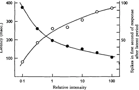
The dark-adapted eye responded to onset of illumination by producing a brief multi-fibre discharge. The greater the intensity of the on-going light, the greater were the initial spike frequency and the duration of the response, and the shorter was the latency. The response rarely lasted more than 2-3 sec, although occasionally it per-sisted at a much reduced frequency throughout the period of illumination. The ' on' response ceased within a few tens of msec, when the eye was darkened (distal nerve cut to abolish 'off' responses). The shortest latency observed for the first spike of the response was about 100 msec, increasing to 500 msec, at threshold.
I
400300
200
100
"V
i
-i -i
- so
8 1
a.
en
01 1 10
Relative intensity
[image:5.451.93.358.219.391.2]100
Fig. 4. On response. Plot of latency ( • — • ) and initial spike frequency (O—O) against intensity of illumination. Each point is the mean of two determinations, one with the stimulus strength increasing, and the other decreasing. Two minutes of darkness between trials. Intensity of 1 = 7 lm./m.1.
The minimum intensity required to evoke a response varied upwards from 0-07 lm./m.2, although in some eyes the response was barely perceptible at intensities 2-3 log units greater. As light is concentrated very little by the lens, the flux at the proximal cells will be approximately the same as at the pupil; the reduction in intensity at any one cell, due to partial occlusion of the field of view by the iris, is offset by the fact that much of the incident light is reflected back through the cells by the argentea.
The size of the response was also affected by how long the eye had been in dark-ness prior to stimulation. After strong illumination (8-5 x 10* lm./m.2) it took 5-10 min. before a response was produced to a test flash of 70 lm./m.2. Where previous illumination was less intense, this desensitizing effect was observable as an increase in latency, compared with responses obtained following prolonged darkness. After an intensity of 70 lm./m.2 a minute in darkness was sufficient for a maximal 'on'
re-sponse to be produced.
88
The 'off' response
The ideas associated with energy transduction, familiar from other types of receptor activity, cannot easily be applied to 'off* responses from cells which are, anatomically, primary receptors. It is pertinent to ask what the role of light actually is, when its effect only becomes apparent at its cessation. It does not simply have an inhibitory effect, as these cells are not spontaneously active in the dark—i.e. there is no intrinsic activity for light to inhibit. The fact that the magnitude of the 'off' response depends to a large extent upon the amount of light the eye has received before it is darkened, suggests that light may have an excitatory as well as an inhibitory effect.
1000
500
200 XI
8
100
o
1000
500
20
t
\
' i
40 60 80
Duration (sec.)
(b) Duration 10 sec •<
——
I I I
- 200
11
e S.
- 100 *
o
20 •40 60
Relative intensity
80
Fig. 5. Off response. Latency ( • — • ) and initial spike frequency (O—O) plotted against (a) duration and (b) intensity of the previous period of illumination. Same eye in both cases. Conditions as in Fig. 4.
Effect of previous illumination. The duration, frequency of spikes, and latency of the
Optic nerve activity in Pecten 89
inversely related; their relation to the parameters of previous illumination are shown in Fig. 5.
Latencies as long as 2 sec. have been recorded at threshold, while the minimum latency, following prolonged illumination, was never less than 70 msec, of which not more than 20 msec, could be accounted for by conduction time. Spike frequencies well in excess of 200/sec. (whole nerve) have been observed, but at these frequencies superimposition of spikes made the records uncountable. It was clear from few-fibre preparations that both the number of active fibres, and the frequency of firing of each fibre, increased with increasing intensity and duration of the stimulus.
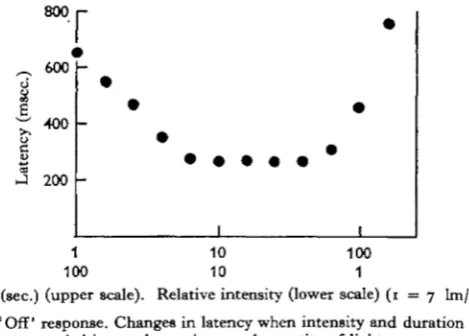
The similarity between the intensity and duration curves in Fig. 5 suggested that the magnitude of the 'off' response is determined by the total quantity of light en-tering the eye during the preceding light period. Fig. 6 shows the result of an experi-ment in which duration and intensity were varied reciprocally.
800r
~ 600
•400
200
I
1 10 100 100 10 1
[image:7.451.118.353.237.405.2]Duration (sec.) (upper scale). Relative intensity (lower scale) ( 1 = 7 lm/m.1) Fig. 6. ' Off' response. Changes in latency when intensity and duration were
varied inversely to give equal quantity of light.
Over a range of about one log unit, duration and intensity were interchangeable, equal quantities of light producing equal responses. Following intensities greater than 200 lm./m.2 the responses were smaller than those produced after the same quantity of light at lower intensities: under these conditions the duration rather than the intensity determined the size of the response. This possibly indicates that some process with a maximum rate of working is involved in the production of the 'off' response, this maximum being reached at intensities greater than 200 lm./m.2. At much lower light levels, intensity and duration similarly cease to be reciprocally effective. In this case equal quantities of light become progressively less effective until an intensity is reached at which no response is produced, however long the duration. For durations longer than 2 min. it is intensity alone which determines the size of the response. In life, where the eye is exposed to relatively constant illumination, the size of the response to complete darkening can only depend upon intensity.
Re-illumination. The dependence of the response upon the amount of light the eye
If, during an 'off' response, the eye was re-illuminated, the production of action potentials ceased within 100-150 msec. When the re-illuminating flash was so short that it would not, by itself, cause an 'off' response at its termination, the existing response was only interrupted, recommencing after a short latency at the end of the flash (Fig. 7).
(b)
Fig. 7. (a) Maximal ' off' response following 1 minute exposure to 70 lm./m.1. (b) as (a), but response 'interrupted' by flashes of 200 msec. duration at 70 lm./m.'. The minimum required to produce an 'off' response at this intensity was 2 sec. Time marks: ioomiec. Stimulus retouched.
Since the re-illuminating flashes were, on their own, subthresold, it is clear that the responses following the flashes are due to excitability remaining from the previous period of illumination. During the temporary cessation of firing caused by the flashes, this residual excitability must exist simultaneously with the inhibitory effect upon the production of action potentials. The definite increase in size of the response after each flash might be caused by release from adaptation occurring during the brief inter-ruption of firing.
Effect of dimming. The eye responded not only to cessation of illumination, but also
to dimming. In contrast to the response to increase in intensity, the dimming response was extremely sensitive, a decrease of 0-05 log units (10%) being usually enough to produce a recognizable response. This contrast may be reflected in the behaviour of the animal: Wenrich (1916) found that the shell-closing reaction in P. gibbus was much more sensitive to decrease than to increase in illumination. Like the magnitude of the full 'off' response, the sensitivity of the eye to dimming increased as the duration of the previous period of illumination increased. This is probably a mani-festation of the same sensitizing process that underlies the 'off' response proper. Provided the eye had been exposed to light for sufficient time (2 min. or more), the sensitivity to dimming was not greatly affected by the absolute light intensity over a range of 2 log units.
Threshold. The absolute threshold for the 'off' response, i.e. the intensity at which
dif-Optic nerve activity in Pecten gi
ference between 'on' and 'ofF thresholds was as much as 1-5 log units either way. It is interesting to compare these threshold values with the conditions of illumination normally experienced by the animal in its natural habitat. P. maximus is trawled at Plymouth from depths of 80-100 m., although individuals are found up to the low water mark. Green light, to which the animal is probably most sensitive, is absorbed least in coastal waters, but still decreases by a factor of 10 for every 20 m. below the surface. Surface illumination of ioslm./m.2 in daylight will therefore be reduced at 100 metres to the green component of white light of intensity 1 lm./m.2. As the light reflected in a horizontal direction will be only a fraction of this, the threshold values obtained for the 'on' and 'off' responses would only just be adequate at this depth for the eyes to be of any value to the animal. It is, of course, possible that the animals used here, which had been out of the sea for a week or more, and exposed to surface illumination, would not reflect fairly the sensitivity of the animals in their normal habitat.
1000 r
800
600
I 400
2
200
200
10 20 Minutes
30
10 20
Minutes
30
Fig. 8. Recovery after strong illumination. Changes in latency of (a) 'off' and (6) 'on' responses following illumination for 5 min. at 8-5 x io4 lm./m.1. Test flashes of 10 sec. dura-tion at 70 lm./m.1 every 5 min. Three of the eyes used in (a) are the same as in (b), plus one other.
Recovery after strong illumination
In these experiments, eyes were illuminated by light from a microscope lamp (intensity at the eye 8-5 x 10* lm./m.2) for 5 min., and test flashes of 10 sec. duration at 70 lm./m.2 were given at 5 min. intervals over the following 40 min. Fig. 8 shows the changes in latency of the 'on' and 'off' responses for four eyes.
complete after 25 min. Such changes are typical of dark adaptation in other photo-receptor systems.
In the 'off' response, on the other hand, recovery was invariably associated with an
increase in latency. If one assumes that the direct effect of bright light is to bleach
photopigment, then this increase presumably results from the recovery of a photo-chemical process whose effect on the response itself is inhibitory. This increase in latency, however, was not always accompanied by a decrease in the initial rate of firing. Out of eight eyes, three showed an increase, four a decrease, and in one the initial spike frequency fluctuated. This is in contrast to the reliably inverse way that latency and spike frequency vary with changes in previous illumination when the eye is fully adapted (Fig. 5). Responses recorded during the early phase of recovery were anomalous in other respects. They often showed a short-latency, vigorous burst of spikes of very short duration, whereas in the fully adapted eye the same stimulus would evoke a response lasting many times as long, even though the latency was much longer, and the initial spike frequency no greater, or even less than in the early responses.
These effects could be explained by assuming that the 'off' response is the result of the interaction of two light-dependent processes, one inhibitory and one excitatory. Independent recovery of the two processes after bleaching could lead to the results described, if the response parameters—latency, rate of firing and duration—depended in a complex way on the balance between the two. Thus, for example, the brief, short-latency bursts occurring early in recovery might have short latencies because of shallow inhibition, and brief durations because residual excitability is also low.
D k strites Responses to pattern and movement
The argentea produces a reflected image only on the cells of the distal retina. It might therefore be expected that 'off' responses would be produced from distal cells, when they are crossed by the image of a dark object.
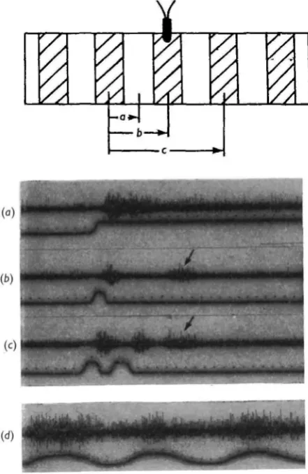
When a black stripe was introduced into the centre of an evenly illuminated field, a small response followed after a latency of a few hundred msec. Like the 'off' response, this lasted only a few seconds. It could be shown that the response was due to the image of the stripe, rather than to the net dimming it caused, by moving the stripe in steps across the field (Fig. 9a). A separate burst of spikes was generated after each movement, even though the light intensity at the eye remained approximately constant once the stripe had entered the field.
In Fig. 96, where the stripe was moved back and forth across the centre of the field, the first event to occur following each movement was a temporary cessation of the response. This is interpreted as an inhibition of the responding cells as they are re-illuminated. This is then followed, after a latent period, by a response from the newly darkened cells.
Optic nerve activity in Pecten 93
cells anywhere on the retina. This is because, in this eye, light has already passed unfocused through the retina once before falling on the distal cells as a focused image. The smallest stripe that produced a recognizable response subtended 2° at the eye, and the smallest angle through which a larger stripe had to be moved to cause firing was about the same. On the image this represents a distance of 9 fi, which is the approximate packing distance of the distal cells.
6° stripe
A
10° stripe D
(b)
-r
10° stripe
8° stripe, different eye
t
Fig. 9. Responses to dark stripes, (a) Movement of stripe in steps across the field. Back-ground illumination 50 cd./m.1. Time marks 100 msec. Upward movement of lower trace indicates darkening of photodiode. (6) Movement of stripe back and forth across centre of field. Arrows on lower record indicate cessation of firing, following each movement.
Light stripes
94
Provided a light stripe had remained in a particular position long enough, moving it a few degrees in either direction always evoked a response (Fig. 10b).
One may conclude from this that the two processes concerned in the 'off' response —build-up of excitability and simultaneous inhibition of the production of action potentials—both occur in the distal retina. This is because removal of light from a particular part of the image is only effective as a stimulus if the same part of the image has previously received a sufficient quantity of light.
Fig. 10. Responses to light stripes, (a) Movement of stripe across centre of field. Stripe had remained i min. in position A before it was moved. Intensity of stripe 50 cd./m.1. Time marks 100 msec, (b) Responses following movement in either direction from a pre-illuminated position.
Optic nerve activity in Pecten gf
re-instated, responses are produced only for the duration of the period(s) in which the previously illuminated cells are darkened. In (b) and (c) small, long-latency bursts (arrowed) follow the initial responses, and these are almost certainly 'off* responses resulting from the transient lightening and darkening of the initially darkened cells.
(b)
•J\.
Fig. I I . Responses to moving a pattern of equally spaced light and dark stripes. In (a), (b) and (c) the pattern was held stationary for i min., then moved through J, i and 2 ' wavelengths' respectively. Each dark stripe subtended 160 at the eye. Brightness of light stripes 50 cd./m.1. In (d) a pattern of 8° stripes was moved continuously after previously remaining stationary for 1 min. Different eye from (a), (6) and (c) and light stripes 10 x brighter. Upward move-ment of lower trace indicates lightening of photodiode. Time scale: 100 msec.
[image:13.451.117.338.116.456.2]individual cell will depend as much upon its ability to respond to slight dimming as upon the definition of the image itself. The fact that the distal cells can respond to dimming of as little as 10% means that quite small changes of image contrast could be readily detected. The contrast between light and dark regions of the image is already reduced by the light which has passed once through the retina before being reflected. All the responses described above are interpretable in terms of the behaviour of the distal cells. They are all essentially 'off' responses, whether they are caused by move-ment of dark or light stripes, and there is no evidence that the proximal retina is con-cerned in any of them. This view is further supported by the fact that these responses are all obtainable during the early stages of recovery from strong illumination, when
'on' responses are absent.
DISCUSSION
This eye differs from most others in two important respects. First, the choice of an optical system based on a concave reflector is, if not unique, a very unusual solution to the problem of producing a spatially resolved representation of the environment. Secondly, the retina consists of two physically and functionally separate arrays of receptors (see below), which respond to light in quite different ways. One responds to lightening, the other to darkening—an 'on' retina and an 'off' retina in fact. This is not like the vertebrate eye, where 'on', 'off' and more complex responses are syn-thesized by ganglion cells from receptor inputs all behaving in essentially the same way. In Pecten, where the fibres of the optic nerve are the axons of the receptors, the two types of response are properties of the two types of receptor.
As a device for extracting information about the optical environment the most important feature of this eye is that only one of the two receptor systems, the distal (off) retina, lies in the plane of the image. Consequently, all information about the
spatial distribution of light and dark in the environment will be confined to the
responses of the distal cells. It has been shown in this paper that this is the case. The proximal (on) retina, placed in a region of the eye where light is entirely diffuse, responds only to changes in overall light intensity.
There are three kinds of stimuli, therefore, which elicit impulses in the optic nerve. (i) Overall decrease in light intensity (all cells of the distal retina respond). (ii) Decrease in light intensity in particular parts of the visual field (particular cells of the distal retina respond). An important corollary of this is that moving objects, light or dark, are effective stimuli, as in either case their movement causes darkening of some part of the distal retina.
(iii) Overall increase in light intensity (all cells of the proximal retina respond) or, in P. irradians where the 'on' discharges have a tonic component (Hartline, 1938), continuous illumination of any absolute intensity.
Optic nerve activity in Pecten 97
since the distal nerve contains fibres from all parts of the distal retina, discharges elicited by moving objects contain potential information not only about the existence of these objects, but also about their position and direction of movement. Whether the C.N.s. actually makes use of this knowledge, and that contained in the discharges from simultaneously stimulated adjacent eyes, is not known. Purely as a detector, however, the efficacy of this system has been demonstrated behaviourally by Budden-brock & Moller-Racke (1953) who showed that P.jacobaeus would react, by closing, to an angular displacement of a dark object by 2-3°—a figure which agrees well with the equivalent sensitivity of the isolated eye, 2-40, as measured here for P. maximus. Objects moving at a wide range of speeds, from one or two to several hundred degrees per second, can be detected by the distal retina, although in any particular case the actual speed range depends on the relative size of the object and whether it is lighter or darker than the background in a way that is too complex to discuss briefly.
So far, only responses of a spatially resolved character have been discussed (category (ii) above). The response to overall dimming (i) is not likely to be biologically impor-tant, as the ambient light intensity on the sea floor will change little over short periods of time. The same applies to the proximal cell responses to increase of light intensity (iii). However, the tonic component of these cells' discharge (rarely found in P.
maximus) may have the biologically important function of providing the animal with a
monitor of light intensity. Several species of scallop, though probably not P. maximus (Dakin, 1928), exhibit light/dark habitat preferences, the animals migrating if necessary to a lighter or darker part of the aquarium. As no information about absolute light intensity is contained in the responses of the distal cells, the tonic, proximal cell responses almost certainly provide the sensory basis of these preferences.
The receptors
Of the two types of receptor found in this eye, only the proximal cells have much in common with photoreceptors found outside the lamellibranchs. They have outer segments consisting of a mass of long microvilli arising from a conical projection of the cell body (Miller, i960), an arrangement found in many invertebrate photoreceptors, including those of coelenterates, planarians, polychaetes, echinoderms and gastropod molluscs (Eakin, 1963). Their response to light is similarly typical of other primary visual cells—the onset of illumination evokes a brief discharge, whose magnitude depends on the strength of the stimulus, and latency inversely so. As with other systems, 'light-adaptation' occurs, the responsiveness decreasing after strong illu-mination, recovering again during the dark.
The distal cells contrast structurally as well as functionally with this 'standard' pattern. In the first place the lamellated structures described by Miller (1958), which are presumably the photoreceptive sites, are situated at the same end of the cell body as the origin of the nerve fibre, and immediately adjacent to it. There are several of these structures in each cell, and the arrangement of membranes within each organelle is unlike that found in any other type of photoreceptor.
The response of these cells has been described as a primary 'off' response, but this is a statement which requires some qualification. Evidence presented in this paper indicates that two distinct light-dependent processes are involved in the 'off' response —a cumulative development of excitability, and a simultaneous inhibition of spike
production—and there is, at present, no evidence that more than one of these pro-cesses occurs actually in the cells from whose axons recordings were made. One type of interaction can, however, be excluded: the 'on' cells of the proximal retina do not play any part in the generation of 'off' responses. Both excitation and inhibition have been shown, in the experiments where stripe patterns were used, to be image-dependent, in other words both effects depend on the conditions of illumination at particular points on the distal retina. Also, strong 'off' responses are recorded in the early stages of adaptation, when ' on' responses are absent, and even in the dark-adapted eye the period over which distal cell excitability continues to summate greatly outlasts the normal duration of the 'on' response. The evidence is thus strongly against there being any interaction between the two retinae. Dr V. Barber (unpublished) has failed to find any evidence from electron micrographs of synaptic junctions between the two cell layers, or for that matter between cells of the distal retina.
Visual responses in other lamellibranchs
Both ' on'-responding and 'off'-responding cells have been found in other lamelli-branchs. Kennedy (i960) recorded 'off' responses from a single photoreceptive fibre in the pallial nerve of Spisula. In this case the response was generated actually in the fibre; but, as in Pecten, Kennedy found evidence of independent excitatory and inhibitory effects of light. I have examined the cockle, Cardium edule, where a number of simple eyes are borne on tentacles around the siphons. These contain receptors of only one type, which, preliminary recordings indicate, produce 'off' responses similar in character to those of Pecten; no ' on' responses have been seen. A weak resting dark-discharge, and a shorter minimum latency (20-30 msec.) are the only differences so far observed; the sensitivity of this eye to dimming is as acute as in Pecten. One of the characteristic behavioural responses of many lamellibranchs is a ' shadow reaction', the animal closing its shells or withdrawing its siphon when momentarily darkened. It may be that primary 'off' receptors evolved in the lamellibranchs, possibly from light-sensitive neurones like that of Spisula, as the quickest, and neurally most economical, way of warning the animal of the approach of danger.
The more conventional ' on' receptors are probably also fairly widespread. Hecht's (1920) pioneer work on photochemical constants was based on a behavioural reaction of My a, mediated by an 'on' response. Kennedy (i960) recorded both 'on' and 'off' responses from receptor axons in the siphonal nerves of Mya and Venus.
In view of the rather general distribution of light sensitivity of the two kinds mentioned, it is odd that very few lamellibranch genera, other than Pecten, have developed optical systems capable of exploiting the properties of either type of receptor as the basis of a directionally sensitive eye. The selective advantage to the possessor of such a device seems obvious—it enables the animal to detect and react to predators long before they are near enough to cast a direct shadow.
SUMMARY
Optic nerve activity in Pecten 99
2. The activity of each retina was investigated by recording from the optic nerve> which contains the axons of both types of receptor. Visual stimuli used included changes in overall illumination, pattern and movement. Dark adaptation was also studied.
3. The 'off' response is generated by cells which are anatomically primary re-ceptors. Two light dependent processes are involved: a slow development of excita-bility and a simultaneous inhibition of action potentials.
4. The reflected image is important in movement perception. Only the distal cells lie in the image plane, and they can respond to moving objects, as these cause dim-ming of successive cells when their images cross the retina. Dark objects subtending 2° or more at the eye, and moving through more than 2°, are effective stimuli. Light objects are also effective, provided they have remained stationary for several seconds before moving. There is no response to stationary pattern. The proximal cells respond only to increase in overall light intensity.
5. These findings are discussed in relation to the behaviour of Pecten.
My thanks are due to my supervisor, Prof. J. A. B. Gray, for much helpful advice, and to the Medical Research Council for a research training scholarship.
REFERENCES
BUDDENBROCK, W. VON & MOLLER-RACKE, I. (1953). Uber den Lichtainn von Pecten. Pubbl. zool. Staz. Napoli. 24, 217-45.
BUTCHER, E. O. (1930). The formation, regeneration and transplantation of eyes in Pecten (Gibbus borealis). Biol. Bull., Woods Hole, 59, 154-64.
DAKIN, W. J. (1928). The eyes of Pecten and allied lamellibranchs. Proc. Roy. Soc. B, 103, 355-65. EAKIN, R. M. (1963). Lines of evolution of photoreceptors. In: General Physiology of Cell
Speciali-zation, ed. D. Mazia and A. Tyler. New York: McGraw-Hill.
HARTLINE, H. K. (1938). The discharge of impulses in the optic nerve of Pecten in response to illu-mination of the eye. J. cell. Comp. Pkysiol. n , 465-77.
HECHT, S. (1920). The photochemical nature of the photosensory process. J. Gen. Physiol. a, 229—46. KENNEDY, D. (i960). Neural photoreception in a lamellibranch mollusc. J. Gen. Physiol. 44, 277—99. LAND, M. F. (1965). Image formation by a concave reflector in the eye of the scallop, Pecten maximus.
J. Physiol. 179, 138-53.
MILLER, W. H. (1958). Derivatives of cilia in the distal sense cells of the retina of Pecten. J. biophys.
biochem. Cytol. 4, 227—8.
MILLER, W. H. (i960). Proximal sense cells of Pecten retina. In The Cell, vol. iv, pp.342-5. Ed. J. Brachet and A. Mirsky. London: Academic Press.
WENRICH, D. H. (1916). Image formation in Pecten. J. Anim. Behav. 6, 297-318.