Original Article
Modified maxillary sinus floor elevation via a
mini-lateral window with simultaneous placement
of dental implants: a clinical and radiographical study
Liqin Zhu1, Liangru He2, Qun Wang1, Xiaoyi Chen1, Junhua Xu1, Huiming Wang1
1Department of Oral Implantology, The First Affiliated Hospital, College of Medicine, Zhejiang University,
Hang-zhou, China; 2Department of E.N.T, Anji Traditional Chinese Medicine Hospital, Zhejiang, China
Received March 5, 2017; Accepted March 31, 2017; Epub June 15, 2017; Published June 30, 2017
Abstract: This study assessed the effectiveness of modified sinus floor elevation via a mini-lateral window with simultaneous placement of implants in 20 patients with severely atrophic maxilla. Patients received mini-round-like or a mini-slot osteotomy which was done to establish an access on the lateral sinus wall for placement of bone grafts depending on the number of inserted implants and the characteristics of sinus floor. Clinical and radiographic parameters were collected. Results showed all implants were well maintained, with 100% cumulative success rate. The mean residual, immediate and 6-month postoperative augmented bone height was 3.0 ± 0.5, 13.6 ± 0.9, and 13.2 ± 0.8 mm, respectively. Intra-operative and 6-month postoperative implant stability quotient (ISQ) were 61.2 ± 3.7 and 76.8 ± 2.4, respectively. No significant differences were found in clinical outcomes between two types of window approaches. Sinus augmentation via a mini window with simultaneous placement of implants has a higher success rate, may improve clinical outcome and thus can be used as a reliable treatment for severely atrophic maxilla.
Keywords: Lateral sinus floor elevation, sinus augmentation, dental implant, vital bone, cone-beam computerized tomography
Introduction
Sinus augmentation using lateral window tech-nique has been a predictable and popular approach in case of bone volume deficiency in the posterior maxilla for patients who require implant-based treatments [1-4]. Moreover, the placement of implants can be completed simul-taneously or in later management to allow for graft maturation [5]. The opening created in the lateral window approach provides an easier access to the sinus membrane as well as good view in the surgery. The Schneiderian mem-brane reflection is extended by direct surgical undermining, and the wider the extent of reflec-tion, the greater the vertical elevation height is [3, 4, 6, 7]. Typically, the lateral window size is determined by the amount of augmentation as well as the number of missing posterior teeth that should be replaced. On an average, a win-dow with mesiodistal width and apicocoronal height of 20 and 15 mm, respectively (also
preservation of a substantial portion of the lat-eral bone may enhance healing and improve graft consolidation and maturation along with vital bone formation. The outcome of implants placed in sinus-grafted regions is closely relat-ed to the vital bone; the vital bone, which is directly interfaced with a considerable portion of the implant surface, plays critical roles in osseointegration and local responses to func-tional loading [11].
In order to reduce complications and retain more cortical bones to promote vital bone for-mation during this sinus augmentation via the lateral window, we introduced a modified sinus floor elevation technique via a mini-lateral win-dow, which is either mini-round-like or mini-slot. This study was conducted to assess the ef- fectiveness of sinus augmentation via the lat-eral window with simultaneous dental implant placement in 20 patients with severely atrophic maxillae.
Materials and methods
Patients
From July 2013 to September 2015, 20 pati- ents (10 women and 10 men) aged 19-78 years (median: 46.1 years) were enrolled into this study. All patients provided written informed consent.
[image:2.612.91.524.84.405.2]The inclusion criteria were as follows: (i) patients required posterior maxillary implants; (ii) there was no rhinitis or sinusitis; (iii) there was tooth extraction at least 3 months before implant surgery; (iv) the residual bone height (RBH) was ≤4 mm; and (v) there was sufficient bone width to maintain the primary stability of implants. Heavy smokers (>10 cigarettes/day) or patients with uncontrolled periodontal dis-eases were excluded from this study. Baseline patients’ characteristics are summarized in
Table 1.
Table 1. Baseline characteristics and clinical outcomes of patients in this study
Patient No. Sex Age (yr) Missing tooth Window Shape
Window Size ISQ
RBH
(mm) Imm-ABH (mm)
6 M Post-ABH
(mm) Max H
(mm) Max W (mm) operationIntra Operation6 M Post
1 F 78 15\16\17 MSW 5 12 64 78 3.1 13.3 12.8 2 F 30 26 MRLW 3.5 3.5 58 74 2.7 14.8 13.8 3R M 23 13\14\15 MRLW 5 5 66 76 3.4 13.2 12.5 3L 23\24\25 MRLW 5 5 60 78 3.6 14.3 13.6 4 M 67 24\25\26\27 MSW 5.5 12.5 68 77 3.8 13.7 13.5 5 M 59 26\27 MSW 3.5 8.5 59 72 2.5 14.7 14.0
6 F 19 16 MRLW 4 4 60 79 2.8 13.1 13.5
7 F 69 25\26\27 MSW 4.5 11.5 62 75 3.0 12.9 13.3 8 M 44 26\27 MSW 3 8 55 76 2.8 12.3 12.0
9 M 42 16 MRLW 4 4 61 80 3.2 13.5 12.2
10R F 33 15\16 MRLW 4.5 4.5 62 75 2.7 14.2 13.8 10L 26 MRLW 3.5 3.5 59 81 3.2 13.8 14.2 11 F 24 26 MRLW 4.5 4.5 68 78 3.7 15.1 14.5 12 M 25 15\16\17 MSW 4 11.5 65 77 3.3 13.8 13.4 13 F 25 15 MRLW 4 4 60 79 2.6 11.8 12.2 14 M 63 26\27 MSW 3 9 57 77 2.1 14.3 13.8 15 M 50 25\26\27 MSW 4.5 12 61 74 2.5 12.4 12.0 16 F 56 36\37 MSW 3.5 10 67 77 3.5 14.5 13.8 17 F 34 36 MRLW 4 4 58 75 2.9 13.9 13.5 18 M 68 16\17 MRLW 5 5 60 80 3.5 12.9 12.5 19 F 66 15\16\17 MSW 4.5 12 61 78 2.8 13.2 12.5 20 M 46 36\37 MRLW 5 5 56 73 3.5 12.6 12.0 Mean ± SD 61.2 ± 3.7 76.8 ± 2.4 3.0 ± 0.5 13.6 ± 0.9 13.2 ± 0.8
Surgical procedures
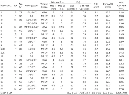
Before sinus augmentation, the number of missing teeth, height of the residual alveolar ridge, anatomy of the sinus, and position of the posterior-superior alveolar artery were precise-ly determined by panoramic radiography and cone-beam computerized tomography (CBCT) (Figure 1A, 1B). The insertion path and shape
above the sinus floor. The maximal nal height of the slot or the maximal apicocoro-nal height and the maximal mesiodistal width of the mini-round-like window was approximate-ly 3-5 mm to allow the placement of elevation instruments into the sinus floor. Moreover, max-imal mesiodistal width of the slot was appropri-ately extended to include implant placement sites. To calculate the approximate window size Figure 1. Radiography and clinical procedures for mini-round-like or mini-slot window ap-proaches. Note: (A, B) Preoperative computed tomography; (C, D) Preparation of the access on the lateral sinus wall; (E, F) Simultaneous placement of the implants; (G, H) Immediate postoperative computed tomography (A, C, E, G: Cases treated using the mini-round-like window approach; B, D, F, H: Cases treated using the mini-slot window approach).
Figure 2. Measurement of approximate window size. Note: Left: Mini-round-like window; Right: Mini-slot window.
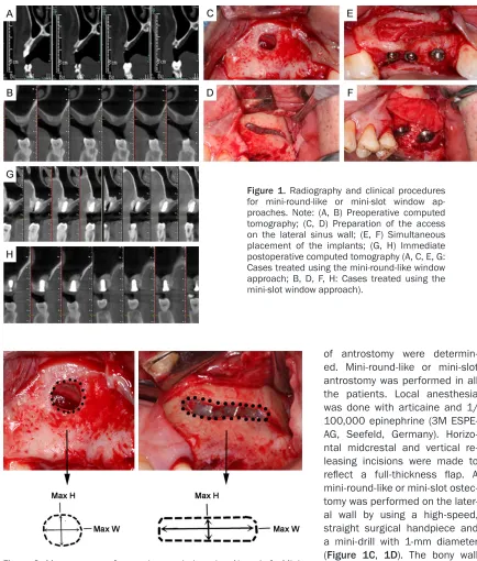
[image:3.612.87.521.67.577.2](AWS), maximal height and width of the win- dow were measured using a Hu-Friedy peri-odontal probe, and the value was rounded to the nearest half millimeter (Figure 2). The ele-vated Schneiderian membrane was maximally extended both mesiodistally and medially over the anticipated drilling site by using a mic- roelevator that was specifically prepared in our department for sinus membrane reflection. Drilling was performed continually while pro-tecting the membrane with a periosteal eleva-tor. Then, the graft materials (Bio-oss, Geis- tlich) were inserted through the mini-round-like or mini-slot window until it filled the whole cavi-ty, and implants were simultaneously placed (Figure 1E, 1F). Resorbable collagen mem-branes (Bio-Gide; Geistlich) were used to cover all lateral windows. After proper reposition, full-thickness flaps were sutured with 5-0 Vicryl (Ethicon) horizontal mattress and interrupted sutures.
After surgery, amoxicillin (500 mg, thrice daily; Xinya Co., Shanghai, China) and metronidazole (400 mg, thrice daily; Xinyiwanxiang, Shanghai, China) were administered for 1 week, along with use of 0.12% chlorhexidine oral rinse (60 seconds, 5-6 times/day for 2 weeks), and the sutures were removed 10-14 days later. Six months after surgery, superstructures of im- plants were prepared and fixed on the alveolar bone.
Clinical and radiological follow-up
Patients were followed up once weekly in first postoperative month, and both intraoperative or postoperative complications were recorded. Intraoperative complications included exces-sive bleeding, perforation of membrane, and benign paroxysmal vertigo. Postoperative com-plications included swelling, ecchymosis, pain, loss of graft materials, and nasal bleeding. CBCT was performed before surgery, immedi-ately after surgery, and 6 months after surgery. Two investigators collected and reviewed the following radiographical parameters in a blind manner: residual bone height before surgery (Pre-RBH), immediate augmented bone height after surgery (Imm-ABH), and postoperative augmented bone height 6 months after surgery (6 M Post-ABH). Averages were calculated and expressed for both sets of measurements (Table 1).
Three-dimensional reconstructions of the CBCT scans were performed to view the three-dimen-sional images of the sinus grafts.
During implant placement and 6 months after surgery, implant stability quotient (ISQ) was determined using resonance frequency analy-sis (RFA) with the Osstell ISQ System (Osstell AB, Goteborg, Sweden) and SmartPeg (Table 1). In all cases, ISQ was calculated as the aver-age of four measurements (facial, lingual, mesi-al, and distal) per implant.
Implant survival was determined by assessing the clinically detectable implant mobility, pain, subjective sensation, recurrent peri-implant infections, and continuous peri-implant radiolu-cency [12, 13].
Statistical analysis
Data are expressed as mean ± standard devia-tion (SD). One-way analysis of variance (ANOVA) (F) and t test (t) were used for comparisons with statistical package for the social sciences (SPSS) (version 11.0; SPSS Inc., Chicago, IL). A value of P<0.05 was considered statistically significant.
Results
Clinical observations and assessments
A total of 20 patients (10 women and 10 men) aged 19-78 years (median: 46.1 years) partici-pated in this study. In addition, bilateral maxil-lary sinus augmentation was performed in two patients, and thus a total of 22 sinus elevation procedures were conducted with placement of 46 implants. All the implants were repaired using superstructures with a cumulative sur-vival rate (CSR) of 100%.
measurements are summarized in Table 1. The mean sizes for the two different windows are presented in Table 2.
Membrane perforation occurred in two proce-dures, and thus a resorbable collagen mem-brane was used to cover the perforation before graft placement in each case. Intraoperative or postoperative excessive bleeding was not observed in all these patients. Moreover, large intraosseous arteries were noted across the lateral sinus wall on CBCT slices during preop-erative analysis and the slot ostectomy was done bypassing the artery. There was no benign paroxysmal vertigo, which occasionally occurs after severe osteotome preparation. Postope- rative facial swelling was relatively milder than that observed after using the conventional lat-eral window approach. Postoperative complica-tions such as ecchymosis, loss of graft materi-als, and nasal bleeding were not observed in these patients.
Radiographical assessment
Postoperative immediate radiography revealed that the augmented bone graft formed a dome
of bone grafts after 6-month follow-up. Table 1
enlists the mean values of all radiographical parameters. Table 2 summarized the compari-sons of these parameters between two differ-ent window approaches. There were no signifi-cant differences in the augmented bone height between two approaches.
Resonance frequency analysis (RFA)
RFA was performed for 46 implants. Table 1
summarizes the mean ISQ. The ISQ at the time of implant placement was 55-68, with a mean of 61.2 ± 3.7. Six months after surgery, mean ISQ increased to 76.8 ± 2.4. Table 2 pres- ents the comparisons of ISQ between two ferent window approaches. No significant dif-ferences were observed in the ISQ between two approaches.
Discussion
The maxillary sinus floor elevation was first described by Tatum [7] in 1977, but the first study on this topic was first published in 1980 by Boyne and James [14]. Since then, it has been widely used in various preprosthetic
sur-Table 2. Clinical outcomes between mini-round-like and the mini-slot window approaches
Window Shape
Window Size Radiographic Parameters ISQ Max H
(mm) Max W (mm) (mm)RBH Imm-ABH (mm) 6 M Post-ABH (mm) F P operationIntra operation6 M Post F p
Mini-slot 4.1 ± 0.8 10.7 ± 1.7 2.9 ± 0.5a 13.5 ± 0.8 13.1 ± 0.7 710.242 .000 61.9 ± 4.2c 76.1 ± 1.9 94.617 .000
Mini-round-like 4.3 ± 0.6 4.3 ± 0.6 3.2 ± 0.4b 13.6 ± 0.9 13.2 ± 0.9 704.268 .000 60.7 ± 3.4d 77.3 ± 2.6 182.119 .000
t 1.089 0.234 0.236 -0.765 1.231
p 0.289 0.818 0.816 0.453 0.233
Notes: Data were statistically analyzed using one-way ANOVA (F) and t test (t). p (probability) <0.05 represents statistically significant differences. No significant differ-ences were observed in augmented bone height and ISQ between mini-round-like and the mini-slot window groups. a, b: vertical height of the sinus floor after surgery was significantly higher than that recorded before surgery in both mini-round-like and mini-slot window approaches (P<0.05). c, d: ISQ at 6 months after surgery was significantly higher than that recorded during surgery in both mini-round-like and mini-slot window groups (P<0.05). ANOVA: analysis of variance; ISQ: implant stability quotient; Max H: Maximum Height; Max W: Maximum Width; RBH: Remaining Bone Height. Imm-ABH: Immediately Postoperative Augmented Bone Height; 6 M Post-ABH: 6-Month Postoperative Augmented Bone Height.
Figure 3. Three-dimensional images of sinus grafts at 6 months after sur-gery. Note: A: Case treated using the mini-round-like window approach; B: Case treated using the mini-slot window approach.
with clear round margins under the elevated Schneiderian me- mbrane. The apex of the impla- nt was not exposed out of the dome or membrane (Figure 1G,
gical procedures. Despite vast clinical research-es and surgical experience, various novel tech-niques and new technology in past 30 years, no consensus has been achieved regarding which surgical technique would provide most favor-able clinical outcomes [15]. However, one of the most important evolutional trends with lat-eral sinus floor elevation is that the surgical procedure should be simpler and minimally invasive, and the risk of complications be mini-mized. In this study, a modified sinus floor ele-vation via a mini-lateral window approach was introduced as a simpler and safer alternative for sinus augmentation, with simultaneous placement of dental implants, and the clinical and radiographical outcomes of this technique were further evaluated in 20 patients with severely resorbable posterior maxilla.
This modified technique is primarily based on the prerequisites that bone regeneration is more predictable when a defect is mainly sur-rounded by the host bone [16], and the cortical bone of the lateral sinus wall is greatly osteo-genic [10]. Establishment of mini-windows on the lateral sinus wall enables greater cortical bone preservation as compared to convention-al-sized window. Keeping this band of bone intact provides an immediate advantage as it helps to hold the biomaterials and expedites the repair of this window: this was confirmed by the absence of leakage of residual particles of bone grafts from the window, optimal recon-struction of the cortical plate at the osteotomy site, and absence of epithelial invaginations on CBCT scans after 6-month follow-up.
Moreover, the substantial preservation of lat-eral bone facilitates better healing by improving graft consolidation and maturation as well as formation of vital bone [9]. The vital bone around the surface of implants plays a critical role in osseointegration as well as in local response to functional loading and thus deter-mines the success of implant placement [11]. The strength of the bone-implant anchorage is usually assessed by the stability of implants measured using RFA. RFA is clinically non-inva-sive and was introduced by Meredith et al. in 1996 [17]. the present study, the preprosthetic ISQ was significantly higher than the initial ISQ, which indicates that the strength of the bone-implant anchorage is enhanced along with more vital bone formation 6 months after surgery.
In this study, two types of mini-windows were established for modified sinus floor elevation: mini-round-like window and mini-slot window. The clinical indication to two different antrosto-mies is primarily determined by the number of implants required and the anatomy of the sinus. Typically, a mini-round-like window is used for single implant placement and a mini-slot window for multiple implant placement. Occasionally, mini-round-like window is used for the placement of multiple implants if the sinus has a wide lateral-to-medial anatomy, and no membrane adhesions are suspected. Compared with the mini-round-like window approach, the mini-slot window approach can be used to gain greater surgical access and also to avoid blood vessels that are visible on CBCT scans to prevent bleeding. Results showed there were no significant differences in the clinical outcomes with respect to augment-ed bone height and ISQ. During the surgery, the obtained windows appeared slightly irregular since the surgical technique is free hand in nature; hence, maximal height and width of the windows were measured using a periodontal probe.
In this study, the augmented bone height was favorable in all cases, although the mini-win-dow provided a small surgical field, which made the surgery more challenging. Notably, no addi-tional operation time was required in this nique when compared with conventional tech-nique. Based on radiography, the augmented bone graft formed a dome with clear round margins under the elevated Schneiderian mem-brane. The apex of the implant was not exposed out of the dome or membrane.
Considering the limitations that the sample size was small and the follow-up period was rela-tively short in this study, our results indicate that simultaneous implant placement along with sinus augmentation via a mini-window achieves clinically favorable outcomes. This suggests that the technique may become a pre-dictable treatment modality with a low preva-lence of complications for severely resorbable posterior maxilla. Nevertheless, additional his-tomorphometric studies are needed to investi-gate the bone maturation, graft consolidation, and vital bone formation following maxillary sinus augmentation, and case-control studies are also required to better illustrate the superi-ority of this modified technique over the tradi-tional technique.
Acknowledgements
This work was supported by the National Natural Science Foundation of China (No. 8137- 1120) and Zhejiang Provincial Natural Science Foundation of China (NO. LQ17H140001). There is no conflict of interest. We thank the native English speaking scientists of Elixigen Company (Huntington Beach, California) for editing our manuscript.
Disclosure of conflict of interest
None.
Address correspondence to: Dr. Huiming Wang, Department of Oral Implantology, The First Affiliated Hospital, College of Medicine, Zhejiang University, 79# Qinchun Road, Hangzhou 310003, China. Tel: +86 571 87236338; Fax: +86 571 87236395; E-mail: [email protected]
References
[1] Aghaloo TL and Moy PK. Which hard tissue augmentation techniques are the most suc-cessful in furnishing bony support for implant placement? Int J Oral Maxillofac Implants 2007; 22 Suppl: 49-70.
[2] Del Fabbro M, Testori T, Francetti L and Wein-stein R. Systematic review of survival rates for implants placed in the grafted maxillary sinus. Int J Periodontics Restorative Dent 2004; 24: 565-577.
[3] Pjetursson BE, Tan WC, Zwahlen M and Lang NP. A systematic review of the success of sinus floor elevation and survival of implants insert-ed in combination with sinus floor elevation. J Clin Periodontol 2008; 35: 216-240.
[4] Wallace SS and Froum SJ. Effect of maxillary sinus augmentation on the survival of endos-seous dental implants. A systematic review. Ann Periodontol 2003; 8: 328-343.
[5] Woo I and Le BT. Maxillary sinus floor eleva-tion: review of anatomy and two techniques. Implant Dent 2004; 13: 28-32.
[6] Geurs NC, Wang IC, Shulman LB and Jeffcoat MK. Retrospective radiographic analysis of si-nus graft and implant placement procedures from the academy of osseointegration consen-sus conference on sinus grafts. Int J Periodon-tics Restorative Dent 2001; 21: 517-523. [7] Tatum H Jr. Maxillary and sinus implant
recon-structions. Dent Clin North Am 1986; 30: 207-229.
[8] Testori T, Del Fabbro M and Weinstein R. Maxil-lary sinus surgery and alternatives in treat-ment. Chicago, IL: Quintessence Publishing; 2009.
[9] Avila-Ortiz G, Wang HL, Galindo-Moreno P, Misch CE, Rudek I and Neiva R. Influence of lateral window dimensions on vital bone for-mation following maxillary sinus augmenta-tion. Int J Oral Maxillofac Implants 2012; 27: 1230-1238.
[10] Johansson LA, Isaksson S, Lindh C, Becktor JP and Sennerby L. Maxillary sinus floor augmen-tation and simultaneous implant placement using locally harvested autogenous bone chips and bone debris: a prospective clinical study. J Oral Maxillofac Surg 2010; 68: 837-844. [11] Zerbo IR, Zijderveld SA, de Boer A, Bronckers
AL, de Lange G, ten Bruggenkate CM and Burg-er EH. Histomorphometry of human sinus floor augmentation using a porous beta-tricalcium phosphate: a prospective study. Clin Oral Im-plants Res 2004; 15: 724-732.
[12] Buser D, Mericske-Stern R, Bernard JP, Behneke A, Behneke N, Hirt HP, Belser UC and Lang NP. Long-term evaluation of non-sub-merged ITI implants. Part 1: 8-year life table analysis of a prospective multi-center study with 2359 implants. Clin Oral Implants Res 1997; 8: 161-172.
[13] Cochran DL, Buser D, ten Bruggenkate CM, Weingart D, Taylor TM, Bernard JP, Peters F and Simpson JP. The use of reduced healing times on ITI implants with a sandblasted and acid-etched (SLA) surface: early results from clinical trials on ITI SLA implants. Clin Oral Im-plants Res 2002; 13: 144-153.
[14] Boyne PJ and James RA. Grafting of the maxil-lary sinus floor with autogenous marrow and bone. J Oral Surg 1980; 38: 613-616.
[16] Retzepi M and Donos N. Guided Bone Regen-eration: biological principle and therapeutic applications. Clin Oral Implants Res 2010; 21: 567-576.
[17] Meredith N, Alleyne D and Cawley P. Quantita-tive determination of the stability of the im-plant-tissue interface using resonance fre-quency analysis. Clin Oral Implants Res 1996; 7: 261-267.
[18] Veltri M, Gonzalez-Martin O and Belser UC. Influence of simulated bone-implant contact and implant diameter on secondary stability: a resonance frequency in vitro study. Clin Oral Implants Res 2014; 25: 899-904.
[19] Katranji A, Fotek P and Wang HL. Sinus aug-mentation complications: etiology and treat-ment. Implant Dent 2008; 17: 339-349. [20] Papa F, Cortese A, Maltarello MC, Sagliocco R,
Felice P and Claudio PP. Outcome of 50 con-secutive sinus lift operations. Br J Oral Maxil-lofac Surg 2005; 43: 309-313.
[21] Schwartz-Arad D, Herzberg R and Dolev E. The prevalence of surgical complications of the si-nus graft procedure and their impact on im-plant survival. J Periodontol 2004; 75: 511-516.
[22] Toscano NJ, Holtzclaw D and Rosen PS. The ef-fect of piezoelectric use on open sinus lift per-foration: a retrospective evaluation of 56 con-secutively treated cases from private practices. J Periodontol 2010; 81: 167-171.