R E V I E W
Open Access
Is antral membrane balloon elevation truly
minimally invasive technique in sinus floor
elevation surgery? A systematic review
Huda Moutaz Asmael
Abstract
Background:Minimally invasive antral membrane balloon elevation was introduced as a less traumatic technique in sinus floor elevation surgery. This is the first systematic review to assess the results of previous studies utilizing this technique.
Aims of the study:The objectives of this study were to assess the bone gain, sinus augmentation success rate, implant survival rate, and complications with minimally invasive antral membrane balloon elevation technique in comparison with the sinus floor elevation by traditional transalveolar technique (Summers’technique).
Materials and methods:An electronic search including MEDLINE (PubMed) and Cochrane database sites was conducted and supported by manual searching for articles on minimally invasive antral membrane balloon elevation from 1945 to 16 January 2017. Sometimes the researchers were contacted to fill the missing information which was not mentioned in their articles.
Results:The extracted articles which involved utilization of balloon technique in maxillary sinus floor elevation surgery were 27 articles, among which only 10 articles met the inclusion criteria. The average of schneiderian membrane perforation with minimally invasive antral membrane balloon elevation (MIAMBE) technique was 6.76%. The sinus augmentation success rate ranged from 100 to 71.4% with average of 91.6%. Bone gain with this technique could reach for more than 10 mm with an average of 6.96 mm.
Conclusions:Minimally invasive antral membrane balloon elevation combined the beneficial points of both lateral
window approach and transalveolar approach in which it produced≥10 mm of gained bone in minimally invasive
manner. Anyhow, long follow-up period is needed to accurately identify the long-term success rate of dental implants placed with this technique.
Keywords:MIAMBE technique, Sinus augmentation, Sinus floor elevation surgery
Review
Several sinus floor elevation techniques had been introduced as a minimally invasive surgical procedure. Among which, minimally invasive antral membrane balloon elevation tech-nique was developed to achieve better results with minimal trauma to the patient also to reduce complications and intra-operative time. Conventionally, sinus augmentation procedure is performed either via lateral approach (modified
Caldwell-Luc approach) [1] or through more conservative transcrestal approach (Summers’technique) [2].
The antral membrane balloon elevation (AMBE) technique was introduced via lateral approach (direct sinus lift surgery) [3,4].
After that, the minimally invasive antral membrane balloon elevation (MIAMBE) technique was described via transcrestal approach (indirect sinus lift) which involved utilization of balloon device through conservative 3-mm osteotomy site [5]. Since then, several articles were pub-lished utilizing this technique. This is the first systematic review for evaluation of the (MIAMBE) technique in sinus lift surgery.
Correspondence:[email protected]
Department of Oral & Maxillofacial Surgery, Dental Teaching Hospital, College of Dentistry, University of Baghdad, Bab- Almoadham, P.O.Box 1417, Baghdad, Iraq
Question in focus
Is the MIAMBE effective in the terms of sinus augmenta-tion success rate, survival rate of dental implants, bone gain, and complication rate compared with the conventional sinus floor elevation by transalveolar technique (Summers’ technique)?
Materials and methods
Search strategies
This study was executed following the PRISMA criteria for the systematic review. An electronic search including MEDLINE (PubMed) and Cochrane database sites was conducted and supported by manual searching for targeted articles through the related journals and web sites from 1945 to 16 January 2017.
Inclusion criteria
1. Prospective, retrospective studies and randomized clinical trials.
2. Articles published in English language only. 3. Human studies.
4. Healthy patients without systemic or local disease that may affect the maxillary sinus health or the sinus lift procedure outcome.
5. Studies which included at least six patients. 6. Sinus floor elevation via the transcrestal approach
only (indirect sinus lift).
7. Follow-up period of at least 6 months.
Exclusion criteria
1. Case reports and studies which included less than six patients.
2. Studies published in other language than English. 3. Experimental (animal studies).
4. Sinus floor elevation via lateral approach.
5. Maxillary sinus pathology or presence of sinus septa. 6. Studies with follow up period of less than 6 months.
The process of extracting articles
The following keywords were involved in the electronic search:
MAILLARY SINUS AUMENATION, SINUS LIFT, INDIRECT SINUS LIFT, ANTRAL MEMEMBRANE
ELEVATION, MINIMALLY INVASIVE ANTRAL
BALLOON ELEVATION, ATROPHIC MAXILLA, SINUS FLOOR ELEVATION, SINUS MEMBRANE ELEVATION
The results (abstracts and articles) were reviewed twice by the same author at different time intervals. Hand searching for the full-text articles bibliographies of the selected studies was established. Sometimes the researchers were contacted to fill the missing information which was not mentioned in
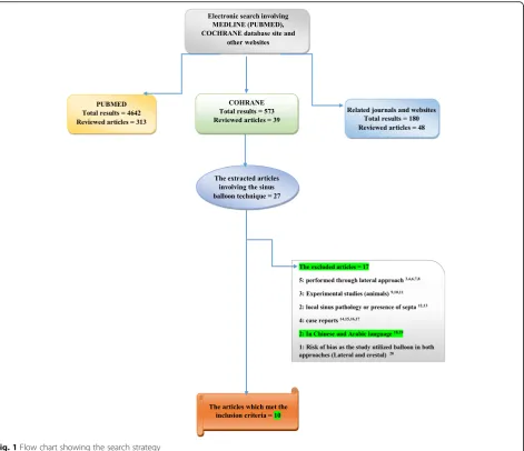
their articles or for more explanation about their results. The search process was demonstrated in (Fig.1).
Results
The total electronic search results were 5395 articles. The reviewed articles were 400, and the extracted articles which involved utilization of balloon technique in the maxillary sinus floor elevation surgery were 27 articles. Siventen articles were excluded from this study [6–20] and only 10 articles met the inclusion criteria.
The results of the selected studies were categorized to assess the success rate of sinus augmentation by MIAMBE technique, to report the perforation rate of schneiderian membrane and to identify the rate of complications associated with MIAMBE technique as shown in Table1. The survival rate and failure rates of dental implants placed in the augmented maxillary sinus were calculated for the selected studies and registered in Table2. The average of schneiderian membrane perforation was calculated for the ten extracted studies utilizing MIAMBE technique, and it was 6.76%. The sinus augmenta-tion success rate reported ranged from 100 to 71.4% with an average of 91.6%. The average of implant survival rate in these studies was 96.62%. Regarding the grafting material, synthetic bone graft was utilized in five studies. Four studies used a mixture of autogenous and synthetic bone graft while one study utilized allogeneic cancellous bone graft. PRF (platelets rich fibrin) mixed with either autogenous or synthetic bone graft was used in the five studies.
Discussion
Sinus floor elevation surgery with balloon is said to be a minimally invasive technique [5],but to date, no systematic review was made to clearly present the study results, authors experience, and surgical outcomes. Results of studies that utilized MIAMBE technique could be discussed under these highlighted points.
Sinus augmentation and bone gain
The success of sinus augmentation procedure with MIAMBE technique was ranged from 100 to 71.4% with an average of 91.6% in these studies. Bone gain with MIAMBE technique could reach for more than 10 mm, it ranged from 3 to 10.8 mm with an average of 6.96 mm. It should be mentioned that some articles failed to report the gained bone in details.
The traditional procedure (Summers’technique) had a limitation of allowing for only a minimal amount of bone gain which is 3–4 mm. While sinus floor elevation surgery via lateral approach produced a huge elevation
≥10 mm [23], it is considered as an invasive technique.
Implants survival rates
Implant survival rate associated with MIAMBE technique was ranged from 90 to 100% with an average of 96.62% as
shown in Table 2. On the other hand, systematic reviews have evaluated the implant survival rate after osteotome-mediated sinus floor elevation surgery which shows an im-plant survival rate higher than 90% [24–26]. In most of MIAMBE studies, dental implant failure occurred early dur-ing the first 6 months after operation, some authors men-tioned the cause for implant failure which was associated with infection, and others did not addressed the cause.
Surgical complications Intra-operative complications
The most common intra-operative complication associated with sinus lift procedure was sinus membrane tear [27]. The rate of schneiderian membrane perforation with MIAMBE technique was ranged from 0 to 21.32% with an average of 6.76%. This rate was similar to the
Table 1 Descriptive statistics demonstrate patient characteristics and outcomes of MIAMBE technique in the selected studies Study Patients Age (years) N of sinus augmentation
Sinus augmentation success
rate
%
Baseline bone height (mm) Bone gain (mm) Antral membrane elevation (mm) Total bone height (mm) Inflate balloon volume
Type of graft Membrane perforation Test for membrane perforation Complications Follow-up period after operation N M:F R M N PR % Kfir et al. [ 5 ] 12 NM 42 ± 9 NM 91.6 3.7 ± 1.4 NM > 10 10 – 17 ≤ 2.5 mL
-PRF -ABP -Bi-Ostetic synthetic bone
graft
1
8.33
Valsalva maneuver Direct visualization
1 membrane tear 1 balloon rupture 1 implant failed 23 months 12 NM NM 100 3.5 ± 1.3 NM > 10 10 – 18 0 0 1 mild periprocedural nosebleed 12 months Kfir et al. [ 29 ]3 6 M = 1 8 F: 18 42 ± 9 36 97.2 3.4 ± 2.1 NM > 10 8 – 18 NM
-PRF -ABP -Bi-Ostetic Synthetic bone
graft -Fisiograft gel 1 2.77 Valsalva maneuver 1 membrane tear 2 implant failed 6 – 8 months Hu et al. [ 30 ] 28 M = 143 F=1 4 40.2 ± 12.35 28 92.85 4.92 ± 1.24 NM Mean 10.9 ± 2.06 NM 0.67 ± 0.17 mL -PRF -Bio-Oss 2 7.14 Valsalva maneuver 1 mild nosebleed after surgery 2 membrane tear and the cases aborted 15.9 ± 2.94 months. Kfir et al. [ 31 ] 112 M = 50 F=6 2 44.1 ± 12.9 NM 97.3% initial procedural success 100% secondary procedural success 3.8 ± 2.1 NM > 10 11 – 18 NM
-PRF -Synthetic bone
graft
-ABP -Fisiograft gel
12
10.71
Valsalva maneuver Direct visualization
1
infection
and
oroantral
fistula
at 4weeks 3membrane
tear
and
the
procedures aborted 9micropuncture
13 months Mazor et al. [ 32 ]2 0 N M 3 7 – 72 24 100 2 – 6 NM 11 NM NM
-PRF -Synthetic bone substitute
0
0
Valsalva maneuver Direct visualization
1 patient was allergic to the antibiotic 18 months Petruzzi et al. [ 33 ] 40 M = 16 F=2 4 41.5 NM NM 8.00 ± 2.19 0.6 ≤ 20
14.66 ±1.48
1
–
ml
Calcium sulphate solution
3
7.5
Microscope (KarlKaps)
1m acrolaceration 2m icrolacerations 4hemifacial edema
1 year Peñarrocha- Diago et al. [ 34 ] 6M = 52 7 – 51 6 83.3% for the all 6 cases 100% for the five cases 2.1 – 4.1 7.2 – 10.8 8.7 NM 11.3 – 14.5 NM
-ABP -BIO-OSS bovine bone grafts
Table
1
Descriptive
statistics
demonstrate
patient
characteristics
and
outcomes
of
MIAMBE
technique
in
the
selected
studies
(Co
ntinued)
Study
Patients
Age (years)
N
of
sinus
augmentation
Sinus augmentation success
rate
%
Baseline bone height (mm) Bone gain (mm) Antral membrane elevation (mm) Total bone height (mm) Inflate balloon volume
Type
of
graft
Membrane perforation
Test
for
membrane perforation
Complications
Follow-up period
after
operation
N
M:F
R
M
N
PR
%
Gonzalez
et
al.
[
35
]
14
M
=
7
F=7
NM
NM
71.4
5.2
8.5
NM
NM
NM
BIO-OSS bovine bone grafts
3
21.32
An operating microscope
Failure
of
four
cases
due
to
mucosa perforation (21.32%)
and
balloon
breakage
(7.14%). 1implant showed
marginal
periimplantitis which
treated
successfully
1
year
Dhandapani et
al.
[
36
]
9N
M
2
5
–
60
10
100
5
–
83
– 5.5
4.34
≥
10
13.5
–
9
1
cc
Irradiated allogeneic cancellous bone
and
marrow graft
0
0
NM
No
complications
6
months
Asmael,
and
Lateef
[
37
]
13
M
=
4
F:9
28
–
57
17
100
2.3
–
7.8
4.9 – 10.6
6.70
NM
9.8
–
17.2
0.5
–
1c
c
Particulate bone
grafts
(
β
Tricalcium
Phosphate)
0
0
Direct vi
ion
Hydraulic pressure
1
mild
nasal
bleeding 1infraorbital ecchymosis
1
year
Total
:
Average
Total
:
Range
91.6 100
–
71.4%
6.968 3– 10.8
mm
6.76 0–21.32%
MIAMBE
minimally
invasive
antral
membrane
balloon
elevation,
N
number,
NM
not
mentioned,
M:F
male:
female
numbers,
M
mean,
R
range,
PRF
platelets
rich
fibrin,
PRP
platelets
rich
plasma,
ABP
autogenous
bone
particles,
PR
perforation
schneiderian membrane perforation rate (0–21.4%) which was reported in the systematic review of sinus floor eleva-tion success via transalveolar approach by Tan [28].
In some of these studies, membrane perforation was treated successfully with collagen membrane and the procedure continued with successful MIAMBE technique; other studies aborted the procedure. Furthermore, some authors demonstrated the causes of sinus membrane perforation which could be due to the too rapidly inflated balloon, balloon rapture, and fracture of the sinus floor during the osteotome procedures. Anyhow, with minimally invasive methods, the accuracy in the diagnosis of sinus membrane perforation is difficult without the availability of endoscope. Therefore, the perforation rates in these studies should be interpreted carefully, and the tests utilized to detect the perforation should be addressed accurately Table1. An important point to the surgeons who executed this procedure is to check for membrane integrity after each surgical step by endoscope, Valsalva maneuver, direct vision, and/or by aspiration with normal saline to accur-ately report the cause of perforation.
Post-operative complications
Complications registered with MIAMBE technique in these studies involved sinus membrane perforation, implant failure, infection, oroantral fistula, balloon rapture, mild self-limiting nose bleeding, and infra-orbital ecchymosis. All studies reported less post-operative pain, bleeding, and discomfort on the patient side. On the surgeon side, it offered short learning curve and less surgical time.
This systematic review detected several shortcomings in the studies utilized (MIAMBE technique), these include:
One study was not critical in the presentation of its results and did not include the failed aborted cases in the total sinus augmentation success rate. Some studies failed to report the number of sinus
augmentation procedures as it differed from the number of the patients enrolled in these studies. Some did not mention the cause of membrane
perforation or implant failure.
Some studies did not mentioned well-defined implant survival or success criteria according to which they depend in reporting the survival rate of implants. Lack of long follow-up period in most of these studies. Lack of randomized clinical trial (RCT) studies as
shown in (Table1).
Conclusion
MIAMBE technique is proved to be a minimally invasive procedure which is associated with low post-operative complications. The amount of gained bone with MIAMBE technique is predictable and comparable with the amount
of bone achieved with the more invasive lateral window technique. Anyhow, long follow-up period is needed to accurately identify the long-term success rate of dental implants placed with this technique.
Abbreviations
ABP:Autogenous bone particles; AMBE: Antral membrane balloon elevation; M: Mean; M:F: Male:female numbers; MIAMBE: Minimally invasive antral membrane balloon elevation; N: Number; NM: Not mentioned; PR: Perforation rate; PRF: Platelets rich fibrin; PRP: Platelets rich plasma; R: Range; RCT: Randomized clinical trial
Acknowledgements
I would like to kindly thank the authors of the original articles who responded instantly upon communication with them to complete the missing data or to clarify the unexplained points in their studies.
Funding
This research did not receive any funding from any funding resources.
Authors’contributions
HMA performed all the aspects of this research which involved writing the research and collecting, interpreting, and analyzing data.
Ethics approval and consent to participate
This is not applicable as this research was a systematic review of the previous studies utilizing the MIMBE technique in the sinus lift surgery.
Consent for publication
Not applicable.
Competing interests
Huda M Asmael declares that she had no competing interests.
Publisher’s Note
Springer Nature remains neutral with regard to jurisdictional claims in published maps and institutional affiliations.
Received: 13 July 2017 Accepted: 8 February 2018
References
1. Tatum H. Lecture presented to the Alabama Implant Congress 1976. 2. Summers RB. The osteotome technique: part 3—less invasive methods of
elevating the sinus floor. Compendium (Newtown, Pa). 1994;15:698–700. 3. Muronoi M, Xu H, Shimizu Y, et al. Simplified procedure for augmentation
of the sinus floor using a haemostatic nasal balloon. Br J Oral Maxillofac Surg. 2003;41:120–1.
4. Soltan M, Smiler DG. Antral membrane balloon elevation. J Oral Implantol. 2005;31:85–90.
5. Kfir E, Kfir V, Mijiritsky E, et al. Minimally invasive antral membrane balloon elevation followed by maxillary bone augmentation and implant fixation. J Oral Implantol. 2006;32:26–33.
6. Yukinobu F, Sennichi S, Kazuko K, et al. Evaluation of subantral membrane balloon elevation technique using cone-beam CT. J Gifu Dent Soc. 2009;36:100–4. 7. Elabbasy SE. Assessment of sinus floor elevation utilizing balloon lift
technique with simultaneous implant placement: a clinical study 2010. 8. Rao GS, Reddy SK. Antral balloon sinus elevation and grafting prior to
dental implant placement: review of 34 cases. Int J Oral Maxillofac Implants. 2014;29:414–8.
9. Stelzle F, Benner KU. Evaluation of different methods of indirect sinus floor elevation for elevation heights of 10 mm: an experimental ex vivo study. Clin Implant Dent Relat Res. 2011;13:124–33.
10. Chan HL, Oh TJ, Fu JH, et al. Sinus augmentation via transcrestal approach: a comparison between the balloon and osteotome technique in a cadaver study. Clin Oral Implants Res. 2013;24:985–90.
12. Kfir E, Goldstein M, Abramovitz I, et al. The effects of sinus membrane pathology on bone augmentation and procedural outcome using minimal invasive antral membrane balloon elevation. J Oral Implantol. 2014;40:285–93.
13. Kfir E, Goldstein M, Rafaelov R, et al. Minimally invasive antral membrane balloon elevation in the presence of antral septa: a report of 26 procedures. J Oral Implantol. 2009;35:257–67.
14. Ziv mazor. The use of minimally invasive antral membrane balloon elevation to treat the posterior maxilla: Aclinical presentation. J Implant Reconstr Dent. 2010;2:26-31.
15. Kfir E, Kfir V, Kaluski E, et al. Minimally invasive antral membrane balloon elevation for single-tooth implant placement. Quintessence Int. 2011;42:645–50.
16. Kfir E, Kfir V, Goldstein M, et al. Minimally invasive subnasal elevation and antral membrane balloon elevation along with bone augmentation and implants placement. J Oral Implantol. 2012;38:365–76.
17. Arroyo R, Cabrera D. Minimally invasive antral membrane balloon elevation (MIAMBE): a 3 cases report. J Oral Res. 2013;2:135–8.
18. Hu XL, Lin Y, Li JH, et al. Clinical study of sinus elevation by water balloon and implant placement. Zhonghua Kou Qiang Yi Xue Za Zhi. 2008;43:226–9.
19. Elyas J, Almdalal Y. Study of activity of maxillary sinus floor lifting with balloon and its range of success,Tishreen university journal for research and scientific studies - health sciences series, vol. 35; 2013.
20. Rodrigues GN, Katayama AY, Cardoso RF. Subantral augmentation utilizing the Zimmer® sinus lift balloon technique; 2010. p. 1–5.
21. Ahn SH, Park EJ, Kim ES. Reamer-mediated transalveolar sinus floor elevation without osteotome and simultaneous implant placement in the maxillary molar area: clinical outcomes of 391 implants in 380 patients. Clin Oral Implants Res. 2012;23(7):866–72.
22. Jesch P, Bruckmoser E, Bayerle A, Eder K, Bayerle-Eder M, Watzinger F. A pilot-study of a minimally invasive technique to elevate the sinus floor membrane and place graft for augmentation using high hydraulic pressure: 18-month follow-up of 20 cases. Oral Surg Oral Med Oral Pathol Oral Radiol. 2013;116(3):293–300. 23. Zitzmann N, Scharer. Sinus elevation procedures in the reabsorbed posterior
maxilla. Comparison of the crestal and lateral approaches. Oral Surg Oral Med Oral Pathol Oral Radiol Endod. 1998;85:8–17.
24. Chen MH, Shi JY. Clinical and radiological outcomes of implants in osteotome sinus floor elevation with and without grafting: a systematic review and a meta-analysis. J Prosthodont. 2017.
25. Călin C, Petre A, Drafta S. Osteotome-mediated sinus floor elevation: a systematic review and meta-analysis. Int J Oral Maxillofac Implants. 2014;29:558–76.
26. Starch-Jensen T, Jensen JD. Maxillary sinus floor augmentation: a review of selected treatment modalities. J Oral Maxillofac Implants. 2017;8:e3. 27. Wallace SS, Mazor Z, Froum SJ, et al. Schneiderian membrane
perforation rate during sinus elevation using Piezosurgery: clinical results of 100 consecutive cases. Int J Periodontics Restorative Dent. 2007;27:413–9.
28. Tan WC, Lang NP, Zwahlen M, et al. A systematic review of the success of sinus floor elevation and survival of implants inserted in combination with sinus floor elevation Part II: transalveolar technique. J Clin Periodontol. 2008;35:241–54. 29. Kfir E, Kfir V, Eliav E, et al. Minimally invasive antral membrane balloon
elevation: report of 36 procedures. J Periodontol. 2007;78:2032–5. 30. Hu X, Lin Y, Metzmacher AR, et al. Sinus membrane lift using a water
balloon followed by bone grafting and implant placement: a 28-case report. Int J Prosthodont. 2009;22:243–7.
31. Kfir E, Goldstein M, Yerushalmi I, et al. Minimally invasive antral membrane balloon elevation—results of a multicenter registry. Clin Implant Dent Relat Res. 2009;11:e83–91.
32. Mazor Z, Kfir E, Lorean A, et al. Flapless approach to maxillary sinus augmentation using minimally invasive antral membrane balloon elevation. Implant Dent. 2011;20:434–8.
33. Petruzzi M, Ceccarelli R, Testori T, et al. Sinus floor augmentation with a hydropneumatic technique: a retrospective study in 40 patients. Int J Periodontics Restorative Dent. 2012;32:205–10.
34. Peñarrocha-Diago M, Galán-Gil S, Carrillo-García C, et al. Transcrestal sinus lift and implant placement using the sinus balloon technique. Med Oral Patol Oral Cir Bucal. 2012;17:e122.
35. Gonzalez AG, Fernández IC, Frechoso SC, et al. Minimally invasive sinus lift elevation with a silicone balloon and simultaneous implant placement. An independent 1-year follow-up clinical study. Clin Oral Implants Res. 2014;25:503. 36. Dhandapani RB, Baskaran S, Arun KV, et al. Minimally invasive maxillary sinus
elevation using balloon system: a case series. 2016.