Original Article
Association between long non-coding RNA MALAT-1
expression and cancer progression: a meta-analysis
Tiancheng Xie*, Yangye Yan*, Binbin Dong, Guanghui Hu, Yunfei Xu
Department of Urology, Shanghai Tenth People’s Hospital, School of Medicine, Tongji University, Shanghai, People’s Republic of China. *Equal contributors and co-first authors.
Received October 18, 2015; Accepted August 14, 2016; Epub December 15, 2016; Published December 30, 2016
Abstract: The aim of this meta-analysis is to investigate the relationship between lncRNA MALAT-1 and cancer progression. We collected all the relevant articles by searching the following database: PubMed, Cochrane Library, OVID, Web of Science and Chinese National Knowledge Infrastructure (CNKI). We calculated the odds ratio (OR) and
its corresponding 95% confidence interval (CI) to assess the strength of the association by using RevMan5.0. In
total, 751 patients from 7 studies were included in this meta-analysis. The subgroup analysis showed that MALAT-1
expression was significantly associated with tumor distant metastasis (Absent subgroup: OR=0.41, 95% CI 0.19-0.91, P=0.03; Present subgroup: OR=2.42, 95% CI 1.09-5.37, P=0.03), but not significantly associated with gender differences (female subgroup: OR=0.86, 95% CI 0.63-1.17, P=0.33; male subgroup: OR=1.17, 95% CI 0.86-1.59, P=0.33), tumor differentiation (Well moderate subgroup: OR=0.79, 95% CI 0.50-1.25, P=0.31; Poor subgroup: OR=1.27, 95% CI 0.80-2.00, P=0.31) and lymphatic metastasis (Absent subgroup: OR=1.09, 95% CI 0.77-1.55, P=0.63; Present subgroup: OR=0.92, 95% CI 0.65-1.30, P=0.63). The results indicated that MALAT-1 could be a
potential biomarker for tumor distant metastasis.
Keywords: MALAT-1, lncRNA, cancer, meta-analysis
Introduction
The human RNA acts not only as a supporting role in the intermediation of genetic informa-tion, but more importantly as a regulator in a variety of physiological and pathological pro-cesses. More than 80% of human RNAs cannot directly encode protein so that they are named as non-coding RNAs (ncRNAs). Long non-coding RNAs (lncRNAs) are ncRNAs with more than 200 nucleotides. Nowadays lncRNA has beco- me a research hotspot after microRNA (miRNA). The structure of lncRNA is similar to mRNA [1, 2]. The vast majority of lncRNAs are transcript-ed by RNA polymerase II and alternative splic-ing. They widely participate in almost all physi-ological and pathphysi-ological processes in view of genetic and transcription level [3].
Using lncRNA microarray, high-throughput se-
quencing and qRT-PCR (real-time fluorescent
quantitative-PCR), researchers have found a large number of lncRNAs which differentially ex- press in human cancer such as prostate
can-Metastasis associated lung adenocarcinoma transcript 1 (MALAT-1), also known as the nu- clear-enriched abundant transcript 2 (NEAT2), exist widely in normal human cells. It is a very important gene which can regulate the function of endothelial cells and angiogenesis [7]. In 2003, Ji et al [8] found that MALAT-1 was a pre-dictive indicator for I stage lung adenocarcino-ma or squamous cell carcinoadenocarcino-ma by using sub-tractive hybridization. Since then, many re-
searchers subsequently confirmed that the
expression level of MALAT-1 was up regulated in tumor cells including liver cancer, breast can-cer, pancreatic cancan-cer, bladder cancer and prostate cancer [9-11].
inva-Recently, the association between long non-coding RNA MALAT-1 expression and tumor has been investigated by some studies. However, there are still some disputes about the results and the sizes of the samples are small. Besides, there is no meta-analysis reporting the associa-tion between long non-coding RNA MALAT-1 expression and cancer progression. Therefore, we conducted this meta-analysis to explore the potential relationship.
Materials and methods
Search strategy
We performed a systematic computer literature search to collect potentially eligible articles. The following electronic databases were used
for finding relevant studies: PubMed, Cochrane
Library, OVID, Web of Science and Chinese National Knowledge Infrastructure (CNKI). The keywords we used were “MALAT1” OR “MALAT-1” OR “metastasis associated lung adenocarci-noma transcript 1”. The literatures were retri- eved for further screening.
Study selection
We used the following criteria to evaluate the retrieval literatures which were consistent with our analysis included in the request: (a) studies investigating the roles of MALAT-1 in the gene-sis and progression of cancer, (b) the expres-sion levels of MALAT-1 in tumor tissues were measured, (c) patients were divided into groups
whether they met the selection criteria and the eligible articles were included in our study. Data extraction
We extracted the following data from the
includ-ed articles: first author, publish date, country,
tumor type, and total number of patients, num-ber of high MALAT-1 expression groups and low expression groups and detection methods of MALAT-1 expression. Two investigators (Xie and Dong) extracted data independently. If there was any disagreement about the extraction, another investigator (Yan) adjudicated the re- sults.
Study quality
Two authors (Xie and Yan) evaluated the quality of retrieved studies independently according to the Newcastle-Ottawa Scale (NOS) for case-control studies. The NOS ranged from zero to nine stars. The controversial part solved by dis-cussion. And the third investigator (Dong) adju-dicated the disagreements.
Statistical analysis
Heterogeneity was analyzed by calculating the Q test statistics with RevMan 5.0 software. Counting data was estimated with Risk Ratio
(RR) and 95% confidence intervals (CI);
Mea-surement data was assessed by pooled Sta- ndard Mean Difference (SMD). If the data
sug-gests P>0.10 with no significant heterogeneity, we will use the fixed effects model, otherwise,
[image:2.612.91.375.75.266.2]use the random effects model. Considering the
Figure 1. Flowchart presenting the steps of literature search and selection.
clinic pathologic parameters were described, (e) there were
sufficient data for us to calcu -late the odds ratios (OR) and
corresponding 95%
approximately equal to OR. Here we used OR instead of HR. Egger’s funnel plot was explored
to find if there is any evidence of publication
bias [14]. Results
Information from the literature
Based on the above search strategy, we ac- hieved 632 relevant studies. 605 articles were excluded by title and abstract information.
Then we further excluded the rest articles by details which we show in Figure 1.
Study characteristics
[image:3.612.91.522.85.204.2]All studies were published from 2012 to 2014. Patients enrolled were all Asian [15-21]. They compared the differences in sex, tumor differ-entiation, lymphatic metastasis and distant metastasis. The average NOS score was 6 indi-cating the reliable quality. The characteristics of included studies were showed in Table 1. Table 1. Characteristics of the eligible studies in this meta-analysis
First author Year Country Ethnicity Cancer type Total number MALAT-1 expression Detection method
High Low
Lai 2012 China Asian LC 60 33 27 qRT-PCR
Liu 2014 China Asian PC 45 26 19 qRT-PCR
Pang 2014 China Asian PC 126 63 63 qRT-PCR
Zhang 2014 China Asian RC 106 56 50 qRT-PCR
Zheng 2014 China Asian CRC 146 73 73 qRT-PCR
Ma 2014 China Asian Glioma 118 59 59 qRT-PCR
Okugawa 2014 Japan Asian GC 150 88 62 qRT-PCR
LC: liver cancer, PC: pancreatic cancer, RC: renal cancer, CRC: colorectal cancer, GC: gastric cancer, qRT-PCR: real-time fluores -cent quantitative-PCR.
[image:3.612.96.522.250.544.2]Tumor differentiation
There were 4 studies with a total of 195 cases and 182 controls comparing the MALAT-1 ex-
pression level in different stage of tumor differ-entiation with that in case-control groups. The
result showed no significant heterogeneity
[image:4.612.100.521.72.317.2](I2=0%, P=0.71), and the pooled OR was 0.79
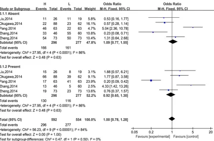
Figure 3. MALAT-1 expression was not significantly associated with lymphatic metastasis.
[image:4.612.99.521.359.637.2](95% CI: 0.50, 1.25; Z=1.02; P=0.31) in the
absent subgroup, 1.27 (95% CI: 0.80, 2.00;
Z=1.02; P=0.31) in the present subgroup. It
indicated that MALAT-1 expression was not
sig-nificantly associated with tumor differentiation
(Figure 2).
Lymphatic metastasis
There were 5 studies with a total of 296 cases and 277 controls comparing the MALAT-1 ex- pression level in cancer with lymphatic metas-tasis with that in case-control groups. There
was significant heterogeneity in both sub -groups (I2=86%, P<0.0001). And the pooled OR was 1.09 (95% CI: 0.77, 1.55; Z=0.48; P=0.63)
in the absent subgroup, 0.92 (95% CI: 0.65,
1.30; Z=0.48; P=0.63) in the present sub -group. The results showed that MALAT-1 ex-
pression was not significantly associated with
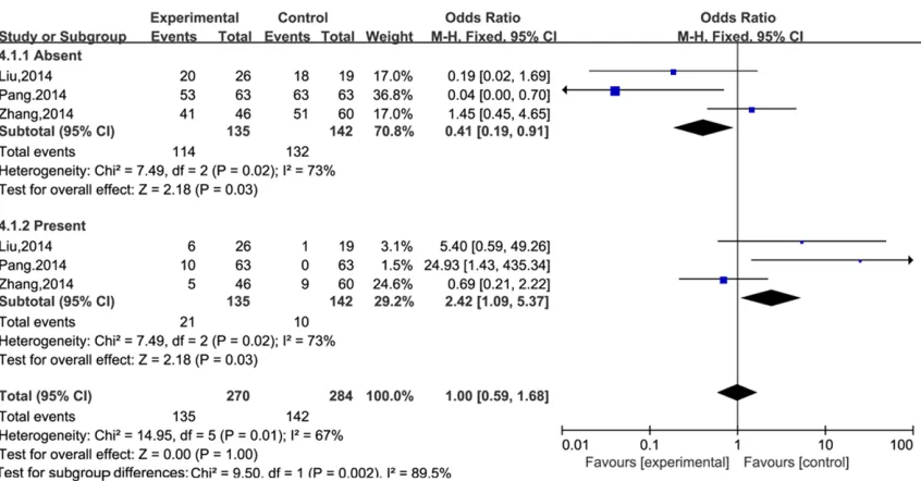
lymphatic metastasis (Figure 3). Distant metastasis
There were 3 studies with a total of 135 cases and 142 controls comparing the MALAT-1 expression level in tumor with distant metasta-sis with that in case-control groups. There was
significant heterogeneity in both subgroups
(I2=73%, P=0.02). And the pooled OR was 0.41 (95% CI: 0.19, 0.91; Z=2.18; P=0.03) in the
absent subgroup, 2.42 (95% CI: 1.09, 5.37;
Z=2.18; P=0.03) in the present subgroup. The
results showed that MALAT-1 expression was
significantly associated with distant metastasis
(Figure 4). Sex
There were 7 studies with a total of 388 cases and 363 controls comparing the MALAT-1 expression level in different sex with
case-con-trol groups. There was significant heterogeneity
in both subgroups (I2=42%, P=0.11). And the pooled OR was 0.86 (95% CI: 0.63, 1.17;
Z=0.98; P=0.33) in the female subgroup, 1.17 (95% CI: 0.86, 1.59; Z=0.98; P=0.33) in the
male subgroup. The results showed there was
no significant difference between female and
male groups (Figure 5).
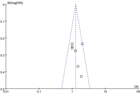
Publication bias and sensitivity analysis
In order to test whether the final result of this
meta-analysis was affected by individual study and gauge the stability of the results, a sensitiv-ity analysis was conducted (Figure 6). The pooled OR in the meta-analysis was not effect by single study. The result of Egger’s regression test showed the asymmetrical distribution in the funnel plot in the expression of MALAT-1 between high and low Gleason score groups. Discussion
[image:5.612.97.520.71.292.2]Our results confirm that the expression level of MALAT-1 is significantly related to the tumor
distant metastasis, however, it is not signifi -cantly associated with sex, tumor differentia-tion and lymphatic metastasis. It reveals that the expression level of MALAT-1 could be a ref-erence indicator for tumor distant metastasis and provides a clinical guide for further treat- ment.
The subgroup analysis showed that MALAT-1
expression was significantly associated with
tumor distant metastasis (Absent subgroup:
OR=0.41, 95% CI 0.19-0.91, P=0.03; Pres-ent subgroup: OR=2.42, 95% CI 1.09-5.37, P=0.03), but not significantly associated with gender differences (female subgroup: OR=0.86, 95% CI 0.63-1.17, P=0.33; male subgroup: OR=1.17, 95% CI 0.86-1.59, P=0.33),
tum-or differentiation (Well moderate subgroup:
OR=0.79, 95% CI 0.50-1.25, P=0.31; Poor sub
-group: OR=1.27, 95% CI 0.80-2.00, P=0.31)
and lymphatic metastasis (Absent subgroup:
OR=1.09, 95% CI 0.77-1.55, P=0.63; Pres-ent subgroup: OR=0.92, 95% CI 0.65-1.30, P=0.63).
The function of MALAT-1 in promoting cancer
progression has been confirmed by many
researches. Yang et al [22] found that MALAT-1 was up-regulated in human primary CRC tis-sues with lymph node metastasis, and MALAT-1 may promote CRC tumor development via its target PRKA kinase anchor protein 9 (AKAP-9). Tumor invasion and metastasis is known asso-ciated with epithelial-mesenchymal transition (EMT). Fan et al [23] found that TGF-beta could
metastasis. First, all the studies we included didn’t put hematogenous metastasis into
con-sideration for its difficulty to detect in clinical
practice. Second, the heterogeneities of the studies were at least moderate. Third, the num-ber of the patients in the included studies was small.
For clinical practice, if the expression of MALAT-1 is found rising, we should be on high alert for the risk of tumor distant metastasis. Some appropriate adjuvant therapies such as system-ic chemotherapies could be considered post operation to reduce the possibility of tumor dis-tant metastasis. This might play an effective role improving the quality of patients’ life, im- proving the prognosis and increasing survival rate.
However, there are still some limitations. Firstly, the literatures we searched may accept their positive results predominantly, leading the re- sults of our meta-analysis expanding. Secondly, samples of this meta-analysis were relatively small which may lead the results unstable. Thirdly, only those studies written in Chinese or English were included in the meta-analysis even though we set no language restriction. Last but not least, most of the included studies were based on the population from Asian co- untries.
Conclusions
Taken together, our meta-analysis showed that
MALAT-1 expression was significantly associat
-Figure 6. Funnel plot of publication bias on the differences of MALAT-1.
induce MALAT-1 expression and EMT in bladder cancer cells in vitro suggested that MALAT-1 was an important mediator of TGF-beta-induced EMT and participated in the progress of tumor metastasis. Although these studies found
significant correlations
betw-een high expression level of MALAT-1 and cancer progres-sion, we found they were not
significantly associated with
[image:6.612.92.372.74.264.2]ed with tumor distant metastasis. Therefore, MALAT-1 could be a potential indicator for tu- mor distant metastasis. However, more well-designed researches with large quantities of samples are need to further verify the results of this meta-analysis.
Acknowledgements
This work was funded by the National Natural Science Foundation of China (81370699). Disclosure of conflict of interest
None.
Address correspondence to: Dr. Yunfei Xu, Depart- ment of Urology, Shanghai Tenth People’s Hospital, School of Medicine, Tongji University, 301 middle Yanchang Road, Shanghai 200072, People’s Re- public of China. Tel: (+86) 13817990948; Fax: (+86) 13817990948; E-mail: [email protected]
References
[1] Erdmann VA, Szymanski M, Hochberg A, Groot N, Barciszewski J. Non-coding, mRNA-like RN- As database Y2K. Nucleic Acids Res 2000; 28: 197-200.
[2] Novikova IV, Hennelly SP, Tung CS, San- bonmatsu KY. Rise of the RNA machines: ex-ploring the structure of long non-coding RNAs. J Mol Biol 2013; 425: 3731-3746.
[3] Jemal A, Bray F, Center MM, Ferlay J, Ward E, Forman D. Global cancer statistics. CA Cancer J Clin 2011; 61: 69-90.
[4] Gutschner T, Hammerle M, Diederichs S. MALAT1--a paradigm for long noncoding RNA function in cancer. J Mol Med 2013; 91: 791-801.
[5] Chen G, Wang Z, Wang D, Qiu C, Liu M, Chen X, Zhang Q, Yan G, Cui Q. LncRNADisease: a data-base for long-non-coding RNA-associated dis-eases. Nucleic Acids Res 2013; 41: 983-986. [6] Spizzo R, Almeida MI, Colombatti A, Calin GA.
Long non-coding RNAs and cancer: a new fron-tier of translational research. Oncogene 2012; 31: 4577-4587.
[7] Michalik KM, You X, Manavski Y, Doddaballapur A, Zornig M, Braun T, John D, Ponomareva Y, Chen W, Uchida S, Boon RA, Dimmeler S. Long noncoding RNA MALAT1 regulates endothelial cell function and vessel growth. Circ Res 2014; 114: 1389-1397.
[8] Ji P, Diederichs S, Wang W, Boing S, Metzger R, Schneider PM, Tidow N, Brandt B, Buerger H, Bulk E, Thomas M, Berdel WE, Serve H, Müller-Tidow C. MALAT-1, a novel noncoding RNA, and
thymosin beta4 predict metastasis and surviv-al in early-stage non-smsurviv-all cell lung cancer. Oncogene 2003; 22: 8031-8041.
[9] Lin R, Maeda S, Liu C, Karin M, Edgington TS. A large noncoding RNA is a marker for murine hepatocellular carcinomas and a spectrum of human carcinomas. Oncogene 2007; 26: 851-858.
[10] Prensner JR, Chinnaiyan AM. The emergence of lncRNAs in cancer biology. Cancer Discov 2011; 1: 391-407.
[11] Wu XS, Wang XA, Wu WG, Hu YP, Li ML, Ding Q, Weng H, Shu YJ, Liu TY, Jiang L, Cao Y, Bao RF, Mu JS, Tan ZJ, Tao F, Liu YB. MALAT1 promotes the proliferation and metastasis of gallbladder cancer cells by activating the ERK/MAPK path-way. Cancer Biol Ther 2014; 15: 806-814. [12] Tripathi V, Shen Z, Chakraborty A, Giri S, Freier
SM, Wu X, Zhang Y, Gorospe M, Prasanth SG, Lal A, Prasanth KV. Long noncoding RNA MALAT1 controls cell cycle progression by reg-ulating the expression of oncogenic transcrip-tion factor B-MYB. PLoS Genet 2013; 9: e1003368.
[13] Hirata H, Hinoda Y, Shahryari V, Deng GR, Nakajima K, Tabatabai ZL, Ishii N, Dahiya R. Long Noncoding RNA MALAT1 Promotes Agg- ressive Renal Cell Carcinoma through Ezh2 and Interacts with miR-205. Cancer Res 2015; 75: 1322-1331.
[14] Peters JL, Sutton AJ, Jones DR, Abrams KR, Rushton L. Comparison of two methods to de-tect publication bias in meta-analysis. JAMA 2006; 295: 676-680.
[15] Lai MC, Yang Z, Zhou L, Zhu QQ, Xie HY, Zhang F, Wu LM, Chen LM, Zheng SS. Long non-cod-ing RNA MALAT-1 overexpression predicts tu-mor recurrence of hepatocellular carcinoma after liver transplantation. Med Oncol 2012; 29: 1810-1816.
[16] Liu JH, Chen G, Dang YW, Li CJ, Luo DZ.
Expression and prognostic significance of ln -cRNA MALAT1 in pancreatic cancer tissues. Asian Pac J Cancer Prev 2014; 15: 2971-2977. [17] Pang EJ, Yang R, Fu XB, Liu YF. Overexpression of long non-coding RNA MALAT1 is correlated with clinical progression and unfavorable prog-nosis in pancreatic cancer. Tumour Biol 2015; 36: 2403-2407.
[18] Zhang HM, Yang FQ, Chen SJ, Che J, Zheng JH. Upregulation of long non-coding RNA MALAT1 correlates with tumor progression and poor prognosis in clear cell renal cell carcinoma. Tumour Biol 2015; 36: 2947-2955.
[20] Ma KX, Wang HJ, Li XR, Li T, Su G, Yang P, Wu JW. Long noncoding RNA MALAT1 associates with the malignant status and poor prognosis in glioma. Tumour Biol 2015; 36: 3355-3359. [21] Okugawa Y, Toiyama Y, Hur K, Toden S, Saigusa
S, Tanaka K, Inoue Y, Mohri Y, Kusunoki M, Boland CR, Goel A. Metastasis-associated long non-coding RNA drives gastric cancer develop-ment and promotes peritoneal metastasis. Carcinogenesis 2014; 35: 2731-2739.
[22] Yang MH, Hu ZY, Xu C, Xie LY, Wang XY, Chen SY, Li ZG. MALAT1 promotes colorectal cancer cell proliferation/migration/invasion via PRKA kinase anchor protein 9. Biochim Biophys Acta 2015; 1852: 166-174.