For Review Only
Techniques for RNA in vivo imaging in plants
Journal: Journal of Microscopy
Manuscript ID: JMI-2014-0134.R1 Wiley - Manuscript type: Invited Review Date Submitted by the Author: n/a
Complete List of Authors: Tilsner, Jens; University of St Andrews, Biomolecular Sciences Research Complex
For Review Only
1
Short Review
Techniques for RNA in vivo imaging in plants
Jens Tilsner
Biomedical Sciences Research Complex, University of St Andrews, BMS Building, North Haugh, St
Andrews, Fife KY 16 9ST, Scotland, U.K.
Cell & Molecular Science, The James Hutton Institute, Invergowrie, Dundee, DD2 5DA, Scotland, U.K.
Tel. +44 (0)1334 464829
Fax +44 (0)1334 462595
Key words. RNA localisation, molecular beacon, MS2, λN, Pumilio
ABSTRACT
Since the discovery of small RNAs and RNA silencing, RNA biology has taken a centre stage in cell and
developmental biology. Small RNAs, but also mRNAs and other types of cellular and viral RNAs are
processed at specific subcellular localisations. To fully understand cellular RNA metabolism and the
various processes influenced by it, techniques are required that permit the sequence-specific
tracking of RNAs in living cells. A variety of methods for RNA visualisation have been developed since
the 1990s, but plant cells pose particular challenges and not all approaches are applicable to them.
On the other hand, plant RNA metabolism is particularly diverse and RNAs are even transported
For Review Only
2
the cellular and tissue level. This Short Review briefly introduces the currently available techniques
for plant RNA in vivo imaging and discusses their suitability for different biological questions.
INTRODUCTION
Many RNAs in cells exhibit dynamic, functional localisations related to processes such as nuclear
export, localised translation, RNA turnover within nuclear and cytoplasmic granules, and intercellular
communication. Fully understanding the functional significance of these localisations and the
mechanisms that underlie them requires the ability to track RNAs sequence-specifically in living cells.
A variety of RNA imaging techniques have been developed, but their use in plants, where RNA
localisations are still little understood, is limited by factors such as the impenetrability of the cell
wall, and sources of auto-fluorescence such as chloroplasts and phenolic cell wall compounds. This
Short Review compares live-cell RNA visualisation techniques that have been used in plants,
highlighting their advantages and limitations.
1. APPROACHES BASED ON FLUORESCENTLY LABELLED NUCLEIC ACIDS
RNA can be fluorescently labelled either by direct incorporation of fluorescent nucleotides, or by
hybridisation with a fluorescent probe. Both of these approaches are feasible in vivo but require
invasive delivery into cells. The permeabilisation and transfection techniques routinely used in
animal cell culture do not work on walled plant cells. In planta, this limits invasive delivery of
fluorescent nucleic acids to microprojectile bombardment or micro-injection, the former of which
causes significant damage to the cell whereas the latter is technically challenging and extremely
low-throughput. Electroporation or PEG-mediated transformation are only possible in protoplasts, i.e.
cells removed from their tissue context and significantly stressed. Nevertheless, these invasive
For Review Only
3
that are unsuitable for genetically encoded sensors, e.g. small RNAs such as siRNAs and miRNAs,
which would become non-functional if tagged with additional sequences, or sequestered by
RNA-binding proteins.
1.1. Direct labelling
RNAs can be rendered fluorescent by incorporation of fluorescent nucleotide derivatives. To do this
in a sequence-specific manner, the fluorescent RNA has to be transcribed or synthesized in vitro. If
unincorporated fluorescent nucleotides are removed after synthesis, direct labelling allows for
essentially background-free RNA imaging.
The main disadvantage of direct labelling is that the invasive introduction of the labelled RNA into
the cell bypasses all nuclear and many cytoplasmic processing steps, resulting in potentially
significantly altered protein associations compared to endogenous transcripts. The amount of
labelled RNA introduced into the cell may also differ significantly from endogenous levels and thus
overload cellular processing and localization machineries.
Direct labelling has been used to study the behaviour of uncapped mRNAs and the early events of
RNA virus infections in plant cells. Fluorescent, non-capped mRNA PEF-transformed into protoplasts
became trapped in the nucleus when nuclear export was inhibited with leptomycin B (Stuger &
Forreiter, 2004). Directly labelled genomic RNA of Tobacco mosaic virus was recruited into
endomembrane/cytoskeleton-associated motile granules in a cap-dependent manner (Christensen
et al., 2009). Directly end-labelled 21 nucleotide siRNA duplexes were used in leaf bombardment
assays to demonstrate that they function as mobile silencing signals between cells (Figure 1A). The
subcellular localisations of these small RNAs were not further analysed in these experiments
For Review Only
4
1.2. Molecular beacons
Sequence-specific visualisation of RNAs by fluorescent in situ hybridisation (FISH) is a common
technique in animal cell biology, though subcellular resolution is more difficult to achieve in plant
samples. Hybridisation-based approaches can also be used in vivo, but because unlike FISH, unbound
probe cannot easily be washed out, this requires probes that allow distinguishing between unbound
and target-bound forms. Many different design variants have been developed for this purpose (Bao
et al., 2009), which are beyond the scope of this Short Review. The only hybridisation-based RNA
probes used in live plant cells to date are molecular beacons (MBs). MBs are short hairpin-structured
nucleic acids coupled to a fluorophore on one end, and a quencher molecule on the other. In the
unbound form, the MB stem-loop brings the fluorophore and quencher into close proximity,
preventing fluorescence. Binding of the MB to its target RNA separates the hairpin stems and thus
leads to unquenching of the fluorophore.
The major benefit of MBs for live-cell imaging is that they permit visualising endogenously expressed
and processed RNAs. Apart from the invasive delivery, their main drawback is that they require
extensive optimisation, especially for in vivo uses. The target sequence has to be accessible and not
hidden by secondary structures. The free energy of hybridisation to the target has to be sufficient to
open the hairpin, but the stem-loop has to be stable enough not to open in the absence of target in
vivo. Nonspecific fluorescence can also occur due to endonuclease processing. One way to overcome
problems with nonspecific fluorescence is to include a MB with no cellular targets as an internal
control for ratiometric imaging. In animal cells, nonspecific nuclear sequestration of beacons often
needs to be prevented by linking them to large carriers such as PEG or streptavidin. In plants,
2’-O-methyl-RNA MBs accumulated in the nucleus whereas DNA beacons did not (Göhring et al., 2014)
(Figure 1B).
Using MBs, Göhring et al. (2014) were able to distinguish different mRNA splice variants in
For Review Only
5
insensitive to nonsense-mediated decay (NMD) were retained in the nucleus, thereby evading the
cytoplasmic NMD machinery. These splice variants also showed reduced mobility in the nucleus,
possibly due to a different ribonucleoprotein complex composition. Subsequently, the authors
further optimised their MB protocol and achieved the first single molecule RNA detection in plants.
2. GENETICALLY ENCODED RNA REPORTERS
Invasive delivery of RNA probes can be avoided by using genetically encoded reporters, thereby
facilitating imaging in a large number of cells within the context of intact tissues. Sequence-specific
RNA detection is possible with sequence-specific RNA-binding proteins (RBPs) fused to fluorescent
proteins (FPs). Generally applicable methods that can be adapted to any RNA of interest require
either tagging of the RNA with an RBP target sequence, or RBPs whose specificity can be predictably
modified. Both types of reporters have been used in plants. It is also necessary to distinguish free
and RNA-bound RBP-FP fusions. Two different strategies have been employed: nuclear retention of
unbound reporter, or fluorescence complementation between two RBP-splitFP fusions binding the
same RNA.
2.1. MS2CP and λλλλN22
The capsid protein of bacteriophage MS2 (MS2CP), and a 22-amino acid peptide of the N protein of
bacteriophage λ (λN22) are both RBPs that recognise 19 and 15 Nt, stem-loop forming-sequences
(MS2 and boxB) with dissociation constants of 6.2 and 22 nM, respectively, and have no targets
within plant genomes. For RNA imaging, either MS2CP or λN22 have been fused to an FP and a
nuclear localisation signal. Thus, the fusion proteins are sequestered in the nucleus in the absence of
a target RNA. The presence of the target RNA leads to a re-localization of the RNA-reporter into the
For Review Only
6
bacteriophage PP7 coat protein with their respective corresponding stem-loops, but neither of these
systems has so far been applied in plants.)
A downside of the MS2CP or λN22 systems is that the RNA of interest has to be tagged with the MS2
or boxB stem-loops, and thus needs to be expressed as a transgene. Preferably the RNA is
transcribed from its native promoter, but due to the random insertion of T-DNAs into the genomes
of higher plants, its expression level and nuclear processing may still differ from the native
transcript, especially if the transgene does not include introns. Additionally, the secondary structure
introduced by the MS2 or boxB tags may disrupt function or location of modified RNAs. On the other
hand, an advantage of these systems is that very high sensitivities can be achieved by using multiple
tandem copies of the stem-loop tags (typically 6-24). In animal systems, a 96xMS2 tag enabled
single-molecule-sensitive RNA imaging, but obviously, increasingly large tags with extensive
secondary structure exacerbate the risk of disrupting RNA processing and localisation.
MS2CP was the first genetically encoded RNA imaging system described (Bertrand et al. 1998) and is
so far the one most extensively used in plants, where its applications have included tracking of
storage protein-coding and other mRNAs, viral RNAs, and analysis of nuclear miRNA processing
bodies. Between 2 and 24 copies of the MS2 tag, and both nuclear-targeted and cytoplasmic
MS2CP-GFP fusions were used in these studies. Recently, Schönberger et al. (2012) have developed
Gateway-based, 35S promoter-driven plant expression vectors for tagging of target RNAs with
6xMS2 or 16xboxB hairpins, respectively, at either the 5’ or the 3’ end. They also constructed
expression constructs for multiple spectral variants of nuclear-targeted MS2CP-FP and λN22-FP
fusions. With these systems, they demonstrated the possibility of simultaneously imaging two
different RNAs in the same plant cell. Both were full genomic transcripts including UTRs and introns.
One RNA encoded a soluble protein, the other a membrane-targeted protein, so their translation
should occur in the cytoplasm and on the ER, respectively. Two-colour imaging showed that both
For Review Only
7
of target RNA, both MS2CP-FP and λN22–FP fusions accumulated in the RNA-rich environment of the
nucleolus despite the absence of natural binding sites in the endogenous transcriptome. The authors
also showed that in plants, insertion of MS2 or boxB stem-loops directly upstream of an open
reading frame prevented its translation. Thus, tagging downstream of the stop codon is the
preferred choice in plants, although in yeast in some cases, 3’ tags have disturbed mRNA
localisations whereas 5’ tags did not.
2.2. Pumilio RNA binding domain
Pumilio/FBF family (PUF) proteins are sequence-specific RBPs found in all eukaryotes (26 in
Arabidopsis). Their RNA binding domain, the Pumilio homology domain (PUMHD) consists of eight
tandem repeat Puf motifs that each bind one nucleotide in an 8 Nt target sequence.
Sequence-specific interactions are mediated by the side chains of two amino acids per Puf repeat and the RNA
nucleobases (a third amino acid side chain in each repeat forms a non-specific stacking interaction
with the RNA). This makes it possible to re-engineer the specificity of the PUMHD with relatively few
modifications. Structural analysis of the native human Pumilio 1 and molecular evolution have
produced a complete code for recognition of the four RNA bases, and a GoldenGate pipeline for
rapid assembly of any PUMHD variant has been developed (Abil et al., 2014). Within certain
limitations (which are beyond the scope of this Short Review), the PUMHD can thus be engineered
to bind any RNA of choice. When used for RNA imaging, this means that in contrast to MS2CP and
λN22 systems, untagged, native RNAs can be imaged. Alternatively, RNAs can be tagged with
recognition motifs of selected PUMHD variants. The benefit in this case is that these tags do not
introduce stable secondary structures. Wild-type PUMHD of human Pumilio 1 also has a very high
affinity to its target sequence (kD = 0.48 nM), and variants with a kD as low as 0.05 nM have been
described. This makes PUMHD-based RNA imaging potentially more sensitive than MS2CP- or λN22–
For Review Only
8
RNA affinity of PUMHD variants modified to bind novel RNA sequences is still unpredictable and can
be an order of magnitude lower than the wild-type protein.
Disadvantages of the PUMHD compared with MS2CP and λN22 are the short length of the 8 Nt target
sequence and the occurrence of natural binding sites in endogenous plant mRNAs. Additionally, the
PUMHD shows degrees of binding preference for target sequence variants, rather than complete
specificity. Because off-target binding in a complex eukaryotic cell in vivo is thus practically
unavoidable, combinations of two PUMHDs are used to increase the specificity of RNA imaging. To
distinguish RNA-bound and free reporter constructs, the two PUMHD variants are coupled to the
two halves of a split FP for bimolecular fluorescence complementation (BiFC). The two modified
PUMHDs bind closely adjacent (5-9 Nt) binding sites on the same RNA, so that fluorescence
reconstitution occurs upon binding of both split FP fusion proteins to the same RNA. PUMHD-BiFC is
unfortunately not background free, as the reconstituted FP is extremely stable and the fluorescent
complex accumulates in plant cells. In animal cells, nuclear-targeted fusions of two PUMHDs coupled
to a FP have also been used to distinguish free and RNA-bound fluorescence similar to the MS2CP
and λN22 systems, but this approach has yet to be tested in plants.
In plants, PUMHD-BiFC imaging has been used to track vial RNAs (Tilsner et al., 2009). This has
enabled the observations that potyvirus replication complexes develop from ER-associated to
chloroplast-associated membrane sites, and that replication and cell-to-cell movement of a
potexvirus are spatially coupled at plasmodesmata (Figure 1D).
CHOICE OF REPORTER
This Short Review has described RNA in vivo imaging systems that have been successfully used in
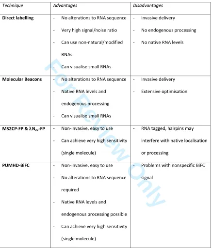
plants, and compared their respective advantages and drawbacks (Table 1), as well as highlighting
For Review Only
9
choice of RNA reporter must depend on the type of RNA that is being studied and the types of
questions that need to be addressed. For small RNAs direct labelling or molecular beacons are
probably the only suitable in vivo approaches. For mRNA imaging, considerations need to include
how abundant these are and which RNA processing pathways need to remain unaffected by the
imaging system, as well as if imaging can be performed in protoplasts or requires intact tissue. The
main consideration for imaging viral RNAs is how tags and RBPs affect their infectivity. As RNA
imaging systems are being developed further, the available toolbox will become even more diverse.
Some promising reporters, such as RNA aptamer tags like “Spinach” (and its improved derivatives
“Spinach2” and “BabySpinach”), “Mango” and IMAGE that selectively bind cell-permeant
fluorophores, have yet to be adapted to use in plants (Paige et al., 2011). Thus, it is to be hoped that
in the near future, studying functional localisations of plant RNAs will become a routine approach.
References
Abil, Z., Denard, C.A. & Zhao, H. (2014) Modular assembly of designer PUF proteins for specific
post-transcriptional regulation of endogenous RNA. J. Biol. Eng. 8, 7. doi:10.1186/1754-1611-8-7
Bao, G., Rhee, W.J. & Tsourkas, A. (2009) Fluorescent probes for live-cell RNA detection. Annu.
Rev. Bioeng. 11, 25-47. doi: 10.1146/annurev-bioeng-061008-124920
Bertrand, E., Chartrand, P., Schaefer, M., Shenoy, S.M., Singer, R.H. & Long, R.M. (1998)
Localization of ASH1 mRNA particles in living yeast. Mol. Cell 2, 437-445. doi:
For Review Only
10
Christensen, N., Tilsner, J., Bell, K., Hammann, P., Parton, R., Lacomme, C. & Oparka, K.J. (2009)
The 5’ cap of Tobacco mosaic virus (TMV) is required for virion attachment to the
actin/endoplasmic reticulum network during early infection. Traffic 10, 536-551. doi:
10.1111/j.1600-0854.2009.00889.x
Dunoyer, P., Schott, G., Himber, C., Meyer, D., Takeda, A., Carrington, J.C. & Voinnet, O. (2010)
Small RNA duplexes function as mobile silencing signals between plant cells. Science 328,
912-916. doi: 10.1126/science.1185880
Göhring, J., Jacak, J. & Barta, A. (2014) Imaging of endogenous messenger RNA splice variants in
living cells reveals nuclear retention of transcripts inaccessible to nonsense-mediated decay in
Arabidopsis. Plant Cell 26, 754-764. doi: 10.1105/tpc.113.118075
Paige, J.S., Wu, K.Y. & Jaffrey, S.R. (2011) RNA mimics of green fluorescent protein. Science 333,
642-646. doi: 10.1126/science.1207339
Schönberger, J., Hammes, U.Z. & Dresselhaus, T. (2012) In vivo visualization of RNA in plants cells
using the λN22 system and a GATEWAY-compatible vector series for candidate RNAs. Plant J. 71,
173-181. doi: 10.1111/j.1365-313X.2012.04923.x
Stuger, R. & Forreiter, C. (2004) Uncapped mRNA introduced into tobacco protoplasts can be
imported into the nucleus and is trapped by leptomycin B. Plant Cell Rep. 23, 99-103. doi:
For Review Only
11
Tilsner, J., Linnik, O., Christensen, N.M., Bell, K., Roberts, I.M., Lacomme, C. & Oparka, K.J. (2009)
Live-cell imaging of viral RNA genomes using a Pumilio-based reporter. Plant J. 57, 758-770. doi:
10.1111/j.1365-313X.2008.03720.x
Figure legends
Fig. 1. Examples of RNA imaging in plant cells.
(A) Direct labelling. Fluorescently end-labelled, double-stranded siRNAs bombarded into an
Arabidopsis epidermal leaf cell have moved into surrounding cells (1.) and there caused silencing of a
GFP transgene (2.) (Dunoyer et al., 2010). (B) Molecular Beacons. A gene-specific (1.) and a control
beacon with no cellular target (2.) were electroporated into an Arabidopsis protoplast (3.). A
ratiometric image (4.) is generated representing the signal/noise ratio in each pixel. From this, a cell
compartment-specific distribution of background-corrected signal intensities can be averaged for a
population of cells (Göhring et al., 2014). (C) MS2CP-CFP and λN22-mVenus. Nuclear-targeted
MS2CP-CFP (1.) and λN22-mVenus (2.) are both recruited into cytoplasmic granules by their
co-expressed target mRNAs tagged with 6xMS2- and 16xboxB, respectively. The MS2CP-imaged mRNA,
encoding a cytoplasmic protein, and the λN22-imaged RNA, encoding a membrane protein, localise to
For Review Only
12
PUMHD-BiFC (Tilsner et al., 2009) localises to small membrane structures at the entrances of
For Review Only
Table 1Properties of RNA in vivo imaging systems used in plants.
Technique Advantages Disadvantages
Direct labelling - No alterations to RNA sequence
- Very high signal/noise ratio
- Can use non-natural/modified
RNAs
- Can visualise small RNAs
- Invasive delivery
- No endogenous processing
- No native RNA levels
Molecular Beacons - No alterations to RNA sequence
- Native RNA levels and
endogenous processing
- Can visualise small RNAs
- Invasive delivery
- Extensive optimisation
MS2CP-FP & λλλλN22-FP - Non-invasive, easy to use
- Can achieve very high sensitivity
(single molecule)
- RNA tagged, hairpins may
interfere with native localisation or processing
PUMHD-BiFC - Non-invasive, easy to use
- No alterations to RNA sequence
required
- Native RNA levels and
endogenous processing possible
- Can achieve very high sensitivity
(single molecule)
- Problems with nonspecific BiFC
For Review Only
For Review Only
1
Short Review
Techniques for RNA in vivo imaging in plants
Jens Tilsner
Biomedical Sciences Research Complex, University of St Andrews, BMS Building, North Haugh, St
Andrews, Fife KY 16 9ST, Scotland, U.K.
Cell & Molecular Science, The James Hutton Institute, Invergowrie, Dundee, DD2 5DA, Scotland, U.K.
Tel. +44 (0)1334 464829
Fax +44 (0)1334 462595
Key words. RNA localisation, molecular beacon, MS2, λN, Pumilio
ABSTRACT
Since the discovery of small RNAs and RNA silencing, RNA biology has taken a centre stage in cell and
developmental biology. Small RNAs, but also mRNAs and other types of cellular and viral RNAs are
processed at specific subcellular localisations. To fully understand cellular RNA metabolism and the
various processes influenced by it, techniques are required that permit the sequence-specific
tracking of RNAs in living cells. A variety of methods for RNA visualisation have been developed since
the 1990s, but plant cells pose particular challenges and not all approaches are applicable to them.
On the other hand, plant RNA metabolism is particularly diverse and RNAs are even transported
For Review Only
2
the cellular and tissue level. This Short Review briefly introduces the currently available techniques
for plant RNA in vivo imaging and discusses their suitability for different biological questions.
INTRODUCTION
Many RNAs in cells exhibit dynamic, functional localisations related to processes such as nuclear
export, localised translation, RNA turnover within nuclear and cytoplasmic granules, and intercellular
communication. Fully understanding the functional significance of these localisations and the
mechanisms that underlie them requires the ability to track RNAs sequence-specifically in living cells.
A variety of RNA imaging techniques have been developed, but their use in plants, where RNA
localisations are still little understood, is limited by factors such as the impenetrability of the cell
wall, and sources of auto-fluorescence such as chloroplasts and phenolic cell wall compounds. This
Short Review compares live-cell RNA visualisation techniques that have been used in plants,
highlighting their advantages and limitations.
1. APPROACHES BASED ON FLUORESCENTLY LABELLED NUCLEIC ACIDS
RNA can be fluorescently labelled either by direct incorporation of fluorescent nucleotides, or by
hybridisation with a fluorescent probe. Both of these approaches are feasible in vivo but require
invasive delivery into cells. The permeabilisation and transfection techniques routinely used in
animal cell culture do not work on walled plant cells. In planta, this limits invasive delivery of
fluorescent nucleic acids to microprojectile bombardment or micro-injection, the former of which
causes significant damage to the cell whereas the latter is technically challenging and extremely
low-throughput. Electroporation or PEG-mediated transformation are only possible in protoplasts, i.e.
cells removed from their tissue context and significantly stressed. Nevertheless, these invasive
For Review Only
3
that are unsuitable for genetically encoded sensors, e.g. small RNAs such as siRNAs and miRNAs,
which would become non-functional if tagged with additional sequences, or sequestered by
RNA-binding proteins.
1.1. Direct labelling
RNAs can be rendered fluorescent by incorporation of fluorescent nucleotide derivatives. To do this
in a sequence-specific manner, the fluorescent RNA has to be transcribed or synthesized in vitro. If
unincorporated fluorescent nucleotides are removed after synthesis, direct labelling allows for
essentially background-free RNA imaging.
The main disadvantage of direct labelling is that the invasive introduction of the labelled RNA into
the cell bypasses all nuclear and many cytoplasmic processing steps, resulting in potentially
significantly altered protein associations compared to endogenous transcripts. The amount of
labelled RNA introduced into the cell may also differ significantly from endogenous levels and thus
overload cellular processing and localization machineries.
Direct labelling has been used to study the behaviour of uncapped mRNAs and the early events of
RNA virus infections in plant cells. Fluorescent, non-capped mRNA PEF-transformed into protoplasts
became trapped in the nucleus when nuclear export was inhibited with leptomycin B (Stuger &
Forreiter, 2004). Directly labelled genomic RNA of Tobacco mosaic virus was recruited into
endomembrane/cytoskeleton-associated motile granules in a cap-dependent manner (Christensen
et al., 2009). Directly end-labelled 21 nucleotide siRNA duplexes were used in leaf bombardment
assays to demonstrate that they function as mobile silencing signals between cells (Figure 1A). The
subcellular localisations of these small RNAs were not further analysed in these experiments
For Review Only
4
1.2. Molecular beacons
Sequence-specific visualisation of RNAs by fluorescent in situ hybridisation (FISH) is a common
technique in animal cell biology, though subcellular resolution is more difficult to achieve in plant
samples. Hybridisation-based approaches can also be used in vivo, but because unlike FISH, unbound
probe cannot easily be washed out, this requires probes that allow distinguishing between unbound
and target-bound forms. Many different design variants have been developed for this purpose (Bao
et al., 2009), which are beyond the scope of this Short Review. The only hybridisation-based RNA
probes used in live plant cells to date are molecular beacons (MBs). MBs are short hairpin-structured
nucleic acids coupled to a fluorophore on one end, and a quencher molecule on the other. In the
unbound form, the MB stem-loop brings the fluorophore and quencher into close proximity,
preventing fluorescence. Binding of the MB to its target RNA separates the hairpin stems and thus
leads to unquenching of the fluorophore.
The major benefit of MBs for live-cell imaging is that they permit visualising endogenously expressed
and processed RNAs. Apart from the invasive delivery, their main drawback is that they require
extensive optimisation, especially for in vivo uses. The target sequence has to be accessible and not
hidden by secondary structures. The free energy of hybridisation to the target has to be sufficient to
open the hairpin, but the stem-loop has to be stable enough not to open in the absence of target in
vivo. Nonspecific fluorescence can also occur due to endonuclease processing. One way to overcome
problems with nonspecific fluorescence is to include a MB with no cellular targets as an internal
control for ratiometric imaging. In animal cells, nonspecific nuclear sequestration of beacons often
needs to be prevented by linking them to large carriers such as PEG or streptavidin. In plants,
2’-O-methyl-RNA MBs accumulated in the nucleus whereas DNA beacons did not (Göhring et al., 2014)
(Figure 1B).
In the only study so far uUsing MBs in plants, Göhring et al. (2014) were able to distinguish different
For Review Only
5
transcripts that were insensitive to nonsense-mediated decay (NMD) were retained in the nucleus,
thereby evading the cytoplasmic NMD machinery. These splice variants also showed reduced
mobility in the nucleus, possibly due to a different ribonucleoprotein complex composition.
Subsequently, the authors further optimised their MB protocol and achieved the first single
molecule RNA detection in plants.
2. GENETICALLY ENCODED RNA REPORTERS
Invasive delivery of RNA probes can be avoided by using genetically encoded reporters, thereby
facilitating imaging in a large number of cells within the context of intact tissues. Sequence-specific
RNA detection is possible with sequence-specific RNA-binding proteins (RBPs) fused to fluorescent
proteins (FPs). Generally applicable methods that can be adapted to any RNA of interest require
either tagging of the RNA with an RBP target sequence, or RBPs whose specificity can be predictably
modified. Both types of reporters have been used in plants. It is also necessary to distinguish free
and RNA-bound RBP-FP fusions. Two different strategies have been employed: nuclear retention of
unbound reporter, or fluorescence complementation between two RBP-splitFP fusions binding the
same RNA.
2.1. MS2CP and λλλλN22
The capsid protein of bacteriophage MS2 (MS2CP), and a 22-amino acid peptide of the N protein of
bacteriophage λ (λN22) are both RBPs that recognise 19 and 15 Nt, stem-loop forming-sequences
(MS2 and boxB) with dissociation constants of 6.2 and 22 nM, respectively, and have no targets
within plant genomes. For RNA imaging, either MS2CP or λN22 have been fused to an FP and a
nuclear localisation signal. Thus, the fusion proteins are sequestered in the nucleus in the absence of
a target RNA. The presence of the target RNA leads to a re-localization of the RNA-reporter into the
For Review Only
6
bacteriophage PP7 coat protein with their respective corresponding stem-loops, but neither of these
systems has so far been applied in plants.)
A downside of the MS2CP or λN22 systems is that the RNA of interest has to be tagged with the MS2
or boxB stem-loops, and thus needs to be expressed as a transgene. Preferably the RNA is
transcribed from its native promoter, but due to the random insertion of T-DNAs into the genomes
of higher plants, its expression level and nuclear processing may still differ from the native
transcript, especially if the transgene does not include introns. Additionally, the secondary structure
introduced by the MS2 or boxB tags may disrupt function or location of modified RNAs. On the other
hand, an advantage of these systems is that very high sensitivities can be achieved by using multiple
tandem copies of the stem-loop tags (typically 6-24). In animal systems, a 96xMS2 tag enabled
single-molecule-sensitive RNA imaging, but obviously, increasingly large tags with extensive
secondary structure exacerbate the risk of disrupting RNA processing and localisation.
MS2CP was the first genetically encoded RNA imaging system described (Bertrand et al. 1998) and is
so far the one most extensively used in plants, where its applications have included tracking of
storage protein-coding and other mRNAs, viral RNAs, and analysis of nuclear miRNA processing
bodies. Between 2 and 24 copies of the MS2 tag, and both nuclear-targeted and cytoplasmic
MS2CP-GFP fusions were used in these studies. Recently, Schönberger et al. (2012) have developed
Gateway-based, 35S promoter-driven plant expression vectors for tagging of target RNAs with
6xMS2 or 16xboxB hairpins, respectively, at either the 5’ or the 3’ end. They also constructed
expression constructs for multiple spectral variants of nuclear-targeted MS2CP-FP and λN22-FP
fusions. With these systems, they demonstrated the possibility of simultaneously imaging two
different RNAs in the same plant cell. Both were full genomic transcripts including UTRs and introns.
One RNA encoded a soluble protein, the other a membrane-targeted protein, so their translation
should occur in the cytoplasm and on the ER, respectively. Two-colour imaging showed that both
For Review Only
7
of target RNA, both MS2CP-FP and λN22–FP fusions accumulated in the RNA-rich environment of the
nucleolus despite the absence of natural binding sites in the endogenous transcriptome. The authors
also showed that in plants, insertion of MS2 or boxB stem-loops directly upstream of an open
reading frame prevented its translation. Thus, tagging downstream of the stop codon is the
preferred choice in plants, although in yeast in some cases, 3’ tags have disturbed mRNA
localisations whereas 5’ tags did not.
2.2. Pumilio RNA binding domain
Pumilio/FBF family (PUF) proteins are sequence-specific RBPs found in all eukaryotes (26 in
Arabidopsis). Their RNA binding domain, the Pumilio homology domain (PUMHD) consists of eight
tandem repeat Puf motifs that each bind one nucleotide in an 8 Nt target sequence.
Sequence-specific interactions are mediated by the side chains of two amino acids per Puf repeat and the RNA
nucleobases (a third amino acid side chain in each repeat forms a non-specific stacking interaction
with the RNA). This makes it possible to re-engineer the specificity of the PUMHD with relatively few
modifications. Structural analysis of the native human Pumilio 1 and molecular evolution have
produced a complete code for recognition of the four RNA bases, and a GoldenGate pipeline for
rapid assembly of any PUMHD variant has been developed (Abil et al., 2014). Within certain
limitations (which are beyond the scope of this Short Review), the PUMHD can thus be engineered
to bind any RNA of choice. When used for RNA imaging, this means that in contrast to MS2CP and
λN22 systems, untagged, native RNAs can be imaged. Alternatively, RNAs can be tagged with
recognition motifs of selected PUMHD variants. The benefit in this case is that these tags do not
introduce stable secondary structures. Wild-type PUMHD of human Pumilio 1 also has a very high
affinity to its target sequence (kD = 0.48 nM), and variants with a kD as low as 0.05 nM have been
described. This makes PUMHD-based RNA imaging potentially more sensitive than MS2CP- or λN22–
For Review Only
8
RNA affinity of PUMHD variants modified to bind novel RNA sequences is still unpredictable and can
be an order of magnitude lower than the wild-type protein.
Disadvantages of the PUMHD compared with MS2CP and λN22 are the short length of the 8 Nt target
sequence and the occurrence of natural binding sites in endogenous plant mRNAs. Additionally, the
PUMHD shows degrees of binding preference for target sequence variants, rather than complete
specificity. Because off-target binding in a complex eukaryotic cell in vivo is thus practically
unavoidable, combinations of two PUMHDs are used to increase the specificity of RNA imaging. To
distinguish RNA-bound and free reporter constructs, the two PUMHD variants are coupled to the
two halves of a split FP for bimolecular fluorescence complementation (BiFC). The two modified
PUMHDs bind closely adjacent (5-9 Nt) binding sites on the same RNA, so that fluorescence
reconstitution occurs upon binding of both split FP fusion proteins to the same RNA. PUMHD-BiFC is
unfortunately not background free, as the reconstituted FP is extremely stable and the fluorescent
complex accumulates in plant cells. In animal cells, nuclear-targeted fusions of two PUMHDs coupled
to a FP have also been used to distinguish free and RNA-bound fluorescence similar to the MS2CP
and λN22 systems, but this approach has yet to be tested in plants.
In plants, PUMHD-BiFC imaging has been used to track vial RNAs (Tilsner et al., 2009). This has
enabled the observations that potyvirus replication complexes develop from ER-associated to
chloroplast-associated membrane sites, and that replication and cell-to-cell movement of a
potexvirus are spatially coupled at plasmodesmata (Figure 1D).
CHOICE OF REPORTER
This Short Review has described RNA in vivo imaging systems that have been successfully used in
plants, and compared their respective advantages and drawbacks (Table 1), as well as highlighting
For Review Only
9
choice of RNA reporter must depend on the type of RNA that is being studied and the types of
questions that need to be addressed. For small RNAs direct labelling or molecular beacons are
probably the only suitable in vivo approaches. For mRNA imaging, considerations need to include
how abundant these are and which RNA processing pathways need to remain unaffected by the
imaging system, as well as if imaging can be performed in protoplasts or requires intact tissue. The
main consideration for imaging viral RNAs is how tags and RBPs affect their infectivity. As RNA
imaging systems are being developed further, the available toolbox will become even more diverse.
Some promising reporters, such as RNA aptamer tags like “Spinach” (and its improved derivatives
“Spinach2” and “BabySpinach”), “Mango” and IMAGE that selectively bind cell-permeant
fluorophores, have yet to be adapted to use in plants (Paige et al., 2011). Thus,and it is to be hoped
that in the near future, studying functional localisations of plant RNAslocalisations in plants will
become a routine approach in the near future.
References
Abil, Z., Denard, C.A. & Zhao, H. (2014) Modular assembly of designer PUF proteins for specific
post-transcriptional regulation of endogenous RNA. J. Biol. Eng. 8, 7. doi:10.1186/1754-1611-8-7
Bao, G., Rhee, W.J. & Tsourkas, A. (2009) Fluorescent probes for live-cell RNA detection. Annu.
Rev. Bioeng. 11, 25-47. doi: 10.1146/annurev-bioeng-061008-124920
Bertrand, E., Chartrand, P., Schaefer, M., Shenoy, S.M., Singer, R.H. & Long, R.M. (1998)
Localization of ASH1 mRNA particles in living yeast. Mol. Cell 2, 437-445. doi:
For Review Only
10
Christensen, N., Tilsner, J., Bell, K., Hammann, P., Parton, R., Lacomme, C. & Oparka, K.J. (2009)
The 5’ cap of Tobacco mosaic virus (TMV) is required for virion attachment to the
actin/endoplasmic reticulum network during early infection. Traffic 10, 536-551. doi:
10.1111/j.1600-0854.2009.00889.x
Dunoyer, P., Schott, G., Himber, C., Meyer, D., Takeda, A., Carrington, J.C. & Voinnet, O. (2010)
Small RNA duplexes function as mobile silencing signals between plant cells. Science 328,
912-916. doi: 10.1126/science.1185880
Göhring, J., Jacak, J. & Barta, A. (2014) Imaging of endogenous messenger RNA splice variants in
living cells reveals nuclear retention of transcripts inaccessible to nonsense-mediated decay in
Arabidopsis. Plant Cell 26, 754-764. doi: 10.1105/tpc.113.118075
Paige, J.S., Wu, K.Y. & Jaffrey, S.R. (2011) RNA mimics of green fluorescent protein. Science333,
642-646. doi: 10.1126/science.1207339
Schönberger, J., Hammes, U.Z. & Dresselhaus, T. (2012) In vivo visualization of RNA in plants cells
using the λN22 system and a GATEWAY-compatible vector series for candidate RNAs. Plant J. 71,
173-181. doi: 10.1111/j.1365-313X.2012.04923.x
Stuger, R. & Forreiter, C. (2004) Uncapped mRNA introduced into tobacco protoplasts can be
imported into the nucleus and is trapped by leptomycin B. Plant Cell Rep. 23, 99-103. doi:
For Review Only
11
Tilsner, J., Linnik, O., Christensen, N.M., Bell, K., Roberts, I.M., Lacomme, C. & Oparka, K.J. (2009)
Live-cell imaging of viral RNA genomes using a Pumilio-based reporter. Plant J. 57, 758-770. doi:
10.1111/j.1365-313X.2008.03720.x
Figure legends
Fig. 1. Examples of RNA imaging in plant cells.
(A) Direct labelling. Fluorescently end-labelled, double-stranded siRNAs bombarded into an
Arabidopsis epidermal leaf cell have moved into surrounding cells (1.) and there caused silencing of a
GFP transgene (2.) (Dunoyer et al., 2010). (B) Molecular Beacons. A gene-specific (1.) and a control
beacon with no cellular target (2.) were electroporated into an Arabidopsis protoplast (3.). A
ratiometric image (4.) is generated representing the signal/noise ratio in each pixel. From this, a cell
compartment-specific distribution of background-corrected signal intensities can be averaged for a
population of cells (Göhring et al., 2014). (C) MS2CP-CFP and λN22-mVenus. Nuclear-targeted
MS2CP-CFP (1.) and λN22-mVenus (2.) are both recruited into cytoplasmic granules by their
co-expressed target mRNAs tagged with 6xMS2- and 16xboxB, respectively. The MS2CP-imaged mRNA,
encoding a cytoplasmic protein, and the λN22-imaged RNA, encoding a membrane protein, localise to
For Review Only
12
PUMHD-BiFC (Tilsner et al., 2009) localises to small membrane structures at the entrances of