STRUCTURAL BASIS OF LASSA FEVER NUCLEOPROTEIN
BINDING PATHOGEN-ASSOCIATED PATTERN MOLECULE
dsRNA
Xue Jiang
A Thesis Submitted for the Degree of MPhil
at the
University of St Andrews
2012
Full metadata for this item is available in
St Andrews Research Repository
at:
http://research-repository.st-andrews.ac.uk/
Please use this identifier to cite or link to this item:
http://hdl.handle.net/10023/3512
Structural basis of Lassa fever nucleoprotein binding
pathogen-associated pattern molecule dsRNA
Xue Jiang
This thesis is submitted in partial fulfillment for the degree of MPhil
at the School of Chemistry
University of St Andrews
1. Candidate’s declarations:
I, Xue Jiang, hereby certify that this thesis, which is approximately 9000 words in length,
has been written by me, that it is the record of work carried out by me and that it has not
been submitted in any previous application for a higher degree.
I was admitted as a research student in September 2010 and as a candidate for the degree
of Mphil in February 2012; the higher study for which this is a record was carried out in
the University of St Andrews between 2010 and 2012.
Date …… …… …… signature of candidate …… …… …… ………
2. Supervisor’s declaration:
I hereby certify that the candidate has fulfilled the conditions of the Resolution and
Regulations appropriate for the degree of Mphil in the University of St Andrews and that
the candidate is qualified to submit this thesis in application for that degree.
Date …… …… …… signature of supervisor …… …… …… ………
3. Permission for electronic publication:
In submitting this thesis to the University of St Andrews I understand that I am giving
permission for it to be made available for use in accordance with the regulations of the
University Library for the time being in force, subject to any copyright vested in the
work not being affected thereby. I also understand that the title and the abstract will be
published, and that a copy of the work may be made and supplied to any bona fide library
or research worker, that my thesis will be electronically accessible for personal or
The following is an agreed request by candidate and supervisor regarding the electronic
publication of this thesis:
Embargo on both all of printed copy and electronic copy for the same fixed period of 2
years on the following grounds:
Publication would be commercially damaging to the researcher, or to the supervisor, or
the University;
Publication would preclude future publication;
Date …… …… signature of candidate ……
signature of supervisor ………
Supporting statement for embargo
My project focuses on a novel protein complex structure with dsRNA. Some competitors
have published the protein’s complex structure with ssRNA but not dsRNA. And the
model of my protein-RNA complex could help elucidating the mechanism of its immune
suppression function.
Abstract
Lassa fever virus (LASV) infects thousands of people and produces more than 5,000 deaths each year in
West Africa. This severe virus is a huge threat, as it transmits between human and rodents, and no effective
vaccine or drug is available currently. One key of getting control of this disease lies in the nucleoprotein
(NP) of LASV, which plays an essential role in viral replication, transcription and immune suppression.
The full length NP crystal structure has been solved, showing a novel structural fold and multi-functions
with unusual mechanisms in immune suppression and viral RNA transcription.
The C-terminal domain of LAVS NP is a 3’-5’ exonuclease, whose activity is essential for viral immune
suppression. This domain alone can suppress an immune response and can degrade dsRNAs with specific
preference higher than for ssRNAs. However, the detail of the mechanism is unclear. To understand the
mechanism while avoiding another domain’s effect (the N-terminal domain), the C-terminal domain of
LASV NP was expressed and purified, and pathogen-associated pattern molecular RNAs were synthesized
chemically and biologically to carry on crystallization and functional testing. The C-domain crystals in
complex with a pathogen-associated pattern molecule, triphosphate 8 nucleotide dsRNA were obtained.
The crystal belongs to the space group P3 with unit cell dimension a=b=177.6 Å, c=56.49Å, α=β=90°,
γ=120°. This crystal structure showed that the dsRNA binds in the 3’-5’ exonuclease active site with one 3’
end of the dsRNA perfectly sitting for cleavage. We are trying to figure out the detailed mechanism by
Acknowledgment
Thanks very much to my supervisor Dr. Changjiang Dong. It is a great honor to study with your guidance. Thank you for providing me the opportunity to this project. Your patience and profound knowledge are a great support to me.
Thanks to School of Chemistry, University of St Andrews, for the financial support and study opportunity to carry out this research.
Thanks to all the group members in CJD group and JHN group, for all the technical support and routine help.
Thanks to Dr. Timothy Smith in University of Aberdeen, for the supporting on ITC method. And thanks to
Dr Janice Bramham in University of Edinburgh, for help with SPR.
Index
1
INTRODUCTION
... 1
1.1
L
ASSA FEVER... 1
1.1.1
Background
... 1
1.1.2
Transmission
... 1
1.1.3
Pathogenesis
... 2
1.1.4
Prevention and treatment
... 2
1.2
L
ASSA VIRUS... 3
1.2.1
Classification
... 3
1.2.2
Shape
... 3
1.2.3
Genome
... 4
1.2.4
Infection mechanism
... 5
1.2.5
Proteins
... 7
1.3
NP
PROTEIN... 7
1.3.1
Background
... 7
1.3.2
Structure
... 8
1.3.3
Function
... 10
2
MATERIALS AND METHODS
... 13
2.1
E
XPRESSION CONSTRUCT... 13
2.1.1
Cloning NP C-‐terminal domain’s nucleotides into pLOU3 (His-‐tag-‐MBP attached)13
2.1.2
Cloning NP C-‐terminal domain into pHisTEV plasmid ... 13
2.1.3
mutagenesis of pHisTEV NP C-‐terminal domain. ... 13
2.2
P
ROTEIN EXPRESSION AND PURIFICATION... 14
2.2.1
for the NP C-‐terminal domain (His-‐tag-‐MBP attached) ... 14
2.2.2
For the His-‐tagged NP C-‐terminal domain (native and mutations) ... 15
2.3
SDS-‐PAGE
GEL... 16
2.4
T
RIPHOSPHATE-‐
DSRNA
CONSTRUCTION... 16
2.4.1
Template DNA preparations:
... 16
2.4.2
Transcription reaction
... 16
2.4.3
RNA purification
... 17
2.4.4
dsRNA preparation
... 17
2.5
C
RYSTALLIZATION... 17
2.6
X-‐
RAY CRYSTAL DATA COLLECTION... 18
2.7
STRUCTURE DETERMINATION
,
REFINEMENT AND MODEL BUILDING... 18
3.1
PROTEIN PURIFICATION
... 19
3.1.1
His-tag-MBP fusion protein
... 19
3.1.2
His-‐tagged NP C-‐terminal protein ... 22
3.2
CRYSTALLIZATION
... 24
3.3
C
RYSTAL STRUCTURE... 26
3.4
M
UTAGENESIS AND MUTANT PROTEIN PURIFICATION... 29
3.4.1
pHisTEV NP C-‐terminal domain plasmid construction ... 29
3.4.2
single site plasmid mutagenesis ... 30
3.4.3
expression and purification
... 31
4
FUTURE WORK
... 32
5
NP N-TERMINAL DOMAIN
... 33
5.1
M
ATERIAL AND METHODS... 33
5.1.1
expression construct
... 33
5.1.2
protein expression and purification ... 33
5.1.3
Crystallization
... 34
5.2
R
ESULT... 34
5.3
FUTURE WORK
... 36
6
APPENDIX I
... 37
7
APPENDIX II
... 38
8
APPENDIX III
... 39
9
APPENDIX IV
... 40
10
APPENDIX V
... 41
1
1
I
NTRODUCTION1.1 LASSA FEVER
1.1.1 BACKGROUND
Lassa fever is a hemorrhagic illness caused by Lassa virus. It is first described in the town of Lassa, Nigeria in
1969, when two missionary nurses died there. It spread endemically across West Africa, mainly in Nigeria,
Guinea, Liberia and Sierra Leone. Lassa fever cases are also been reported in Europe, America and Asia, due to
people travelling to epidemic areas. (Ogbu O, et al., 2007)
Lassa virus (LAV) has high mortality and no effective vaccine. It causes 300,000 to 500,000 infections and
5000 deaths yearly, (Walter HH, et al., 2003) with 15% to 20% hospitalized patients’ death and 50% fatality
during epidemics.The Center of Disease Control and Prevention of USA classified Lassa fever virus as a
biosafety level 4 agent, for it is airborne transmission. (Illick MM, et al., 2003)
1.1.2 TRANSMISSION
The wide distribution of Lassa fever is mainly caused by infected Mastomys rodents. They can instigate the
virus' spreading by direct contacting human, contaminating foods with their secretions, or spreading tiny virus
particles from excretions in the air which might inhaled by people. Since rodent distributes very close to human
[image:10.595.191.407.551.696.2]living range and lives undetectably, Lassa fever transmission becomes hard to control.
2 Lassa fever can also transmit from person to person, via blood, tissue, secretions, or excretions of an individual
infected with the Lassa virus. However, casual contact, such as skin to skin without exchange of body fluids
might not spread the disease. On the other hand, both men and women, young and old can be infected. And the
pregnant women and infants are among the most risky group. (The center of Disease Control and Prevention,
December 3, 2004)
Fig 2: Risk Map of Lassa fever in West Africa (Picture from Fichet E, Rogers DJ. 2009)
1.1.3 PATHOGENESIS
The incubation period of Lassa fever ranges from 6 to 21 days, and the onset duration is 1to 4 weeks. This acute
illness will harm several organs in the body, such as the liver, spleen and kidneys. The disease usually begins
with fever, general weakness, and malaise, following by headache, sore throat, muscle pain, chest pain, nausea,
vomiting, diarrhea, cough, and abdominal pain in a few days later. In extreme situations, patients may suffer
facial swelling, fluid in the lung cavity, bleeding from mouth, nose, vagina or gastrointestinal tract, and low
blood pressure. Fatal cases always end up in death within 14 days.
1.1.4 PREVENTION AND TREATMENT
Lassa fever has many kinds of unspecific symptoms, which make it hard to be diagnosed out from other diseases
during the first stage. Malaria, shigellosis, typhoid fever, yellow fever and other viral hemorrhagic fevers may
have similar signs as Lassa fever. Therefore, the most often detection method is using enzyme-linked
3 At an early stage, giving ribavirin, a general antiviral drug, could be effective. But there is no evidence to show
ribavirin could be a prophylactic treatment. When the disease becomes severe, ribavirin is no longer efficient.
General prevention is by trying all means to eliminate rodents from the human community.(World Health
Organization, Fact sheet N°179)
1.2 LASSA VIRUS
1.2.1 CLASSIFICATION
Lassa virus belongs to old world arenaviruse, the Arenaviridae family. The Arenaviridae family contains many
life threatening members such as the hemorrhagic fever viruses Machupo, Junin, and the Lymphocytic
Choriomeningitis virus (LCMV). Arenaviruses are divided into two groups, the Old World and New World,
based on their geographical distribution and genetic differences. The Old World is found in the Eastern
Hemisphere in places such as Europe, Asia, and Africa. While New World is found in the Western Hemisphere,
in places such as Argentina, Bolivia, Venezuela, Brazil, and the United States. (Buchmeier MJ, et al., 2007)
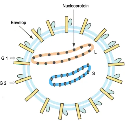
1.2.2 SHAPE
Like all other Arenaviruses, Lassa virus is enveloped and has a bisegmented single-strained RNA with a unique
ambisense genomic organization, and is coated by a nucleoprotein to form a nucleocapsid. Under cryoelectron
microscopy, the virion looks like a grainy particle, due to its beaded nucleocapsid. That is how arenavirus gets
its name, arena in the Latin root meaning sand.
4 Fig 3: Lassa virus structure (Image taken from Swiss Institute of Bioinformatics,
http://viralzone.expasy.org/viralzone/all_by_species/501.html)
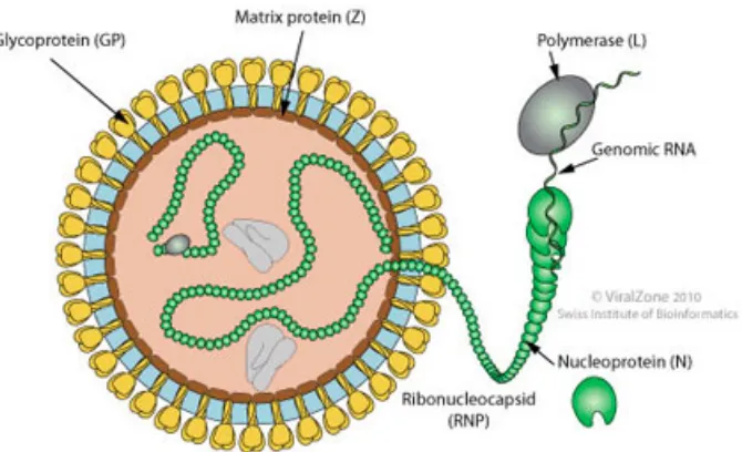
1.2.3 GENOME
Including Lassa virus, Arenaviruses’ two genomic RNA segments are large RNA (L-RNA, 7.2kb) and small
RNA (S-RNA, 3.4kb), and they encode four known proteins: a nucleoprotein NP (60 kDa), a matrix protein Z
(~11kDa), a RNA-dependent polymerase L (∼200 kDa), and a glycoprotein GP. Their ambisense coding
strategy allows the polypeptides to be synthesized in two opposite directions, with the help of a noncoding
intergenic region (IGR) folding into a hairpin structure.
The small RNA encodes NP and immature GP precursor. While large RNA is responsible for L and Z. In the S
segment, the NP coding region is transcribed from viral sense strand 3’ to 5’ into genomic complementary
mRNA, while the GP coding region is transcribed from 5’ to 3’ into a genomic sense mRNA. The same rule
applies to the L gene. The L protein’s mRNA is genomic complimentary and Z protein’s is genomic sense. This
is the ambisense-coding stratagem that helps viral mRNAs being transcribed without overlapping. As a result of
this arrangement, only after viral genomic RNA can the replication begin, opening reading frames of GPC and Z
being transcribed.
For precursor GP (82kDa), during the translation in the endoplasmic reticulum, a signal peptidase cleaves it into
a signal peptide and GP-C (76kDa). After translation, subtilase SKI-1/S1P cleaves this GP-C into N-terminal
5 Fig 4: Lassa fever virus’ virion and genome structure.
(Image taken from Swiss Institute of Bioinformatics,
http://viralzone.expasy.org/all_by_species/83.html)
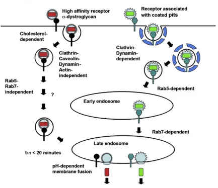
1.2.4 INFECTION MECHANISM
Lassa virus infects hosts mainly by contacting, but how virions break through the epithelial barrier for
initializing host infection is still unclear. When it reaches the host cell, the α-dystroglycan (α-DG), a
multipurpose receptor for the extracellular matrix protein, becomes the essential receptor and helps Lassa virus’
endocytosis to enter the cell. The GP protein plays the role for receptor recognition. GP1 (N-terminal) locates at
the glycoprotein spike to bind receptor α-DG on the cell surface. GP2 (C-terminal) binds to membrane and is
6 followed by the viral nucleocapsid entering a late endosomal partition via vesicular trafficking. This process is
independent from molecular motors like clathrin, caveolin, dynamin or actin. However, some cholesterol from
the membrane is required. The unusual endocytosis pathway allows Lassa virus particles being delivered to late
endosomes more rapidly, thus makes infection more effective. (Schlie K, et al., 2010)
Old world arenavirus LASV and LCMV share the same cell-entering mechanism. However, new world
arenaviruses recognize different cell receptors and need molecular motors when entering a cell, and depend on
Rab5 for delivering into early endosome, and require Rab7 as well for transmitting to late endosome (Fig 5).
[image:15.595.83.516.322.696.2](Kunz S. 2009)
7
1.2.5 PROTEINS
Glycoprotein GP
The GP protein is encoded by the S-segment RNA. Its function is to recognize and bind to the receptor of host
cell surface and fuse into the cell. The process has been introduced above.
Ring finger protein Z
The Z protein is a matrix protein, which plays essential roles in virus assembly and budding. Expressing only Z
without other viral proteins can be enough to form and release the enveloped Z-containing particles, which is
not notably different from Lassa virus particles in morphology and size. Z acts as the driving force during virus
particle releasing. Z also has strong association with the membrane. In addition, Z is assumed to interact with
NP during viral assembling. (Shtanko O, et al., 2010)
RNA-dependent polymerase L
L protein is a putative RNA-dependent RNA polymerase. It controls the synthesis of mRNA terminating in the
intergenic region, and noncapped genomic or antigenic RNA forming a full-length genome copy. (Lelke M, et
al., 2010)
The L protein consists of 2300 amino acids, which can be divided into three domains, N-terminal domain,
central domain and C-terminal domain. It is believed to harbor several enzymatic functions in the N and C
terminals, but this is yet unproven. Cap-snatching is supposed to be one of the L protein’s significant functions.
With NP, L protein can form the minimal trans-acting factors in genome replication and replication. Both L and
NP N-terminal domain are believed to cooperate with each other in a cap-snatching mechanism. The central
domain (residues 1000-1500) of L is regarded as the RNA-dependent RNA polymerase. (Lelke M, et al., 2010)
1.3 NP PROTEIN
1.3.1 BACKGROUND
NP is the most abundant viral protein in an LASV infected cell. NP associates with RNA to form the
8 protein for RNA replication and transcription. NP also interacts with the matrix protein Z during viral assembly.
(Eichler R, et al., 2004)
However, the most interesting function of NP is to suppress the host cell immune responses. Moreover, LAVS
NP structure is the only NP structure reported among Arenaviridae family.
In human cells, there are several receptors, which play essential roles in sensing infections by detecting
pathogen-associated pattern molecules (PAPM) and trigger immune response pathways. It is well known that
cytoplasm receptors retinoic acid-inducible I receptor and melanoma differentiation-associated 5(MDA-5), and
membrane toll-like receptors can detect PAMP molecules. PAMP, such as triphosphate dsRNAs, long chain
dsRNAs or short chain dsRNAs, trigger immune response pathways to produce interferon, when virus infections
occur. Virus infection usually produces dsRNA in the infected cell. Once retinoic acid-inducible I (RIG-I),
melanoma differentiation-associated 5 (MDA-5) or other cellular immune receptors detect dsRNA, signaling
will trigger IFNs production. LAVS NP has been demonstrated to play an essential role in immune suppression,
and its 3’-5’ exonuclease activity has been shown to be crucial for the function. Our hypothesis is that the LAVS
NP can specifically recognize and degrade the PAMP RNA molecules generated from the virus infection.
Therefore the receptors cannot detect the infection, and no immune response occurs. (Kathryn MH, et al., 2011)
1.3.2 STRUCTURE
The NP coated viral genomic RNA to formed nucleocapsid, a beads-like viral particle under microscopy.The
LAVS NP consists of two parts: amino (N) terminal and carboxy (C) terminal domains. A positively charged
groove locates between the two domains and the viral genomic RNAs are expected to bind in the groove. All the
structures of published nucleoprotein structures among all negative-stranded RNA viruses share the same
organization.
In each NP protomer, a Zn2+ locates in the C-terminal domain, forming a zinc finger structure. As a 3’-5’
exonuclease, one Mn2+ molecule was identified in the C-terminal domain active site. In the N-terminal domain,
9 Fig 6: NP monomer structure
Yellow region represents N-domain. Blue region represents C-domain, PDB code 3MWP. (Picture taken from Qi X, et al., 2010)
Fig 7: The ring shape form of the NP trimer
[image:18.595.197.402.467.684.2]10
1.3.3 FUNCTION
The N-terminal domain of LAVS NP forms a deep cavity for cap binding. The 5’ terminal m7G cap is presented
on most eukaryotic mRNAs. For most RNAs, elimination of the cap structure causes a loss of stability,
especially against exonuclease degradation, and a decrease in the formation of the initiation complex of mRNAs
for protein synthesis. In addition, a cap requirement has been observed for splicing eukaryotic substrate RNAs.
Only mRNAs with a ‘cap’ structure at its 5’ end can be translated into protein by the ribosome. But arenavirus
cannot produce cap from their own viral mRNA, therefore Lassa virus has to snatch cellular mRNAs’ caps.
During the cap-snatching, L protein’s N terminal domain is an endonuclease that cleaves off the 5’ end of
cellular mRNAs, while the NP’s N terminal domain holds the 5’ end cap. The holding function has not been
[image:19.595.200.382.317.590.2]reported in any equivalent protein among any other virus family.
(Qi X, et al., 2010)
11 Fig 9: A cap analogue molecule dTTP located in the cap-binding site. The initial FoFc difference density map for dTTP is in blue, with pink atoms representing carbon. Yellow residues are for the deep cavity site. Green residues are the pass for another mRNA to enter, PDB code 3MX2. (Picture taken from Qi X, et al., 2010)
The C terminal domain structure is similar to 3’-5’ exonucleases, especially the human DNA 3’-5’ exonuclease
enzyme TREX1. Experiments also show the C terminal domain alone can digest RNA substrates and suppresses
[image:20.595.88.512.73.333.2]immune responses, which means that the C-terminal domain is the functional part for the exonucleases activity.
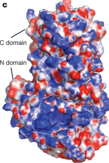
12 Fig 11: NP C-terminal domain with Mn2+, showing the essential residues for exonuclease activity. Purple colored residues match to DEDDh exonuclease, PDB code 3Q7C(Modified from Kathryn MH, et al., 2011)
However, how NP binds and degrades PAMP molecules, such as dsRNA is not exactly clear, for no complex
crystal has been reported. Therefore, my work focuses on expression, purification and crystallization of LAVS
NP C-terminal domain in complex with PAMP molecules, figuring out the novel mechanism.
I have built an expression construct of the native NP C-domain and carried out mutagenesis on designed sites.
Protein expression and purification were successful. I’ve set up crystallization trials and get nice crystal
complex of native NP C-domain with RNA ligand. The crystals have been sent to Diamond IO3 for X-ray
13
2
M
ATERIALS AND METHODS2.1 EXPRESSION CONSTRUCT
2.1.1 CLONING NPC-TERMINAL DOMAIN’S NUCLEOTIDES INTO PLOU3(HIS-TAG-MBP ATTACHED)
The nucleotides encoding the LAVS NP C-terminal domain from residue 364 to 569 was cloned into the
pMAL-c2X-derived plasmid pLou3, with a 6His-tag at N terminus of MBP (maltose binding protein) and a
TEV cleavage site between the MBP and the target protein. This construct was transformed into Rosetta cells
(Novagen). The transformed cells have antibiotic resistance against ampicillin and chloramphenicol. Xiaoxuan
Qi previously built this construct in our group.
2.1.2 CLONING THE NPC-TERMINAL DOMAIN INTO PHISTEV PLASMID
The nucleotides encoding the NP C-terminal domain were also inserted into pHisTEV plasmid. The forward and
reverse primers containing the NCOI and HindIII enzyme cutting sites were synthesized in Eurogentec. The
C-terminal nucleotides were amplified by PCR using a program of heating at 95°C for 2 mins, annealing at
58°C for 1 min and extending at 72°C for 2 min for 32 cycles. The pHisTEV plasmid was cut by restriction
enzymes NCOI and HindIII. The NP C-terminal domain PCR product was then inserted into the pHisTEV
plasmid with the help of T4 DNA ligase (10 µl reaction system in 37°C for 2 hour. See appendix Ⅳ).
This reconstructed plasmid was transformed into TAM1 cells for producing and harvesting more plasmids
(transform method and plasmid extraction with miniprep method see appendixⅤ). Enzyme digestion was used
to confirm the correct insertion (NCOI and HindIII cutting reconstructed plasmid). Afterwards, the correct
plasmid was transferred into expression system Rosetta cells.
2.1.3 MUTAGENESIS OF PHISTEVNPC-TERMINAL DOMAIN.
DNA primers were designed based on QuikChange™ Site-Directed Mutagenesis and Huangting Liu’s protocol,
14 The mutant plasmids were generated using the primers and Promega Pfu DNA Polymerase. Five units of Dpn I
were used to remove the original template plasmids by incubation at 37°C for 2 hrs.
Agarose gel electrophoresis was used to separate the C-terminal mutant plasmid DNA band from others. One
micro liter of each mutant plasmid was transformed into TAM1 cells, and the transformed cells were spread on
LB agar gel containing 100µg/ml ampicillin. Two colonies from each mutant were picked and each colony was
inoculated into 10 ml of LB containing 100µg/ml ampicillin and cultured overnight. The mutant plasmids were
extracted with Qiagen miniprep kit, and 5µl of each plasmid was sent to University of Dundee for sequencing.
The correct mutant genes were transformed into Rosetta cells for expression.
2.2 PROTEIN EXPRESSION AND PURIFICATION
2.2.1 FOR THE NPC-TERMINAL DOMAIN (HIS-TAG-MBP ATTACHED)
The single colony of the Rosetta transformed cells was inoculated into 500ml LB containing 50µg/ml ampicillin
and 34 µg/ml chloramphenicol and was cultured overnight in incubator at 200 rpm in 37°C. This overnight
culture was then subcultured to 10 liters of LB (containing 50µl/ml ampicillin and 34 µg/ml chloramphenicol) at
200 rpm at 37°C. When OD600 reached 0.6, the cells were induced with 0.03mM final concentration of IPTG
(isopropyl-beta-d-thiogalactopyranoside). And the protein expressions were induced at 20°C for around 20
hours.
The cells were harvested by centrifugation at 10000g for 15 mins. The Cell pellet was resuspended in loading
buffer (20mM Tris-HCl, pH7.5, 10mM imidazole, 300mM NaCl, 10% glycerol), 1 tablet of EDTA-free protease
inhibitor (Roch), 1µM DNase (Sigma), 1µM lysosome (Sigma) and 1mM PMSF (phenylemethylsulfonyl
fluoride, Sigma). Cells had gone through cell disrupter twice for an adequate disruption. The debris is removed
by centrifugation at 40000g for 30 mins. Supernatant was collected and applied to 10 ml Ni-NTA agarose
(Qiagen) beads, which was pre-equilibrated with the loading buffer. The beads were washed with 40ml of
loading buffer (however, even 30mM imidazole wash buffer would wash away some target protein), and were
eluted with 22ml elution buffer consisting of 20mM Tris-HCl, pH 7.5, 500mM imidazole, 300mM NaCl
15 Then the protein buffer was changed to a buffer containing 20mM Tris-HCl, pH 7.5, 300mM NaCl and 10%
glycerol, by going through a desalting column (Hiprep 26/10, GE). The His-MBP-NPC fusion protein was
cleaved by TEV proteinase at room temperature for around 18 hours. Afterwards, the cleavage sample was
applied to Ni-NTA agarose beads (pre-equilibrated with desalt buffer). The His-MBP was removed from the
sample by Ni-NTA, while the target protein went through the beads column
.
The sample was concentrated to 7.5ml for gel filtration. The gel filtration column was pre-equilibrated with GF
buffer (20mM Tris-HCl, pH7.5, 300mM NaCl, 10% glycerol for high salt concentration condition, or 20mM
Tris-HCl, pH7.5, 100mM NaCl, 10% glycerol for low salt concentration). The fractions containing the NP
C-terminal domain were pooled. The protein was concentrated to 10mg/ml and frozen in liquid nitrogen, then
stored at -80°C.
2.2.2 FOR THE HIS-TAGGED NPC-TERMINAL DOMAIN (NATIVE AND MUTATIONS)
The His-tagged NP C-terminal domain was expressed in Rosetta E. coli cells. The overnight cultures grew in
incubator at 37 °C and 200rpm in 500 mL LB (supplemented with 50 µg/ml kanamycin and 34 µg/ml
chloramphenicol). The overnight culture was inoculated into 10 L LB containing 50 µg/ml kanamycin, 34 µg/ml
chloramphenicol and 100 µM ZnCl2. When OD600 reached around 0.4, the protein was induced with 500 µM
IPTG for around 20 hours at 25 °C and 200 rpm.
The cells were harvested by centrifugation at 10000g for 15 min. The cell pellet was resuspended in loading
buffer (50 mM Tris-HCl, pH 8.0, 300 mM NaCl, 20 mM imidazole), 1 tablet of EDTA-free protease inhibitor
(Roch), 1µM DNase (Sigma), 1µM lysosome and 1mM phenylemethylsulfonyl fluoride (PMSF, Sigma). Cells
had gone through cell disrupter twice for an adequate disruption. The debris is removed by centrifugation at
40000g for 30mins.
The supernatant was pooled and applied to 10 ml Ni-NTA agarose (Qiagen) beads (pre-equilibrated with
loading buffer). The Ni-NTA beads were washed three times with 50 mL of wash buffer (50 mM Tris-HCl, pH
16 pH 7.5, 500mM imidazole, 300mM NaCl). The protein was concentrated and purified by gel filtration using a
Superdex 200 (GE Healthcare) column, which was equilibrated with GF buffer (20 mM Tris-HCl, pH 7.5, 300
mM NaCl and 10% glycerol). The NP C-terminal domain samples were pooled, concentrated and stored at
-80 °C for binding future affinity trials.
2.3 SDS-PAGE GEL
SDS-PAGE was used for checking protein purity in specific fractions after gel filtration. The gel was pre-cast
NuPAGE 4-12% Bis-Tris gel, and the power supply was Powerease 500. A15µl sample was mixed with 5µl of
SDS loading buffer and incubated at 95 °C for 5 min. Protein molecular standard (Mark12) was used to indicate
protein molecular weight.
2.4 TRIPHOSPHATE-DSRNA CONSTRUCTION
The triphosphate dsRNA was produced with MEGAshortscript Kit according to the manufacture’s manual. The
DNA templates for RNA were synthesized by Eurogentec.
2.4.1 TEMPLATE DNA PREPARATIONS:
The 50µl 100µM T7poly_sense_common and the 50 µl 100µM T7_anti_polyGC8 were mixed together,
incubated at 95°C for 3min, and then cooled down to room temperature.
2.4.2 TRANSCRIPTION REACTION
The reaction takes place in a sterilized 1.5ml eppendorf tube. The 20µl reaction contains 2 µl T7 10X reaction
buffer, 2µl T7 ATP solution (75 mM), 2µl T7 CTP solution (75 mM), 2µl T7 GTP solution (75 mM), 2µl T7
UTP solution (75 mM), 2µl T7 enzyme Mix, 3µl template DNA and 5µL water (nuclease-free).
The 20µl reaction system was mixed thoroughly, and incubated at 37°C overnight.
In order to degrade the template DNA, 1µl of TURBO DNase was added to the system and mixed well, with
incubation at 37°C for more than 1 hour.
17
2.4.3 RNA PURIFICATION
The RNAs were extracted with an equal volume (151µl) of phenol/chloroform (water-saturated), and then with
an equal volume (151 µl) of chloroform. The aqueous phase was collected into a new tube. The RNAs were
precipitated by adding 2 volumes of ethanol (approximately 600µl) and mixing well and keeping at –20°C for
least 15 minutes.
The RNA was pelleted by centrifugation at 4°C for 15 minutes at maximum speed (≥10,000 x g), and the
supernatant was carefully removed. The RNA pellet was washed with 70% ethanol at -20 °C and dried at room
temperature.
2.4.4 DSRNA PREPARATION
The synthesized RNA was dissolved in TEA buffer (10 mM Tris-HCl, pH 8, 1mM EDTA, 0.1M NaCl), around
10µl for 5 20µl-scale reactions. The RNA sample was heated at 95 °C for 3min and annealed at room
temperature, forming triphosphate double strand RNA. The RNA samples were quantified by Nano drop and
store at -80°C for crystallization and assays.
2.5 CRYSTALLIZATION
Crystallization conditions were screened on a SWISSCI 'MRC' 2-Well Crystallization Plates (Douglas) with the
sitting drop vapor diffusion method set by the Honeybee system. Each drop contained 0.15µl protein-RNA
complex and 0.15µl buffer, while 85µl buffer was loaded in each reservoir. The crystallization screens are
stochastic screens, made in JHN laboratory. The NP C-terminal domain RNA complex was formed by mixing
protein and dsRNA at 1:1 molar ratio (approximately 100µl 10mg/ml protein with 4.2µl 10mM dsRNA).
Initial crystals were obtained in 18.5% (w/v) PEG MME 5000, 0.1M Na-citrate, pH 4.5, 2.4% (w/v) PEG
MME350 at room temperature for 2 weeks. Crystallization optimization was carried out with varying the
concentration of PEG MME 5000 and PEG MME350 and pH. The best crystals grew in18%-19.5% (w/v) PEG
18
2.6 X-RAY CRYSTAL DATA COLLECTION
The crystals were frozen in liquid nitrogen using paraffin oil as a cryoprotectant. To obtain the NP C-terminal
domain in complex with dsRNA and Mn2+, the crystals were soaked in a solution containing additional 100 mM
MnCl2 and 20% glycerol for 1 min or 1.5 min or 5 min or 10 min before being frozen.
The diffraction data were collected at the Diamond Light Source UK. Crystals were screened at beamline IO3
with Pilatus 6M-F detector. Some of the good crystals were selected for data collection. Approximately 800
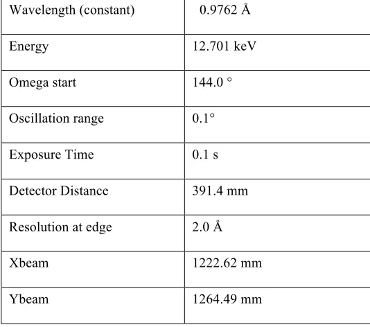
frames have been recorded for each crystal. The parameters used for data collection for NP C-terminal domain
[image:27.595.163.430.292.528.2]in complex with dsRNA are listed in Table 1.
Table 1: Data collection parameters for a crystal of NP C-terminal domain in complex with dsRNA Wavelength (constant) 0.9762 Å
Energy 12.701 keV
Omega start 144.0 °
Oscillation range 0.1°
Exposure Time 0.1 s
Detector Distance 391.4 mm
Resolution at edge 2.0 Å
Xbeam 1222.62 mm
Ybeam 1264.49 mm
2.7 STRUCTURE DETERMINATION, REFINEMENT AND MODEL BUILDING
All the data were indexed and integrated using Mosflm (Geoff GB, et al., 2011), and the data were scaled using
Scala
(Evans P, 2006). The structure was determined by molecular replacement using Phaser (McCoy AJ. 2007).
The initial search model was NP C-terminal domain (PDB code 3MWP). The models were built using Coot
19
3
R
ESULT AND DISCUSSION3.1 PROTEIN PURIFICATION
3.1.1 HIS-TAG-MBP FUSION PROTEIN
The NP C-terminal domain was successfully expressed as a His-tagged MBP fusion protein. The entire fusion
protein was purified using 10 ml of nickel beads. After washing with 30 mM imidazole, we could obtain
relatively pure fusion protein using 22 ml of the elution buffer as shown in Fig 12. The His-tagged MBP is
cleaved off by TEV proteinase. To find the best time for cleavage, we did a time course and found that 15 hours’
TEV cleavage is insufficient. We therefore always tried to cleave the MBP for 18 to 20 hours.
Fig 12: Purification of the MBP and N-terminal domain fusion protein and TEV proteinase cleavage.
However, gel filtration cannot separate MBP and NP C-terminal domain because their molecular weight is
[image:28.595.176.420.310.557.2]20 Fig 13: The C-terminal domain could not be separated from MBP by gel filtration. Lane 1 is marker, lane 2 to 9 are different fractions from the gel filtration.
In order to remove MBP from the C-terminal domain, an additional nickel column was used before gel filtration.
One effective way was to connect extra Ni-NTA columns (5ml) along with the gel filtration column, and we
found that two Ni-NTA columns gave us the best result (Fig 14 and Fig 15).
Fig. 14: NP C-terminal domain purification by a Ni-NTA column connecting to gel filtration column.
21 We have found that it did not reduce the protein yield using the two Ni-NTA columns to remove the MBP,
compared with using one Ni-NTA column.
Two different gel filtration buffers were applied in this experiment: high salt condition (20mM Tris-HCl, pH 7.5,
300mM NaCl, 10% glycerol), and low salt condition (20mM Tris-HCl, pH 7.5, 100mM NaCl, 10% glycerol)
[image:30.595.148.450.310.547.2]Gel filtration result and SDS-PAGE gel picture under high salt condition:
Fig.16: gel filtration graph. There are two peaks, and both of them were the NP C-terminal domain
Fig. 17: NP C-terminal domain purification after gel filtration in high salt buffer.
Gel filtration result and SDS-PAGE gel picture under low salt condition:
Xpress 3008:Sample1_UV Xpress 3008:Sample1_Fractions
0 50 100 150 mAU
0 20 40 60 80 100 120 140 ml
A2 A4 A6 A8 A10 A12 B11 B9 B7 B5 B3 B1 C2 C4 C6 C8 C10C12 D11 D9 D7 D5 D3 D1 E2 E4 E6 E8 E10 E12 F10 F8 F6 F4 F3
Fig.18: Gel filtration graph under low salt concentration. There are two major peaks, both of them were NP C-terminal domain. The third tiny peak shows nothing on SDS-PAGE gel, which could be just jam signal.
np c 16122010 GF run(1292497992)001:Sample1_UV np c 16122010 GF run(1292497992)001:Sample1_Fractions
0 50 100 150 200 mAU
0 50 100 150 ml
22 Fig 19: SDS-PAGE gel of gel filtration purification in low salt buffer
Both peaks were confirmed by SDS-PAGE and mass spectrometry as they both were the NP C-terminal domain.
However, the second peak contains much less protein and we hardly got enough amount of protein from this
peak after concentration. Therefore, we mainly focused on the first peak for crystallization. And the yield was
around 1mg protein per liter culture in high salt buffer, or a bit less around 0.7mg protein per liter culture in low
salt buffer.
3.1.2 HIS-TAGGED NPC-TERMINAL PROTEIN
The His-tagged NP C-terminal domain was expressed successfully. The protein yield is higher, around 6mg
protein per liter cell culture, while the MBP-fusion one got 1mg protein per liter cell culture. But the purity of
the purified protein was not as good as the His-tagged MBP fusion protein. We did not cleave the His-tag away
as the protein would be used for surface plasmon resonance (SPR) assays. To improve the protein purity, we
23 Fig.20: SDS-PAGE shows His-tagged NP C-terminal domain purification by a nickel column
The gel filtration graph showed that there is mainly one peak (Fig. 21). We obtained 5ml of the purified
C-terminal domain with a concentration of 5mg/ml.
np(1310735126)001:Sample1_UV np(1310735126)001:Sample1_Fractions
0 200 400 600 800 mAU
0 20 40 60 80 100 ml
[image:32.595.237.358.71.290.2]A2 A3 A4 A5 A6 A7 A8 A9 A11 B12 B10B8 B7 B6 B5 B4 B3 B2 B1 C1 C3C4C5C6C7C8C9C11 D12 D10 D8D7D6D5D4D3D2 D1
Fig. 21: gel filtration graph.
The SDS-PAGE revealed that we had obtained a reasonably pure protein (Fig 22).
[image:32.595.148.450.371.496.2]24
3.2 CRYSTALLIZATION
The NP C-terminal domain, purified from a His-tagged MBP fusion protein was used for crystallization.
We obtained the native crystals of NP C-terminal domain as shown in Fig 23. As our goal is to determine the
NP C-terminal domain in complex with PAPM molecules, we focused on the crystals of NP C-terminal domain
[image:33.595.138.458.211.370.2]in complex with PAPM molecules, such as dsRNA and triphosphate dsRNAs.
Fig 23: Crystal pictures of the native NP C-terminal protein.
Left one was screen result using honeybee system. Right one was optimization trials result.
A 6 nucleotide-length dsRNA, an 8 nucleotide-length dsRNA and a 12 nucleotide-length dsRNA were
chemically synthesized in Eurogentec. Only the 8 nucleotide-length dsRNA formed crystals with NP C-terminal
domain. The crystals of the NP C-terminal domain with dsRNA are shown in Fig 24. These crystals were thin.
Although intensive optimization was carried out, we could not improve the crystals. We therefore tried to
synthesize 8 and 12 nucleotide-length triphosphate dsRNAs with MEGA shortscript kit. We have tried to
co-crystallize the triphosphate dsRNA with the C-terminal domain.
Fig 24: NP C-terminal domain protein with dsRNA 8 complex crystal
[image:33.595.100.498.596.706.2]25 The crystals of the NP C-terminal domain in complex with triphosphate 8 nucleotide length dsRNA are shown
in Fig 25. Those crystals can grow very fast and big in Crystal Clear P (Douglas) plates. Protein-RNA complex
crystals appeared in less than 24 hours, and these crystals took about a week to grow large enough for X-ray
diffraction.
Fig 25: Crystals of NP C-terminal domain protein in complex with triphosphate dsRNA 8. The crystals that grew from the screening are shown on the left and the crystals after optimization were shown on the right.
[image:34.595.113.487.171.347.2]The crystal that was used to collect data on Diamond beam line IO3 is shown in Fig 26. The rhombus shaped crystal is approximately 150µm long and 40µm wide.
Fig 26: Crystal of NP C-terminal domain protein in complex with triphosphate dsRNA 8 in loop for X-ray diffraction
The diffraction pattern of the crystal is shown in Fig 27. The crystal diffracted to 1.73 Å and shows clearly
defined diffraction spots. This diffraction pattern shows no strong ice rings and the frequency of the diffraction
[image:34.595.112.482.441.592.2]26
Fig 27: One of the diffraction images, collected on Diamond Light Source UK, beamline IO3. The resolution of the outer circle is 1.7 Å. Picture on the right side is the enlarged area of red square on the left side picture.
3.3 CRYSTAL STRUCTURE
The collected data were processed, and the results were listed in Table 2. After molecular replacement and a few
cycles of refinement, the electron density for the dsRNA was very clear. However, we did not see electron
density for the triphosphate. A dsRNA (GGGC/CCCG) was built in the electron density, which shows that the
3’-end nucleotide (cytosine) of chain GGGC is directly located in the 3’-5’ exonuclease active site (Fig 28, 29).
Table 2: The crystallographic statistics of the NP C-terminal domain in complex with triphosphate dsRNA
Data Statistics
Space Group
Unit Cell parameters
P3
a = b 177.16 Å
c 56.49 Å
α=β 90°
γ 120°
Resolution range 46.5-1.73 Å
[image:35.595.73.526.498.747.2]27
Unique reflections 184629 (5543)
I/σI 16.4 (4.2)
Completeness 99.7% (99.9%)
Rmerge 7.3% (39.3%)
Refinement Statistics
Rfactor 17.80%
Rfree 19.87%
Rmsd bonds
Number of protein atoms
0.03 Å / 2.45°
9076
Mean B-factor 25.27 Å
Residues in Ramachandran core
Residues in disallowed regions
96.83%
[image:36.595.152.447.437.666.2]0
28 Fig 29: LAVS NPC and dsRNA complex structure in cartoon, the manganese ion shows as magentas ball and Zinc ion shows as grays ball.
From the structure we can see that one strand (5’GGGC3’) of the dsRNA interacts extensively with NP residues
D466, D389, Gln462, D533, R492, E391, H528 and S430, in which most of them are catalytic residues, while
another strand (5’GCCC3’) interacts with side chains of NP residues Y429, D426, Q425, Q422, R393 and D465.
Particularly, the side chain of residue Y429 stacks against the guanidine ring (Fig 30, 31).
[image:37.595.109.490.462.662.2]
29 Fig 31: Interactions between LAVS NPC and triphosphate dsRNA and dsRNA cleavage activities of LAVS NP-C mutants. a, Residues are involves in the dsRNA binding and cleavage. The cleavage chain of the dsRNA in purple and the binding chain in cyan. b. Schematic representation of LAVS NPC and triphosphate dsRNA interactions. The scissile bond is indicated with a scissor.
In order to validate the functions of the binding residues in dsRNA recognition and cleavage, we have generated
several alanine substitute mutant constructs (name of mutant sites and primer details were shown in appendix II),
and we are purifying the mutant proteins. We will try ITC and SPR to check the mutant binding affinities with
different RNA ligands, such as dsRNA, triphosphate dsRNA and ssRNA. In the same time, we will check
whether the mutant proteins process the PAPM RNA differently in efficiency.
3.4 MUTAGENESIS AND MUTANT PROTEIN PURIFICATION
3.4.1 PHISTEV NPC-TERMINAL DOMAIN PLASMID CONSTRUCTION
The gene fragment encoding the NP C-terminal was inserted into pHisTEV plasmid successfully,
confirmed by the restriction enzyme digestion result (Fig 32). The constructed plasmid was digested
with NCO
Ⅰand Hind
III. And the digested plasmid had been run through a 10% agarose gel. The gel
showed that the NP C-terminal domain encoding gene band was around 600bp, corresponding to the
exactly size for the NP C-terminal domain encoding gene 615bp
.30 Fig 32: The electrophoresis gel of digested NPC-pHisTEV
3.4.2 SINGLE SITE PLASMID MUTAGENESIS
The PCR result of a mutation is shown in Fig 33. Those plasmids were around 6kbp, equivalent to the length of
pHisTEV vector.
Fig 33: PCR result for mutations
The DNAMan software had been used to compare the mutation sequences results with NP C-terminal domain
[image:39.595.134.462.443.636.2]31
3.4.3 EXPRESSION AND PURIFICATION
The mutants were obtained by using the same expression and purification method as for the native pHisTEV NP
C-terminal domain. The single site mutation proteins were expressed well as the native protein, around 5 to 6mg
protein per liter culture. Fig 36 and 37 were S430A result features as an example of all mutants.
npc6 02082011 GF run(1312302419)001:Sample1_UV npc6 02082011 GF run(1312302419)001:Sample1_Fractions
0 20 40 60 80 100 120 140 mAU
0 20 40 60 80 100 ml
[image:40.595.133.467.180.301.2]A2 A3 A4 A5 A6 A7 A8 A9 A11 B12 B10 B8 B7 B6 B5 B4 B3 B2 B1 C1 C2 C3 C4 C5 C6 C7 C8 C9 C11 D12 D10 D8 D7D6D5 D4 D3 D2 D1
Fig 34: gel filtration graph on S430A mutant. There is one main peak of protein.
Fig 35: SDS-PAGE gel of gel filtration of S430A mutant. Lane C9 to D9 were different fractions of main peak from gel filtration.
[image:40.595.175.422.341.519.2]32
4
F
UTURE WORKLAVS NP and other arenavirus’s NPs use a totally novel way to evade the human immune responses. To find
the detail of the mechanism is important for the controlling of such deadly infectious diseases caused by some
arenaviruses. Our collaborators at Emory University have shown that the LAVS NP can process dsRNA much
quicker than those of ssRNA (approximate 20 times faster), and our structure of dsRNA and C-terminal domain
sheds an important light on the mechanism.
In order to elucidate the NP C-terminal’s immune suppression function, the mutant proteins have been designed,
expressed and purified. Their binding affinities with different PAPM RNAs will need to be tested in future
experiments. I’ll use florescence labeled RNAs to identify the cleavage efficiency of different mutants. Each
mutant will interact with several designed RNAs under certain time and temperature. Then polyacrylamide gel
electrophoresis will be used to separate bands and recognize the cleavage extent. On the other hand, a standard
gel filtration chromatography method will be used to determine whether the NP C-terminal protein we obtained
33
5
T
HENP
N-
TERMINAL DOMAINThe N-terminal domain of LAVS plays an essential role for cap binding. To investigate the detailed mechanism,
we have cloned, expressed and crystallized the N-terminal domain.
5.1 MATERIAL AND METHODS
5.1.1 EXPRESSION CONSTRUCT
The N-terminal domain consists of residues 1 to 338. The gene segment is derived from Lassa virus (Josiah
strain), S genome. Similar way as dealing with the C-terminal domain, the N-terminal domain gene segment was
cloned into pMAL-c2X-derived pLou3 plasmid, with a 6His-tag at the N terminus of MBP and TEV cleavage
site between MBP and the target domain. This construct was transformed into Rosetta cells (Novagen).
Xiaoxuan Qi previously built this construct in our group.
5.1.2 PROTEIN EXPRESSION AND PURIFICATION
The expression and purification process was very similar to those of the C-terminal domain.
The transformed cells were cultured in LB media with 50mg/ml ampicillin and 34mg/ml chloramphenicol. The
cells were induced with 0.03mM IPTG when the cell OD600 reached 0.6. And then the cells were cultured for
about 20 hours at 20°C.
The cells were harvested by centrifugation for 15min at 10000g. Cell pellet was resuspended in loading buffer
(20mM Tris-HCl, pH7.5, 10mM imidazole, 300mM NaCl and 10% glycerol) with1 tablet of EDTA-free
protease inhibitor (Roch), 1µM DNase (Sigma), 1µM Lysozome and 1mM PMSF (Sigma). The cells were
disrupted using a cell disruptor. The cell debrides were removed by centrifugation for 30min at 40000g. The
supernatant was collected and applied to 10 ml of Ni-NTA agarose (Qiagen) beads, which pre-equilibrated with
loading buffer. The beads were washed with 20ml loading buffer (wash buffer with 30mM imidazole will wash
away target protein), and the fusion protein was eluted with 22ml elution buffer consisting of 20mM Tris-HCl,
34 The sample buffer was changed to desalt buffer (20mM Tris-HCl, pH 7.5, 300mM NaCl and 10% glycerol),
using a desalting column (Hiprep 26/10, GE). The His-MBP-NPC fusion protein was cleaved by TEV
proteinase at room temperature for 16 hours. The His-MBP was removed by Ni-NTA agarose beads
(pre-equilibrated with desalt buffer).
The sample was concentrated to 7.5ml for the gel filtration. The gel filtration column was pre-equilibrated with
GF buffer (20mM Tris-HCl, pH7.5, 300mM NaCl and 10% glycerol as high salt concentration condition, or
20mM Tris-HCl, pH7.5, 100mM NaCl and 10% glycerol for low salt concentration). The fractions containing
the NP N-terminal domain were checked by SDS-PAGE gel (Invitrogen, Mark12) and pooled. The purified
N-terminal domain was concentrated to 20mg/ml, frozen by liquid nitrogen and stored at -80°C.
5.1.3 CRYSTALLIZATION
Crystallization conditions were screened on MRC plates with the sitting drop vapor-diffusion method using a
Honeybee robot. Drops contained 0.15µl protein solution and 0.15µl buffer with 100µl buffer in each reservoir.
The crystallization screens were stochastic screens, made by JHN lab.
Initial crystals were obtained around 2~3 weeks at 20 °C in 0.5M magnesium formate dihydrate, 0.1M HEPES,
pH 7.5. Optimizations were carried out, and the crystallization conditions were optimized to 0.2M~0.8M
magnesium formate dihydrate, 0.1M HEPES, pH 7.1-7.9.
5.2 RESULT
The gel filtration shows that the N-terminal domain displays two peaks. Mass spectra data is needed to
determine whether both peaks are the same N-terminal domain. The two peaks gain almost same amount of
35 npn 07022011 GF run(1297108826)001:Sample1_UV npn 07022011 GF run(1297108826)001:Sample1_Fractions
0 100 200 300 400 500 mAU
0 20 40 60 80 100 120 ml
[image:44.595.111.484.73.394.2]A2 A3 A5 A7 A9 A11 B12 B10 B8 B6 B5 B3 B1 C2 C4 C6 C8 C10 C12 D11 D9 D7 D5 D3 D1 E2 E3
Fig 36: NP N-terminal domain gel filtration graph
Fig 37: SDS-PAGE gel gel of gel filtration fractions of N-terminal domain. Lane 6 to 8 were the first peak, lane 9 to 11 were the second peak.
Crystals
Fig 38 shows the obtained crystals of the N-terminal domain. On the left figure is the initial crystal from the
screen and the right figure shows the crystals grown under optimized conditions. We are trying to improve the
crystal quality.
[image:44.595.111.485.550.719.2]
36
5.3 FUTURE WORK
The N-terminal domain is very important for the cap-snatching mechanism, and this mechanism is different
from that of bunyavirus or influenza virus. To find out the detailed mechanism, I will try to improve the crystal
quality, test whether the crystal diffracts. If I gain good diffraction data, I will then try to evaluate the cap
binding function by obtaining the m7GpppG and N-terminal domain complex structure. At the same time, I will
set up the in vitro assays using the NP and the N-terminal domain of the L to test how the virus from the host
37
6
A
PPENDIx I
Short chain RNA sequence details
For crystallization trials
ds RNA 6:GAC-GCU
ds RNA 8: CGC-AUG-CG
ds RNA 12: GAC-GCU-AGC-GUC
ppp ds RNA GC8: GGG-CGC-CC
For protein-RNA binding affinity test
ds RNA 16 forward: UCU-CUC-UCU-CUC-UCC-C
ds RNA 16 reverse: GGG-AGA-GAG-AGA-GAG-A
ppp ds RNA GC14: GGC-GCG-CGC-GCG-CCT-ATA-GTG-AGT-CG
DNA template sequence details
(For RNA synthesis)
common: AAT-TTA-ATA-CGA-CTC-ACT-ATA-GG,
GC8: GGG-CGC-CCT-ATA-GTG-AGT-CGT-ATT-AAA-TT
38
7
A
PPENDIXII
DNA primer sequence details for mutants
39
8
A
PPENDIXIII
PCR protocol based on that provided by Dr Huangting Liu.
Pfu buffer 5µl dNTP 5µl primer Forward 1µl primer Reverse 1µl template DNA ~10ng pfu enzyme 1µl H2O to final volume of 50µl
95°C 5min 95°C 1min
12 cycles 61°C 1min (Tm -5°C) 72°C 12min
95°C 1min 61°C 1min 72°C 30min 4°C forever
PCR protocol based on QuikChange™ Site-Directed Mutagenesis method
Pfu buffer 5µl dNTP 5µl primer Forward 1µl primer Reverse 1µl template DNA ~20ng pfu enzyme 1µl H2O to final volume of 50µl
95°C 1min 95°C 30s 13 cycles 55°C 1min
40
9
APPENDIXIV
Restriction enzyme reaction
buffer E 1µl
BSA 1µl
HindIII 0.3µl
NCOI 0.3µl
Plasmid 5µl
H2O 3.3µl
Reaction in 37°C for 2 hours
Run 1% agarose gel for result checking
T4 DNA ligase protocol
vector DNA 100ng insert DNA 17ng
Ligase 10Xbuffer 1µl 10 µl
T4 DNA ligase (Weiss units) 0.1~1u Nuclease-Free water to final volume of 10µl
41
10
APPENDIXV
TRANSFORM PLASMID TO COMPETENT CELLS
1. At 4ºC, add 1µl of plasmid to a microtube with 50µL of TAM1/Rosetta competent cells and mix well; keep in ice for 30min
2. Heat shock at 42ºC for 90sec, and put in ice for 2min
3.
Add 1ml of LB medium to transformed cells. Incubate at 200rpm, 37ºC for 1hour
4. Spin down at 13000 rpm for 2min in a microcentrifuge
5. Reject 850ml supernatant and resuspend cells pellet
6. Spread cells on LB/KANA agarose plate (TAM 1 cell) or LB/KANA/CHLO agarose plate (Rosetta). Inserted plasmid contains kanamycin resistance, and Rosetta cell itself has chloramphenicol resistance.
7. Grow cells overnight by putting the plate up side down at a 37ºC still incubator
Plasmid extraction by QIAGEN miniprep kit
1. Culture target cells overnight in 10ml LB media (supplemented with 34 µg/ml kanamycin for TAM1 cell, or 34 µg/ml kanamycin and 34 µg/ml chloramphenicol for Rosetta cell). Cell colony is picked from agarose plate.
2. Transit overnight culture to 15ml centrifuge tube. Spin down 4500rpm for 10min. Discard supernatant
3. Resuspend pelleted bacterial cells in 250 µl Buffer P1 and transfer to a microcentrifuge tube.
4. Add 250 µl Buffer P2 and gently invert the tube 4–6 times to mix.
5. Add 350 µl Buffer N3 and invert the tube immediately but gently 4–6 times.
6. Centrifuge for 10 min at 13,000 rpm in microcentrifuge.
7. Apply the supernatants from last step to the QIAprep spin column by decanting or pipetting.
8. Centrifuge for 1min. Discard the flow-through.
9. Wash the spin column by adding 0.5 ml Buffer PB and centrifuging for 1min. discard the flow-through.
10. Wash spin column by adding 0.75 ml Buffer PE and centrifuging for 30–60 s.
11. Discard the flow-through, and centrifuge for an additional 1 min to remove residual wash buffer.
42
11
R
EFERENCEOgbu O, Ajuluchukwu E, Uneke CJ. Lassa fever in West African sub-region: an overview. Journal of vector borne diseases 2007 Mar;44(1):1-11.
Haas WH, Breuer T, Pfaff G, Schmitz H, Köhler P, Asper M, Emmerich P, Drosten C, Gölnitz U, Fleischer K, Günther S. Imported Lassa Fever in Germany: Surveillance and Management of Contact Persons.
Clinical Infectious Diseases
2003 May 15;36(10):1254-8.
Illick MM, Branco LM, Fair JN, Illick KA, Matschiner A, Schoepp R, Garry RF, Guttieri MC. Uncoupling GP1 and GP2 expression in the Lassa virus glycoprotein complex: implications for GP1 ectodomain shedding. Virology journal 2008 Dec 23;5:161.
Fichet E, Rogers DJ. Risk Maps of Lassa Fever in West Africa. PLoS Neglected Tropical Diseases. 2009 Mar;3(3):e388.
Schlie K, Maisa A, Freiberg F, Groseth A, Strecker T, Garten W. Viral protein determinants of Lassa virus entry and release from polarized epithelial cells. Journal of virology 2010 Apr;84(7):3178-88. Eichler R, Strecker T, Kolesnikova L, ter Meulen J, Weissenhorn W, Becker S, Klenk HD, Garten W, Lenz
O. Characterization of the Lassa virus matrix protein Z: electron microscopic study of virus-like particles and interaction with the nucleoprotein (NP). Virus research 2004 Mar 15;100(2):249-55. Lelke M, Brunotte L, Busch C, Günther S. An N-terminal region of Lassa virus L protein plays a critical
role in transcription but not replication of the virus genome. Journal of virology 2010 Feb; 84(4): 1934-44.
Buchmeier MJ, de la Torre JC, Peters CJ. Arenaviridae: the viruses and their replication. In Fields Virology, Vol 2. Edited by Knipe DM, Howley PM. Lippincott Williams & Wilkins, a Wolters Klumer Business; 2007:1791-1828.
Bowen MD, Rollin PE, Ksiazek TG, Hustad HL, Bausch DG, Demby AH, Bajani MD, Peters CJ, Nichol ST.
Genetic diversity among Lassa virus strains. Journal of virology 2000 Aug;74(15):6992-7004.
Kunz S. Receptor binding and cell entry of Old World arenaviruses reveal novel Dects of virus-host interaction. Virology 2009 May 10;387(2):245-9.
Shtanko O, Imai M, Goto H, Lukashevich IS, Neumann G, Watanabe T, Kawaoka Y.A role for the C terminus of Mopeia virus nucleoprotein in its incorporation into Z protein-induced virus-like particles. Journal of virology 2010 May;84(10):5415-22.
Qi X, Lan S, Wang W, Schelde LM, Dong H, Wallat GD, Ly H, Liang Y, Dong C. Cap binding and immune evasion revealed by Lassa nucleoprotein structure. Nature
2010 Dec 9;468(7325):779-83. Hastie KM, Kimberlin CR, Zandonatti MA, MacRae IJ, Saphire EO. Structure of the Lassa virus
nucleoprotein reveals a dsRNA-specific 3′ to 5′ exonuclease activity essential for immune suppression. Proceedings of the National Academy of Sciences 2011 Feb 8;108(6):2396-401.
Wang Y, Ludwig J, Schuberth C, Goldeck M, Schlee M, Li H, Juranek S, Sheng G, Micura R, Tuschl T, Hartmann G, Patel DJ. Structural and functional insights into 5ʼ-ppp RNA pattern recognition by the innate immune receptor RIG-I. Nature structural and molecular biology 2010 Jul;17(7):781-7. Cheng A, Wong SM, Yuan YA. Structural basis for dsRNA recognition by NS1 protein of influenza A
43 Battye TG, Kontogiannis L, Johnson O, Powell HR, Leslie AG. iMOSFLM: a new graphical interface for diffraction-image processing with MOSFLM. Acta Crystallogr D Biol Crystallogr 2011 Apr;67(Pt 4):271-81.
Evans P. Scaling and assessment of data quality. Acta Crystallogr D Biol Crystallogr 2006 Jan;62(Pt 1):72-82.
McCoy AJ. Solving structures of protein complexes by molecular replacement with Phaser. Acta Crystallogr D Biol Crystallogr
2007 Jan;63(Pt 1):32-41.
Emsley P, Lohkamp B, Scott WG, Cowtan K. Features and development of Coot. Acta Crystallogr D Biol Crystallogr 2010 Apr;66(Pt 4):486-501.
Murshudov GN, Skubák P, Lebedev AA, Pannu NS, Steiner RA, Nicholls RA, Winn MD, Long F, Vagin AA. REFMAC5 forthe refinement of macromolecular crystal structures. Acta Crystallographica Section D: Biological Crystallography
2011 Apr;67(Pt 4):355-67..
Rey FA. Virology: One protein, many functions, Nature 2010 Dec 9;468(7325):773-5.
Leung DW, Prins KC, Borek DM, Farahbakhsh M, Tufariello JM, Ramanan P, Nix JC, Helgeson LA, Otwinowski Z, Honzatko RB, Basler CF, Amarasinghe GK. Structural basis for dsRNA recognition and interferon antagonism by Ebola VP35. Nature structural & molecular biology 2010 Feb;17(2):165-72..
Morin B, Coutard B, Lelke M, Ferron F, Kerber R, Jamal S, Frangeul A, Baronti C, Charrel R, de Lamballerie X, Vonrhein C, Lescar J, Bricogne G, Günther S, Canard B. The N-terminal domain of the arenavirus L protein is an RNA endonuclease essential in mRNA transcription. PLoS pathogens 2010 Sep 16;6(9):e1001038.
McCartney SA, Colonna M. Viral sensors: diversity in pathogen recognition. Immunology review 2009 Jan;227(1):87-94.
Shah NR, Sunderland A, Grdzelishvili VZ. Cell type mediated resistance of vesicular stomatitis virus and Sendai virus to ribavirin. PloS one
2010 Jun 22;5(6):e11265.
Tayeh A, Tatard C, Kako-Ouraga S, Duplantier JM, Dobigny G. Rodent host cell/Lassa virus interactions: evolution and expression of α-Dystroglycan, LARGE-1 and LARGE-2 genes, with special emphasis on the Mastomys genus. Infection, genetics and evolution journal of molecular epidemiology and evolutionary genetics in infectious diseases 2010 Dec;10(8):1262-70.
Mendenhall M, Russell A, Juelich T, Messina EL, Smee DF, Freiberg AN, Holbrook MR, Furuta Y, de la Torre JC, Nunberg JH, Gowen BB. T-705 (favipiravir) inhibition of arenavirus replication in cell culture. Antimicrobial agents and chemotherapy 2011 Feb;55(2):782-7..
Capul AA, de la Torre JC, Buchmeier MJ. Conserved residues in Lassa fever virus Z protein modulate viral infectivity at the level of the ribonucleoprotein. Journal of virology 2011 Apr;85(7):3172-8. Albariño CG, Bird BH, Chakrabarti AK, Dodd KA, Erickson BR, Nichol ST. Efficient rescue of
recombinant Lassa virus reveals the influence of S segment noncoding regions on virus replication and virulence. Journal of virology 2011 Apr;85(8):4020-4.
Macher AM, Wolfe MS. Historical Lassa fever reports and 30-year clinical update. Emerging Infectious Diseases 2006 May;12(5):835-7.
Feig AL. Studying RNA-RNA and RNA-protein interactions by isothermal titration calorimetry.