A DFT/ECP-Small Basis Set Modelling of Cisplatin:
Molecular Structure and Vibrational Spectrum
Nicolay I. Dodoff
Acad. R. Tsanev Institute of Molecular Biology, Bulgarian Academy of Sciences, Sofia, Bulgaria Email: [email protected]
Received March 25,2012; revised April 25, 2012; accepted May 2, 2012
ABSTRACT
A DFT conformational and vibrational analysis of a single molecule of cisplatin (cis-[Pt(NH3)2Cl2]) was performed by
means of PW91 functional and LANL08 ECP basis set for the Pt atom. 3-21G and 3-21G* Basis sets were used for the remaining atoms. All the initially chosen conformations were found to converge to the global minimum conformation of C2v symmetry with H atoms lying in the coordination plane and pointing to the Cl atoms. The computational results were compared with the newest experimental structural data and with the vibrational spectroscopic data for cisplatin, obtained by other workers. The chosen level of theory was found to describe satisfactory the molecular structure (r. m. s. of the relative deviations ≤ 6%) and the harmonic vibrational frequencies (r. m. s. of the relative deviations ≤ 5%) of cisplatin.
Keywords: Cisplatin; DFT Calculations; Basis Set; Effective Core Potential; Molecular Structure; Vibrational Analysis
1. Introduction
Cisplatin (cis-diamminedichloroplatinum(II)) is the first inorganic compound introduced in clinical use for the treatment of cancer [1-4]. It is a prototype of several thousands platinum [5-12] and other metal [6,8,9,13-16] coordination compounds synthesized and tested so far in the search for novel cytostatic agents with improved the- rapeutic characteristics with respect to the parent com- pound. Among them, only five more Pt(II) complexes— carboplatin, oxaliplatin, nedaplatin, lobaplatin and hepta-platin—have gained international or local marketing ap-proval [8,17]. At present, cisplatin remains amongst the most widely used platinum chemotherapeutics, with par-ticular effectiveness in the treatment of tespar-ticular, ovarian and bladder cancer [9,18,19].
Quantum-chemical studies of the molecular and elec-tronic structure, and prediction of spectroscopic charac-teristics of pharmacologically active compounds, have always been challenging for computational chemists. Moreover, such data are quite useful for better under-standing the reactivity of the drugs with physiological target molecules. Cisplatin molecule has been studied at different levels of theory using the Effective Core Poten-tial [20] (ECP) approximation: Hartree-Fock (HF) [21- 24], Møler-Plesset (MP) [22,23] and Density Functional Theory (DFT) [22-25], as well as by the all-electron DFT approach [26]. The small molecule of cisplatin serves as a useful example for testing the computational accuracy
and effectiveness of different computational schemes in predicting molecular geometry and vibrational frequen-cies [22-25].
Although the vibrational spectra of cisplatin have been studied by many researchers [22-25,27-29], the assign-ment of some low-frequency bands is ambiguous. Curi-ously, for more than forty years, the only available ex-perimental structure of cisplatin was that of Milburn and Truter of 1966 [30]. Last year, Weller et al. [31] pub-lished a re-examination of the crystal structure of cis-platin, and showed that it exists in two polymorphic modifications. They found, inter alia, that the values of the Pt-N bond lengths were underestimated in the earlier study. Moreover, Weller et al. firstly located the posi-tions of the hydrogen atoms by neutron powder diffrac-tion, thus revealing the orientation of the NH3 groups
with respect to the coordination plane.
geome-try optimization and vibrations; Amado et al. [24] also recommend mPW1PW functional for structural and vi-brational calculations. Regarding the basis sets, the com-bination of LANL2DZ [36] (Pt), LANL2MB [35,37] (N and Cl) and 3-21G [38,39] (H) has given the best vibra-tional results, whereas LANL2DZ (Pt)/CEP-4G (N and Cl)/3-21G (H) has produced the best structural parame-ters. Finally, Gao et al. [25] have found the combinations LSDA functional [40]/SDD basis set [33,41] and PBE- 1PBE [42]/SDD to give the best results for structural and vibrational calculations, respectively.
Here we present our computational results on the ge-ometry optimization and vibrational analysis of a single cisplatin molecule obtained with the Perdew-Wang ex-change-correlation functional PW91 [43,44] and small basis sets for the Pt and the remaining atoms, and com-pare them with the new experimental data and with the computational results of other researchers.
2. Methods
The DFT calculations were performed on a personal computer (2.29 GHz, 2.96 GB RAM), as well as on a HPC Cluster Platform Express 7000 (36 blades BL 280 c, dual Intel Xeon X5560 at 2.8 GHz and 24 GB RAM per blade), using the Firefly quantum chemistry package [45], which is partially based on the GAMESS (US) [46] source code. All structures were optimized using the al-gorithm of direct inversion in the iterative subspace and gradient convergence tolerance of 1 × 10⁻⁵ Hartree/Bohr.
The final values of the maximum and r. m. s. gradients were below 8 × 10⁻⁶ and 3 × 10⁻⁶ Hartree/Bohr,
respec-tively. The optimized structures were further subjected to vibrational analysis at the same theory level, using nu-merical calculation of Hessian matrix elements (dis-placement size 1 × 10⁻³ Bohr). No negative eigenvalues
were obtained, thus assuring that actual maxima on the potential energy surface were located. The Hessian ma-
trix and the total energy distribution matrix were ex- pressed in internal coordinates (the complete set of bond lengths, bond angles, torsion angles and out-of-plain an-gles). The vibrational modes were visualized by means of the Molekel programme [47]. The calculated vibrational wavenumbers were compared with the FT Raman data of Amado et al. [24] for solid cisplatin.
The PW91 functional [43,43] was used in the DFT calculations. The relativistic ECP and associated non- contracted basis set LANL08 [48] were used for Pt atom. The H and N atoms were described by the all electron split-valence 3-21G basis set [38,39]. For the description of the Cl atoms, the 3-21G basis set and the supple-mented with d-type functions 3-21G* basis set [49] were applied. The ECP and the basis sets were taken from the EMSL Basis Set Exchange Library [50-52].
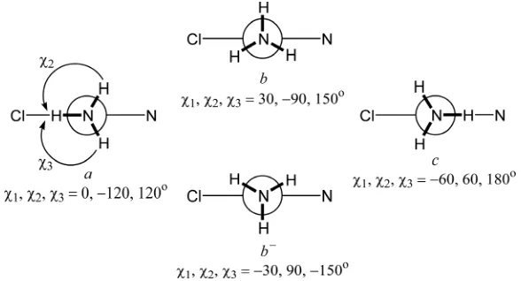
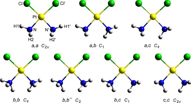
Initial conformations and their minimization. Four con- formations (a, b, b– and c) differing in the torsion angles
H-N-Pt-Cl around one of the N-Pt bonds were considered (Figure 1). Their combination in couples gives seven
non-eqivalent and non-enantiomorphic conformations as depicted in Figure 2. Each of these starting conforma-
tions were subjected to DFT minimization and all of them were found to converge to the a,a comformer (C2v simmetry). The calculated molecular geometry was com- pared with the single crystal X-ray and neutron powder diffraction data of Weller et al. [31] for the two poly-morphic modifications of cisplatin.
All calculations concern a single molecule in gas phase. Input and output files are deposited as Supplementary material.
3. Results and Discussion
3.1. Conformational Considerations
The initial structures of cisplatin (Figure 2) were
mini-mized with the two protocols: PW91/LANL08/3-21G (A) and PW91/LANL08/3-21G* (B) (vide supra). All of
[image:2.595.151.445.551.710.2]them converged to the a,a conformer (Figure 2), with
energies (corrected with the vibrational zero-point energy) of –1147.5051 and –1147.9213 Hartree, respectively. Thus, within the given level of theory, we found a single minimum on the potential energy surface of the cisplatin molecule. Amado et al. [23,24] have pointed out that the number of local minima (1, 2 or 3) varies with the com-bination of density functional and basis sets. In all cases, however, including all electron basis set DFT calculations [26], the lowest energy conformation is of the a,a type.
Methods A and B give the same set of torsion angles around the N-Pt bonds (0, –117, 117). The calculated torsion angles are compared (Table 1) with the
experi-mental data of Weller et al. [31] about the α- and β- polymorphs of cisplatin, the later containing two crystal-lographically non-equivalent molecules. As seen, in the solid state the torsion angles vary considerably, and no one conformation resembles enough the gas-phase minimum-
energy conformation of a,a type. This is a consequence of the extended intermolecular H-bonding network of N-HCl type [31]. Thus, it seems in the solid state the conformational preferences are governed by the H-bond- ing rather than by the torsion potential.
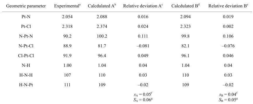
3.2. Bond Lengths and Bond Angles
The bond lengths and bond angles calculated by compu-tational methods A and B are compared with the ave- raged structural data for the two crystal modifications of cisplatin [31]. The relative deviations of the individual structural parameters, as well as the arithmetic mean (s) and root mean square deviations (S) are summarized in
Table 2. The accuracy obtained by the chosen theory
[image:3.595.130.464.320.501.2]levels is similar to or slightly better than that reported by Amado et al. [23,24]. Adding d-orbitals on the Cl atoms (method B) gives somewhat better overall description of the bond lengths and angles. The application of method
[image:3.595.135.463.556.715.2]Figure 2. View of the seven non-equivalent and non-enantiomorphic conformations of cisplatin arising from the combination of the conformations a, b, b and c in couples. Each of them was subjected to DFT minimization.
Table 1. Torsion angles (deg) for cisplatin.
Experimentala
β-Polymorph Torsion angle
α-Poly-morph
Molec. a Molec. b
Calcd. Ab Calcd. Bc
H1NPtCl 20.3 39.8 –37.5 0.0 0.0
H2NPtCl –101.2 –98.2 81.9 –117.1 –117.1
H3NPtCl 130.4 142.5 –153.0 117.1 117.1
H1’N’PtCl’ –39.5 –9.6 16.8 0.0 0.0
H2’N’PtCl’ 86.1 112.2 –106.2 117.1 117.1
H3’N’PtCl’ –154.7 –131.7 124.2 –117.1 –117.1
aData from [31]; bValues calculated at PW91/LANL08/3-21G level; cValues calculated at PW91/LANL08/3-
Table 2. Bond lengths (Å) and bond angles (deg) for cisplatin.
Geometric parameter Experimentala Calcdulated Ab Relative deviation Ac Calcdulated Bd Relative deviation Be
Pt-N 2.054 2.088 0.016 2.094 0.019
Pt-Cl 2.318 2.374 0.024 2.323 0.002
N
H
s f
SA = 0.06g
s f
SB = 0.05g
-Pt-N 90.2 100.2 0.111 99.8 0.106
N-Pt-Cl 88.9 81.7 –0.081 82.1 –0.076
Cl-Pt-Cl 91.9 96.4 0.049 96.1 0.046
N-H 1.00 1.04 0.04 1.04 0.04
-N-H 107 110 0.03 110 0.03
H-N-Pt 111 109 –0.02 109 –0.02
A = 0.05 B = 0.04
aAveraged da or α- and β-polymorphs of cisplatin [31] bAveraged calc at PW91/LANL /3-21G level (a
cl e
devia-tion (δa) of the calculated (ta f a ) from the experimental (; a ) value: ulated valuesδa = (a – a )/a ; dAveraged calculated values at PW91/ 08 dA); cRelativ
A cldA exp A cldA exp cldA
LANL08/3-21G* level (a
cldB); eRelative deviation (δaB) of the calculated (acldB) from the experimental (aexp) value: δaB = (acldB – aexp)/acldB;
fArithmetic mean of the relative deviations’ modules:
1
1 N
A Ai
i
s a
N
, 1
1 N
B Bi
i
s a
N
, N = number of geometric parametes; gRoot mean
square of the relative deviations:
1 2
N
2
1
1
A A
i
S a
N
i ,1 2 2
1
1 N
B B
i
S a
N
i .results in Pt-Cl bond length very close to the
experi-ational Analysis
rations of a single cisplatin
however, a factor group analysis of the number and B
mental (2.323 vs. 2.318 Å, respectively), but worsens the results for the Pt-N bond as compared to method A. Both protocols overestimate the values of the angles N-Pt-N and N-Pt-Cl, and underestimating the N-Pt-Cl value with respect to the experimental data [31], the discrepancy being largest for N-Pt-N (ca. 10˚). The same trend has been noticed in the HF [23] and DFT [23-25] results with other ECP basis sets, and even when all-electron bases were applied [26]. These results can be explained by the existence of intramolecular interactions (H-bonds) Cl
H-N which open the angles N-Pt-Nand Cl-Pt-Cl [23-26]. Indeed, the distances ClH calculated by the A and B schemes are short: 2.354 and 2.349 Å, respectively. In the solid state it seems, however, that the intermolecular H-bonding network [31] leads to conformational changes which diminish the effect of the intramolecular ClH-N contacts.
3.3. Vibr
The number of the normal vib
molecule is 27, distributed in C2v point group as follows:
9A1 + 5A2 + 8B1 + 5B2. All of them are Raman active
vibrations. The assignments of the vibrational spectra of cisplatin available in the literature [22-25], are made un-der the approximation of a single molecule, and the ef-fects of the solid state have not been considered. Because of comparability considerations, here we also hold to this approach. In Table A of the Supplementary material,
symmetry of the expected vibrational modes in the crys-talline state is given.
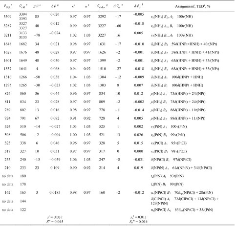
In Tables 3 and 4, the harmonic vibrational modes of
cisplatin calculated by methods A and B, respectively are compared with the experimental Raman sprecrtroscopic data of Amado et al. [24]. The coorespondence between the theoretical and experimental wave number is good even without scaling, the r. m. s. of the relative deviations (S) being 4.5%. Following the approach of other workers [22-25], the vibrational modes were divided in groups and the wavenumbers scaled. The individual scaling fac-tors (ν̃exp/ν̃cld) within each group were averaged to obtain
the group scaling factor σ (see the footnotes d-f to Table 3). For both protocols A and B, we have applied
group-ing into three sets with the correspondgroup-ing scalgroup-ing factors as follows: 1) δa(NH3) B1, ρ(NH3) A2 and δ(N-P-tN) – σ1
= 0.92; 2) νa(NH3), δa(NH3) A1, δa(NH3) A2, δs(NH3),
ρ(NH3) A1, ρ(NH3) B2, ρ(NH3) B1, ν(Pt-Cl) and πs(Pt) – σ2
= 0.97; 3) νs(NH3), δs(NH3), ν(Pt-Ν) and δ(N-Pt-Cl) –σ3
= 1.03. The scaled wave numbers fit closely to the ex-perimental data as indicated from the arithmetic mean (s) and the r. m. s. (S) of the relative deviations: 1.1% and 1.4%, respectively (Tables 3 and 4). The former was
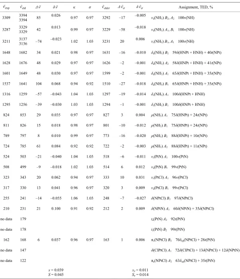
Table 3. Calculated (PW91/LANL08/3-21G) normal vibrational modes (wavenumbers , cm–1) of cisplatin molecule, com-pared with the experimental data.
exp a
cld
b Δc δd κe σf
cld
g Δ
h δ
i Assignmentj, TEDk, %
3309 3394 3393 85 0.026 0.97 0.97 3292 –17 –0.005 νa(NH3) B2, A2 100ν(NH)
3287 3327 0 0 9 3227 .018 νa(NH3) A1, B1
H) + 40t(NPt)
H) + 41t(NPt)
–30
–15 l)
34δ(NPtCl)
no data
no data
0.0185 0. 0. 160 –2 –0. 2 PtCl) + 28t(PtN)
A tCl) + 13δ(NPtCl) +
no data
sl =
Sm = 0.045 sσ
l = 0.011
Sσm = 0.014 3327
3133
4 0.012
–0.024
.9 0.97 –60 –0
0.005
100ν(NH)
3211 3133 –78 1.02 1.03 3227 16 νs(NH3) B1, A1 100ν(NH)
1648 1682 34 0.021 0.98 0.97 1631 –17 –0. 0 01 δa(NH3) B2 59δ(HNPt+HN
1628 1676 48 0.0 9 2 0.97 0.97 1626 –2 –0. 01 0 δa(NH3) A2 58δ(HNPt + HN
1601 1649 48 0.030 0.97 0.97 1599 –2 –0.001 δa(NH3) A1 65δ(HNPt + HNH) + 35t(NPt)
1537 1641 4 0.068 0.94 0.92 1510 –27 –0.018 δa(NH3) B1 65δ(HNPt + HNH) + 35t(NPt)
1316 1266 –50 0.038 1.04 1.03 1304 –12 –0.009 δs(NH3) A1 100δ(HNPt + HNH)
1295 1265 –0.023 1.02 1.03 1303 8 0.007 δs(NH3) B1 100δ(HNPt + HNH)
824 860 36 0.044 0.96 0.97 834 10 0.012 ρ(NH3) A1 75δ(HNPt) + 24t(NPt)
811 834 23 0.028 0.97 0.97 809 –2 –0.002 ρ(NH3) B1 75δ(HNPt) + 24t(NPt)
789 802 13 0.016 0.98 0.97 778 –11 –0.014 ρ(NH3) B2 88δ(HNPt) + 10t(NPt)
724 791 67 0.092 0.91 0.92 728 4 0.005 ρ(NH3) A2 88δ(HNPt) + 11t(NPt)
524 510 –14 –0.027 1.03 1.03 525 1 0.002 νs(PtΝ) A1 100ν(PtΝ)
508 506 –2 –0.004 1.00 1.03 521 13 0.026 νa(PtΝ) B1 99ν(PtΝ)
323 338 6 0.046 0.96 0.97 328 5 0.015 νs(PtCl) A1 95ν(PtCl)
317 327 10 0.031 0.97 0.97 317 0 0.000 νa(PtCl) B1 98ν(PtCl)
255 240 –0.059 1.06 1.03 247 –8 –0.031 δ(NPtCl) B1 97δ(NPtC
210 233 23 0.109 0.90 0.92 214 4 0.019 δ(NPtΝ) A1 61δ(NPtΝ) +
180 ta(PtN) A2 93t(PtN)
178 ts(PtN) B2 99t(PtN)
162 165 3 98 97 01 πs(NPtCl) B2 70δop(N
no data 144 δ(ClPtCl) 1 72δ(ClP
12δ(NPtN)
122 πa(NPtCl) A2 63δop(NPtCl) + 35t(PtN)
0.037
aExperimental Raman wavenumbers [24]; bTheo tical va es; re lu cDeviation of t e theoreticalh values from the experimental ones:
cld exp
; dRelative
devia on of thti e theoret al vaic lues from the experimental ones: δ
cldexp
exp; Individue al scaling factor: expcld; fGroup scaling factor:1
1
i ij
j
N
i n
,where ni is the number of the individual scaling factors in the i-th group. Τhus σ1 = 0.92 for δa(NH3) B1, ρ(NH3) A2 = 0.97 forνa(NH3), δa(NH3) A1, δa(NH3) A2, δs(NH3), ρ(NH3) A1, ρ(NH3) B2, ρ nd πs(NPtCl); σ3 = 1.03 for νs(N ), ν(PtΝ) and δ(NPtCl);
caled values; hDeviation of the theoretical scaled values from the experimental ones:
cld exp
and δ(NPtN); σ2
(NH3
(NH3) B1, ν(PtCl) a H3), δs
gTheoretical s ; iRelative deviation of the theoretical
scaled values from the experimental ones:
cldexp
exp; jNotations for the modes: ν—s ng, ρ—roc nebend-l he relative deviations’ m
tretching, δ—bendi king, π—out-of-pla ing, t— torsion; The indices s and as stand for symmetric and asymmetric respectively, and the index op denotes out-of-plane; kTotal energy distribution over
internal coordinates; Arithmetic mean of t odules:
1
1 Ν i
s
N
;1
1 Ν i
s N
; N = 27 is the number of vibrational modes;mRoot mean square of the relative deviations:
1 2
n
2
1
1 i
S
N
,
1 2
n
2
1
1 i
S N
Table 4. Calculated (PW91/LANL083-21G*) normal vibrational modes ( avenumbers w , cm ) of cisplatin molecule, com-pared with the experimental dataa.
exp
–1
cld Δ δ κ σ cld Δ δ Assignment, TED, %
3309 3394 3394 85 0.026 0.97 0.97 3292 –17 –0.005 νa(NH3) B2, A2 100ν(NH)
3287 3329 3329 42 0.013 0.99 0.97 3229 –58 –0.018 νa(NH3) A1, B1 100ν(NH)
3211 3137 –74 –0.023 1.02 1.03 3231 20 006 ν(NH ) B, A1 100ν(NH)
1648 1682 34 0.98 0.97 1631 –16 δa(NH3) B2 59δ(HNPt + HNH) + 40t(NPt)
1628 1676 48 0. 0.97 0.97 1626 –2 –0. 1 δa(NH3) A2 58δ(HNPt + HNH) + 41t(NPt)
0. –0. 1 a 3 1 ) + 35t(NPt)
l)
35δ(NPtCl)
no data
no data
tCl) + 13δ(NPtCl) + 12δ(NPtN) 3136
0.
s 3 1
0.021 –0.010
029 00
1601 1649 48 030 0.97 0.97 1599 –2 00 δ(NH ) A 65δ(HNPt + HNH
1537 1641 104 0.068 0.94 0.92 1510 –27 –0.018 δa(NH3) B1 65δ(HNPt + HNH) + 35t(NPt)
1316 1259 –57 –0.043 1.04 1.03 1297 –19 –0.014 δs(NH3) A1 100δ(HNPt + HNH)
1295 1256 –39 –0.030 1.03 1.03 1294 –1 –0.001 δs(NH3) B1 100δ(HNPt + HNH)
824 853 29 0.035 0.97 0.97 827 3 0.004 ρ(NH3) A1 75δ(HNPt) + 24t(NPt)
811 826 15 0.018 0.98 0.97 801 –10 –0.012 ρ(NH3) B1 75δ(HNPt) + 24t(NPt)
789 797 8 0.010 0.99 0.97 773 –16 –0.020 ρ(NH3) B2 88δ(HNPt) + 10t(NPt)
724 785 61 0.084 0.92 0.92 722 –2 –0.003 ρ(NH3) A2 88δ(HNPt) + 11t(NPt)
524 503 –21 –0.040 1.04 1.03 518 –6 –0.011 νs(PtΝ) A1 100ν(PtΝ)
508 499 –9 –0.018 1.02 1.03 514 6 0.012 νa(PtΝ) B1 99ν(PtΝ)
323 343 20 0.062 0.94 0.97 333 10 0.031 νs(PtCl) A1 96ν(PtCl)
317 330 13 0.041 0.96 0.97 320 3 0.009 νa(PtCl) B1 99ν(PtCl)
255 241 –14 –0.055 1.06 1.03 248 –7 –0.027 δ(NPtCl) B1 97δ(NPtC
210 231 21 0.100 0.91 0.92 212 2 0.009 δ(NPtΝ) A1 60δ(NPtΝ) +
179 ta(PtN) A2 92t(PtN)
178 ts(PtN) B2 99t(PtN)
162 168 6 0.037 0.96 0.97 163 1 0.006 πs(ΝPtCl) B2 70δop(NPtCl) + 28t(PtN)
no data 147 δ(ClPtCl) A1 72δ(ClP
no data 122 πa(NPtCl) A2 63δop(NPtCl) + 35t(PtN)
s S = 0.045
sσ
Sσ = 0.014
= 0.039 = 0.011
aThe notations are the same as in Table 3.
δ(N-Pt-Cl) and δ(N-Pt-N) modes as compared to the data of Amado et al. [23,24], orm ith th earlie -signments of Pavankum . [21]. The other dis-(N-Pt-Cl) and δ(Cl-Pt-Cl)
modes: our results correlate the experimental band at 162
cm– th π
s(N-Pt-Cl), whereas δ(Cl-Pt-Cl) vi-bra ld be expected at ca. 140 cm–1 (
Tables 3 and 4).
and conf w e r as ar et al
crepany concerns the order of πs
1 [23,24] wi
As concerns the two protocols, adding d-orbitals to Cl later, judging by the force constants given in Table 5.
vi-br
4. Conclusion and Perspective
lving the PW91 atoms (protocol B), slightly worsens the fit to the
ex-perimental wavenumbers, especially with respect to the
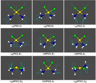
ν(Pt-N) and ν(Pt-Cl) vibrations. The shape of the skeletal vibrations of cisplatin molecule is visualized in Figure 3.
Selected harmonic force constants for cisplatim molecule are presented in Table 5. In agreement with the finding
that adding d-orbitals to the Cl atoms shortens the bond Pt-Cl and lengthens the bond Pt-N (cf. Table 2), it is
seen that it also strengthens the former and weakens the
Typically the tasks on geometry optimization and ational analysis take totally 15 min on a PC.
The DFT computational protocol invo
exchange-correlation functional, the relativistic ECP and associated non-contracted basis set LANL08 for descrip-tion of the Pt atom and 3-21G or 3-21G* basis sets for the remaining atoms, gives satisfactory results for the
[image:7.595.130.464.219.510.2]Figure 3. View of the nine skeletal vibrations of cisplatin molecule with their symmetry. The arrows represent the displace-ment vectors. The vibrational modes were visualized with the aid of Molekel programme using the Firefly output.
Table 5. Selected harmonic force constantsa for cisplatin.
Internal coordinate Force constant, Ab Force constant, Bc
Pt-N stretching 2.473 2.409
Pt-Cl stretching 1.950 2.010
Cl-Pt-Cl in-plane bending 0.627 0.628
N-Pt-N/N-Pt-C 08
N-Pt- nal
l’ off-diagonal
ane bending
N-Pt-N in-plane bending 0.638 0.634
N-Pt-Cl in-plane bending 1.138 1.134
l off-diagonal –0.213 –0.2
N/Cl-Pt-Cl off-diago –0.211 –0.217
N-Pt-Cl/N’-Pt-C –0.715 –0.720
N-Pt-Cl out-of-pl 0.021 0.021
aU hing and mdyn·Å·rad e bending and off-diagonal nding force
constanits: mdyn·Å 21G level; cPW91/LA -21G* level. –1 for the stretc
nts; b
PW91/LANL08/3-–2 for th
NL08/3
[image:7.595.137.461.562.713.2]equilibrium geomet ule. The calcu-lated wave numbers ns of the
mole-cule are in very go perimental
data. The procedure ood
compro-mise between accu resources. I
will be used for DF inum com
plexes that are of i ents,
and in particular to our laboratory
NCES
079-6603(01)67026-0
ry of cisplatin molec of the normal vibratio
od agreement with the ex used seems to be a g
racy and computational t T modelling of other plat -nterest as potential cytostatic ag wards some of the newly synthesized Pt(II) [53] and Pt(IV) complexes with in
sulfonamide ligands.
5. Acknowledgements
Thanks are due to Professor Alex Granovsky for kindly providing us with the Firefly programme. The work is part of the FP7 HP-SEE Project (Contract No RI-261499) and of the bilateral cooperation between the Bulgarian Academy of Sciences and the Aristotle University of Thessaloniki (2012-14).
REFERE
[1] B. Lippert “Chemistry and Biochemistry of a Leading Anticancer Drug,” Verlag Helvetica Chimica Acta, Zürich, 1999.
[2] S. M. Cohen and S. J. Lippard, “Cisplatin: From DNA Damage to Cancer Chemotherapy,” Progress in Nucleic
Acids Research and Molecular Biology, Vol. 67, 2001, pp.
93-130. doi:10.1016/S0
[3] R. A. Alderden, M. D. Hall and T. W. Hambley, “The
Discovery an latin,” Journal of
Chemical Edu 06, pp. 728-734.
d Development of Cisp
cation, Vol. 83, No. 5, 20 doi:10.1021/ed083p728
[4] A.-M. Florea and D. Büsselberg, “Cisplatin as an Anti- Tumor Drug: Cellular Mechanisms of Activity, Drug Re-sistance and Induced Side Effects,” Cancers, Vol. 3, 2011, pp. 1351-1371. doi:10.3390/cancers3011351
[5] T. Boulikas and M. Vougiouka, “Cisplatin and Platinum Drugs at the Molecular Level,” Oncology Reports, V
emy of Sciences of the USA
ol. 10, No. 6, 2003, pp. 1663-1682.
[6] J. Reedijk, “New Cues for Patinum Atitumor Chemistry: Kinetically Controlled Metal Binding to DNA,”
Pro-ceedings of the National Acad ,
Vol.100, No. 7, 2003, pp. 3611-3616. doi:10.1073/pnas.0737293100
[7] I. Kostova, “Platinum Complexes as Anticancer Agents,”
Recent Patents on Anti-Cancer Drug Discovery, Vol. 1,
istry to a Post-Genomic No. 1, 2006, pp. 1-22.
[8] M. J. Hannon, “Metal-Based Anticancer Drugs: From a Past Anchored in Platinum Chem
Future of Diverse Chemistry and Biology,” Pure and Ap-plied Chemistry, Vol. 79, No. 12, 2007, pp. 2243-2261. doi:10.1351/pac200779122243
[9] T. Boulikas, A. Pantos, E. Bellis and P. signing Platinum Compounds
Christofis, “De-in Cancer: Structures and
0] X. Wang and Z. Guo, “Towards the Rational Design of Platinum(II) and Gold(III) Complexes as Antitumour Agents,” Dalton Transactions, No. 12, 2008, pp. 1521-32. doi:10.1039/B715
Mechanisms,” Cancer Therapy, Vol. 5, 2007, pp. 537- 583.
[1
903J
[11] J. Reedijk, “Platinu ancer Coordination Compounds: Study of DNA Bindi Inspires New Drug Design,”
European Journal ganic Chemistry, Vol. 2009, No,
10, 2009, pp. 1303-1312. doi:10.1002/ejic.200900054 m Antic
ng
of Inor
of Platinum Anti-Chemistry,” Pure and Applied Chemistry, Vol. 83,
719.
[12] J. Reedijk, “Increased Understanding cancer
No. 9, 2011, pp. 1709-1
doi:10.1351/PAC-CON-10-11-03
[13] I. Kostova, “Gold Coordination Complexes as Anticancer Agents,” Anti-Cancer Agents in Medicinal Chemistry, Vol. 6, No. 1, 2006, pp. 19-32.
[14] I. Ott and R. Gust, “Non Platinum Metal Complexes as Anti-Cancer Drugs,” Archiv der Pharmazie, Vol. 340, No. 3, 2007, pp. 117-26. doi:10.1002/ardp.200600151 [15] E. R. T. Tiekink, “Anti-Cancer Potential of Gold
Com-plexes,” Inflammopharmacology, Vol. 16, No. 3, 2008, pp. 138-142. doi:10.1007/s10787-007-0018-5
[16] L. Cattaruzza, D. Fregona, M. Mongiat, L. Ronconi, A. Fassina, A. Colombatti and D. Aldinucci, “Antitumor Ac-tivity of Gold(III)-Dithiocarbamato Derivatives on Pros-tate Cancer Cells and Xenografts,” International Journal of Cancer, Vol. 128, No. 1, 2011, pp. 206-215.
doi:10.1002/ijc.25311
[17] N. J. Wheate, S. Walker, G. E. Craig and R. Oun, “The Status of Platinum Anticancer Drugs in the Clinic and Clinical Trials,” Dalton Transactions, Vol. 39, No. 35, 2010, pp. 8113-8127. doi:10.1039/C0DT00292E
[18] C. W. Helm and J. C. States “Enhancing the Efficacy of Cisplatin in Ovarian Cancer Treatment—Could Arsenic Have a Role,” Journal of Ovarian Research, Vol. 2, No. 2, 2009, pp. 1-7. doi:10.1186/1757-2215-2-2
[19] S. Usanova, A. Piée-Staffa, U. Sied, J. Thomale, A. Schneider, B. Kaina and B. Köberle, “Cisplatin Sen- sitivity of Testis Tumour Cells Is Due to Deficiency in Interstrand-Crosslink Repair and Low ERCC1-XPF Ex-pression,” Molecular Cancer, Vol. 9, No. 248, 2010, pp. 1-11. doi:10.1186/1476-4598-9-248
M. Dolg, “Effective Core Potentials,” In: J. G
[20] rotendorst,
Ed., Modern Methods and Algorithms of Quantum Che-
mistry, John von Neumann Institute for Computing,
Jülich, 2000, pp. 507-540.
[21] P. N. V. Pavankumar, P. Seetharamulu, S. Yao, J. D. Saxe, D. G. Reddy and F. H. Hausheer, “Comp
ab Initio Quantum M
rehensive echanical and Molecular Orbital (MO) Analysis of Cisplatin: Structure, Bonding, Charge Density, and Vibrational Frequencies,” Journal of Com-putational Chemistry, Vol. 20, No. 3, 1999, pp. 365-382. doi:10.1002/(SICI)1096-987X(199902)20:3<365::AID-J CC8>3.0.CO;2-1
[22] R. Wysokiński and D. Michalska, “The Performance of Different Density Functional Methods in the Calculation of Molecular Structures and Vibrational Spectra of Plati-num(II) Antitumor Drugs: Cisplatin and Carboplatin,”
Journal of Computational Chemistry, Vol. 22, No. 9,
[23] A. M. Amado, S. M. Fiuza M. P. M. Marques and L. A. E. Batista de Carvalho, “Conformational and Vibrational Study of Platinum(II) Anticancer Drugs cis-Diammine- dichloroplatinum(II) as a Case Study,” The Journal of
Chemical Physics, Vol. 127, No. 18, 2007, Article ID:
185104. doi:10.1063/1.2787528
[24] S. M. Fiuza, A. M. Amado, M. P. M. Marques and L. A. E. Batista de Carvalho, “Use of Effective Core Potential Calculations for the Conformational and Vibrational Study of Platinum(II) Anticancer Drugs. cis-Diammine- dichloroplatinum(II) as a Case Study,” The Journal of
Physical Chemistry, Vol. 112, No. 14, 2008, pp. 3253-
3259. doi:10.1021/jp710868p
[25] H. Gao, F. Y. Xia, C. J. Huang and K. Linc, “Density Functional Theory Calculations on the Molecular Struc -tures and Vibration Spectra of Platinum(II) Antitumor Drugs,” Spectrochimica Acta Part A: Molecular and
Biomolecular Spectroscopy, Vol. 78, No. 4, 2011, pp.
1234-1239. doi:10.1016/j.saa.2010.12.003
[26] R. C. de Berrêdo and F. E. Jorge, “All-Electron Double Zeta Basis Sets for Platinum: Estimating Scalar Relativis-tic Effects on Platinum(II) AnRelativis-ticancer Drugs,” Journal of
Molecular Structure: THEOCHEM, Vol., 961, No. 1-3,
2010, pp. 107-112. doi:10.1016/j.theochem.2010.09.007 [27] K. Nakamoto, P. J. McCarthy, J. Fujita, R. A. Condrate
and G. T. Behnke, “Infrared Studies of Ligand-Ligan In-teraction in Dihalogenodiammineplatinum(II) Complexes,”
Inorganic Chemistry, Vol. 4, No. 1, 1965, pp. 36-43. doi:10.1021/ic50023a008
[28] G. Raudaschl, B. Lippert, J. D. Hoeschele, H. E. How-ard-Lock, C. J. L. Lock and P. Pilon, “Adduct Formation of cis-(NH3)2PtX2 (X = Cl–, I–) with Formamides and the Crystal Structures of cis-(NH3)2PtCl2(CH3)2NCHO. Ap-plication for the Purification of the Antitumor Agent Cis-platin,” Inorganica Chimica Acta, Vol. 106, No. 3, 1985, pp. 141-149. doi:10.1016/S0020-1693(00)87550-7 [29] I. A. Degen and A. J. Rowlands, “The Fourier Transform
Raman Spectra of a Series of Platinum(II), Palladium(II) and Gold(III) Square-Planar Complexes,” Spectrochimica
Acta Part A: Molecular and Biomolecular Spectroscopy,
Vol. 47, No. 9-10, 1991, pp. 1263-1268. doi:10.1016/0584-8539(91)80213-3
[30] G. H. W. Milburn and M. R. Truter, “The Crystal Struc- tures of cis- and trans-Dichlorodiammineplatinum(II),”
Journal of the Chemical SocietyA, 1966, pp. 1609-1616. doi:10.1039/J19660001609
[31] V. P. Ting, M. Schmidman, C. C. Wilson and M. T. Weller, “Cisplatin: Polymorphism and Structural Insight into an Important Chemotherapetic Drug,” Angewan Chemie, International Edit
dte ion, Vol. 49, No. 49, 2011, pp. 9408-9411. doi: 10.1002/anie.201003185
[32] C. Adamo and V. Barone, “Exchange Functionals with Improved Long-Range Behavior and Adiabatic Connec-tion Methods without Adjustable Parameters: The mPW and mPW1PW Models,” The Journal of Chemical Phy- sics, Vol. 108, No. 2, 1998, pp. 664-675.
doi:10.1063/1.475428
[33] T. H. Dunning and P. J. Hay, “Gaussian Basis Sets for Molecular Calculations,” In: H. F. Schaefer, Ed., Methods
in Electronic Structure Theory (Modern Theoretical Che-
mistry), Plenum Press, New York, 1977, pp. 1-28.
[34] P. J. Hay and W. R. Wadt, “Ab Initio Effe tentials for Molecular Calculations
ctive Core Po-. Potentials for the Transition Metal Atoms Sc to Hg,” The Journal of Che- mical Physics, Vol. 82, No. 1, 1985, pp. 270-283. doi:10.1063/1.448799
[35] W. R. Wadt and P. J. Hay, “Ab Initio Effective Core Po-tentials for Molecular Calculations. PoPo-tentials for Main Group Elements Na to Bi,” The Journal of Chemical Physics, Vol. 82, No. 1, 1985, pp. 284-298.
doi:10.1063/1.448800
[36] P. J. Hay and W. R. Wadt, “Ab Initio Effective Core Potentials for Molecular Calculations. Potentials for K to Au Including the Outermost Core Orbitals,” The Journal of Chemical Physics, Vol. 82, No. 1, 1985, pp. 299-310. doi:10.1063/1.448975
[37] W. J. Hehre, R. F. Stewart and J. A. Po sistent Molecular Orbi
ple, “Self-Con- tal Methods. I. Use of Gaussian Expansions of Slater—Type Atomic Orbitals,” The Jour-
nal of Chemical Physics,Vol. 51 No. 6, 1969, pp. 2657-
2664. doi:10.1063/1.1672392
[38] J. S. Binkley, J. A. Pople and W. J. Hehre, “Self-Consis- tent Molecular Orbital Methods. 21. Small Split-Valence Basis Sets for First-Row Elements,” Journal of the Ame-
rican Chemical Society, Vol. 102, No. 3, 1980, pp. 939-
947. doi:10.1021/ja00523a008
[39] M. S. Gordon, J. S. Binkley, J. A. Pople, W. J. Pietro and W. J. Hehre, “Self-Consistent Molecular-Orbital Methods. 22. Small Split-Valence Basis Sets for Second-Row Ele-ments,” Journal of the American Chemical Society, Vol. 104, No. 10, 1982, pp. 2797-2803.
doi:10.1021/ja00374a017
[40] S. H. Vosko, L. Wilk and M. Nusair, “Accurate Spin- Dependent Electron Liquid Correlation Energies for Lo-cal Spin Density Calculations: A CritiLo-cal Analysis,”
Ca-nadian Journal of Physics, Vol. 58, No. 8, 1980, pp.
1200-1211. doi:10.1139/p80-159
[41] D. Andrae, U. Häussermann, M. Dolg, H. Stoll and H. Preuss, “Energy-Adjusted ab Initio Pseudopotentials for the 2nd and 3rd Row Transition-Elements,” Theoretical Chemistry Accounts, Vol. 77, No. 2, 1990, pp. 123-141. doi:10.1007/BF01114537
[42] J. P. Perdew, K. Burke and M. Ernzerhof, “Generalized Gradient Approximation Made Simple,” Physical Re- views Letters, Vol. 77, No. 18, 1996, pp. 3865-3868. doi:10.1103/PhysRevLett.77.3865
[43] J. P. Perdew, J. A. Chevary, S. H. Vosko, K. A. Jackson, M. R. Pederson, D. J. Singh and C. Fiolhais, “Atoms, Molecules, Solids, and Surfaces: Applications of the Ge- neralized Gradient Approximation for Exchange and Cor- relation,” Physical Review B, Vol. 46, No. 11, 1992, pp. 6671-6687. doi:10.1103/PhysRevB.46.6671
[44] J. P. Perdew, K. Burke and Y. Wang, “Generalized Gra-dient Approximation for the Exchange-Correlation Hole of a Many-Electron System,” Physical Review B, Vol. 54, No. 23, 1996, pp. 16533-16539.
doi:10.1103/PhysRevB.54.16533
omic and Molecular Electronic http://classic.chem.msu.su/gran/firefly/index.html [46] M. W. Schmidt, K. K. Baldridge, J. A. Boatz, S. T. Elbert,
M. S. Gordon, J. H. Jensen, S. Koseki, N. Matsunaga, K. A. Nguyen, S. Su, T. L. Windus, M. Dupuis and J. A. Montgomery, “General At
Structure System,” Journal of Computational Chemistry, Vol. 14, No. 11, 1993, pp. 1347-1363.
doi:10.1002/jcc.540141112 [47] Molekel, 2009.
http://molekel.cscs.ch/wiki/pmwiki.php
[48] L. E. Roy, P. J. Hay and R. L. Martin, “Revised Basis Sets for the LANL Effective Core Potentials,” Journal of
Chemical Theory and Computations, Vol. 4, No. 7, 2008,
pp. 1029-1031. doi:10.1021/ct8000409
[49] W. J. Pietro, M. M. Francl, W. J. Hehre, D. J. DeFrees, J. A. Pople and J. S. Binkley, “Self-Consistent Molecular Orbital Methods. 24. Supplemented Small Split-Valence Basis Sets for Second-Row Elements,” Journal of the
American Chemical Society, Vol. 104, No. 19, 1982, pp.
5039-5048. doi:10.1021/ja00383a007 [50] EMSL Basis Set Exchange Library.
https://bse.pnl.gov/bse/portal
[51] D. Feller, “The Role of Databases in Support of Compu-tational Chemistry Calculations,” Journal of Computa-tional Chemistry, Vol. 17, No. 13, 1996, pp. 1571-1586. doi:10.1002/(SICI)1096-987X(199610)17:13<1571::AID -JCC9>3.0.CO;2-P
[52] K. L. Schuchardt, B. T. Didier, T. Elsethagen, L. Sun, V. Gurumoorthi, J. Chase, J. Li and T. L. Windus, “Basis Set Exchange: A Community Database for Computational
mation and
Model-52. Sciences,” Journal of Chemical Infor ing, Vol. 47, No. 3, 2007, pp. 1045-10 doi:10.1021/ci600510j
[53] N. I. Dodoff, M. Lalia-Kantouri, M. Gdaniec, A. Czapik, N. G. Vassilev, L. S. Markovaand M. D. Apostolova, “trans-Dichloro(η2-ethylene)(N-3-pyridinylmethanesulfo- namide)platinum(II). Crystal Structure, Spectroscopic, and Thermoanalytical Characterization, and Cytotoxicity Assays,” Journal of Coordination Chemistry, Vol. 65, No. 4, 2012, pp. 688-704.
doi:10.1080/00958972.2012.659729
[54] R. Beattie, “Vibrational Spectra of Solids,” In: A. J. Barnes and W. J. Orville-Thomas, Eds., Vibrational
Modern Trends, (Russian Edition), Mir, Moscow, 1981, pp. Spectroscopy.
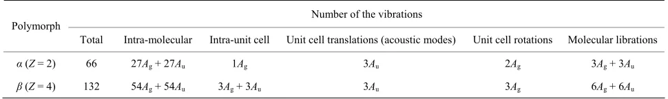
ppendix
actor Group Analysis of the Vibrations of rystalline Cisplatin
eller et al. [31] found that cisplatin exists in two poly-orphs: low-temperature α- and high-temperature β- odifications. The crystals of both forms are triclinic,
space group P1̃ with two and four asymmetric mole- cules for the α- and β-form, respectively. The relation be-tween the point, site and factor group symmetry for both crystal forms is: C1 C1 Ci, respectively. A factor
[image:10.595.55.540.566.638.2]group analysis of the number and symmetry of the vibra-tional modes [54] of the two polymorphs is given in Table A.
Table A. Factor group analysis for the crystals of α- and β-polymorphs of cisplatin.
Number
341-358.
A
F C
W m m
of the vibrations Polymorph
Total Intra-molecular Intra-unit cell Unit cell translations (acoustic modes) Unit cell rotations Molecular librations
α(Ζ= 2) 66 27Ag + 27Au 1Ag 3Au 2Ag 3Ag + 3Au