THE
*1
Dr. Angana Pal,
2Prof. Dr. Dipanjit Singh,
5
Dr. Tarun Kumar Swarnakar and
1,2, 3, 4,6
Department of Prosthodontics, Crown and Bridge and Implantology, Maharana Pratap College of
Dentistry and Research
5
Department of Paediatric and Preventive Dentistry, Burdwan Dental College and Hospital, Burdwan,
6
Department of Prosthodontics, Crown and Bridge and Implantology, Kothiwal Dental College and
ARTICLE INFO ABSTRACT
Immediate Implant placement is becoming a popular
dentistry. The main challenge for the clinician has been the preservation of the residual alveolar bone and prevention of its resorption following the removal of tooth.
represents
time of immediate implant placement to preserve
review discusses all the literatures available on the socket prognosis and biological acceptability
Copyright © 2018, Angana Pal et al. This is an open access
distribution, and reproduction in any medium, provided
INTRODUCTION
The residual alveolar bone undergoes a continuous resorption process following removal of tooth (Amler et al.,
and Lindhe, 2005; Fickl et al., 2008; Pietrokovski
1967; Schropp et al., 2003).Preservation of roots of teeth have proved to be positive factor in retarding the resorption process (Hurzeler et al., 2010). Several research made before have shown that retaining healthy roots, vital or endodontically treated (Salama et al., 2007) preserves the residual alveolar bone from resorption (Filippi et al., 2001;
2003; Sapir and Shapira, 2008; Malmgren, 2000
of implants placed in contact or close to retained pieces of roots were also investigated (Buser et al., 1990;
1993; Gray and Vernino, 2004; Jahangiri et al.,
showed growth or regeneration of periodontal ligament and cementum on implant surfaces. In modern times where esthetics becoming the prime factor, attention and emphasis is being given in preserving the buccal bone while an implant is placed following the removal of a tooth. Therefore an innovative technique of placing implants in close contact with
*Corresponding author: Dr. Angana Pal,
Department of Prosthodontics, Crown and Bridge and Implantology, Maharana Pratap College of Dentistry and Research, Gwalior, India
ISSN: 0975-833X
Article History:
Received 07th December, 2017
Received in revised form 23rd January, 2018
Accepted 24th February, 2018
Published online 30th March, 2018
Citation: Dr. Angana Pal, Prof. Dr. Dipanjit Singh, Dr. Atul Bhandari, Dr. Pratibha Rawat, Dr. Tarun Kumar Swarnakar and Dr. Arka Swarn
2018. “The socket shield technique”, International Journal of Current Research
Key words:
Dental implant, socket shield technique, immediate placement, osseointegration.
REVIEW ARTICLE
THE SOCKET SHIELD TECHNIQUE
Prof. Dr. Dipanjit Singh,
3Dr. Atul Bhandari,
4Dr. Tarun Kumar Swarnakar and
6Dr. Arka Swarnakar
Department of Prosthodontics, Crown and Bridge and Implantology, Maharana Pratap College of
Dentistry and Research Centre, Gwalior, India
Department of Paediatric and Preventive Dentistry, Burdwan Dental College and Hospital, Burdwan,
India
Prosthodontics, Crown and Bridge and Implantology, Kothiwal Dental College and
Research Centre, Moradabad, India
ABSTRACT
Immediate Implant placement is becoming a popular treatment modality of modern day implant dentistry. The main challenge for the clinician has been the preservation of the residual alveolar bone and prevention of its resorption following the removal of tooth.
represents an alternative approach in which a thin section of the remnant root is retained facially at the time of immediate implant placement to preserve buccal periodontal ligament and bundle bone. This review discusses all the literatures available on the socket-shield technique and judge its clinical prognosis and biological acceptability.
access article distributed under the Creative Commons Attribution the original work is properly cited.
The residual alveolar bone undergoes a continuous resorption
et al., 1960; Araujo
Pietrokovski and Massler, .Preservation of roots of teeth have proved to be positive factor in retarding the resorption process earch made before have shown that retaining healthy roots, vital or endodontically preserves the residual alveolar 2001; Andersson et al.,
, 2000).The impact of implants placed in contact or close to retained pieces of 1990; Warrer et al.,
et al., 2005). Results
growth or regeneration of periodontal ligament and/or In modern times where esthetics becoming the prime factor, attention and emphasis is being given in preserving the buccal bone while an implant is placed following the removal of a tooth. Therefore an of placing implants in close contact with
Department of Prosthodontics, Crown and Bridge and Implantology, Maharana Pratap College of Dentistry and Research, Gwalior, India
planned retained roots was developed thus protecting the buccal bone from resorption (Hurzeler
al., 2015; Kan and Rungcharassaeng
2014; Cherel and Etienne, 2014; Hurzeler et al.coined this as the socket
(Hurzeler et al., 2010). In this the root is sectioned and the buccal piece is retained while the remaining root is removed. This retained buccal piece of root acts as a shield against the resorption. An immediate implan
palatal to this root fragment. Histological studies on animals has confirmed the formation of cementum on implant surfaces placed in contact with intentionally retained roots
al., 2010).Similar animal histologic stud
the formation of a fibrous capsule around implants
al., 2005). At present, all clinical human studies currently
available on implants placed in close proximity to intentionally retained root fragments using this technique are
hierarchy of evidence. Hence the purpose of this article review the available literature with regard to socket shield technique.
History
Esthetics has always been a determining factor in the success of any prosthodontic treatment plan.
International Journal of Current Research
Vol. 10, Issue, 03, pp.67200-67204, March, 2018
Dr. Angana Pal, Prof. Dr. Dipanjit Singh, Dr. Atul Bhandari, Dr. Pratibha Rawat, Dr. Tarun Kumar Swarnakar and Dr. Arka Swarn International Journal of Current Research, 10, (03), 67200-67204.
4
Dr. Pratibha Rawat,
Dr. Arka Swarnakar
Department of Prosthodontics, Crown and Bridge and Implantology, Maharana Pratap College of
Department of Paediatric and Preventive Dentistry, Burdwan Dental College and Hospital, Burdwan,
Prosthodontics, Crown and Bridge and Implantology, Kothiwal Dental College and
treatment modality of modern day implant dentistry. The main challenge for the clinician has been the preservation of the residual alveolar bone The socket-shield technique (SST) in which a thin section of the remnant root is retained facially at the buccal periodontal ligament and bundle bone. This hield technique and judge its clinical
License, which permits unrestricted use,
planned retained roots was developed thus protecting the Hurzeler et al., 2010; Baumer et
Rungcharassaeng, 2013; Siormpas et al.,
, 2014; Glocker et al., 2014). this as the socket-shield technique . In this the root is sectioned and the buccal piece is retained while the remaining root is removed. This retained buccal piece of root acts as a shield against the resorption. An immediate implant placement is carried out Histological studies on animals has confirmed the formation of cementum on implant surfaces placed in contact with intentionally retained roots (Hurzeler et
.Similar animal histologic study have demonstrated the formation of a fibrous capsule around implants (Parlar et
. At present, all clinical human studies currently available on implants placed in close proximity to intentionally retained root fragments using this technique are lower in the hierarchy of evidence. Hence the purpose of this article is to review the available literature with regard to socket shield
Esthetics has always been a determining factor in the success of any prosthodontic treatment plan.
INTERNATIONAL JOURNAL OF CURRENT RESEARCH
It is an established fact that deficiency of soft and hard tissues in the esthetic zone can interfere with optimal implant positioning and hamper the overall aesthetic outcome of implant-supported prostheses (Hurzeler et al., 2010). In order to overcome the negative consequences of tooth extraction, various treatment approaches such as immediate implants placement (Botticelli et al., 2004; Arau´jo et al., 2005) graft materials (Carmagnola et al., 2003; Nevins et al., 2006; Arau´jo et al., 2008; Fickl et al., 2008; Arau´jo etv al., 2009) and/or barrier membranes (Lekovic et al., 1997; Lekovic et al.,
1998) have been advocated and described in the literature. As a conclusion, the majority of the studies show that socket preservation is a suitable technique for socket augmentation with the ability to maintain the ridge dimension to a certain amount (Arau´jo et al., 2008; Fickl et al., 2008; Arau´jo etv al., 2009). However, a complete preservation and/or entire regeneration of the extraction socket have not been documented yet. The marked alterations after tooth extraction appear to be attributable to the loss of periodontal ligament and the consecutive trauma in particular at the buccal bone plate (Arau´jo and Lindhe, 2005). Thus, it can be assumed that root retention may have an influence on the occurring resorption process. Clinical studies have tested the hypothesis that root retention, either of vital or pulpless teeth, may be able to avoid tissue alterations after tooth extraction. Filippi et al. in his case report described that decoronation of an ankylosed tooth preserved the alveolar bone before implant placement (Filippi
et al., 2001). Few studies have demonstrated that the
preservation of decoronated roots in the alveolar process not only helps maintaining existing bone volume but also enables vertical bone growth, which can be observed coronally to the decoronated root (Malmgren et al., 1984 Andersson et al.,
2003). Bjo¨rn (1963) confirmed regeneration of alveolar bone around endodontic ally treated teeth that were submerged and covered by a surgical flap (Bjo¨rn, 1963).
Reames et al. (1975) demonstrated in an animal study that even though epithelium commonly occurred over the amputation sites of submerged teeth, bone formation coronal to the submerged roots was evident (Reames et al., 1975).O’Neal et al. (1978) showed histological and radiographic evidence that new cementum and connective tissue will form over the coronal surface of submerged roots separating the dentin from the new bone (Neal et al., 1978). Conclusively, histological and radiographic evidences suggest few inflammatory changes and bone apposition around roots that had been submerged for alveolar bone preservation. Bowers et al. (1989) submerged vital teeth with infrabony defects in nine patients and created notches at regions on the root that had been covered with dental calculus. After 6 months, no root resorption, ankyloses, or pulp death was observed (Bowers et al., 1989). Salama et al. (2007) reported that the Root Submergence Technique (RST) maintains the natural attachment apparatus of the tooth in the pontic site, which in turn allows for complete preservation of the alveolar bone frame and assists in the creation of an aesthetic result in adjacent multiple-tooth-replacement cases (Salama et al., 2007). Davarpanah and Szmukler-Moncler (2009) reported implant placement in contact with ankylosed root fragments in a five-case-report study without any specific pathological sign after a period of 12–42 months of loading (Davarpanah and Szmukler-Moncler, 2009). The “socket-shield technique” described by Hürzeler et al. (2010) ( Hurzeler et al., 2010), used the retained buccal root in an attempt to preserve the buccal bone and tissues, which is the mainly desired effect, after immediate implant placement
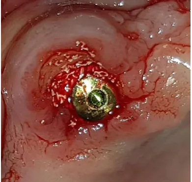
[image:2.595.342.524.248.416.2] [image:2.595.336.532.450.635.2](Figure 1). This approach allowed the buccal cortical bone to be successfully preserved after placement of the implant (Figure 2). Another modification of the socket-shield technique has been described by Baumer et al. which may offer a feasible treatment option to procedures using the socket-shield technique in vertically fractured teeth. The case report indicates that it may also be used without severe adverse events and that the desired effect of buccal maintenance might also be achieved in human tissues (Baumer et al., 2015).In 2014 Troiano et al. introduced the Root-T-Belt technique consists of placing the implant in the preserved tooth root, which will surround the implant entire circumference thereof tooth structure, formed by periodontium, dentin and cement will create a protective structure as a belt, which prevents any movement and maintains the peri-implant system structure (Troiano et al., 2014).
Figure 1: Preserved buccal root fragment after sectioning
Figure 2. Implant placement palatal to the buccal root fragment which inturns protect the buccal cortical bone
HISTOLOGICAL EVIDENCES
A fibrous capsule covered their surfaces and there was absence of any osseointegration. Cellular cementum was deposited on the surfaces of two out of nine implants as well as on the dentinal walls of the chamber. One implant had an exposed edge whereas two implants showed clinical signs of inflammation (Parlar et al., 2005). Hurzeler et al. intentionally left a buccal portion of the remnant root coated with enamel matrix derivative (Emdogain, Straumann), to preserve the buccal cortical plate from resorption during an immediate implant placement (Hurzeler et al., 2010).
They were the first to name this noble and innovative technique as ‘socket-shield’. Histological examination of 4 implants placed in a beagle dog demonstrated cementum formation on implant surface where a direct root-implant contact was noted. When the dental implant and the root piece were in close proximity with no physical contact, a 0.5 mm connective tissue band was found between the implant and the buccal root piece. They also presented a clinical case report using this technique wherein the implant was immediately loaded and followed up for 6 months. They justified the socket-shield technique as a viable option to preserve the buccal bone and achieve satisfactory esthetics with osseointegration and without any inflammatory or resorptive response. Baumer et al. further investigated this technique by employing a similar study design but with a larger sample size (Baumer et al., 2015). Their histologic evaluation showed osseointegration and bone formation between the fragments and the implants after 4 months of healing. They proposed that the socket-shield prevented the resorption of the buccal cortical plate after tooth extraction. Additionally, they also presented a clinical case report, which was followed up for a period of 6 months with no apparent adverse effects. A case-control study on the socket-shield was carried out by Abadzhiev et al. where 26 implants were immediately placed in 25 patients. Though the socket-shield group had better results in terms of bone loss, esthetics and soft tissue volume, a mean bone loss of 0.8mm (2%) was noted at 24 months (Abadzhiev and Velcheva, 2016).
Kan and Rungcharassaeng in 2013 carried out an immediate implant placement in a patient where the implant was in contact with the tooth fragment. The implant was immediately loaded and no adverse reaction was seen after 12 months (Kan and Rungcharassaeng, 2013).Chen and Pan in 2013 published their clinical case report in which they carried out an immediate implant placement in proximity to remaining tooth fragment and delayed loading was done after 4 months. They observed 0.72mm horizontal loss on buccal alveolar bone after 12 months (Chen and Pan, 2013). In 2014 Cherel and Etienne placed two immediate implants in the patient’s mouth followed by immediate loading. After 11 months when the temporary crowns were removed they noticed small coronal part of root fragment was visible through mucosal bed (Cherel et al.,
2014). Siormpas et al. in 2014 placed 46 immediate implants in 46 different patients without any contact with the retained root fragment. The implants were immediately loaded. They were observed over a period of 24-60 months. It was found that the mean crestal bone loss on the mesial side was 0.18±0.09 and on the palatal side it was 0.21±0.09. 1 case of apical root resorption was also reported (Siormpas et al., 2014). Glocker et al. in 2014 placed one implant each in three different patients after 6 months following delayed implant protocol in proximity to roots. No adverse reaction was recorded after 6 months of loading (Glocker et al., 2014).
Troiano et al. in 2014 placed 10 implants in 7 patients immediately and in contact with the retained root fragment. Loading was delayed by 3 months. An average bone loss of 1.3±0.2 mm was observed after 6 months of follow up (Troiano et al., 2014). In 2015 Al Dary and Al Hadadi (Al Dary and Al Hadidi, 2015) and Gluckman et al. (Gluckman et
al., 2015) separately carried out an immediate implant
placement in close proximity to the root fragment with immediate loading. No adverse reaction was recorded after 12 months of observation in both the studies. In 2015 Wadhwani et al. mentioned in his case report the immediate placement of an implant following socket shield procedure. Loading was delayed by 4 months no negative result after 4 months of follow up (Wadhwani et al., 2015).
DISCUSSION
In the past it has been observed that retaining root fragments in situ and keeping them covered by mucosa serves as an alternative technique for alveolar ridge preservation. Studies supported the fact that root fragments assisted in both the preservation of root volume as well as in vertical bone growth coronally. Thus, a planned preservation of root fragments appears to be an approach towards successful alveolar ridge preservation. Various recent studies have confirmed that the socket shield technique has the potential to reduce bone resorption after removal of tooth followed by immediate implantation, mainly through the retention of the buccal segment of the root (Hurzeler et al., 2010,16,44,47-49). The common factor in all these studies was immediate implant placement at the time of preparation of the socket-shields. But everyone had a different loading protocol and follow-up duration. Other modifications to the original technique were in terms of time of implant placement (Glocker et al., 2014), and location of the shield (Kan and Rungcharassaeng, 2013; Troiano et al., 2014). The studies which were conducted on humans were carried out with single implant placement in the anterior esthetic area with no periodontal pathology(Hurzeler
et al., 2010, Baumer et al., 2015, Siormpas et al., 2014;
Glocker et al., 2014; Abadzhiev and Velcheva , 2014; Kan and Rungcharassaeng, 2013; Chen and Pan Cherel and Etienne, 2014; Troiano et al., 2014; Al Dary and Al Hadidi , 2015; Gluckman et al., 2015) the human studies were carried out over a period of 12 months to five years which not sufficient to establish the success of this technique.
The requirement was a simple economical technique which can be carried out with minimum surgical intervention.Socket shield technique allowed us to preserve the bone at the proposed implant site, while the thin and prone to resorption buccal bundle bone was retained (Schropp et al., 2003; Arau´jo
et al., 2009). The lingual portions of the bundle bone are
But it is not the same every time. Warrer et al. showed that new cement is deposited on the aspect of the dentin shield facing the former socket. This cement layer should be regarded as a protection against resorption by osteoclasts (Warrer et al.,
1993). Periodontal membrane formation around the implant will occur when the implant-root interface has a loose structure and a larger gap is left (Hurzeler et al., 2010), and when the periodontal ligament of the root fragment is in contact (Warrer
et al., 1993) with the cement-coated implant surface. There
have been multiple studies in the past which have documented the fate of root pieces left after undetected root fractures at the time of extraction. Recently, complications of infection and bone loss were also recorded when implants were placed in contact with left over root debris at the time of extraction. Therefore it will not be too early to think that the socket-shield is full proof and does pose a risk of infection to implants placed in proximity. Boss loss was also found in few cases, especially on the buccal aspect (Troiano et al., 2014; Chen and Pan, 2013). Failure of the socket-shield due to infection and deficiency of alveolar ridge was also reported leading to loss of the buccal bone that was to be preserved, exposing the implant surface. The dental implants used in the studies documented in this review belonged to different manufacturers and possessed dissimilar designs and surface treatments. In spite of diverse implants, similar success results were observed establishing the fact that the implant surface or design may not be so critical in the success or failure of this technique. Within the limitations of this review article every effort was made to review all available literature on the subject, it possible to have missed certain articles describing similar technique but with a different name. Also, certain studies which could not be translated into English were kept out of this review.
Conclusion
In spite of lack of randomized control trials, cohort studies, and better histological study designs, the long-term prognosis and success of the socket-shield technique stands premature. The available literature, the overall evidence in support of the socket-shield technique is too limited.Only three studies at present has histologic evidence which point out towards the formation of either cementum, periodontal ligamament or a periodontal ligamant like fibrous tissue, on implant surfaces in proximity to the shield, all of which are unfavourable for osseointegration and questions the biologic pausibilty of this technique.Additionally, short term follow-ups and limited case selectiveness provided by most case reports are insufficient to certify this technique a successful and safer one. Though this technique has shown a new direction towards preserving residual bone and improving esthetics, but when the clinical success is still questionable and biologic principles are yet to fully established, more studies of higher hierarchy of evidence are required to be done. Until such evidence is available, the clinician should exercise caution when using this technique.
REFERENCES
Abadzhiev, MN, P. and Velcheva, P. 2014. Conventional immediate implant placement and immediate placement with socket-shield technique – Which is better. Int J Clin
Med Res., 1(5):176-80.
Al Dary, H. and Al Hadidi, A. 2015. The Socket Shield Technique using Bone Trephine: A Case Report. Int J Dent
Oral Sci., 5(001):1-5.
Amler, MH,, Johnson, PL. and Salman, I. 1960. Histological and histochemical investigation of human alveolar socket healing in undisturbed extraction wounds. J Am Dent
Assoc., 61:32-44.
Andersson, L., Emami-Kristiansen, Z. and Hogstrom J. 2003. Single-tooth implant treatment in the anterior region of the maxilla for treatment of tooth loss after trauma: a retrospective clinical and interview study. Dent Traumatol., 19(3):126-131.
Andersson, L., Emami-Kristiansen, Z. and Hogstrom, J. 2003. Single-tooth implant treatment in the anterior region of the maxilla for treatment of tooth loss after trauma: a retrospective clinical and interview study. Dental
Traumatology, 19, 126–31.
Arau´jo, M. G. and Lindhe, J. 2005. Dimensional ridge alterations following tooth extraction. An experimental study in the dog. Journal of Clinical Periodontology, 32, 212–8.
Arau´jo, M., Linder, E. and Lindhe, J. 2009. Effect of a xenograft on early bone formation in extraction sockets: an experimental study in dog. Clinical Oral Implants
Research, 20, 1–6.
Arau´jo, M., Linder, E., Wennstro¨m, J. and Lindhe, J. 2008. The influence of Bio-Oss collagen on healing of an extraction socket: an experimental study in the dog. International Journal of Periodontics and Restorative Dentistry 28, 123–35.
Arau´jo, M., Sukekava, F., Wennstrom, J. and Lindhe, J. 2005. Ridge alterations following implant placement in fresh extraction sockets: an experimental study in the dog.
Journal of Clinical Periodontology ,32, 645–52.
Araujo, MG. and Lindhe, J. 2005. Dimensional ridge alterations following tooth extraction. An experimental study in the dog. J Clin Periodontol., 32(2):212-218. Baumer, D., Zuhr, O., Rebele, S., Schneider, D., Schupbach, P.
and Hurzeler, M. 2015. The socket-shield technique: first histological, clinical, and volumetrical observations after separation of the buccal tooth segment - a pilot study. Clin
Implant Dent Relat Res., 17(1):71-82.
Bjo¨rn, H. 1963. Free transplantation of gingival propria. Sven
Tandlak Tidskr, 22: 684.
Botticelli, D., Berglundh, T. and Lindhe, J. 2004. Hard tissue alterations following immediate implant placement in extraction sites. Journal of Clinical Periodontology, 31, 820–828.
Bowers, G., Chadroff, B. and Carnevale, R. 1989. Histologic evaluation of new attachment apparatus formation in humans. Part II. Journal of Periodontology ,60, 675–82. Buser, D., Warrer, K. and Karring, T. 1990. Formation of a
periodontal ligament around titanium implants. J
Periodontol., 61(9):597-601.
Carmagnola, D., Adriaens, P. and Berglundh, T. 2003. Healing of human extraction sockets filled with Bio-Oss.
Clinical Oral Implants Research, 14, 137–43.
Chen, C. and Pan, Y. 2013. Socket Shield Technique for Ridge Preservation: A Case Report. J Prosthodont
Implantol., 2(2):16-21.
Cherel, F. and Etienne, D. 2014. Papilla preservation between two implants: a modified socket-shield technique to maintain the scalloped anatomy? A case report.
Quintessence Int., 45(1):23-30.
Cherel, F. and Etienne, D. 2014. Papilla preservation between two implants: a modified socket-shield technique to maintain the scalloped anatomy? A case report.
Davarpanah, M. and Szmukler-Moncler, S. 2009. Unconventional implant treatment I. Implant placement in contact with ankylosed root fragments. A series of five case reports. Clinical Oral Implants Research, 20, 851–6. Fickl, S., Zuhr, O., Wachtel, H., Stappert, CF., Stein, JM. And
Hurzeler, MB. 2008. Dimensional changes of the alveolar ridge contour after different socket preservation techniques.
J Clin Periodontol., 35(10):906-913.
Fickl, S., Zuhr, O.,Wachtel, H., Bolz, W. and Huerzeler, M. 2008a. Hard tissue alterations after various socket preservation techniques – an experimental study in the beagle dog. Clinical Oral Implants Research, 19, 1111–8. Filippi, A., Pohl, Y. and von Arx, T. 2001. Decoronation of an
ankylosed tooth for preservation of alveolar bone prior to implant placement. Dent Traumatol., 17(2):93-95.
Filippi, A., Pohl, Y. and von Arx, T. 2001. Decoronation of an ankylosed tooth for preservation of alveolar bone prior to implant placement. Dental Traumatology, 17, 93–5. Glocker, M., Attin, T. and Schmidlin, PR. 2014. Ridge
Preservation with Modified “Socket-Shield” Technique: A
Methodological Case Series. Dent J., 2(1):11-21.
Gluckman, H., Du Toit, J. and Salama, M. 2015. The socket-shield technique to support the buccofacial tissues at immediate implant placement. Int Dent Afr Ed., 5(3):6-14. Gray, JL. And Vernino, AR. 2004. The interface between
retained roots and dental implants: a histologic study in baboons. J Periodontol.,75(8):1102-1106.
Hurzeler, MB., Zuhr, O., Schupbach, P. and Rebele, SF. 2010. Emmanouilidis N, Fickl S. The socket-shield technique: a proof-of-principle report. J Clin Periodontol., 37(9):855-862.
Jahangiri, L., Hessamfar, R. and Ricci, JL. 2005. Partial generation of periodontal ligament on endosseous dental implants in dogs. Clin Oral Implants Res., 16(4):396-401. Kan, JY and Rungcharassaeng, K. 2013. Proximal socket
shield for interimplant papilla preservation in the esthetic zone. Int J Periodontics Restorative Dent., 33(1):e24-31. Kan, JY. and Rungcharassaeng, K. 2013. Proximal socket
shield for interimplant papilla preservation in the esthetic zone. Int J Periodontics Restorative Dent., 33(1):e24-31. Lekovic, V., Carmargo, P., Klokkevold, P., Weinlaender, M.,
Kenney, E., Dimitrijevic, B. and Nedic, M. 1998. Preservation of alveolar bone in extraction sockets using bioabsorbable mebranes. Journal of Periodontology, 69, 1044–9.
Lekovic, V., Kenney, E., Weinlaender, M., Han, T., Klokkevold, P., Nedic, M. and Orsini, M. 1997. A bone regenerative approach to alveolar ridge maintenance following tooth extractions. Report of 10 cases. Journal of
Periodontology, 68, 563–70.
Malmgren, B. 2000. Decoronation: how, why, and when? J
Calif Dent Assoc., 28(11):846-854.
Malmgren, B., Cvek, M., Lundberg, M. and Frykholm, A. 1984. Surgical treatment of ankylosed and infrapositioned reimplanted incisors in adolescents. Scandinavian Journal
of Dental Research, 92, 391–9.
Nevins, M., Camelo, M., De Paoli, S., Friedland, B., Schenk, R. K., Parma-Benfenati, S., Simion, M., Tinti, C. and Wagenberg, B. 2006. A study of the fate of the buccal wall of extraction sockets of teeth with prominent roots. International Journal of Periodontics and Restorative
Dentistry, 26, 19–29.
O’Neal, R. B., Gound, T., Levin, M. P. and del Rio, C. E. 1978. Submergence of roots for alveolar bone preservation. I. Endodontically treated roots. Oral Surgery, Oral
Medicine and Oral Pathology, 45, 803–10.
Parlar, A, Bosshardt, DD., Unsal, B., Cetiner, D., Haytac, C. and Lang, NP. 2005. New formation of periodontal tissues around titanium implants in a novel dentin chamber model.
Clin Oral Implants Res., 16(3):259-67.
Parlar, A., Bosshardt, DD., Unsal, B., Cetiner, D., Haytac, C. and Lang, NP. 2005. New formation of periodontal tissues around titanium implants in a novel dentin chamber model.
Clin Oral Implants Res., 16(3):259-267.
Pietrokovski, J. and Massler, M. 1967. Alveolar ridge resorption following tooth extraction. J Prosthet Dent.,
17(1):21-27.
Reames, R. L., Nickel, J. S., Patterson, S. S., Boone, M. and el-Kafrawy, A. H. 1975. Clinical, radiographic, and histological study of endodontically treated retained roots to preserve alveolar bone. Journal of Endodontics., 1, 367–73.
Salama, M., Ishikawa, T., Salama, H., Funato, A. and Garber D. 2007. Advantages of the root submergence technique for pontic site development in esthetic implant therapy. Int
J Periodontics Restorative Dent., 27(6):521-527.
Sapir, S. and Shapira, J. 2008. Decoronation for the management of an ankylosed young permanent tooth. Dent
Traumatol., 24(1):131-135.
Schropp, L., Wenzel, A., Kostopoulos, L. and Karring, T. 2003. Bone healing and soft tissue contour changes following single-tooth extraction: a clinical and radiographic 12-month prospective study. Int J
Periodontics Restorative Dent., 23(4):313-323.
Siormpas, KD., Mitsias, ME., Kontsiotou-Siormpa, E., Garber, D. and Kotsakis, GA. 2014. Immediate implant placement in the esthetic zone utilizing the "root-membrane" technique: clinical results up to 5 years postloading. Int J
Oral Maxillofac Implants., 29(6):1397-1405.
Troiano, M., Benincasa, M., Sánchez, P. and Calvo-Guirado, J. 2014. Bundle bone preservation with Root-T-Belt: Case study. Ann Oral Maxillofac Surg., 2(1):7.
Troiano, M., Benincasa, M., Sánchez, P., Guirado, J.L.C., 2014. Bundle Bone Preservation with Root-T-Belt: Case Study. Annals Oral Maxillofac Surgery, 2, 7.
Wadhwani, P., Goyal, S., Tiwari, S., Syed, S., Paul, T., Komal, A. 2015. Socket Shield Technique: A New Concept of Ridge Preservation. Asian J Oral Health Allied Sci., 5(2):55-8.
Warrer, K., Karring, T. and Gotfredsen, K. 1993. Periodontal ligament formation around different types of dental titanium implants. I. The self-tapping screw type implant system. J Periodontol., 64(1):29-34.