Determination of 8 Diuretics and Probenecid in Human
Urine by Gas Chromatography-Mass Spectrometry:
Confirmation Procedure
Olga Zaporozhets, Iuna Tsyrulneva, Mykola Ischenko Taras Shevchenko National University of Kiev, Kiev, Ukraine
Email: [email protected]
Received February 16,2012; revised March 15, 2012; accepted March 25, 2012
ABSTRACT
A fast and sensitive method for determination of 8 diuretics (acetazolamide, bendroflumethiazide, bumetanide, chlorthali- done, furosemide, hydrochlorothiazide, metolazone, triamterene) and masking agent (probenecid) in human urine using gas-chromatography with mass spectrometric detection is described. The extraction of the substances as function of the nature of organic solvent, mixing time and pH of aqueous phase was studied. The tandem mass spectrometry was used to increase selectivity of diuretics determination due to elimination of background interferences. Fragmentation reac- tions were studied for each compound and their collision energies were optimized to obtain the best selectivity. The results of method’s validation demonstrate its suitability in routine analysis for confirmation purposes.
Keywords: Diuretics; Urine; Gas Chromatography; Tandem Mass Spectrometry; Confirmation
1. Introduction
It is known [1] that athletes misuse diuretics for several reasons: to reducebody weight in order to be qualified for a lower weight category, to decrease fluid retention in body caused by the application of steroids and to reduce the concentration of other prohibited substances in urine to avoid positive doping results. International Olympic Committee and World Anti-Doping Agency (WADA) pro- hibited the use of diuretics in sport. WADA established the requirement for anti-doping laboratories to control the presence of these compounds in urine at the level less or equal to minimum required performance limit (MRPL), which is 0.25 µg·ml–1 [2]. Most of the banned substances
should not be present at any concentration in the speci- mens (mainly urine) collected for the analysis. Therefore, they are often detected only on a qualitative basis.
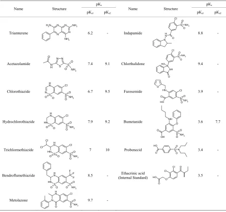
Diuretics belong to non-threshold substances, which means the absence of specific threshold concentration in urine. Table 1 shows that diuretics include compounds which have differences in molecular structures and chemical properties [3], and this fact complicates the elaboration of the only method of their extracting from biological matrix and their detection at the necessary selectivity level.
According to WADA requirements the only methods of analysis that should be applied at anti-doping labora- tories are liquid or gas chromatography with mass spec-
trometric detection. Though liquid chromatography with mass spectrometric detection (LC-MS) causes the de- crease in sensitivity due to the suppression of ionization by biological matrix [4,5] and shift in retention times in case of direct injection of sample [6] it also allows to achieve low detection limits and short time of analysis [7]. Because of relatively high cost of LC-MS this method is more preferable for quantative determination of diuret- ics in biological objects and for screening purpose. Gas chromatography with mass detection (GC-MS) is suit- able for multicomponent, selective and sensitive diuretics determination in human urine, as well as liquid chroma- tography. GC-MS method which includes two-step liq- uid-liquid extraction and derivatization was proposed for quantative determination of 16 diuretics [8]. Long pre- liminary sample preparation of urine limits the use of the method in routine analysis. Moreover, this technique does not provide high recovery of chlorthalidone and tri- amterene. At the same time GC-MS demonstrates the best results for qualitative determination and for confir- mation.
Table 1. General characteristics of diuretics.
рКа рКа
Name Structure рКа1 рКа2
Name Structure
рКа1 рКа2
Triamterene Cl O N N N N NH2 NH2 N H2
6.2 - Indapamide
N N H O S O NH2 8.8 - Acetazolamide Cl O S N N S NH2 O O N H O
7.4 9.1 Chlorthalidone
NH O H
S NH2
O
O
9.4 -
Chlorothiazide N S N H Cl S O O
O O NH2
6.7 9.5 Furosemide
S N H Cl O O O NH2 O H O 3.9 - Hydrochlorothiazide S N H Cl S O O
O O NH2
NH N
H 7.9 9.2 Bumetanide O
O S O NH2 O OH 3.6 7.7 Trichlormethiazide N H S N H Cl
Cl Cl O
S O
O NH2
7 10 Probenecid S N
O O O O O H 3.4 -
Bendroflumethiazide NH
N S H F F F S NH2 O O O O
8.5 - (Internal Standard)Ethacrinic acid
O O Cl Cl O H O 3.5 -
Metolazone N N H Cl 9.7 - O S
O ONH2
Despite the great number of methods proposed, gas or liquid chromatography with mass spectrometric detection do not always provide the necessary selectivity towards the urine components [10,12]. In case of liquid chroma- tography, the use of tandem mass-spectrometry allowed to pass over these problems and to ensure the satisfactory result [13-15].
Despite liquid chromatography is more preferable for screening purposes, gas chromatography with mass de- tection demonstrates the best results for confirmation. According to WADA requirements the optimal method for confirmation of presence of diuretics is the method different from the screening method. Therefore, the goal of the present work was to develop a method of confir- mation of presence of diuretics and probenecid in urine by gas chromatography with tandem mass spectrometric
detection.
2. Experimental Procedure
2.1. Preparation of Reagent Solutions
Stock standard solutions were prepared for 12 diuretic drugs (acetazolamide, bendroflumethiazide, bumetanide, chlorthalidone, chlorothiazide, ethacrinic acid, furosemide, hydrochlorothiazide, indapamide, metolazone, triamterene, trichlormethiazide) and a masking agent (probenecid) (all by SIGMA, St. Louis, MO, USA) by dissolving in metha- nol at concentration of 1.0 mg·ml–1.
USA), sodium sulfate (Merck, Darmstadt, Germany) were used without further purification, potassium carbonate (Merck, Darmstadt, Germany) was used after calcination in oven. Hydrochloric acid and sodium hydroxide were prepared by dilution of the concentrated initial solution (Merck, Darmstadt, Germany).
2.2. Instruments and Materials
Gas chromatograph Varian 3800 (Australia) equipped with an automatic sample injector system Combipal and Fac- torFour Capillary Column VF-DA 12 m, 0.20 mm I.D. for mass spectrometric determination of narcotic drugs and pharmaceuticals. Mass spectrometer Varian 1200 (Australia), directed by computer system of data proc- essing Varian MsWorkstation 6.9, with a system of three quadruples. Auxiliary equipment: thermostat VLM EC1 (Germany), evaporator in nitrogen stream Liebisch (Ger- many), centrifuge OPN-3Y4.2 (Russia), shaker Heidolph Vibramax 110 (Germany), oven Binder (Germany), pH- meter inoLab pH level 1 (Germany), analytical balance KERN ABS (Germany), pipettes of adjustable volume Eppendorf (Germany) 0 - 20 µl, 20 - 200 µl, 100 - 1000µl, 500 - 5000 µl.
2.3. Instrumental Parameters
Instrumental parameters of chromatographic determina- tion: carrier gas—helium, flow rate—1 ml·min–1, injector
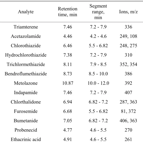
temperature—290˚C, injector mode—split 20; volume of sample injected—2 µl; flushing liquid—acetone, source temperature—225˚C, interface temperature—250˚C; SIM Width—0.7; Scan Time—0.2 s; start of the ion current registration—5.3 min. Instrumental parameters of tandem mass spectrometry: auxiliary gas pressure (argon) on the second quadruple—2.10 - 1.65 mTorr, electron energy at the source—20 eV. Registration mode: selected ion moni- toring (SIM) or multiple reaction monitoring (MRM). In experiment mass spectrometry detection was carried out in SIM mode using ions listed in Table 2 [12].
2.4. Sample Preparation
Negative urine samples were taken from volunteers who did not consume banned substances; the positive (spiked) urine samples were prepared by adding mixtures of stan- dard diuretics solutions to the negative urine samples prior to the sample preparation. The urine samples were stored in polypropylene bottles at a temperature of –20˚C. Before conducting the experiment samples were defreezed, centrifuged for 10 min at 1500 rpm, and the pH value was measured. Ethacrinic acid (internal standard, IS), mixture of standard diuretics, 200 mg of Na2SO4 and 5.0
ml of urine were transferred in a 12 ml glass tube. The pH value was adjusted by adding NaOH or buffer solu- tion and 5.0 ml of organic solvent. The tubes were shaken in a shaker for 5 - 20 min and then centrifuged.
The top layer was totally transferred to a 10 ml glass tubes. Solutions were evaporated in nitrogen stream at 40˚C (for solvents based on diethyl ether) or 80˚C (for solvents based on ethyl acetate) to obtain solid residue, the tubes were cooled to room temperature. The deri- vatization (methylation by methyl iodide in acetone in an alkaline environment) was performed according to [16]. Afterwards the samples were immediately sent to gas- chromatographic analysis. The degree of substances re- covery from matrix achieved by liquid-liquid extraction was investigated at a concentration of 0.9 MRPL (0.225 µg·ml–1). The mixture of standard diuretics and IS were
added to the urine samples and sample preparation was performed by the method described. Simultaneously, the derivatization of mixture of diuretics standards with the same concentration was conducted. All samples were analyzed by chromatographic-mass spectrometric method. The recovery degree was calculated by taking the ratio of peak height obtained at the retention time for the analyte of interest compared to that obtained from an IS in spiked urine and mixture of standards.
2.5. Method Validation
[image:3.595.309.538.506.736.2]To determine all parameters (capability to identification, selectivity, detection limit), that are necessary for suit- ability of verification in accordance with ISO/IEC 17025: 2006 [17], the investigation was performed. Linearity, accuracy and precision were not examined since the me- thod is intended for confirmation and is not intended for quantitative determination of diuretics, as established in [17,18]. Therefore more consideration was given to defi- nite identification of the analyte in urine.
Table 2. Characteristic ions of diuretics in Selected Ion Mo- nitoring mode.
Analyte Retention time, min
Segment range,
min
Ions, m/z
Triamterene 7.46 7.2 - 7.9 336
Acetazolamide 4.46 4.2 - 4.6 249, 108
Chlorothiazide 6.46 5.5 - 6.82 248, 275
Hydrochlorothiazide 7.38 7.2 - 7.9 310
Trichlormethiazide 8.11 7.9 - 8.5 352, 354
Bendroflumethiazide 8.73 8.5 - 10.0 386
Metolazone 10.87 10.0 - 12.0 392
Indapamide 7.46 7.2 - 7.9 407
Chlorthalidone 6.94 6.82 - 7.2 287, 363
Furosemide 6.68 5.5 - 6.82 81, 372
Bumetanide 7.05 6.82 - 7.2 406, 363
Probenecid 4.77 4.6 - 5.5 270
Identification by the retention time. A series of spiked urine samples and mixture of standards were simultane- ously analyzed to check the capability of identification. Absolute and relative differences in retention times were
calculated. y, %
Mass spectrometry identification in tandem mass spec- trometry (MS-MS) mode. A series of extracts of spiked urine samples and mixture of standards at concentrations 0.5, 1.0 and 5.0 MRPL were simultaneously analyzed to prove the possibility of mass spectrometric identification of the analytes. The relative intensity of the diagnostic ion was calculated on the base of the chromatographic peaks’ area or height, integrated by corresponding ion.
R
ecover
The selectivity of method was tested by analyzing six different urine samples after sample preparation without analytes.
The limit of detection was evaluated according to [19]. The samples were prepared by addition of the mixture of standards to urine to achieve the concentrations 0.2, 0.5, 1.0, 2.0 MRPL, and analyzed by marking the number of positive determinations regarding to total number of de- terminations. That concentration level, which corresponded to an unambiguous detection in all the samples, was con- sidered as a limit of detection.
2.6. Quality Control
While carrying out the experiment, the mixture of diu- retics standards at 0.9 MRPL (standard sample), the urine sample obtained from a volunteer who did not consume prohibited substances (negative sample), spiked urine sample with concentration equal to 0.9 MRPL (positive sample) and reagent blank (distilled water) were also analyzed using the same method of sample preparation for each analytical run. All samples, except for the re- agent blank, were prepared with the addition of IS.
3. Results and Discussion
3.1. Optimization of Gas-Chromatographic Method
The method of diuretics determination includes their ex- traction by organic solvents, subsequent derivatization and following chromatographic separation and detection. In this work the recovery degree as the function of the nature of organic solvent, mixing time and pH of aque- ous phase were investigated to optimize the conditions of diuretics determination.
The liquid-liquid extraction of diuretics from urine was carried out with the use of diethyl ether, ethyl acetate and mixtures of organic solvents (ratio): diethyl ether and isopropanol (19:1), diethyl ether and methanol (19:1), ethyl acetate and isopropanol with different ratio of components (19:1, 9:1, 17:3). The obtained results show (Figure 1) that the higher recovery degree for most of
y, %
R
ecove
r
R
ecover
y,
[image:4.595.321.521.86.523.2]%
Figure 1. The recovery of diuretics as function of solvents. Vsolv = 5 ml, Vaq = 5 ml, рН = 7.0, Т = 295 ± 1 K. 1: Diethyl
ether; 2: Diethyl ether:Isopropanol = 19:1; 3: Diethyl ether: Methanol = 19:1; 4: Ethylacetate; 5: Ethylacetate:Isopropa- nol = 19:1; 6: Ethylacetate:Isopropanol = 9:1; 7: Ethylace-tate:Isopropanol = 17:3.
diuretics has been achieved at their extraction from mix- ture ethyl acetate:isopropanol = 17:3. The extraction de-gree of all substances is satisfactory, though the recovery of metolazone and trichlormethiazide under these condi- tions is 53.7% and 3.1%, respectively, but the signal in-tensity is sufficient enough to detect these compounds at the MRPL level. The extraction equilibrium in the systems is reached for 5 minutes at given conditions.
chlorthalidone), strong acids (loop diuretics such as fu- rosemide) and base compounds (triamterene). Consider- ing this fact extraction of selected diuretics as the func- tion of pH value of the aqueous phase was investigated. It was found experimentally that weakly acidic diuretics are effectively extracted in molecular form in the range of pH 6 - 8 (pH of urine is ~5.5 - 7.0). The degree of their extraction exceeds 50%. Extraction degree of triamterene also increases. Extraction degree of more acidic diuretics such as furosemide, bumetanide and probenecid is lower under these conditions, but high chromatographic yield of characteristic ions of these compounds allows to detect them at the MRPL level. Further the acidity of aqueous phase was adjusted by NaOH addition to achieve pH 6 - 8.
Considering the demands made for chromatographic column, such as providing maximum separation of ana- lytes in a short time and compatibility with mass detector, capillary polymethacrilate column FactorFour VF-DA 12 m, 0.20 mm I.D., which was successfully used for deter- mination of narcotic drugs and pharmaceuticals, was cho- sen. To increase the efficiency of chromatographic sepa- ration of diuretics and to provide high rate of the analysis the optimization of temperature program was carried out. The following parameters were varied: initial and final temperature, heating rate and held. The optimal tem- perature program that meets all the requirements has fol- lowing parameters: initial and final temperatures 120 and 300˚C, respectively; first held for 0.5 min, followed by heating rate 30˚C·min–1 and subsequent held for 8.5 min.
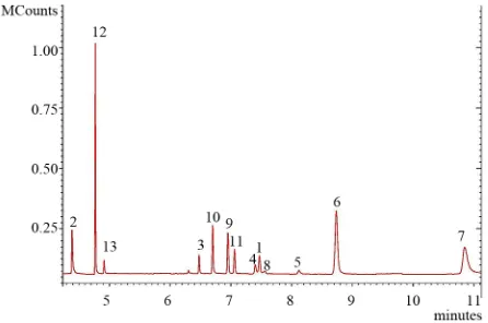
[image:5.595.62.285.506.654.2]The chromatogram of diuretics standards mixture under optimal conditions is shown in Figure 2. It is seen that complete chromatographic separation of hydrochlorothi- azide, triamterene and indapamide at this temperature pro- gram is not achieved. However, these compounds can be easily discriminated due to different mass spectral fea-
Figure 2. The chromatogram of the diuretics standard mix- ture, analyzed in SIM acquisition mode. С = 0.225 µg·ml–1.
1: Triamterene; 2: Acetazolamide; 3: Chlorothiazide; 4: Hy- drochlorothiazide; 5: Trichlormethiazide; 6: Bendroflume-thiazide; 7: Metolazone; 8: Indapamide; 9: Chlorthalidone, 10: Furosemide; 11: Bumetanide; 12: Probenecid; 13: Etha- crinic acid.
tures of diuretics. Besides, the presence of various diure- tics in the same sample is unlikely. Time of chromatog- raphy is 12 min under the optimal conditions.
3.2. Optimization of MS-MS Conditions
One of the major disadvantages of mass spectrometric methods is low selectivity in the analysis of biological objects. The identification of analytes in biological ma- trices is complicated due to misrepresentation of mass spectrum caused by concomitant substances. It can lead to appearance of false-positive results. In this case tan- dem mass spectrometry is more preferable method [20]. The combination of mass selection of the precursor fol- lowed by potentially unique collision-induced dissocia- tion and mass selection or scanning of the product ion provides higher specificity, since coincidence of spec- trum of dissociated ion and ion with known structure is a conclusive proof of ions structure identity. The tandem mass spectrometry method can be used either in full scan acquisition mode or multiply reaction monitoring mode. The second technique is more sensitive in comparison with the first one. Typically, two transitions precursor- product must be monitored, but in some cases the com- bination of a single precursor-product ion pair is suffi- ciently unique to be definitive.
For each diuretic molecular ions, product ions and col- lision energy (E) were chosen. Collision energy should ensure maximum yield of product ions [21]. The data for transition reaction molecular ion—product ion are pre- sented in Table 3.
Chromatograms of spiked with bendroflumethiazide and triamterene chosen as examples urine samples and negative urine samples at these conditions are presented in Figure 3.
It is shown that the compounds may be detected in each of the reactions. Besides, peaks with signal-to-noise ratio
≥ 3 at the time which corresponds to analyte retention time, are absent in chromatogram of negative sample. This result was observed for other diuretics and probenecid as well. This fact proves sufficient selectivity of method.
3.3. Method Validation
According to the requirements of WADA [21] the rela- tive intensities of any of the ions in mass shall not differ by more than the amount in Table 4 from the relative intensities of the same ions acquired from spiked urine.
Table 3. The diagnostic reactions and optimal collision energies for diuretics determination in Multiple Reaction Monitoring mode.
Analyte (molecular ion) Reaction, m/z E, eV Reaction, m/z Е, eV Reaction, m/z Е, eV
Triamterene (336) 336→336a –10 336→320 –23 336→292 –20
Acetazolamide (249) 249→249a –6.5 249→83 –12 249→108 –13
Chlorothiazide (248) 248→248 –6.5 248→141a –10 248→140 –28
Hydrochlorothiazide (353) 353→353 –6.5 353→288a –10 353→180 –22
Trichlormethiazide (399) 399→399 –6.5 399→192a –28 399→90 –15
Bendroflumethiazide (386) 386→386a –8 386→278 –24 386→279 –17
Metolazone (392) 392→392a –15 392→285 –23 392→284 –25
Indapamide (407) 407→407 –5 407→161a –21 407→132 –10
Chlorthalidone (287) 287→287a –8 287→255 –15 287→220 –20
Furosemide (372) 372→372 –5 372→96 –12 372→81a –17
Bumetanide (406) 406→406a –6 406→363 –10 406→318 –17
Probenecid (270) 270→270a –5 270→199 –10 270→135 –15
Ethacrinic acid, IS (261) 261→261 –10
[image:6.595.55.285.313.691.2]aDiagnostic Ion.
Table 4. The maximum tolerance windowsa for relative ion
intensities.
Relative abundanceb (%) Tolerance windows
>50% ±10% (absolute)
25% to 50% ±20% (relative) 5% to <25% ±5% (absolute)
<5% ±50% (relative)
aTolerance window—range for relative ion intensities to ensure appropriate
uncertainty in identification; bRelative abundance—the abundance of a par-
ticular ion relative to the most abundant ion monitored expressed as a per-centage.
Peak of small intensity with signal-to-noise ratio ≥ 3 is observed for probenecid in chromatogram of negative urine sample (m/z 270→135, Table 3). This fact is pos- sibly caused by the influence of matrix. But the inte- grated intensity of this peak is negligible which accord- ing to WADA recommendations [2] can not be a cause of false identification. Besides, there are no peaks with sig- nal-to-noise ratio ≥ 3 in the chromatograms of negative urine sample obtained using other diagnostic ions.
The ratio of intensities of diagnostic ions of acetazo- lamide, chlorothiazide, furosemide, chlorthalidone, bume- tanide, hydrochlorothiazide, triamterene and probenecid in the standard solutions and spiked urine samples meets WADA requirements (Tables 3 and 5). In case of inda-pamide and trichlormethiazide this parameter does not fit the tolerance window. Moreover, significant change of relative intensities dependent on concentration is ob- served which obviously can be explained by matrix in- fluence at relative intensity. Thereby, this method can not be recommended for confirmation of indapamide and tri- chlormethiazide in urine.
[image:6.595.307.538.389.444.2]Table 5. Relative abundance of diagnostic ions in Multiple Reaction Monitoring mode, established during the method valida- tion.
The relation of ions intensity in spiked urine/standard solution (%) Analyte
Reaction, m/z Ion 1 Reaction, m/z Ion 2 Reaction, m/z Ion 3
Triamterene 336→336 100/100 336→320 26.8/29.0 336→292 23.9/23.7 Acetazolamide 249→83 100/100 249→108 60.9/62.3 249→249 35.1/35.0 Chlorothiazide 248→141 100/100 248→140 53.5/53.4 248→248 26.7/22.1 Hydrochlorothiazide 353→180 100/100 353→288 37.8/38.0 353→353 7.7/6.1 Trichlormethiazide 399→90 100/100 399→192 73.7/69.8 399→399 49.9/25.4 Bendroflumethiazide 386→278 100/100 386→386 89.1/90.3 386→279 30.5/29.4 Metolazone 392→284 100/100 392→392 34.3/32.4 392→285 18.1/18.2 Indapamide 407→132 100/100 407→161 50.3/59.0 407→407 0.9/0.8 Chlorthalidone 287→255 100/100 287→287 104.8/100 287→220 72.0/71.7 Furosemide 372→81 100/100 372→96 30.4/30.8 372→372 3.0/1.8 Bumetanide 406→318 100/100 406→363 53.4/56.0 406→406 12.0/10.7 Probenecid 270→135 100/100 270→199 25.5/35.0 270→270 4.6/4.7
Limits of detection of acetazolamide, probenecid, fu- rosemide, chlorthalidone, bumetanide, hydrochlorothi- azide, triamterene, bendroflumethiazide, metolazone by tandem mass spectrometry are 0.050 µg·ml–1. Limit of
detection of chlorothiazide which is 0.500 µg·ml–1 does
not meet WADA requirements. High limit of detection may be explained by its partial dissociation at the column.
4. Conclusions
The developed method proves the suitability of gas chro- matography and tandem mass spectrometry for confirma- tion of presence of 8 diuretics and probenecid in urine. It is selective towards acetazolamide, furosemide, bumet- anide, hydrochlorothiazide, bendroflumethiazide, meto- lazone, probenecid, chlorthalidone and triamterene, since the components of urine do not interfere with the detec- tion of diuretics. The method can not be recommended for determination of indapamide, trichlormethiazide and chlorothiazide.
The reduction of time for sample preparation and suf- ficient recovery of all diuretics merit the method proposed. Optimization of temperature program for diuretics deter- mination at chromatographic column which wasn’t pre- viously used for this purpose allowed to achieve chro- matographic separation of all diuretics in short time. Se- lection of diagnostic fragmentation reactions and optimal collision energies allowed to achieve high selectivity.
Limits of detection of diuretics and probenecid by MS-MS are lower than that regulated by WADA and are 0.050 µg·ml–1.
The method was validated that proves the possibility of its application in anti-doping laboratories.
REFERENCES
[1] WADA, “The world Anti-Doping Code—The 2009 Pro-
hibited List: International Standard,” World Anti-Doping Agency, Montreal, 2009.
[2] WADA, “Minimum Required Performance Limits for Detection of Prohibited Substances (Technical Document TD2009MRPL),” World Anti-Doping Agency, Montreal, 2009.
[3] R. Ventura and J. Segura, “Detection of Diuretic Agents in Doping Control,” Journal of Chromatography B,Vol. 687, No. 1, 1996, pp. 127-144.
doi:10.1016/S0378-4347(96)00279-4
[4] M. Mazzarino, X. de la Torre and F. Botrè, “A Screening Method for the Simultaneous Detection of Glucocorti- coids, Diuretics, Stimulants, Anti-Estrogens, Beta-Adren- ergic Drugs and Anabolic Steroids in Human Urine by LC-ESI-MS/MS,” Analytical and Bioanalytical Chemis- try,Vol. 392, No. 4, 2008, pp. 681-698.
doi:10.1007/s00216-008-2292-5
[5] O. J. Pozo, P. Van Eenoo, K. Deventer and F. T. Delbeke, “Development and Validation of a Qualitative Screening Method for the Detection of Exogenous Anabolic Ster- oids in Urine by liquid Chromatography-Tandem Mass Spectrometry,” Analytical and Bioanalytical Chemistry, Vol. 389, No. 4, 2007, pp. 1209-1224.
doi:10.1007/s00216-007-1530-6
[6] K. Deventer, O. J. Pozo, P. Van Eenoo and F. T. Delbeke, “Qualitative Detection of Diuretics and Acidic Metabo- lites of Other Doping Agents in Human Urine by High- Performance Liquid Chromatography-Tandem Mass Spec- trometry. Comparison between Liquid-Liquid Extraction and Direct Injection,” Journal of Chromatography A,Vol. 1216, No. 31, 2009, pp. 5819-5827.
doi:10.1016/j.chroma.2009.06.003
[7] R. Ventura, M. Roig, N. Monfort, P. Saez, R. Berges and J. Segura, “High-Throughput and Sensitive Screening by Ultra-Performance Liquid Chromatography Tandem Mass Spectrometry of Diuretics and Other Doping Agents,”
European Journal of Mass Spectrometry, Vol. 14, No. 3, 2008, pp. 191-200. doi:10.1255/ejms.920
and Masking Agents in Human Urine. Development and Validation of a Productive Screening Protocol for Anti- doping Analysis,” Journal of Chromatography A, Vol. 1135, No. 2, 2006, pp. 219-229.
doi:10.1016/j.chroma.2006.09.034
[9] L. Amendola, C. Colamonici, M. Mazzarino and F. Botrè, “Rapid Determination of Diuretics in Human Urine by Gas Chromatography-Mass Spectrometry Following Micro- wave Assisted Derivatization,” Analytica Chimica Acta, Vol. 475, No. 1-2, 2003, pp. 125-136.
doi:10.1016/S0003-2670(02)01223-0
[10] C. Brunelli, C. Bicchi, A. Di Stilo, A. Salomone and M. Vincenti, “High-Speed Gas Chromatography in Doping Control: Fast-GC and Fast-GC/MS Determination of β- Adrenoceptor Ligands and Diuretics,” Journal of Separa- tion Science, Vol. 29, No. 18, 2006, pp. 2765-2771. doi:10.1002/jssc.200500387
[11] Y.-L. Tseng, M.-H. Shieh, C.-T. Lin and F.-H. Kuo, “De-tection of Diuretics in Urine during Sports Events in Taiwan,” Tzu Chi Medical Journal,Vol. 16, No. 2, 2004, pp. 69-77.
[12] P. Van Eenoo and F. T. Delbeke, “Criteria in Chromato- graphy and Mass-Spectrometry—A Comparison between Regulations in the Field of Residue and Doping Analy- sis,” Chromatographia, Vol. 59, Suppl. 1, 2004, pp. 39- 44. doi:10.1365/s10337-004-0198-8
[13] L. Politi, L. Morini and A. Polettini, “A Direct Screening Procedure for Diuretics in Human Urine by Liquid Chro- matography-Tandem Mass Spectrometry with Informa- tion Dependent Acquisition,” Clinica Chimica Acta, Vol. 386, No. 1-2, 2007, pp. 46-52.
doi:10.1016/j.cca.2007.07.018
[14] C. Goebel, G. Trout and R. Kazlauskas, “Rapid Screening Method for Diuretics in Doping Control Using Auto- mated Solid Phase Extraction and Liqud Chromatogra- phy-Electrospray Tandem Mass Spectrometry,” Analytica Chimica Acta, Vol. 502, No. 1, 2004, pp. 65-74. doi:10.1016/j.aca.2003.09.062
[15] D. Thieme, J. Grosse, R. Lang, R. K. Mueller and A. Wahl, “Screening, Confirmation an Quantification of Diuretics in Urine for Doping Control Analysis by High-Perform- ance Liquid Chromatography-Atmospheric Pressure Ioniza- tion Tandem Mass Spectrometry,” Journal of Chroma- tography B: Biomedical Sciences and Applications, Vol. 757, No. 1, 2001, pp. 49-57.
doi:10.1016/S0378-4347(01)00058-5
[16] S. J. Park, H. S. Pyo, Y. J. Kim, M. S. Kim and J. Park, “Systematic Analysis of Diuretic Doping Agents by HPLC Screening and GC/MS Confirmation,” Journal of Analytical Toxicology,Vol. 14, No. 2, 1990, pp. 84-90. [17] ISO/IEC 17025, “General Requirements for the Compe-
tence of Testing and Calibration Laboratories,” 2006. [18] Eurachem Working Group, “A Laboratory Guide to Meth-
od Validation and Related Topics,” Eurachem, Tedding-ton, 1998.
[19] WADA, “The International Standard for Laboratories, Ver- sion 6.0,” World Anti-Doping Agency, Montreal, 2009. [20] E. Hoffmann and V. Stroobant, “Mass Spectrometry. Prin-
ciples and Applications,” 3rd Edition, WILEY-VCH Ver- lag GmbH & Co. KGaA, Weinheim, 2007.