warwick.ac.uk/lib-publications
Original citation:
Collins, Teresa, Bergenholm, Linnéa, Abdulla, T., Yates, J. W. T., Evans, N. D., Chappell, M. J.
(Michael J.) and Mettetal, J. T. (2015) Modeling and simulation approaches for
cardiovascular function and their role in safety assessment. CPT : Pharmacometrics &
Systems Pharmacology, 4 (3). pp. 175-188.
Permanent WRAP URL:
http://wrap.warwick.ac.uk/97686
Copyright and reuse:
The Warwick Research Archive Portal (WRAP) makes this work of researchers of the
University of Warwick available open access under the following conditions.
This article is made available under the Creative Commons Attribution 4.0 International
license (CC BY 4.0) and may be reused according to the conditions of the license. For more
details see:
http://creativecommons.org/licenses/by/4.0/
A note on versions:
The version presented in WRAP is the published version, or, version of record, and may be
cited as it appears here.
REVIEW
Modeling and Simulation Approaches for Cardiovascular
Function and Their Role in Safety Assessment
TA Collins1, L Bergenholm2, T Abdulla2, JWT Yates3, N Evans2, MJ Chappell2* and JT Mettetal4*
Systems pharmacology modeling and pharmacokinetic-pharmacodynamic (PK/PD) analysis of drug-induced effects on
cardiovascular (CV) function plays a crucial role in understanding the safety risk of new drugs. The aim of this review is to
outline the current modeling and simulation (M&S) approaches to describe and translate drug-induced CV effects, with an
emphasis on how this impacts drug safety assessment. Current limitations are highlighted and recommendations are made
for future effort in this vital area of drug research.
CPT Pharmacometrics Syst. Pharmacol.
(2015)
4
, 175–188; doi:
10.1002/psp4.18
; published online on 11 March 2015.
Cardiovascular (CV) function is essential for life; the heart efficiently pumps blood containing vital nutrients and oxy-gen through vessels to cells throughout the body and removes waste products and carbon dioxide. CV function is controlled by the central nervous system, autonomic nerv-ous system, and endocrine system, and is able to respond to a variety of external stimuli, while feedback mechanisms maintain homeostatic control. It adapts to the body’s needs; for example, during exercise the heart rate (HR) increases, causing an increase in cardiac output (CO), and the propor-tion of CO is increased to muscles. Drug-induced CV side effects are undesirable because they may cause long-term CV damage, which puts the patient at greater risk of mor-tality and morbidity.
CV function declines with age: stiffened arteries lead to increased systolic arterial pressure,1 and a reduction in maximum HR causes a compensatory stroke volume (SV) increase in order to maintain CO during, e.g., exercise.2 Therefore, as the general population ages, more patients are likely to present with preexisting CV conditions, which when combined with drug-induced CV changes could result in even greater risks. Understanding and predicting the consequences of these safety changes are challenges for the development of new drugs.
Mathematical modeling of drug effects and the CV sys-tem can aid this understanding, and there are numerous examples of pharmacokinetic-pharmacodynamic (PK/PD), mechanistic, and systems pharmacology approaches in the literature. In this review, modeling analyses are explored in these three approaches defined as:
1. Traditional ‘‘top-down’’ PK/PD modeling and simulation (M&S) that utilizes empirical or descriptive models to describe the linkage between drug concentration and observed response, such as change in functional or structural CV biomarkers.
2. Mechanistic or systems biology ‘‘bottom-up’’ approaches that com-bine knowledge of the system from cellular targets to their impact at a cellular, tissue, or whole-body level.
3. Systems pharmacology3 ‘‘middle-out’’ approaches that sit at the interface between the other two categories. These combine aspects of both PK/PD and systems biology, and incorporates physiological processes and mechanism of action at targets.
M&S approaches promise greater impact in the CV safety space by integratingin silico,in vitro, andin vivopreclinical data with mechanism-based models to anticipate and predict the effects of new drugs in humans.4The application of these principles is now beginning to become reality in CV safety research, although it is important not to overlook earlier empirical PK/PD models that have been used in cross-species comparison of CV PK/PD relationships. Prospective predictions of human effects at expected therapeutic doses is a vital component of preclinical CV safety risk assessment for potential new drugs and is a rapidly developing area of interest. However, this is only valuable if clinical data can be obtained and compared to the model-based predictions. If differences between predicted and observed magnitude and kinetics of drug-induced effects can be better understood, then progress in refinement of prospective predictions of CV safety endpoints can be achieved.
OVERVIEW OF DRUG-INDUCED CARDIOVASCULAR EFFECTS
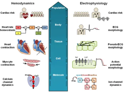
A wide panel of electrophysiological and mechanical func-tional markers is collected during clinical and preclinical in vivo testing. These are outlined in Figure 1 and typically include:
1. Electrocardiograph (ECG) measurements. Common ECG interval measurements include QT interval, QRS duration, and PR intervals, all measured in milliseconds (ms). The QT interval is often cor-rected for HR, defined as corcor-rected QT (QTc).
2. Hemodynamic measurements including HR and blood pressure (BP), measured in beats min-1and mmHg, respectively.
1Drug Safety and Metabolism, AstraZeneca, Alderley Park, Macclesfield, UK;2School of Engineering, University of Warwick, UK;3Oncology, AstraZeneca, Alderley Park,
Macclesfield, UK;4Drug Safety and Metabolism, AstraZeneca, Waltham, Massachusetts, USA. Correspondence: JT Mettetal ([email protected]) or MJ
Chappell ([email protected])
3. Indices of contractility including the maximum and minimum rate of left ventricular pressure change (dP/dt max (1) and dP/dt min (–)), measured in mmHg s-1, and QA interval, which is an inverse index measured in ms (time between Q wave on ECG and onset of arte-rial wave (blood pressure)). Ejection fraction (EF) is a common clin-ical biomarker measured as the percentage of the diastolic volume of blood in the ventricle that is ejected with each beat.
4. In preclinical pathological studies, chronic changes such as histopa-thology of the heart and/or vessels are also monitored.
Functional changes of ECG and hemodynamics,
although not typically life-threatening in themselves, are readily monitorable, although they may be associated with more serious but rare events. Most famously, QT/QTc pro-longation is known to be a risk factor for the potentially fatal arrhythmia Torsades de Pointes (TdP), which caused one-third of drug withdrawals between 1990 and 2006.5In addi-tion, prolongation of the PR and QRS intervals are linked to CV mortality and morbidity in cardiac risk populations.6HR and BP changes are known to increase risk to all causes of mortality, including CV complications.7,8 Contractility changes typically require measurements through more technical or invasive techniques and are therefore more dif-ficult to obtain clinically, but can lead to heart failure, hyper-trophic cardiomyopathy, and sudden cardiac death.9 Other examples of cardiac safety liability include the withdrawal of rofecoxib, a selective Cox-2 inhibitor, from the market after long-term use showed increased risk of serious thrombotic events including myocardial infarction and stroke.10
Cardiovascular complications were the leading cause of drug withdrawals from the EU market during 2002 and 201111 across a range of therapy areas. Any unintended
drug-induced effects on the CV system represent a poten-tial safety concern, and in order to avoid costly late-stage failures, a variety of early preclinical experiments were per-formedin vitro,in silico,andin vivoto rule out unsafe com-pounds or to understand and quantify risk with those that do progress. Novel therapeutics must distinguish them-selves from standard of care agents either in terms of improved efficacy or safety risk, so a thorough assessment of CV effects is essential.
Preclinical Safety studies with CV endpoints include:
1. ‘‘Off-target’’ activity assessed in vitro on molecular targets (enzymes, GPCRs, ion channels) associated with effects on CV structure and function.
2. In silico and quantitative structure–activity relationships (QSAR) models built using data from suchin vitro‘‘off-target’’ data. 3. In vitrofunctional assessments using traditional tissue-based studies
(e.g., isolated heart/blood vessel pharmacology), supplemented with new technologies such as human stem cell-based cardiomyocytes. 4. In vivo functional assessment is mainly run in conscious animals
and measures ECG, BP, and HR via implantable telemetry in single-dose, safety pharmacology studies at the therapeutic range and above.12
5. In vivo assessment of the effects on CV structure in repeat-dose toxicology studies assessing clinical pathology, histopathology, and clinical observations. Functional assessments of HR and ECG can also be made in such studies using jacketed telemetry.
[image:3.613.71.545.87.336.2]All of these endpoints contribute to an integrated CV risk assessment of the drug before it is first administered to humans. In the following sections we cover modeling approaches applied to drug-induced effects on ECG
intervals, hemodynamics (including contractility), and car-diac damage, and where appropriate, highlight examples of translation from preclinical to humans.
DRUG-INDUCED CHANGES ON ECG INTERVALS
The ECG reflects the electrical depolarization and repolari-zation that cardiomyocytes undergo during an action poten-tial (AP). It represents a combined view of the spread of excitation that occurs across the cardiac tissue. Changes in ECG intervals can indicate changes in cardiac electrophysi-ology, for example, resulting from ion channel inhibition.
One of the most frequently assessed ECG intervals is QT, and with increasing concern regarding QT/QTc prolon-gation, the ICH announced in 2005 preclinical (S7B)13 and clinical (E14)14 regulatory guidance for new drugs. These focused on QT prolongation and blockade of the ion chan-nel (Kv11.1) encoded by the human ether-a-go-go-related gene (hERG). The E14 guidelines states that in thorough QT/QTc (TQT) studies, the threshold level of regulatory concern is at 5 ms, as evidenced by an upper bound of the 95% confidence interval (CI) around the mean effect on QTc exceeding 10 ms.15PK/PD modeling of QTc prolonga-tion has been beneficial in this setting for predicting QTc prolongation at doses that were not directly studied in the TQT study and to support statistical analysis.
Biomarkers such as QT prolongation are sensitive, but not specific, predictors of ventricular proarrhythmia (i.e., TdP), which can be complicated by further mechanisms, for example, multiple ion channel blockade or effects on traf-ficking of the ion channel to the cell membrane. PK/PD modeling of QT prolongation began in the late 1970s (see
Table 1 and Supplementary Material for references). Arguably, this is the most characterized of all PK/PD rela-tionships of ECG interval changes due to the regulatory focus on the link between hERG, QT, and TdP. In earlier reviews of drug-induced QT effects,16,17 the following fac-tors have been identified as important to consider during the modeling of QT, but could also remain relevant for other ECG endpoints:
1. Heart rate correction, preferably individual-specific.
2. Variability of baseline, both interindividual and intraindividual, as in circadian rhythm.
3. Subject demographic information such as age, sex.
4. Genetics, e.g., rare polymorphisms causing long QT syndrome. 5. Environmental or other factors such as obesity, physical activity,
electrolyte levels, blood pressure, blood glucose, and alcoholism.
QT interval duration is strongly dependent on HR and the use of correction methods aim to remove the influence of heart rate, providing a more stable measure: QTc. HR cor-rection formulas include linear and fixed exponential (Bazett, QTcB, Fridericia, QTcF, and individual, QTcI). QTcI and QTcF have been shown to perform best in humans, while QTcI performed best in preclinical species including dog, guinea pig, and cynomolgus monkey.18 Cosine func-tions19 have been used to account for within-subject vari-ability in baseline due to homeostatic mechanisms, external
factors, or circadian rhythm (regular diurnal fluctuation), either using typically single or where necessary multiple cosine functions.20,21 In one example of QTc prolongation modeling, the model structure utilized three baseline cosine functions with time periods of 4, 8, and 24 hours in the dog, and 2, 4, and 24 hours in human, combined with effect compartment models for both species and for the concentration–response relationship an Emax model for the dog and a linear model for human.22
PK/PD modeling efforts have focused on QT and hERG inhibition, but there are other examples of other mechanisms affecting ECG intervals,Table 1describes the type of struc-tural models used in the descriptive PK/PD modeling of drug-induced ECG interval changes for both preclinical and clinical studies. Currently, PK/PD modeling of clinical or non-clinical data is routinely modeled using population (mixed effects) approaches rather than individual or pooled data-sets.22,23Interindividual variability is often included on base-line parameters and drug effect. This reflects the availability of high-quality, rich ECG data combined with more readily available software to conduct these analyses.
Linear, Emax, and sigmoidal Emax models have all been used to describe the relationship between concentration and effect for ECG intervals.19 58% of the compounds listed could be adequately described with simple linear drug effect models. While it is expected that drug effects will eventually saturate with exposure, linear models may be more prevalent for ECG changes, as maximal effects are often not achieved in safety studies where doses are selected based on margins to therapeutic exposure not to characterize the full concentration–response relationship. Studies can also be halted before reaching a maximum level due to lack of tolerability at these exposures. However, Emax or sigmoidal Emax models are often used with antiar-rhythmic compounds when these exposures are tolerated. Once in the clinic, concentrations required to reach maxi-mal effect are less often reached for these same reasons, leading to even fewer saturable models being observed.24 Where Emax models have been utilized for clinical QTc
changes, the maximal activity level appears to be
compound-specific rather than reaching a system-specific or physiological upper limit (for example, 20 ms for verna-kalant25and 170 ms for N-acetylprocainamide26).
and degradation is uncommon for ECG intervals. One potential reason for this is that the effect of the drug on ion channel activity is expected to be rapid once the compound has reached the myocytes, not requiring a turnover process to have an effect. In addition to delays in reaching the site of action, another potential cause of observed time delays could be the production of an active metabolite.27
Although PK/PD modeling examples of intervals other than QT are limited, it appears most other endpoints are typically treated similar to QT, although heart rate correc-tion may not always be required for other ECG intervals. While not as well studied, CV complications are still
poten-tially hazardous with these endpoints. Cav1.2 inhibition leads to increased PR interval and is linked to AV block, while Nav1.5 inhibition leads to increased QRS duration and is linked to ventricular tachycardia. The concentration– QRS relationship of a number of compounds has been investigated in dog23using a population approach, and this enabled comparison across compounds and investigation of the therapeutic window. The data were modeled as per-cent change from baseline and the size of the estimated Emaxfor QRS change varied from 8 to 57%.
In contrast to the descriptive PK/PD modeling
[image:5.613.59.555.104.569.2]approaches, drug-induced ECG effects have also been
Table 1 Overview of the composition of PK/PD models used for modeling of ECG intervals in preclinical species and human, indicating selected
concentration–effect relationship, model for capturing potential time delays and baseline function
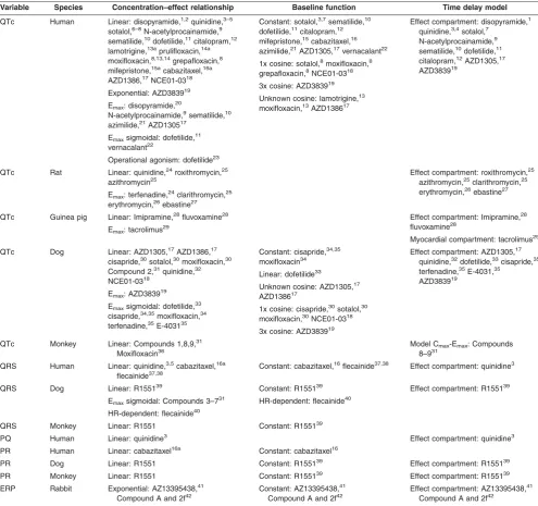
Variable Species Concentration–effect relationship Baseline function Time delay model
QTc Human Linear: disopyramide,1,2quinidine,3–5
sotalol,6–8N-acetylprocainamide,9
sematilide,10dofetilide,11citalopram,12
lamotrigine,13aprulifloxacin,14a
moxifloxacin,8,13,14grepafloxacin,8
mifepristone,15acabazitaxel,16a
AZD1386,17NCE01-0318
Exponential: AZD383919
Emax: disopyramide,20
N-acetylprocainamide,9sematilide,10
azimilide,21AZD130517
Emaxsigmoidal: dofetilide,11
vernacalant22
Operational agonism: dofetilide23
Constant: sotalol,3,7sematilide,10
dofetilide,11citalopram,12
mifepristone,15cabazitaxel,16
azimilide,21AZD1305,17vernacalant22
1x cosine: sotalol,8moxifloxacin,8
grepafloxacin,8NCE01-0318
3x cosine: AZD383919
Unknown cosine: lamotrigine,13
moxifloxacin,13AZD138617
Effect compartment: disopyramide,1
quinidine,3,4sotalol,7
N-acetylprocainamide,9
sematilide,10dofetilide,11
citalopram,12AZD1305,17
AZD383919
QTc Rat Linear: quinidine,24roxithromycin,25
azithromycin25
Emax: terfenadine,24clarithromycin,25
erythromycin,26ebastine27
Effect compartment: roxithromycin,25
azithromycin,25clarithromycin,25
erythromycin,26ebastine27
QTc Guinea pig Linear: Imipramine,28fluvoxamine28
Emax: tacrolimus29
Effect compartment: Imipramine,28
fluvoxamine28
Myocardial compartment: tacrolimus29
QTc Dog Linear: AZD1305,17AZD1386,17
cisapride,30sotalol,30moxifloxacin,30
Compound 2,31quinidine,32
NCE01-0318
Emax: AZD383919
Emaxsigmoidal: dofetilide,33
cisapride,34,35moxifloxacin,34
terfenadine,35E-403135
Constant: cisapride,34,35
moxifloxacin34
Linear: dofetilide33
Unknown cosine: AZD1305,17
AZD138617
1x cosine: cisapride,30sotalol,30
moxifloxacin,30NCE01-0318
3x cosine: AZD383919
Effect compartment: AZD1305,17
quinidine,32dofetilide,33cisapride,35
terfenadine,35E-4031,35
AZD383919
QTc Monkey Linear: Compounds 1,8,9,31
Moxifloxacin36
Model Cmax-Emax: Compounds
8–931
QRS Human Linear: quinidine,3,5cabazitaxel,16a
flecainide37,38
Constant: cabazitaxel,16flecainide37,38 Effect compartment: quinidine3
QRS Dog Linear: R155139
Emaxsigmoidal: Compounds 3–731
HR-dependent: flecainide40
Constant: R155139
HR-dependent: flecainide40
Effect compartment: R155139
QRS Monkey Linear: R1551 Constant: R155139
PQ Human Linear: quinidine3 Effect compartment: quinidine3
PR Human Linear: cabazitaxel16a Constant: cabazitaxel16
PR Dog Linear: R1551 Constant: R155139 Effect compartment: R155139
PR Monkey Linear: R1551 Constant: R155139 Effect compartment: R155139
ERP Rabbit Exponential: AZ13395438,41 Compound A and 2f42
Constant: AZ13395438,41 Compound A and 2f42
Effect compartment: AZ13395438,41 Compound A and 2f42
aStudies where no ECG effect was found. References contained in Supplementary Material.
modeled using detailed, “bottom-up” mechanistic models. Drug effects on ion channels are described mathematically to predict morphology changes in the AP or ECG. Cellular cardiac AP models have been developed for different spe-cies including human,28–31dog,32guinea pig,33and rabbit.34 These models represent the relevant electrophysiological aspects of the cellular system: transmembrane conduct-ance, ion channels and their inhibition by drugs, as well as other pumps/exchangers and intracellular ion concentra-tions, and integrate the influence of these factors over time on cellular ion concentrations. Drug effects are modeled by altering the ion conductance term, which represents the gat-ing (open/closed, etc.) of the relevant ion channel.
Such cardiac AP models have been applied to predict the effects of antiarrhythmic drugs that alter ion channel activities35–37and better describe the effects of multiple ion channel inhibition than focusing purely on potency values. Davies et al.35 calibrated the Hund-Rudy canine AP model32to predict change in AP duration in dog cardiomyo-cytes solely fromin vitrodata of five ion channels (Nav1.5, Cav1.2, Kv4.3, Kv7.1, and Kv11.1), demonstrating a predic-tivity of 68% (ratio of sum of true positive and true negative results to total number examined). Similarly, Miramset al.37 showed that a human AP model could be trained to classify TdP risk based on predicted therapeutic Cmaxand Nav1.5, Cav1.2, and Kv4.3 channel median inhibitory concentration
(IC50) values with markedly improved accuracy compared to safety margins between hERG IC50 and therapeutic Cmaxalone. A recent comparison of predicted effect of ion channel block in AP models from human and preclinical species highlights the importance of cautious extrapolation between species.38For example, a 70% block of the hERG ion channel resulted in an 80% AP prolongation in humans but only a 30% and 20% change in dog and guinea pig, respectively.
[image:6.613.89.525.84.412.2]While these ion flux models can capture information relating to the membrane potential in a single cell, the resulting ECG are at the tissue/whole-body level (Figure 2) and depend in part on the spatial orientation of myocytes in the heart and the AP propagation in tissue. Models of car-diac tissue have therefore been constructed to describe the propagation of the AP in 1, 2, and 3 dimensions39by linking multiple cellular models in a spatially relevant way. These tissue models have been used to study the effects of single and multiple ion channel blockade on Purkinje fibers.40 Sotalol-induced effects41 have been studied via pseudo-ECG generated from the simulation of a one-dimensional fiber representing proportional distribution of cells of the epicardium, midmyocardium, and endocardium. At the whole-heart level, cardiac structure and electrophysiology have been integrated with whole-body geometry to translate ion channel effects through simulation of cardiac AP
propagation to calculate 12-lead ECG and QT prolongation as measured in the torso.42A 12-lead ECG was also calcu-lated by Wilhelmset al.43 for comparison of the two QTc-prolonging drugs cisapride (proarrhythmic) and amiodarone (antiarrhythmic), identifying the effect of amiodarone alone on AP conduction as the mechanism behind the drug being anti- rather than proarrhythmic.
The usefulness of mechanistic in silico models in drug discovery and development now needs to be demonstrated, as a recent study44that investigated the ability of AP mod-els to predict the QT change in TQT studies and showed the models in general underpredicted the TQT outcome. These became more reliable predictions of TQT outcome when the comparison range was relaxed to within a 100-fold range of concentration reached in the TQT study,
suggesting simple concentration comparisons between
modeled AP changes and in vivo plasma exposure–
response requires improvement by considering other factors such as tissue distribution and intracellular concentrations.
One tool built specifically to do this is the Cardiac Safety Simulator (CSS) (Certara), a commercially available tool designed to increase the ability of nonmodelers to test the effect of ion channel activity on the ECG incorporating pop-ulation variability on both exposure (through SimCYP, Cer-tara) and ECG prediction. As a test of predictivity of the simulator, the QTc effects of six antipsychotic drugs45were investigated using the CSS, showing good agreement between predicted and observed mean QTc change, although the predictions did not account for all of the observed variability. This approach represents a bottom-up approach based purely onin vitro data and it is potentially tempting to adopt this as a predictive strategy but not enough is yet known about the predictive performance of this tool. With a vast number of inputs and settings in such a model, this should be used cautiously until more validated examples are demonstrated, and it is hoped this will explore more diverse situations such as mixed ion channel inhibition, combined effect of parent and metabolite, and mechanisms not caused by ion channel inhibition.
In our experience AP changes can be routinely predicted once in vitro ion channel inhibition assay data are gener-ated, forming a key part of the early risk assessment prior toin vivo data being generated. During the drug discovery process ion channel inhibition liability can often be reduced or removed via structural modification of potential drug enti-ties, meaning only small changes will be observed in vivo
or only at high safety margins. PK/PD modeling of in vivo
ECG changes is then applied wherever statistically signifi-cant changes are observed. This PK/PD modeling then defines the underlying concentration–response relationship and better quantifies safety margins. If a compound is then selected to enter clinical studies, a prediction will be made at expected therapeutic doses in human combining the pre-dicted human PK and the PD model including any time delays to assess the expected magnitude of effect.
While mechanistic modeling provides a powerful lens, empirical PK/PD modeling will still remain a valuable approach to quantifying the concentration–effect relation-ship of ECG intervals from both the clinical and preclinical studies in the near future, because it is an effective way to
assess CV safety risk and the models are straightforward to implement with little mechanistic understanding. Mecha-nistically based models of ECG change have focused pri-marily on ion channel inhibition as the typical mechanism of effect, but due to earlyin vitrosafety screens it is likely that in the future observedin vivo effects on ECG intervals will be small, and may be driven by other, more unusual mech-anisms. While much effort has been invested in the devel-opment of mechanistic models that replicate the time course of ECG changes, it is unclear how and at what point these will be used in the drug development process.In vitro
data becomes available early in drug evaluation, when there is limited understanding of likely exposure in human. Later, once the human dose and pharmacokinetic predic-tions are better defined and one could use the mechanistic model of ECG change, thein vivoCV results become avail-able and could take precedence over thein vitro-based pre-diction in the integrated risk assessment.
DRUG-INDUCED CHANGES ON HEMODYNAMICS
Hemodynamics is the study of blood circulation, which is governed by pressures and resistances in different parts of the cardiovascular system, as well as the force and rate of contraction of the heart. Typically, HR and BP are moni-tored including mean arterial, diastolic, and systolic blood pressure (MAP, DBP, and SBP, respectively). Other impor-tant variables include stroke volume (SV), cardiac output (CO), total peripheral resistance (TPR), contractility, and in some cases compliance and central venous pressure. The anatomy and physiology of the CV system result in funda-mental interrelationships existing between these variables, including that CO is the product of HR and SV, MAP is the product of CO and TPR and others; for example, HR and contractility are known to be highly correlated in vivo.46 Despite this understanding, PK/PD modeling of drug effects is often conducted separately on individual parameters.23
Table 2 describes the application of PK/PD models to blood pressure, heart rate, and contractility effects. Base-line functions are widely used to explain the inherent vari-ability and circadian rhythm, such as cosine functions. 76% of compounds adopt indirect response models to describe the time delay between concentration and effect, rather than a simple effect compartment model more commonly used for ECG intervals. In contrast to the observed time delays with ECG intervals, the time delays on hemody-namic parameters are far more varied, and half-lives of the indirect response parameter kout could be within a few minutes to greater than 50 days. This is likely to reflect the variety and timescales of mechanisms by which a com-pound can affect hemodynamics. The use of indirect response models may better capture biological processes downstream of receptor activity, but upstream of CV func-tion, for example, nitric oxide production. The underlying exposure response models most typically include linear or Emax/sigmoidal Emaxpharmacodynamic models.
HR changes were observed with an anti-HIV agent with
baseline daily variation and a direct linear drug effect. The mean drug effect slope was 2.3-fold steeper in dog com-pared to that observed in human and therefore the predic-tion of human response was made by using the dog slope and combining it with human PK, which slightly overesti-mated the clinical HR change. As a predictive strategy this is a sensible approach and represents a worst-case sce-nario, although in order to assess applicability across a range of mechanisms additional drugs would need to be assessed in a similar manner in order to ascertain if there are any consistencies in cross-species differences in drug effect for HR.
Since many hemodynamic measurements result from interacting mechanical and physiological processes, exam-ples of mathematical descriptions of hemodynamics have been constructed with a systems approach in mind. These include the regulation of CO by the peripheral tissues of the body48 and the 2-element Windkessel model,49 which describes the heart and circulation as a closed system cir-cuit, including a pump and chamber containing a pocket of air.
A computational physiological model was published in 1972 by Guyton et al.50 describing long-term blood pres-sure and cardiac output control and was later expanded.51
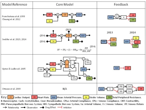
These modeling works were instrumental in illustrating the role of the renal system in long-term fluid balance and BP control. More recent efforts to develop systems pharmacol-ogy models (Figure 3) have generally focused on shorter-term drug effects, and therefore attempt to combine the relationships between HR, BP (also incorporated as mean arterial pressure, or MAP), CO, TPR, and SV, and can be further expanded to include contractility and compliance. Homeostatic feedback from MAP is most commonly imple-mented on HR, CO, SV, and via sympathetic activity to vasculature and is analogous to the mechanism of barore-ceptor feedback, which regulates arterial pressure.
One such example has been applied to the clinical effects of nicardipine and nifedipine,52which are L-type cal-cium channel blockers. The model introduces an additional feature by capturing this feedback through dual mecha-nisms of the proportional and rate-sensitive functions of MAP. Since only HR and MAP are typically observed, while the model includes additional state variables for CO, TPR, and SV, the system suffers structural identifiability issues. A more recent structural identifiability analysis53resulted in a parameter reduction of the model, resolving this issue.
[image:8.613.56.553.102.448.2]Systems pharmacology models have been applied in rat54,55 that try to resolve the identifiability issues by the
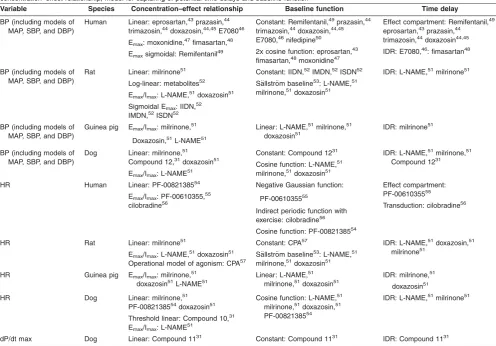
Table 2Overview of the composition of PK/PD models used for modeling of hemodynamic parameters in preclinical species and human, indicating selected
concentration–effect relationship, model for capturing of potential time delays and baseline function
Variable Species Concentration–effect relationship Baseline function Time delay
BP (including models of MAP, SBP, and DBP)
Human Linear: eprosartan,43prazasin,44
trimazosin,44doxazosin,44,45E708046
Emax: moxonidine,47fimasartan,48
Emaxsigmoidal: Remifentanil49
Constant: Remifentanil,49prazasin,44
trimazosin,44doxazosin,44,45
E7080,46nifedipine50
2x cosine function: eprosartan,43
fimasartan,48moxonidine47
Effect compartment: Remifentanil,49
eprosartan,43prazasin,44
trimazosin,44doxazosin44,45
IDR: E7080,46: fimasartan48
BP (including models of MAP, SBP, and DBP)
Rat Linear: milrinone51
Log-linear: metabolites52
Emax/Imax: L-NAME,51doxazosin51
Sigmoidal Emax: IIDN,52
IMDN,52ISDN52
Constant: IIDN,52IMDN,52ISDN52
S€allstr€om baseline53: L-NAME,51
milrinone,51doxazosin51
IDR: L-NAME,51milrinone51
BP (including models of MAP, SBP, and DBP)
Guinea pig Emax/Imax: milrinone,51
Doxazosin,51L-NAME51
Linear: L-NAME,51milrinone,51 doxazosin51
IDR: milrinone51
BP (including models of MAP, SBP, and DBP)
Dog Linear: milrinone,51
Compound 12,31doxazosin51
Emax/Imax: L-NAME51
Constant: Compound 1231
Cosine function: L-NAME,51 milrinone,51doxazosin51
IDR: L-NAME,51milrinone,51
Compound 1231
HR Human Linear: PF-0082138554
Emax/Imax: PF-00610355,55
cilobradine56
Negative Gaussian function:
PF-0061035555
Indirect periodic function with exercise: cilobradine56
Cosine function: PF-0082138554
Effect compartment: PF-0061035555
Transduction: cilobradine56
HR Rat Linear: milrinone51
Emax/Imax: L-NAME,51doxazosin51
Operational model of agonism: CPA57
Constant: CPA57
S€allstr€om baseline53: L-NAME,51 milrinone,51doxazosin51
IDR: L-NAME,51doxazosin,51
milrinone51
HR Guinea pig Emax/Imax: milrinone,51
doxazosin51L-NAME51
Linear: L-NAME,51
milrinone,51doxazosin51
IDR: milrinone,51
doxazosin51
HR Dog Linear: milrinone,51
PF-0082138554doxazosin51
Threshold linear: Compound 10,31 Emax/Imax: L-NAME51
Cosine function: L-NAME,51
milrinone,51doxazosin,51
PF-0082138554
IDR: L-NAME,51milrinone51
dP/dt max Dog Linear: Compound 1131 Constant: Compound 1131 IDR: Compound 1131
monitoring of CO during the model building process. In this approach data from six compounds were combined in order to estimate the rat model parameters. The advantage of these types of models is the ability to determine the site of drug effect, and these have so far been applied to TPR, HR, SV, and CO, although notably direct drug effects on contractility and BP have not been studied in a mechanistic manner.
These hemodynamic models only account for total CO and MAP; however, the blood pressure profile (Figure 1) contains much more information about the heart contraction as driven by the action potential. Calcium plays a critical role in modulating contraction56: its cellular influx following depolarization is an indirect activator of myofilaments ( Fig-ure 2). This process is sensitive to myofilament stretching as the heart fills with blood, resulting in a stronger contrac-tion, and is an important autoregulatory mechanism. Cellu-lar models have therefore been used in describing this interplay between the kinetics of calcium gradient and the dynamics of myocyte contraction57–59 as well as their con-trol via the autonomic nervous system. Adrenergic and muscarinic receptors mediate this process, and drugs can be antagonists at these receptors which may cause
changes in indexes of contractility or other effects, such as beta-blockers (beta1 adrenoreceptor antagonists), which are used in the treatment of hypertension. To further link AP with hemodynamics, models have been produced describing the electromechanics of the whole heart,60,61 and these have combined cell excitation/contraction (EC) coupled with heart mechanics, system circulation,62 and autonomic control.63,64 While great progress has been made defining multiscale systems approaches (Figure 2), these have not yet been fully utilized for linking drug expo-sure with cardiovascular changes for the assessment of safety or efficacy.
[image:9.613.72.544.85.438.2]In our experience, modeling hemodynamic changes for safety assessment often consists of two stages depending on the questions to be addressed. First application of a top-down PK/PD approach to the observable of greatest concern allows us to quantify the effects and reveals the steady-state concentration–response relationship. With this information in hand, one can either assess the margin between projected efficacious and safe exposures or use predicted human pharmacokinetics to generate a predicted magnitude of response at therapeutic doses. PK/PD model-ing of hemodynamic parameters has been demonstrated
successfully and is useful for studies when only a single hemodynamic parameter changes over time and when little or none is known about the underlying mechanism.
Second, existing systems pharmacology models can be applied to HR and BP data to provide insight into the mech-anism of drug effect and the effects on the system as a whole. If system parameters already exist,54 we can fix these and only vary the drug-specific properties. When modeling species without preexisting systems parameters, we may need to develop a unique set of system parame-ters prior to using the system or modify the system param-eters from other species. There is not yet feedback from the clinic to understand how successful these systems approaches are in clinical predictions.
In the future it is expected that systems pharmacology models will incorporate the simple interrelationships between BP and HR, and may overcome the identifiability issues experienced in some existing models. There will
also be future opportunities to explore bottom-up
approaches that have not been applied to drug-induced changes in hemodynamics.
LINK BETWEEN ACUTE FUNCTIONAL EFFECTS AND CARDIAC DAMAGE
Long-term damage or risk of CV failure is often associated
with perturbations of the cardiovascular parameters
reviewed above. Structural damage can arise as a conse-quences of direct drug toxicity (such as necrosis of heart tissue including the valves of the heart), but can also be an indirect consequence of drug-induced dysfunction of
hemo-dynamic or ion channel effects over time. These effects, however, are often poorly characterized. For example, even the link between concentration, QTc prolongation, and TdP, the incidence of drug-induced TdP in all patients taking the drug is likely to be very low and is not well quantified, with less than 4% of all TdP reporting fatalities.5Other forms of cardiovascular damage may also have similarly low inciden-ces of life-threatening or fatal events, and are equally diffi-cult to link back to exposure of the drug.
Cardiotoxic agents, which are characterized by dysfunc-tion of cardiac or vascular smooth muscle, can cause65:
1. Myocardial infarction 2. Venous thromboembolism 3. Cardiac arrest
4. Necrosis (e.g., cocaine) 5. Valve damage66
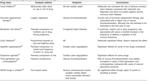
[image:10.613.55.557.93.367.2]Most analyses of the risk of cardiovascular damage and other events concentrate on environment or diet-induced effects and markers of disease progression (Table 3). How-ever, there are a number of examples of statistical analyses associating drug use with cardiovascular changes. Almost all of these studies considered treatment by a particular drug, or the actual dose level, and in no case were PK- or concentration-dependent effects explored. These studies also tended to be long-term and/or retrospective across multiple drugs, and so PK analysis was most likely unfeasi-ble. These were frequently retrospective studies and so exact dose levels were also probably missing, explaining the lack of detailed exposure-driven analysis. Systems approaches to cardiovascular biomarkers for heart failure
Table 3Mathematical approaches to predict cardiac damage
Drug class Analysis method Endpoint Conclusions
Cox2 inhibitors58 Multivariate odds ratios
on use or not of drug
MI and cardiac death Rofecoxib use increases the risk of serious coronary heart disease compared with celecoxib use. Naproxen use does not protect against serious coronary heart disease
Hormone replacement therapy59
Logistic regression Venous thromboembolism Current use of hormone replacement therapy was associated with a higher risk of venous
thromboembolism, although the risk seemed to be restricted to the first year of use.
Bendectin and others60 Pairwise comparison on
mothers use of drug during pregnancy
Congenital Heart Disease In particular, aspirin use in early pregnancy was associated with about a twofold increase in the frequency of defects in septation of the truncus arteriosus
Cox2 inhibitors61 Proportional hazards on
use and high/low dose
MI Rofecoxib significant effect. Aspirin reduces the effect
Appetite suppressants62 Pairwise comparison vs. control and frequency of event vs. drug use
Cardiac valve regurgitation Significant effects for some of the drugs considered
Dopamine agonists63 Pairwise comparison of risk Cardiac valve regurgitation Significant effects for some drugs
Third generation oral contraceptives64
Pairwise comparisons Venous thromboembolism Risk of venous thromboembolism was slightly increased in users of third generation oral contraceptives compared with users of second generation products.
ADHD drugs in children65 Cox hazard ratios Serious cardiovascular events
(sudden cardiac death, acute myocardial infarction, and stroke)
No significant effect though upper CI points to doubling of events
are now being recognized,67 although there are no exam-ples known to date that link such biomarkers to PK/PD modeling. This is potentially an important application of sys-tems modeling to enable the discovery of cardiovascular damage signature based on blood-borne biomarkers: Many markers of cardiovascular disease suffer, on their own, from a lack of specificity versus sensitivity.
TRANSLATIONAL APPROACHES
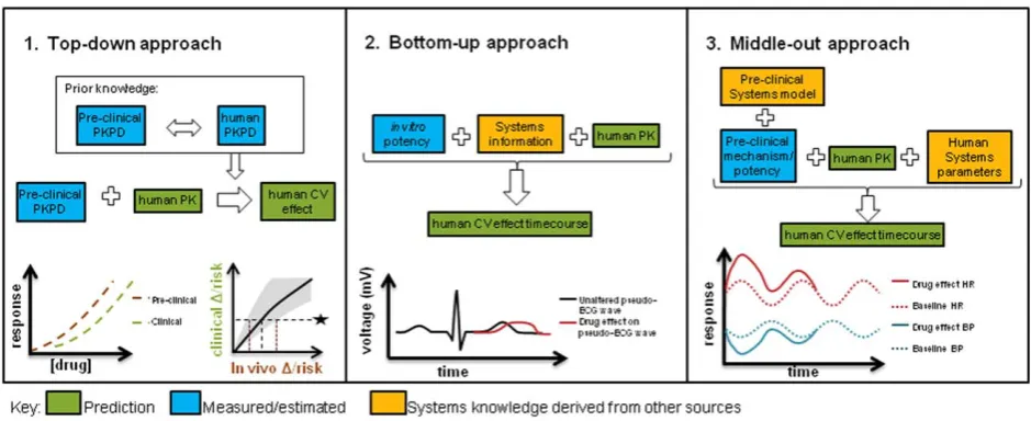
Several types of modeling approaches have been brought to bear on cardiovascular safety, employing various levels of mechanistic insight. One of the major values of modeling for safety assessment is as translational tools to make pro-spective predictions about drug effects in humans.Figure 4
describes some of the model-based translational approaches that have been investigated to date, namely, phenomenologi-cal PK/PD methods, bottom-up systems models, and semi-mechanistic systems pharmacology approaches. Each of these has specific utilities and benefits in the translational context.
Cross-species comparative assessment using descriptive PK/PD modeling and simulation, or “top-down” approaches
The traditional PK/PD framework allows quantification of the biological exposure–response relationship for better determining safe exposures. By integrating data across doses and timepoints, it can capture delayed effects, noisy data, or baseline variation over time that would be more dif-ficult to analyze through the application of standard statisti-cal approaches. PK/PD models have been fitted to data with multiple compounds from preclinical species (dog) and human and then the concentration–response relationship compared.20They proceeded to use the relationship to
pre-dict the likely QTc change of a compound over a specific concentration range that was about to enter clinical studies; however, the concentrations required to reach 10 ms were never reached in human to fully support the prediction made. In similar cross-species comparisons21,68 the proba-bility of reaching 10 ms was estimated for dog and human and this could also be used for prospective predictions. We noticed the reported mean slopes were 7–20-fold higher in human than dog, and had concerns that the dog model was less sensitive than that used in Parkinson et al.20; therefore, we adopted the Parkinson et al. comparative assessment as our predictive strategy for compounds showing QTc prolongation in dog studies.
By carrying out these PK/PD modeling analyses it can help to identify a pharmacokinetic driver of the safety response, for example, Cmax, AUC, or moving average of drug exposure, providing a means to potentially optimize the therapeutic index through appropriate study design.69 This is particularly important when effects have a slow onset (days or weeks) in comparison to daily concentration fluctuations, and the data collected over a prolonged time-scale may necessitate a simplification of exposure to drive the PD model.
While a PK/PD modeling and simulation approach is val-uable in making predictions about an alternative study design under similar conditions, one limitation is that they do not inherently take into account underlying physiological differences across species such as expression levels, impact of different baseline values to level of absolute change, and different turnover rates. It is likely that homeo-static mechanisms, especially for hemodynamic parame-ters, could obscure the underlying concentration–response relationship to different extents across species.
[image:11.613.73.543.86.278.2]Incorporation of drugs with varied or even multiple mech-anisms20 in these cross-species analyses can mitigate sit-uations when a signal is observed preclinically and a
human prediction is required, but the mechanism is unknown or not well understood.
Bottom-up systems pharmacology model usingin vitro
potency
The bottom-up approach utilizes mechanistic knowledge of the system, allowing the input of in vitrodata for prediction of in vivo effects, similar to in vitro to in vivoextrapolation (IVIVE) in PK prediction. However, purely in vitrosystems approaches must prove their value as translational tools and part of the challenge is to understand how these scale
from cellular dynamics to the whole-body system
(Figure 2). While understanding how the single endpoint of
in vitroinhibition compared to in vivo change is a standard part of the integrated preclinical risk assessment,70 now in silicopredictions allow assessment of the combined effects on multiple molecular targets. This is done by combining models at a cellular level (for example, with AP models),44 with a multiscale approach that scales the cellular results up to the in vivo situation (pseudo-ECG). In the example with antipsychotics61 the predicted QTc effects reasonably replicated the mean observed effects. Another example predicts (through a combination of different models and techniques) the PK and PD (QTc prolongation) of domperi-done (metabolized by CYP3A) and ketoconazole (CYP3A inhibitor) in a patient population.71 The effect of co-dosing on PK and PD was investigated, and despite having some issues predicting baseline, and some inconsistencies in the predicted and observed effects, it reproduced the study conclusions fairly well.
To date, this approach has been used to demonstrate translation largely for ion channel inhibition and QTc effects,45,71 although there is systems biology knowledge and therefore the potential for a similar approach to be applied incorporating contractility and hemodynamics. A purely in vitro-driven approach to predicting clinical CV changes has not yet been attempted by us but we look for-ward to the time when this would sit alongside the more established PK/PD approach.
Systems pharmacology, or “middle-out” approaches
The final type of approach represents a middle-out3 approach that attempts to combine the best properties of the purely descriptive top-down and reductionist bottom-up approaches. For example, comprehensive in vivo systems pharmacology models are in existence for CV system behavior, particularly for hemodynamics,52,54,55 but these have so far only been implemented in a single species, making translational predictions from preclinical species to human difficult. We do not have feedback from the clinic yet to understand how successful human predictions have been and there are no reports of cross-species comparisons with these models. Alternatively, simplified model structures can be applied that capture the relevant process governing information flow without over-parameterization.23,47 Models of cardiovascular function with similar structure, but with species-specific parameters could allow for further refine-ment of the predictive power of these approaches. In these approaches, substituting human physiological parameters into the preclinical model is a key part of the translation. For
example, when HR changes have been translated from dog to human using a PK/PD approach,47the sensitivity to drug in addition to a baseline typical for human were applied. In another example where BP was translated using an indirect response model, a human baseline and rate of turnover (kout) for SBP and DBP was obtained from the literature and combined with a PK prediction and the drug sensitivity obtained in dog to successfully predict effects in first-time-in-human studies.23
In contrast to the purely phenomenological approaches, a key component of both the bottom-up and middle-out approaches is the distinction between drug-specific and physiological relevant system-specific parameters. While drug-specific pharmacodynamic parameters describe the interaction of the drug in terms of target affinity and target activation, system-specific parameters describe the proc-esses of the biological system.72 Physiologically based PK models are an example that have been used in this sense and have successfully been used to make human PK pre-dictions and estimate doses.73
There is great opportunity to expand on existing knowl-edge to bring translational understanding to the forefront of preclinical safety pharmacology assessment through a sys-tems pharmacology approach, but there should be a bal-ance between complexity and simplicity (what can actually be ascertained from the measurements or data available in practice). While a systems approach is preferable for trans-lational purposes, it requires understanding or derivation of the drug effect mechanism. It must be acknowledged that when the drug effect mechanism is understood and well
characterized, complex translational models can be
adopted to give a robust translational prediction with certain underlying assumptions. However, when a mechanism is less understood or is a combination of effect mechanisms, it will be more challenging to make translational predictions and simpler approaches should be adopted that rely on fewer assumptions.
DISCUSSION
channel inhibition) and therefore great progress has been made in this area towards reaching well-understood transla-tion to human.
While physiologically based systems approaches for hemodynamics have been in use since the 1970s, there are very few applications of such models for understanding drug effects on the system. So far investigations of human translation have been limited, which may reflect the com-plexity of mathematical models required as well as the rela-tively recent need to reduce attrition in the clinic due to such safety issues. There are several factors that on a practical level may contribute to the limited progress in the area of hemodynamics: first, the lack of appropriate moni-torable biomarkers in this complex system that potentially give rise to difficulties in parameter estimation or structural identifiability issues. Second, the investigation of effects on functional hemodynamic parameters is typically over a rela-tively short (24-hour) timescale, which may not allow drug-induced effects or feedback/disease processes over longer timescales to be observed or quantified in a systems model. Finally, the link to mechanism is often less clear for hemodynamic and contractility effects: the number and nature of molecular targets that could be involved is varied and therefore may be difficult to combine in an in
vitro-driven systems model.
As mechanistic models become further advanced and gain traction in the coming years, we also expect that empirical modeling approaches will remain relevant in the safety space. This is partly because many of the known mechanisms of action for adverse events can be built into
in vitroscreens, and therefore in discovery compounds can be selected that do not interact with these targets. When effects are then observed in vivo, they may frequently be due to as yet unidentified mechanisms, making the use of bottom-up models more difficult.
While standard dog telemetry is the model of choice to investigate preclinical CV risk and make a quantitative trans-lation to human, it is largely set up to investigate acute, func-tional, tightly PK-driven effects such as ECG effects. This type of study was originally designed for statistical analyses looking for significant differences between vehicle and com-pound dosed groups, and the standard designs have not changed considerably in recent years. It is typically a cross-over design in which each dog receives a vehicle dose and three different doses of the drug under investigation with monitoring for up to 24 hours postdose. In order to maximize the utility of dog data there is a need for a strong, collabora-tive, working relationship between those conducting the study and those performing modeling on the ensuing data, to ensure that the study design is appropriate for the effect and the expected model structure if known. Further chal-lenges to model building include incorporating the impact of feeding effects and blood sampling on the CV endpoints that can introduce unexplained or random error in a model if not appropriately accounted for.55 The relative sparsity of blood sampling for pharmacokinetic assessment during telemetry experiments can prove difficult when trying to build a robust pharmacokinetic model.
In addition to increased interpretability and translatability, modeling approaches can provide a strong 3R’s (reduction,
refinement, replacement of animal usage) benefit, as they allow for greater insight to be drawn from smaller numbers of animals and can use prediction to avoid unnecessary studies. Much discussion has taken place concerning the replacement ofin vivomodels entirely within silicoapproaches.74
The most informed translation of drug-induced human changes will require knowledge of underlying physiology to be combined with systems pharmacology approaches so drug potency can be obtained from experimental data. In these scenarios, drug potency can be identified in a preclin-ical setting and would then be combined with human sys-tem parameters and other relevant information to make a well-validated prediction of effects in humans, and this has been shown in other areas such as myelosuppression.75 This approach has enormous potential to aid decision-making and risk assessment on the progression of new drugs into the clinic through their predictive capacity. Fur-ther, application of these systems models promise to increase in-depth understanding of the mechanism of drug effects when an interaction at a specific target has not yet been identified.
Going forward, it will be important that modeling with regard to CV safety focuses increased attention on CV effects such as contractility and structural damage, as efforts have been relatively concentrated on CV parameters that are readily measurable in a longitudinal manner from short-term experiments. Much of the modeling space has been dictated by the availability of data rather than the merit of CV parameters to predict long-term safety risks. The enormous interest and progress in QT has been driven by a large number of molecules entering the clinic that could be explained largely by a single mechanism. As such, the hERG channel role in QT prolongation is now much better understood and can readily be screened in vitro. On the other hand, cardiovascular structural effects (damage) downstream of ECG and hemodynamic changes are relatively difficult to measure and predict, tend to be chronic in nature, and arise from a greater number of potential mechanisms. These therefore represent a poten-tially large safety hazard. However, these effects could be modeled at least empirically through application of techni-ques for categorical variable modeling to preclinical cardio-vascular data.
The relative rarity of cardiovascular damage in the clinic normally means that a strong safety signal will only emerge in large pivotal trials or investigations undertaken through postmarketing surveillance. At this stage this is costly to the sponsor. The challenge is therefore to quantify and pre-dict risk of long-term CV safety issues from more frequently observed CV parameter changes.
Systems modeling may be able to help predict these chronic effects in the clinic as preclinical pathology read-outs are further quantified and combined with short-term parameter changes that have successfully been modeled to date.
Advances through Collaborative Training’’ and T.A. through the NC3Rs/ EPSRC project No. NC/K001205/1 ‘‘Structural Identifiability and Indistin-guishability Analysis as Tools for Quantitative and Systems Pharmacology to Support the 3Rs.’’
Conflict of Interest. T.A.C., J.W.T.Y., and J.T.M. are employees of AstraZeneca Pharmaceuticals.
1. Chen, C., Nakayama, M., Nevo, E., Fetics, B., Maughan, W. & Kass, R. Coupled systolic-ventricular and vascular stiffening with age. Implications for pressure regula-tion and cardiac reserve in the elderly.J. Am. Coll. Cardiol.32, 1221–1227 (1998). 2. Rodeheffer, R.J., Gerstenblith, G., Becker, L.C., Fleg, J.L., Weisfeldt, M.L. & Lakatta,
E.G. Exercise cardiac output is maintained with advancing age in healthy human sub-jects: cardiac dilatation and increased stroke volume compensate for a diminished heart rate.Circulation69, 203–213 (1984).
3. van der Graaf, P.H. CPT: pharmacometrics and systems pharmacology.CPT Phar-macometrics Syst. Pharmacol.1, e8 (2012).
4. Mager, D.E. & Jusko, W.J. Development of translational pharmacokinetic-pharmacodynamic models.Clin. Pharm. Ther.83, 909–912 (2008).
5. Yap, Y.G. & Camm, A.J. Drug induced QT prolongation and Torsades de Pointes.
Heart89, 1363–1372 (2003).
6. Nada, A.et al. The evaluation and management of drug effects on cardiac conduc-tion (PR and QRS intervals) in clinical development. Am. Heart J.165, 489–500 (2013).
7. Perret-Guillaume, C., Joly, L. & Benetos, A. Heart rate as a risk factor for cardiovas-cular disease.Prog. Cardiovasc. Dis.52, 6–10 (2009).
8. Miura, K.et al. Relationship of blood pressure to 25-year mortality due to coronary heart disease, cardiovascular diseases, and all causes in young adult men. The Chicago Heart Association Detection Project in Industry.Arch. Int. Med.161, 1501–1508 (2001). 9. Marian, A.J. & Roberts, R. The molecular genetic basis for hypertrophic
cardiomyopa-thy.J. Mol. Cell. Cardiol.33, 655–670 (2001).
10. FDA. Vioxx (rofecoxib) questions and answers.<http://www.fda.gov/drugs/drugsafety/ postmarketdrugsafetyinformationforpatientsandproviders/ucm106290.htm>(2004). 11. McNaughton, R., Huet, G. & Shakir, S. An investigation into drug products withdrawn
from the EU market between 2002 and 2011 for safety reasons and the evidence used to support the decision-making.BMJ Open4, e004221 (2014).
12. ICH. Safety pharmacology studies for human pharmaceuticals (S7A) (2000). 13. ICH. The non-clinical evaluation of the potential for delayed ventricular repolarization
(QT interval prolongation) by human pharmaceuticals (S7B) (2005).
14. ICH. The clinical evaluation of QT/QTc interval prolongation and proarrhythmic poten-tial for non-antiarrhythmic drugs (E14) (2005).
15. Stockbridge, N., Zhang, J., Garnett, C. & Malik, M. Practice and challenges of thor-ough QT studies.J. Electrocardiol.45, 582–587 (2012).
16. Piotrovsky, V. Pharmacokinetic-pharmacodynamic modeling in the data analysis and interpretation of drug induced QT/QTc prolongation.AAPS J.7, E609–E624 (2005). 17. Polak, S., Wisniowska, B., Fijorek, K., Glinka, A. & Mendyk, A. In vitro-in vivo
extrap-olation of drug-induced proarrhythmia predictions at the population level.Drug Discov. Today19, 275–281 (2014).
18. Holzgrefe, H.et al. Preclinical QT safety assessment: cross-species comparisons and human translation from an industry consortium.J. Pharmacol. Toxicol. Methods69, 61–101 (2014).
19. Upton, R.N. & Mould, D.R. Basic concepts in population modeling, simulation, and model-based drug development: part 3. Introduction to pharmacodynamic modeling methods.CPT Pharmacometrics Syst. Pharmacol.3, e88 (2014).
20. Parkinson, J.et al. Translational pharmacokinetic-pharmacodynamic modeling of QTc effects in dog and human.J. Pharmacol. Toxicol. Methods68, 357–366 (2013). 21. Chain, A., Dubois, V., Danhof, M., Sturkenboom, M. & Della Pasqua, O. Identifying
the translational gap in the evaluation of drug-induced QTc-interval prolongation.Br. J. Clin. Pharmacol.76, 708–724 (2013).
22. Sparve, E.et al. Prediction and modeling of effects on the QTc interval for clinical safety margin assessment, based on single-ascending-dose study data with AZD3839.J. Pharmacol. Exp. Ther.350, 469–478 (2014).
23. Caruso, A., Frances, N., Meille, C., Greiter-Wilke, A., Hillebrecht, A. & Lave, T. Translational PK/PD modeling for cardiovascular safety assessment of drug candi-dates: methods and examples in drug development.J. Pharmacol. Toxicol. Methods
70, 73–85 (2014).
24. Anon. ICH S7A. ICH Guidelines; 2001 (2001).
25. Mao, Z., Wheeler, J.J., Townsend, R., Gao, Y., Kshirsagar, S. & Keirns, J.J. Popula-tion pharmacokinetic-pharmacodynamic analysis of vernakalant hydrochloride injecPopula-tion (RSD1235) in atrial fibrillation or atrial flutter.J. Pharmacokinet. Pharmacodyn.38, 541–562 (2011).
26. Piergies, A.A., Ruo, T.I., Jansyn, E.M., Belknap, S.M. & Atkinson, A.J. Effect kinetics of N-acetylprocainamide-induced QT interval prolongation. Clin. Pharm. Ther. 42, 107–112 (1987).
27. Mathot, R.A.et al. Pharmacokinetic-pharmacodynamic relationship of the cardiovascu-lar effects of adenosine A1 receptor agonist N6-cyclopantyladenosine in the rat.J. Pharmacol. Exp. Ther.268, 616–624 (1994).
28. Priebe, L. & Beuckelmann, D.J. Simulation study of cellular electric properties in heart failure.Circ. Res.82, 1206–1223 (1998).
29. ten Tusscher, K.H.W.J., Noble, D., Noble, P.J. & Panfilov, A.V. A model for human ventricular tissue.Am. J. Physiol. Heart Circ. Physiol.286, H1573–1589 (2004). 30. O’Hara, T., Virag, L., Varro, A. & Rudy, Y. Simulation of the undiseased human
car-diac ventricular action potential: model formulation and experimental validation.PLoS Comp. Biol.7, e1002061 (2011).
31. Iyer, V., Mazhari, R. & Winslow, R.L. A computational model of the human left-ventricular epicardial myocyte.Biophys. J.87, 1507–1525 (2004).
32. Hund, T.J. & Rudy, Y. Rate dependence and regulation of action potential and calcium transient in a canine cardiac ventricular cell model.Circulation110, 3168–3174 (2004). 33. Noble, D., Varghese, A., Kohl, P. & Noble, P. Improved guinea-pig ventricular cell
model incorporating a diadic space, IKr and IKs, and length- and tension-dependent processes.Am. J. Cardiol.14, 123–134 (1998).
34. Shannon, T.R., Wang, F., Puglisi, J., Weber, C. & Bers, D.M. A mathematical treat-ment of integrated Ca dynamics within the ventricular myocyte. Biophys. J. 87, 3351–3371 (2004).
35. Davies, M.R.et al. An in silico canine cardiac midmyocardial action potential duration model as a tool for early drug safety assessment.Am. J. Physiol. Heart Circ. Physiol.
302, H1466–1480 (2012).
36. Moreno, J.D.et al. A computational model to predict the effects of class I anti-arrhythmic drugs on ventricular rhythms.Sci. Transl. Med.3, 98ra83 (2011). 37. Mirams, G.R.et al. Simulation of multiple ion channel block provides improved early
prediction of compounds’ clinical torsadogenic risk.Cardiovasc. Res.91, 53–61 (2011). 38. O’Hara, T. & Rudy, Y. Quantitative comparison of cardiac ventricular myocyte electro-physiology and response to drugs in human and nonhuman species.Am. J. Physiol. Heart Circ. Physiol.302, H1023–1030 (2012).
39. Clayton, R.H.et al. Models of cardiac tissue electrophysiology: progress, challenges and open questions.Prog. Biophys. Mol. Biol.104, 22–48 (2011).
40. Brennan, T., Fink, M. & Rodriguez, B. Multiscale modelling of drug-induced effects on cardiac electrophysiological activity.Eur. J. Pharm. Sci.36, 62–77 (2009). 41. Brennan, T.P., Fink, M., Stokeley, D., Rodriguez, B. & Tarassenko, L. Modelling
effects of sotalol on T-wave morphology.IEEE; 2007.
42. Zemzemi, N.et al. Computational assessment of drug-induced effects on the electro-cardiogram: from ion channel to body surface potentials. Br. J. Pharmacol.168, 718–733 (2013).
43. Wilhelms, M., Rombach, C., Scholz, E.P., D€ossel, O. & Seemann, G. Impact of amio-darone and cisapride on simulated human ventricular electrophysiology and electro-cardiograms.Europace14(suppl. 5), v90–v96 (2012).
44. Mirams, G.R.et al. Prediction of thorough QT study results using action potential sim-ulations based on ion channel screens.J. Pharmacol. Toxicol. Methods70, 246–254 (2014).
45. Glinka, A. & Polak, S. The effects of six antipsychotic agents on QTc—an attempt to mimic clinical trial through simulation including variability in the population.Comp. Methods Biol. Med.47, 20–26 (2014).
46. Norton, K., Iacono, G. & Vezina, M. Assessment of the pharmacological effects of inotropic drugs on left ventricular pressure and contractility: an evaluation of the QA interval as an indirect indicator of cardiac inotropism.J. Pharmacol. Toxicol. Methods
60, 193–197 (2009).
47. Langdon, G.et al. Translational pharmacokinetic-pharmacodynamic modelling; appli-cation to cardiovascular safety data for PF-00821385, a novel HIV agent.Br. J. Clin. Pharmacol.69, 336–345 (2010).
48. Starling, E.H. & Visscher, M.B. The regulation of the energy output of the heart.J. Physiol.62, 243–261 (1927).
49. Sagawa, K., Lie, R.K. & Schaefer, J. Translation of Otto Frank’s paper ‘‘Die grund-form des arteriellen pulses’’ Zeitschrift f€ur biologie 37:483–526 (1899).J. Mol. Cell. Cardiol.22, 255–277 (1990).
50. Guyton, A.C., Coleman, T.G. & Granger, H.J. Circulation: overall regulation.Annu. Rev. Physiol.34, 13–46 (1972).
51. Guyton, A.C. Long term arterial pressure control: an analysis from animal experi-ments and computer and graphic models.Am. J. Physiol.259, R865–R877 (1990). 52. Francheteau, P., Steimer, J.L., Merdjan, H., Guerret, M. & Dubray, C. A mathematical
model for dynamics of cardiovascular drug action: application to intravenous dihydro-pyridines in healthy volunteers.J. Pharmacokinet. Biopharm.21, 489–514 (1993). 53. Cheung, S.Y.A., Majid, O., Yates, J.W.T. & Aarons, L. Structural identifiability analysis
and reparameterisation (parameter reduction) of a cardiovascular feedback model.
Eur. J. Pharm. Sci.46, 259–271 (2012).
54. Snelder, N.et al. PKPD modeling of the interrelationship between mean arterial blood pressure, cardiac output and total peripheral resistance in conscious rats.Br. J. Phar-macol.169, 1510–1524 (2013).
55. Snelder, N. et al. Drug effects on the cardiovascular system in conscious rats: separating cardiac output into heart rate and stroke volume using PKPD modeling.
Br. J. Pharmacol.171, 5076–5092 (2014).
57. Mullins, P.D. & Bondarenko, V.E. A mathematical model of the mouse ventricular myocyte contraction.PLoS One8, 1–15 (2013).
58. Negroni, J.A. & Lascano, E.C. A cardiac muscle model relating sarcomere dynamics to calcium kinetics.J. Mol. Cell. Cardiol.28, 915–929 (1996).
59. Rice, J.J., Wang, F., Bers, D.M. & de Tombe, P.P. Approximate model of cooperative activation and crossbridge cycling in cardiac muscle using ordinary differential equa-tions.Biophys. J.95, 2368–2390 (2008).
60. Trayanova, N.A. & Rice, J.J. Cardiac electromechanical models: from cell to organ.
Front. Physiol.2, 43 (2011).
61. Land, S.et al. An analysis of deformation-dependent electromechanical coupling in the mouse heart.J. Physiol.590, 4553–4569 (2012).
62. Kim, Y.T., Lee, J.S., Youn, C.H., Choi, J.S. & Shim, E.B. An integrative model of the cardiovascular system coupling heart cellular mechanics with arterial network hemo-dynamics.J. Korean Med. Sci.28, 1161–1168 (2013).
63. Heldt, T., Shim, E.B., Kamm, R.D. & Mark, R.G. Computational modeling of cardio-vascular response to orthostatic stress.J. App. Physiol.92, 1239–1254 (2002). 64. Shim, E.B., Jun, H.M., Leem, C.H., Matusuoka, S. & Noma, A. A new integrated method
for analyzing heart mechanics using a cell-hemodynamics-autonomic nerve control coupled model of the cardiovascular system.Prog. Biophys. Mol. Biol.96, 44–59 (2008). 65. Megarbane, B., Aslani, A.A., Deye, N. & Baud, F.J. Pharmacokinetic/ pharmacodynamic modeling of cardiac toxicity in human acute overdoses: utility and limitations.Exp. Opin. Drug Metab. Toxicol.4, 569–579 (2008).
66. Force, T. & Kolaja, K.L. Cardiotoxicity of kinase inhibitors: the prediction and transla-tion of preclinical models to clinical outcomes.Nat. Rev. Drug Discov.10, 111–126 (2011).
67. Azuaje, F.J., Dewey, F.E., Brutsaert, D.L., Devaux, Y., Ashley, E.A. & Wagner, D.R. Systems-based approaches to cardiovascular biomarker discovery.Circ. Cardiovasc. Genet.5, 360–367 (2012).
68. Dubois, V., Yu, H., Danhof, M. & Della, Pasqua O. Model-based evaluation of drug-induced QT(c) prolongation for compounds in early development.Br. J. Clin. Pharma-col.79, 148–161 (2014).
69. Patel, M., Palani, S., Chakravarty, A., Yang, J., Shyu, W.C. & Mettetal J.T. Dose schedule optimization and the pharmacokinetic driver of neutropenia.PLoS One9, e109892 (2014).
70. Jonker, D.M., Kenna, L.A., Leishman, D., Wallis, R., Milligan, P.A. & Jonsson, E.N. A pharmacokinetic-pharmacodynamic model for the quantitative prediction of dofetilide clinical QT prolongation from human ether-a-go-go-related gene current inhibition data.Clin. Pharm. Ther.77, 572–582 (2005).
71. Mishra, H., Polak, S., Jamei, M. & Rostami-Hodjegan, A. Interaction between dom-peridone and ketoconazole: toward prediction of consequent QTc prolongation using purely in vitro information.CPT Pharmacometrics Syst. Pharmacol.3, e130 (2014). 72. Danhof, M., de Lange, E.C., Della Pasqua, O.E., Ploeger, B.A. & Voskuyl, R.A.
Mechanism-based pharmacokinetic-pharmacodynamic (PK-PD) modeling in transla-tional drug research.Trends Pharmacol. Sci.29, 186–191 (2008).
73. Jones, H.M., Mayawala, K. & Poulin, P. Dose selection based on physiologically based pharmacokinetic (PBPK) approaches.AAPS J.15, 377–387 (2013). 74. Mirams, G.R. & Noble, D. Is it time for in silico simulation of drug cardiac side
effects?Ann. N. Y. Acad. Sci.1245, 44–47 (2011).
75. Friberg, L.E., Sandstrom, M. & Karlsson, M.O. Scaling the time-course of myelosup-pression from rats to patients with a semi-physiological model.Invest. New Drugs28, 744–753 (2010).
VC 2015 The Authors CPT: Pharmacometrics & Systems Pharmacology published by Wiley Periodicals, Inc. on behalf of American Society for Clinical Pharmacology and Therapeutics. This is an open access article under the terms of the Creative Commons Attribution License, which permits use, distribution and reproduction in any medium, provided the original work is properly cited.