Original Article
Differential expression of GSK3β and pS9GSK3β in
normal human tissues: can pS9GSK3β be an
epithelial marker?
Hojung Lee1, Jae Y Ro2
1Department of Pathology, Eulji General Hospital, Eulji University School of Medicine, Seoul, Korea; 2Department of Pathology and Genomic Medicine, Houston Methodist Hospital, Weill Medical College of Cornell University, Houston, Texas, USA
Received January 19, 2015; Accepted March 20, 2015; Epub April 1, 2015; Published April 15, 2015
Abstract: Glycogen synthase kinase 3β (GSK3β) and phosphorylated GSK3β at Ser9 (pS9GSK3β) are crucial in cel -lular proliferation and metabolism. GSK3β and pS9GSK3β are deregulated in many diseases including tumors. Data on altered expression of GSK3β and pS9GSK3β are mainly limited to tumor tissues, thus the expression of GSK3β and pS9GSK3β in normal human tissue has been largely unknown. Thus, we examined the immunohistochemical localization of GSK3β and pS9GSK3β in human fetal and adult tissues, and also compared the expression pattern of GSK3β and pS9GSK3β with that of the CK7 and CK20. We found GSK3β expression in neurons of brain, myenter -ic plexus in gastrointestinal tract, squamous epithelium of skin, and mammary gland. The expression of pS9GSK3β was restricted to the epithelial cells of breast and pancreaticobiliary duct, distal nephron of kidney, gastrointestinal tract, fallopian tube, epididymis, secretory cell of prostatic gland, and umbrella cell of urinary tract. The staining pat -tern of pS9GSK3β and CK7 was overlapped in most organs except for gastrointestinal tract where CK7 was negative and CK20 was positive. Our results show that the expression of GSK3β may be associated with differentiation of ectodermal derived tissues and pS9GSK3β with that of epithelial cells of endodermal derived tissues in human. In addition, the expression of pS9GSK3β in the selective epithelial cells may indicate its association with secretory or barrier function of specific cells and may serve as another immunohistochemical marker for epithelial cells.
Keywords: GSK3β, pS9GSK3β, keratin, normal tissues, immunohistochemical study
Introduction
Glycogen synthase kinase 3 (GSK3) is a serine (Ser)/threonine (Thr) kinase involved in multi-ple cellular processes, including proliferation, differentiation, and cell cycle regulation [1]. GSK3 exists as α and β isoforms, and GSK3β has lower molecular weight (47 kDa) than GSK3α (51 kDa) due to lack of glycine-rich N-terminal domain [1]. Their activity is inhibited by Ser/Thr phosphorylation (Ser21 in GSK3α and Ser9 and Thr390 in GSK3β) and activated by tyrosine phosphorylation (Tyr279 in GSK3α and Tyr216 in GSK3β) [1]. Of the two isoforms, GSK3β has been known to be associated with the development of variable diseases including cancer [2-4]. Main regulators of GSK3β activity are the phosphoinositide 3-kinase (PI3K)/Akt pathway and mitogen activated protein
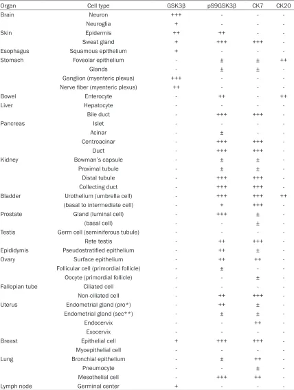
Table 1. Expression of GSK3β, pS9GSK3β, CK7 and CK20 in normal human adult tissues
Organ Cell type GSK3β pS9GSK3β CK7 CK20
Brain Neuron +++ - -
-Neuroglia + - -
-Skin Epidermis ++ ++ -
-Sweat gland + +++ +++
-Esophagus Squamous epithelium + - -
-Stomach Foveolar epithelium - ± ± ++
Glands - ± ±
-Ganglion (myenteric plexus) +++ - -
-Nerve fiber (myenteric plexus) ++ - -
-Bowel Enterocyte - ++ - ++
Liver Hepatocyte - - -
-Bile duct - +++ +++
-Pancreas Islet - - -
-Acinar - ± -
-Centroacinar - +++ +++
-Duct - +++ +++
-Kidney Bowman’s capsule - ± ±
-Proximal tubule - ± ±
-Distal tubule - +++ +++
-Collecting duct - +++ +++
-Bladder Urothelium (umbrella cell) - +++ +++ ++
(basal to intermediate cell) - + +++
-Prostate Gland (luminal cell) - +++ ±
-(basal cell) - - ±
-Testis Germ cell (seminiferous tubule) - - -
-Rete testis - ++ +++
-Epididymis Pseudostratified epithelium - ++ ±
-Ovary Surface epithelium - ++ ++
-Follicular cell (primordial follicle) - ± -
-Oocyte (primordial follicle) - - ±
-Fallopian tube Ciliated cell - - -
-Non-ciliated cell - ++ +++
-Uterus Endometrial gland (pro*) - ++ ±
-Endometrial gland (sec**) - ± ±
-Endocervix - - ++
-Exocervix - - -
-Breast Epithelial cell + +++ +++
-Myoepithelial cell - - -
-Lung Bronchial epithelium - ± ++
-Pneumocyte - - ±
-Mesothelial cell - +++ ++
-Lymph node Germinal center + - -
--, undetectable; ±, < 5% positive cells; +, mild intensity in most cells; ++, moderate intensity in most cells; +++, strong intensity in most cells, *, proliferative phase, **, secretory phase.
dishevelled (Dsh) and β-catenin accumulates in
filament protein cytokeratin (CK), CK7 and CK20.
Materials and methods
Tissue samples and arrays
The list of human tissues was obtained from tissue archives within the Department of Pa- thology at Eulji General Hospital, Eulji University School of Me- dicine. The tissues were collect-ed with informcollect-ed consent prior to each operation and the study was performed with the approv-al of the Institutionapprov-al review board of Eulji General Hospital. Slides of normal adult tissues from surgical specimens and fetal tissues from autopsy were reviewed. Two fetuses were at 21 weeks and 38 weeks of ges-tation, respectively.
leading to transcription of specific target genes [8].
GSK3β has paradoxical role either as a tumor suppressor or as a tumor promoter [3]. GSK3β is a promoter of glioblastoma multiforme (GBM) by protecting the tumor cells from apoptosis via the inactivation of p53- and/or Rb-mediated pathways [3]. In GBM, breast and colon cancer patients, high level expression of GSK3β has been reported [2, 4, 9]. On the contrary, GSK3β function as a tumor suppressor in squamous cell carcinoma (SCC) of skin [10]. The overex-pression of pS9GSK3β has been observed in adenocarcinoma of lung [11] and pancreas [12].
Although the expression level of GSK3β and pS9GSK3β in human tumors has been studied widely, their expression pattern in normal human tissue has been only sporadically reported and received a little attention. The comparison of distribution of GSK3β and pS9GSK3β in normal human tissues would pro-vide better understanding of physiologic and functional role of these proteins. Therefore, we performed an immunohistochemical analysis of GSK3β and pS9GSK3β in normal human adult and fetal tissues, and also compared their expression with that of the intermediate
Representative areas of normal tissues were taken from the paraffin blocks and tissue microarray (TMA) was constructed as previous-ly described [13]. In case of cores of TMA insuf-ficient for representing the tissue, the sections of normal tissue samples were separately prepared.
Immunohistochemistry
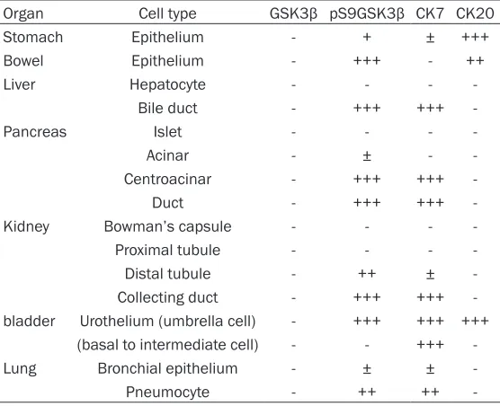
[image:3.612.91.369.97.323.2]Immunohistochemical staining was performed using DAKO Autostainer (DakoCytomation, Carpinteria, CA, USA). Four micron-thick tissue sections were obtained from TMA blocks and transferred onto poly-L-lysine coated slides. After deparaffinization and rehydration, antigen retrieval was performed using citrate buffer (pH 6.0) at 121°C for 10 minutes. Endogenous per-oxidase activity was blocked with 3% hydrogen peroxide for 5 minutes, and the sections were incubated with antibodies against GSK3β (BD Biosciences, Lexington, KY, 1:20), pS9GSK3β (Abcam, Cambridge, UK, 1:250), CK7 (Dako, Carpinteria, CA, 1:100) and CK20 (Dako, 1:50). Color was developed with diaminobenzidine, and the slides were counterstained with hema-toxylin. The tissue section of GBM was used as a positive control for GSK3β and that of pancre-atic adenocarcinoma was used as a positive control for pS9GSK3β. Cases omitted primary Table 2. Expression of GSK3β, pS9GSK3β, CK7 and CK20 in
normal human fetal tissues
Organ Cell type GSK3β pS9GSK3β CK7 CK20
Stomach Epithelium - + ± +++
Bowel Epithelium - +++ - ++
Liver Hepatocyte - - -
-Bile duct - +++ +++
-Pancreas Islet - - -
-Acinar - ± -
-Centroacinar - +++ +++
-Duct - +++ +++
-Kidney Bowman’s capsule - - -
-Proximal tubule - - -
-Distal tubule - ++ ±
-Collecting duct - +++ +++
-bladder Urothelium (umbrella cell) - +++ +++ +++ (basal to intermediate cell) - - +++
-Lung Bronchial epithelium - ± ±
-Pneumocyte - ++ ++
antibodies were served as negative control. The cytoplasmic and/or membranous expres-sion of GSK3β, pS9GSK3β, CK7 and CK20 was approved as positive staining. The staining intensity with the number of positive cells was scored as: -, undetectable; ±, < 5% positive cells; 1+, mild intensity in most cells; 2+, mod-erate intensity in most cells and 3+, strong intensity in most cells.
Results
We found tissue-specific distribution of GSK3β and pS9GSK3β in normal human tissues and corresponding expression of pS9GSK3β and CK7 in epithelia of many organs. Tables 1 and 2 summarize the patterns of GSK3β, p pS9GSK3β, CK7, and CK20 expression in nor-mal adult and fetal tissues, respectively.
Expression of GSK3β in normal human tissues
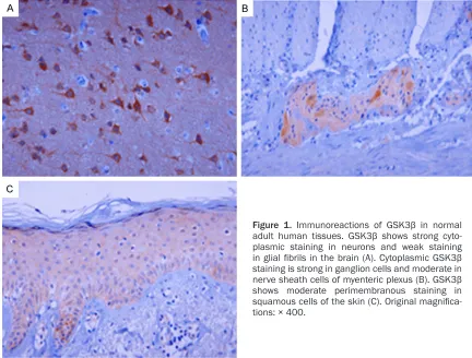
GSK3β expression was found in brain, myen-teric plexus in gastrointestinal tract, skin, mam-mary gland, and lymphoid tissues. Neurons in brain showed strong GSK3β immunoreactivity, while glial fibrils were weakly stained for GSK3β
(Figure 1A). GSK3β expression was strong for ganglion cells of myenteric plexus and moder-ate for nerve sheath cells (Figure 1B). GSK3β was moderately expressed in squamous epi-thelial cells of skin (Figure 1C) and weakly stained in squamous mucosa of the esopha-gus, but not detected in uterine cervix. Sweat glands of skin, mammary gland, and germinal center cells of lymph node showed weak GSK3β staining.
Expression of pS9GSK3β in normal human
tissues
The expression of pS9GSK3β was mainly restricted to the epithelial cells, which lined ductal structures within organs such as breast, prostate, pancreas, liver, kidney, bladder, fallo-pian tube, and epididymis. The luminal epithe-lial cells of the ducts and acini of the breast (Figure 2A) and prostate (Figure 2B) displayed prominent pS9GSK3β expression, whereas basal/myoepithelial layer cells were unreactive. In the pancreas (Figure 2C), the immunoreac-tion of pS9GSK3β was intense in ductal and centroacinar cells and occasionally weakly seen in acinar cells, but negative in Langerhans
[image:4.612.88.520.72.400.2]islets. In the liver, pS9GSK3β expression was only observed in bile duct cells. In the kidney (Figure 2D), pS9GSK3β expression was found strongly in distal tubules and collecting ducts, and occasionally in Bowman’s capsule and proximal tubules. In the bladder, pS9GSK3β expression was strong in urothelial superficial cells and weakly seen in basal to intermediate cells. Strong pS9GSK3β staining in umbrella cells was accentuated in fetal bladder (Figure
2E).
In the female reproductive tract, ovarian sur-face epithelial cells were moderately
[image:5.612.91.522.72.397.2]immuno-reactive for pS9GSK3β. Follicular cells of some primordial follicles were positive for pS9GSK3β, however, oocytes and more mature form of fol-licles, and stromal cells were all negative for this protein In the fallopian tube (Figure 2F), secretory cells were strongly positive for pS9GSK3β, whereas ciliated cells were nega-tive for pS9GSK3β. In the endometrium of the uterus, pS9GSK3β was constantly positive with moderate intensity in proliferative phase glands, but variable in secretory phase glands. The columnar cells of endocervix and squa-mous cells of exocervix were all negative for pS9GSK3β.
In the male reproductive tract, rete testis showed moderate pS9GSK3β staining, but seminiferous tubules showed negative stain-ing. In the epididymis (Figure 2G), the epithelial cells showed diffusely moderate membranous pS9GSK3β staining with strong cytoplasmic staining of pS9GSK3β in scattered cells of the luminal side.
The expression pattern of pS9GSK3β was simi-lar in most organs of adult and fetal tissues, but the difference was noted in the gastrointes-tinal and respiratory tracts. In fetal digestive tract, pS9GSK3β was strongly expressed in the crypts of small and large intestine (Figure 2H) and weakly in the foveolar pits of the stomach. In adult tissue, the intensity of pS9GSK3β was weaker than that in the fetal tissue with addi-tional staining in the surface epithelium of gas-trointestinal tract and body glands of stomach. In fetal lung, pS9GSK3β expression was occa-sionally seen in bronchial epithelium and grad-ually intensified in terminal alveolar unit, where-as, in adult lung, pS9GSK3β expression was faintly seen in certain bronchial epithelia and negative in pneumocytes. Pleural mesothelial cells were strongly positive for pS9GSK3β. The expression of pS9GSK3β was different in the squamous epithelium from mucosa and skin. In the squamous mucosa such as esopha-gus and uterine cervix, pS9GSK3β was not detectable. However, in the epidermis (Figure
2I), pS9GSK3β was moderately stained in inter-cellular bridges of squamous cells. In addition, pS9GSK3β was strongly stained in secretory cells of sweat gland with cytoplasmic and mem-branous patterns. The lymphoid tissue, neural tissue and mesenchymal elements including fibroblasts, endothelial cells, and muscle cells were unreactive for pS9GSK3β.
Comparison of GSK3β, pS9GSK3β, CK7, and
CK20 expression pattern
GSK3β, pS9GSK3β and CK7 showed common expression pattern only in epithelial cells of sweat glands and breast with stronger intensity in CK7 and pS9GSK3β than GSK3β. The expression pattern of pS9GSK3β was mostly corresponding to that of CK7 in epithelia of breast, pancreas, liver, kidney, fallopian tube, and stomach. In the prostate, pS9GSK3β expression was limited to the luminal secretory cells of glands, whereas CK7 expression was
observed sporadically in the basal cells as well as luminal cells. In the bladder, the difference between pS9GSK3β and CK7 expression was that pS9GSK3β was strongly stained in the umbrella cells but CK7 was in the whole urothe-lial layer. In the epididymis, CK7 showed similar scattered staining pattern as seen in pS9GSK3β stain. The ovarian surface epithelia and meso-thelial cells were positive for CK7, similar to pS9GSK3β. Interestingly, CK7 was negative in follicular cells of primordial follicles, but dot-like CK7 immunoreaction was noted in the cyto-plasm of primary oocytes of the ovary.
CK20 expression was restricted in the gastroin-testinal epithelia and umbrella cells of urinary bladder. In the gastrointestinal tract, the expression site of pS9GSK3β and CK20 was slightly different. The expression of pS9GSK3β seen in fetal gastric foveolar pits and intestinal crypts extended to the surface epithelia of the gut in the adult tissue, whereas CK20 was localized in the surface epithelia of gastric and intestinal mucosa in both fetal and adult tis-sues. Distinct CK20 expression in umbrella cell of bladder corresponded to that of pS9GSK3β, which was intensified in fetal tissue than in adult tissue.
Discussion
gland showed the expression of pS9GSK3β as well as GSK3β, suggesting the physiologic requirement of balanced expression of GSK3β and pS9GSK3β in these tissues. Previously, Ma et al [10] report colocalization of GSK3β and pS9GSK3β in the cytoplasm of human kerati-nocytes, which is different from our result showing cytoplasmic or occasional nuclear GSK3β expression and pS9GSK3β expression in intercellular bridges of keratinocytes. In normal breast tissue, GSK3β is shown to be strongly stained in the cytoplasm of mammary gland epithelium [9], however, there is no study of pS9GSK3β expression in breast tissue. In dairy cow, GSK3β and pS9GSK3β regulate pro-liferation of mammary epithelial cells and milk synthesis via the mTOR/S6K1 signaling path-way [6]. The inhibition of GSK3β by lithium chlo-ride, a known inhibitor of GSK3β, promotes GSK3β phosphorylation and increases the expression of mTOR, p-mTOR, S6K1, p-S6K1, CyclinD1, SREBP1, and β-casein, leading to cell growth and synthesis of protein and lipid [6]. In our study, the staining intensity of pS9GSK3β was higher than that of GSK3β in mammary epithelial cells, however, comparative expres-sion level of these two proteins is supposed to be variable depending on the physiological con-dition and hormonal status of the host. The luminal epithelial localization of pS9GSK3β was also observed in secretory cells of sweat gland and prostate in our study. These com-bined findings may imply that the luminal local-ization of pS9GSK3β is associated with the secretory function of the cells of these organs.
We observed intense expression of pS9GSK3β and CK7 in centroacinar and ductal cells of the pancreas, which is corresponding to the result of Pham et al [12]. Pancreatic ductal cell differ-entiation is known to be mediated by PI3K/Akt pathway, while ductal cell proliferation is by the MEK-ERK1/2 pathway [18]. Taken together, PI3K/Akt mediated inactivation of GSK3β appears to be involved in pancreatic ductal cell differentiation.
In the bladder, pS9GSK3β staining was distinc-tively membranous with strong intensity in umbrella cells, corresponding with the staining pattern of CK20 in the urothelium. In mouse urothelium [19], CK20 expression appears in superficial cells on late embryonic days than CK7 expression. CK20 is known as differentia-tion marker of umbrella cells [20] and
contrib-utes to the elastic properties of cytokeratin net-work [19]. The umbrella cells form tight barrier to urine, toxin, and pathogen [21]. Sharp mem-branous staining of pS9GSK3β in umbrella cells may show its association with barrier function of umbrella cells. In addition to CK20, pS9GSK3β is considered to be a possible dif-ferentiation marker of umbrella cell.
results was that GSK3β staining was negligible in the kidney in our study. Further studies would be needed to define the pattern of GSK3β expression in the kidney by comparing various anti-GSK3β antibodies.
We found different expression pattern of pS9GSK3β in the gastrointestinal and respira-tory tracts from fetal and adult tissues. In adult gut mucosa, surface epithelial cells were stained for pS9GSK3β, which was not detected in fetal surface cells of gastrointestinal tract. The intestinal epithelial monolayer provides barrier against various luminal insults and defect in this layer necessitates a rapid repair. In response to wounding, PI3K-dependent GSK3β (Ser9)-phosphorylation is involved in the intestinal epithelial restitution [7]. The expression of pS9GSK3β in the intestinal sur-face epithelia in our result may demonstrate the involvement of pS9GSK3β in maintenance of epithelial barrier, which is more important in adult tissue. Epithelial barrier function is main-tained by GSK-3 regulating apical junctional complex transmembrane proteins such as occludin, claudin-1 and E-cadherin [26].
In the respiratory tract, pS9GSK3β was not detected in pneumocytes of adult lung, where-as moderately stained in those of fetal lung. In fetal mice [27], the activation of PI3K-Akt-mTOR pathway in the lung is associated with delayed maturation of the lung epithelial cells and reduced alveolar capillary density. Thus, down-regulation of the PI3K-Akt-mTOR pathway is required for normal lung epithelial maturation [27]. Although the link of pS9GSK3β with PI3K-Akt-mTOR axis during the development of lung remains unclear, lower expression of pS9GSK3β in the adult than in the fetal lung tissue in our study suggests an association between down-regulated pS9GSK3β and pneumocyte matura-tion. In addition, the expression of pS9GSK3β is highest in adenocarcinoma compared with other types of lung carcinomas [11]. Taken together, pS9GSK3β is suspected to be critical for the fetal alveolar structuring to the certain point in fetal development and tumor histogen-esis in the lung.
The correlation between pS9GSK3β and CK is not known, however, Akt/mTOR signaling path-way, in which GSK3β is a downstream mole-cule, is shown to be closely linked with CK in keratinocyte repair [28]. In wounded keratino-cyte, CK17 is induced and binds with the
adap-tor protein 14-3-3σ, leading to mTOR activity and cell growth [28]. Recently, 14-3-3σ is reported to regulate embryonic stem cell prolif-eration by binding, phosphorylating and sequestrating GSK3β in a PI3K/Akt-dependent manner and enhancing β-catenin translocation into nucleus and proliferative transcription [29]. In our study, CK7 and pSGSK3β were colo-calized in epithelial cells of pancreas, kidney, breast, fallopian tube, and CK20 and pSGSK3β in umbrella cells of bladder. Simultaneous expression of CK and pSGSK3β in certain epi-thelial cells may be coincidental finding. However, in that CK is involved in cell growth and GSK3β is a downstream molecule of PI3K/ Akt/mTOR signaling, further study clarifying the relation between CK and 14-3-3σ mediated pS9GSK3β will be of interest.
In summary, we showed GSK3β expression in ectodermal derived tissues such as, neural tis-sue, skin, sweat gland and mammary gland, and pS9GSK3β expression in the epithelial cells of endodermal derived tissues, such as gastrointestinal tract, pancreaticobiliary tract, and urogenital tract. The cytoplasmic and/or membranous expression of pS9GSK3β in the specific epithelial cells may implicate its asso-ciation with secretory or barrier function depending on the tissue and cell type.
Our comprehensive study on normal adult and fetal tissues would give invaluable information for the future studies on different disease con-ditions, and pS9GSK3β may serve as another immunohistochemical marker for epithelial cells.
Disclosure of conflict of interest
None.
Address correspondence to: Dr. Jae Y Ro, Department of Pathology and Genomic Medicine, Houston Methodist Hospital, Weill Medical College of Cornell University, 6565 Fannin Street, Houston 77030, TX. Tel: 713-441-2263; Fax: 713-793-1603; E-mail: [email protected]
References
[1] Doble BW and Woodgett JR. GSK-3: tricks of the trade for a multi-tasking kinase. J Cell Sci 2003; 116: 1175-1186.
Takahashi Y and Minamoto T. Deregulated GSK3beta activity in colorectal cancer: its as -sociation with tumor cell survival and prolifera-tion. Biochem Biophys Res Commun 2005; 334: 1365-1373.
[3] Mishra R. Glycogen synthase kinase 3 beta: can it be a target for oral cancer. Mol Cancer 2010; 9: 144.
[4] Miyashita K, Kawakami K, Nakada M, Mai W, Shakoori A, Fujisawa H, Hayashi Y, Hamada J and Minamoto T. Potential therapeutic effect of glycogen synthase kinase 3beta inhibition against human glioblastoma. Clin Cancer Res 2009; 15: 887-897.
[5] Cross DA, Alessi DR, Cohen P, Andjelkovich M and Hemmings BA. Inhibition of glycogen syn-thase kinase-3 by insulin mediated by protein kinase B. Nature 1995; 378: 785-789. [6] Zhang X, Zhao F, Si Y, Huang Y, Yu C, Luo C,
Zhang N, Li Q and Gao X. GSK3beta regulates milk synthesis in and proliferation of dairy cow mammary epithelial cells via the mTOR/S6K1 signaling pathway. Molecules 2014; 19: 9435-9452.
[7] Karrasch T, Spaeth T, Allard B and Jobin C. PI3K-dependent GSK3ss(Ser9)-phosphoryla tion is implicated in the intestinal epithelial cell wound-healing response. PLoS One 2011; 6: e26340.
[8] Clevers H and van de Wetering M. TCF/LEF fac -tor earn their wings. Trends Genet 1997; 13: 485-489.
[9] Mylona E, Vamvakaris I, Giannopoulou I, Theohari I, Papadimitriou C, Keramopoulos A and Nakopoulou L. An immunohistochemical evaluation of the proteins Wnt1 and glycogen synthase kinase (GSK)-3beta in invasive breast carcinomas. Histopathology 2013; 62: 899-907.
[10] Ma C, Wang J, Gao Y, Gao TW, Chen G, Bower KA, Odetallah M, Ding M, Ke Z and Luo J. The role of glycogen synthase kinase 3beta in the transformation of epidermal cells. Cancer Res 2007; 67: 7756-7764.
[11] Zheng H, Saito H, Masuda S, Yang X and Takano Y. Phosphorylated GSK3beta-ser9 and EGFR are good prognostic factors for lung car-cinomas. Anticancer Res 2007; 27: 3561-3569.
[12] Pham NA, Schwock J, Iakovlev V, Pond G, Hedley DW and Tsao MS. Immunohistochemical analysis of changes in signaling pathway acti-vation downstream of growth factor receptors in pancreatic duct cell carcinogenesis. BMC Cancer 2008; 8: 43.
[13] Lee H, Choi SK and Ro JY. Overexpression of DJ-1 and HSP90alpha, and loss of PTEN asso -ciated with invasive urothelial carcinoma of urinary bladder: Possible prognostic markers. Oncol Lett 2012; 3: 507-512.
[14] Diekmann U, Lenzen S and Naujok O. A reli -able and efficient protocol for human pluripo -tent stem cell differentiation into the definitive endoderm based on dispersed single cells. Stem Cells Dev 2015; 24: 190-204.
[15] Emily-Fenouil F, Ghiglione C, Lhomond G, Lepage T and Gache C. GSK3beta/shaggy me -diates patterning along the animal-vegetal axis of the sea urchin embryo. Development 1998; 125: 2489-2498.
[16] Itoh K, Tang TL, Neel BG and Sokol SY. Specific modulation of ectodermal cell fates in Xenopus embryos by glycogen synthase kinase. Development 1995; 121: 3979-3988.
[17] Kim L and Kimmel AR. GSK3, a master switch regulating cell-fate specification and tumori -genesis. Curr Opin Genet Dev 2000; 10: 508-514.
[18] Uzan B, Figeac F, Portha B and Movassat J. Mechanisms of KGF mediated signaling in pancreatic duct cell proliferation and differen-tiation. PLoS One 2009; 4: e4734.
[19] Erman A, Veranic P, Psenicnik M and Jezernik K. Superficial cell differentiation during embry -onic and postnatal development of mouse uro-thelium. Tissue Cell 2006; 38: 293-301. [20] Moll R, Lowe A, Laufer J and Franke WW.
Cytokeratin 20 in human carcinomas. A new histodiagnostic marker detected by monoclo-nal antibodies. Am J Pathol 1992; 140: 427-447.
[21] Apodaca G. The uroepithelium: not just a pas -sive barrier. Traffic 2004; 5: 117-128.
[22] Li J, Fadare O, Xiang L, Kong B and Zheng W. Ovarian serous carcinoma: recent concepts on its origin and carcinogenesis. J Hematol Oncol 2012; 5: 8.
[23] Li J, Ning Y, Abushahin N, Yuan Z, Wang Y, Wang Y, Yuan B, Cragun JM, Chambers SK, Hatch K, Kong B and Zheng W. Secretory cell expansion with aging: risk for pelvic serous car-cinogenesis. Gynecol Oncol 2013; 131: 555-560.
[24] Shum WW, Ruan YC, Da Silva N and Breton S. Establishment of cell-cell cross talk in the epi-didymis: control of luminal acidification. J Androl 2011; 32: 576-586.
[25] Kjaersgaard G, Madsen K, Marcussen N, Christensen S, Walter S and Jensen BL. Tissue injury after lithium treatment in human and rat postnatal kidney involves glycogen synthase kinase-3beta-positive epithelium. Am J Physiol Renal Physiol 2012; 302: F455-465.
[27] Ikeda H, Shiojima I, Oka T, Yoshida M, Maemura K, Walsh K, Igarashi T and Komuro I. Increased Akt-mTOR signaling in lung epithelium is asso -ciated with respiratory distress syndrome in mice. Mol Cell Biol 2011; 31: 1054-1065. [28] Kim S, Wong P and Coulombe PA. A keratin cy
-toskeletal protein regulates protein synthesis and epithelial cell growth. Nature 2006; 441: 362-365.