Original Article
Survivin, MMP-2, and MMP-9 expression in different
types of cervical lesions and correlation analysis
Jin Yu1,2*, Qingsheng Xie1*, Hui Zhou1, Yongpai Peng1, Huaiwu Lu1, Tingting Yao1, Zhongqiu Lin1,2
1Department of Gynecological Oncology, Sun Yat-sen Memorial Hospital, Sun Yat-sen University, Guangzhou,
China; 2Key Laboratory of Malignant Tumor Gene Regulation and Target, Therapy of Guangdong Higher Education
Institutes, Sun Yat-sen University, Guangzhou, China. *Equal contributors.
Received November 16, 2015; Accepted March 9, 2016; Epub May 1, 2016; Published May 15, 2016
Abstract: To analyze Survivin, MMP-2, and MMP-9 expression in different types of cervical lesions and discuss their correlation and clinical significance. Patients with chronic cervicitis, cervical intraepithelial neoplasia, or cervical cancer in our hospital were collected. ELISA was applied to detect the level of Survivin, MMP-2 and MMP-9 in serum. Immunohistochemistry was used to measure Survivin, MMP-2 and MMP-9 expression in tissue. Their correlation was analyzed. Survivin, MMP-2, and MMP-9 were overexpressed in chronic cervicitis, cervical intraepithelial neo-plasia, and cervical cancer (P < 0.05). Their levels were related to pathological grade, clinical stage, differentiation, vascular invasion, and lymph node metastasis (P < 0.05), but not age, gross type, and histological type (P > 0.05). Their levels were elevated significantly following increased pathologic classification, clinical upstage, lower differ-entiation degree, vascular invasion and lymph node metastasis emergence. Survivin, MMP-2, and MMP-9 showed escalating trend in the serum and cervical tissue of chronic cervicitis, cervical intraepithelial neoplasia, and cervical cancer patients. They were highly expressed in cervical cancer patients’ serum and tissue. Their levels were cor-related with pathological grade, clinical stage, differentiation, vascular invasion, and lymph node metastasis, and showed obvious elevation.
Keywords: Cervical cancer, Survivin, MMP-2, MMP-9
Introduction
Cervical cancer is a common malignant tumor in female. There were 500,000 new cases around the world every year, and developing countries accounted for 80%. Its mortality rate is high with 233,000 patients died every year. In China, new cases accounted for about 28. 8% of the total amount, which are about 140,000 cases every year [1]. Recent survey showed that cervical cancer presented young trend. Because of its inconspicuous symptoms in early stage and low attention, cervical cancer is often misdiagnosed and missed diagnosed seriously affects the prognosis. In the process of cervical cancer development, there is a type of lesion called time precancerous lesions characterized as long interval and reversible. That is from benign lesions becoming cervical intraepithelial neoplasia (CIN) after a series of pathological development. Cancer develop-ment has multiple stages and steps. Oncogenes
their correlation with different types of cervical lesions.
Materials and methods
General information
60 patients with early cervical invasive cancer between in January 2014 and January 2015 in Sun Yat-sen Memorial Hospital were enrolled. The mean age of patients was 52.5 ± 4.8 (30-70) years old. There were 11 cases in stage Ia, 17 cases in stage Ib, Including 11 cases in stage I, 18 cases in stage IIa, and 14 cases in stage IIb. 10 patients were in G1, 12 patients in G2, and 38 patients in G3. 43 cases were squa-mous cell carcinoma and 19 cases were ade-nocarcinoma, with 16 cases of vascular inva-sion and 10 cases of pelvic lymph node metastasis. 50 CIN patients received surgical treatment in Sun Yat-sen Memorial Hospital were selected with mean age at 51.8 ± 4.0 (35-65) years old. 30 patients with chronic cervici-tis were chose as control with average age at 52.6 ± 4.2 (35-70) years old. The enrolled patients showed no significant difference in gender, age, and weight among different groups (P > 0.05). All of the patients had signed the informed consent, and the study was approved by Sun Yat-sen Memorial Hospital medical eth-ics committee.
Inclusion criteria: All patients were diagnosed by pathology without combining with connec-tive tissue diseases or immune system dis-ease. No patients received preoperative
radio-lene, anhydrous ethanol, paraffin wax, hema-toxylin, neutral balsam (Changzhou Mountain Chemical co., LTD, China).
Clean bench (Formal, USA), centrifuge (Pigeon, Shanghai), inverted microscope (Olympus), microplate reader (TECNA, UK), tissue embed-der (SAKURA, Japan); dehydrator (TIYODA, Japan), ultramicrotome (Leica, Germany), tem-perature vibrator (Jinghong, Shanghai), image analysis system (HP, USA).
ELISA
Fasting venous blood was extracted from patients and centrifuged to collect supernatant and store in refrigerator. ELISA was applied to test Survivin, MMP-2, and MMP-9 content in blood: the kit was put in room temperature for 30 min. standard substance was diluted and added to five repeat holes at each concentra-tion. After sample adding, dosing fluid, wash-ing, developwash-ing, and termination, the plate was read absorbance on microplate reader at 450 nm. Linear regression equation was drawn to calculate the sample concentration.
Immunohistochemistry
Formalin fixation; conventional dehydration, hyalinization; paraffin, embedding; slicing, patch, baking; dewaxing, dehydration; high pressure hot repair; hydrogen peroxide block-ing; goat serum blockblock-ing; 50 μl primary anti-body incubation at room temperature for 1 h; 50 μl horse radish peroxidase labeled
second-Table 1. ELISA detection of Survivin, MMP-2, and MMP-9 content in
serum
Group Cases Survivin (ng/ml) MMP-2 (ng/ml) MMP-9 (ng/ml) Cervical cancer 60 1.16 ± 0.04*,# 1.47 ± 0.07*,# 1.58 ± 0.04*,#
CIN 50 0.09 ± 0.03# 0.08 ± 0.01# 0.07 ± 0.02#
Chronic cervicitis 30 0.03 ± 0.01 0.02 ± 0.01 0.02 ± 0.02
[image:2.612.90.387.97.151.2]*P < 0.05, compared with CIN; #P < 0.05, compared with chronic cervicitis.
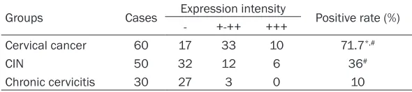
Table 2. Immunohistochemistry detection of Survivin expression in
cervical tissue
Groups Cases Expression intensity Positive rate (%) - +-++ +++
Cervical cancer 60 17 33 10 71.7*,#
CIN 50 32 12 6 36#
Chronic cervicitis 30 27 3 0 10
*P < 0.05, compared with CIN; #P < 0.05, compared with chronic cervicitis.
therapy, chemotherapy, im- mune therapy, freezing or laser treatment.
Reagents and instruments
Na2EDTA anticoagulation,
[image:2.612.89.384.211.277.2]ary antibody incubation for 10 min; 50 μl strep-tavidin-peroxidase incubation for 10 min; DAB development; termination; hematoxylin re-stain, hydrochloric acid and alcohol differentia-tion; conventional dehydration, hyalinizadifferentia-tion; sealing; image system collected 5 random fields from each section and recorded.
Criteria
The known immunohistochemical staining slice was treated as positive control and PBS buffer instead of primary antibody was used as nega-tive control to observe HE staining slice. MMP-2 and MMP-9 positive results were judged negative (-) as no staining in nucleus, while brown or tan particles in cell membrane or cyto-plasm. Weak positive (+), staining cells less than 10%; positive (++), staining cells were 26%-50%; strong positive (+++), staining cells were > 50%.
Survivin positive results were judged negative (-) as no staining in nucleus, while brown or tan particles in cell membrane or cytoplasm. Weak positive (+), staining cells were ≤ 10%; positive (++), staining cells were 26%-50%; strong posi-tive (+++), staining cells were > 50%.
(-) was deemed as negative, while (+)-(+++) were positive. Image system collected 5 ran-dom fields from each section and recorded.
Statistical analysis
All statistical analysis was performed on SPSS19.0 software. Data were expressed as Mean ± SD. Enumerate data was tested by
chi-square test, whereas measurement data was presented as mean ± standard deviation and tested by ANOVA. Logistic regression model was applied for multivariate analysis. P < 0.05 was considered as statistically significant.
Results
ELISA detection of Survivin, 2, and MMP-9 content in serum
Peripheral venous blood was extracted from the patients to detect serum level of Survivin, MMP-2 and MMP-9 using ELISA. The results showed that the level of Survivin was 1.16 ± 0.04 ng/ml, MMP-2 was 1.47 ± 0.07 ng/ml, and MMP-9 was 1.58 ± 0.04 ng/ml in cervical cancer patients, which was obviously higher than that in CIN group and chronic cervicitis group, with statistically significant difference (P < 0.05). Serum Survivin, MMP-2 and MMP-9 levels in CIN group were significantly higher than that in chronic cervicitis group (P < 0.05) (Table 1).
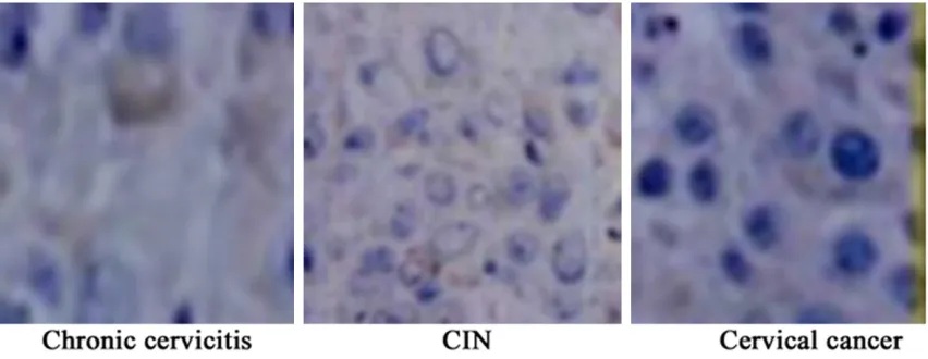
Immunohistochemistry detection of Survivin expression in cervical tissue
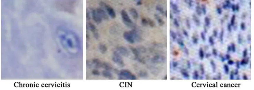
[image:3.612.94.520.74.239.2]Immunohistochemistry detection of MMP-2 and MMP-9 expression in cervical tissue
MMP-2 and MMP-9 expression in cervical tis-sue were tested using immunohistochemistry. The results showed that MMP-2 expression was strongly positive in 10 cases and positive expressed in 38 cases of cervical cancer, while MMP-9 expression was strongly positive in 11 cases and positive in 36 cases of cervical can-cer. Their positive rate achieved 80% and 78.3%, which were obviously higher compared
with CIN and chronic cervicitis (P < 0.05). Furthermore, their expressions in CIN were markedly higher than that in chronic cervicitis (P < 0.05) (Table 3; Figures 2 and 3).
Relationship between Survivin, MMP-2, and MMP-9 expression levels in cervical cancer group and clinicopathological feature
[image:4.612.89.524.88.152.2]The relationship between Survivin, MMP-2, and MMP-9 expression levels in cervical cancer group and clinicopathological features were
Table 3. Immunohistochemistry detection of MMP-2 and MMP-9 expression in cervical tissue
Groups Cases MMP-2 expression intensity MMP-9 expression intensity - +-++ +++ Positive rate (%) - +-++ +++ Positive rate (%) Cervical cancer 60 12 38 10 80*,# 13 36 11 78.3*,#
CIN 50 35 14 1 30# 40 10 0 20#
Chronic cervicitis 30 27 3 0 10 27 3 0 10
[image:4.612.93.520.188.356.2]*P < 0.05, compared with CIN; #P < 0.05, compared with chronic cervicitis.
Figure 2. Immunohistochemical detection of MMP-2 expression in cervical tissue (×400).
[image:4.612.92.523.404.558.2]analyzed, including age, pathologic grade, clini-cal stage, gross type, histologiclini-cal type, differ-entiation degree, vascular invasion, and lymph node metastasis. Their levels were found to be related to pathological grade, clinical stage, dif-ferentiation, vascular invasion, and lymph node metastasis (P < 0.05), but not age, gross type, and histological type (P > 0.05). Their levels elevated significantly following the pathologic classification increase, clinical upstage, lower
MMP-2, and MMP-9 expression using ELISA. It was found that levels of Survivin, MMP-2, and MMP-9 were elevated in cervical cancer com-pared with CIN and chronic cervicitis. They also increased in CIN compared with chronic cervici-tis. We further collected cervical tissue and tested Survivin, MMP-2, and MMP-9 expression by immunohistochemistry. The results present-ed that Survivin, MMP-2, and MMP-9 positive rates were elevated in cervical cancer
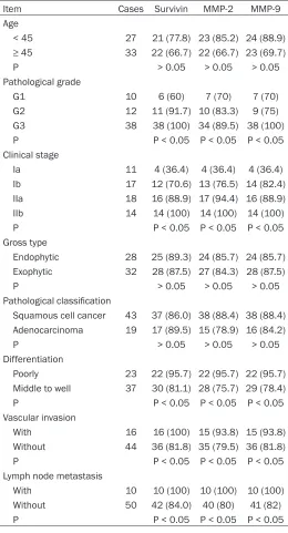
com-Table 4. The relationship between Survivin, MMP-2, and
MMP-9 expression levels in cervical cancer group and clini-copathological feature
Item Cases Survivin MMP-2 MMP-9 Age
< 45 27 21 (77.8) 23 (85.2) 24 (88.9) ≥ 45 33 22 (66.7) 22 (66.7) 23 (69.7) P > 0.05 > 0.05 > 0.05 Pathological grade
G1 10 6 (60) 7 (70) 7 (70) G2 12 11 (91.7) 10 (83.3) 9 (75) G3 38 38 (100) 34 (89.5) 38 (100) P P < 0.05 P < 0.05 P < 0.05 Clinical stage
Ia 11 4 (36.4) 4 (36.4) 4 (36.4) Ib 17 12 (70.6) 13 (76.5) 14 (82.4) IIa 18 16 (88.9) 17 (94.4) 16 (88.9) IIb 14 14 (100) 14 (100) 14 (100) P P < 0.05 P < 0.05 P < 0.05 Gross type
Endophytic 28 25 (89.3) 24 (85.7) 24 (85.7) Exophytic 32 28 (87.5) 27 (84.3) 28 (87.5) P > 0.05 > 0.05 > 0.05 Pathological classification
Squamous cell cancer 43 37 (86.0) 38 (88.4) 38 (88.4) Adenocarcinoma 19 17 (89.5) 15 (78.9) 16 (84.2) P > 0.05 > 0.05 > 0.05 Differentiation
Poorly 23 22 (95.7) 22 (95.7) 22 (95.7) Middle to well 37 30 (81.1) 28 (75.7) 29 (78.4) P P < 0.05 P < 0.05 P < 0.05 Vascular invasion
With 16 16 (100) 15 (93.8) 15 (93.8) Without 44 36 (81.8) 35 (79.5) 36 (81.8) P P < 0.05 P < 0.05 P < 0.05 Lymph node metastasis
With 10 10 (100) 10 (100) 10 (100) Without 50 42 (84.0) 40 (80) 41 (82) P P < 0.05 P < 0.05 P < 0.05
differentiation degree, vascular invasion and lymph node metasta-sis emergence (Table 4).
Discussion
Chronic cervicitis is a common gynecological disease with high incidence and younger trend. It seri-ous impacts on women’s physical and mental health by causing pelvic inflammatory disease and adverse pregnancy that are high risk factors of cervical cancer [6]. CIN showed high canceration rate, containing cervical erosion, cervical polyp, and cervical adenocele. It developed to malignancy through multiple onco-genes and tumor suppressor onco-genes, leading to neovascularization and cell apoptosis [7, 8]. Cervical cancer is the most common female malig-nant diseases in developing coun-tries. In recent years, its incidence gradually increased especially in women younger than 35. It accounts for the leading type in female malig-nancy in our country and showed younger trend [9]. Early cervical cancer can appear invasion, metas-tasis, and poor prognosis without timely treatment. Finding and treat-ing cervical cancer in time to pre-vent tumor invasion and metastasis can prolong the survival time [10]. This study selected chronic cervici-tis, CIN, and cervical cancer patients in our hospital to detect Survivin, MMP-2, and MMP-9 in serum and tissue, and further analyzed their correlation with different types of cervical lesions.
[image:5.612.90.350.108.589.2]pared with CIN and chronic cervicitis, while their levels increased in CIN compared to chronic cervicitis. It is suggested that Survivin, MMP-2, and MMP-9 showed a rising trend in the serum and cervical tissue of chronic cervici-tis, CIN, and cervical cancer. Survivin locates in 17q25 and encodes cytoplasm protein contain-ing 142 amino acids. It showed strong anti-apoptosis ability with the molecular weight reached 16.5 kd [11]. Survivin was overex-pressed in solid tumor and hematological malignancy. It was related to tumor invasion and metastasis and affected prognosis [12]. MMPs can degrade ECM, destroy basement membrane barrier, and decompose local tissue structure, leading to tumor cells invasion and metastasis gradually to the surrounding tissue. Moreover, it also can promote neovasculariza-tion to provide favorable condineovasculariza-tion for tumor cell migration through blood and lymphatic channel [13]. As the most important members of MMPs superfamily, MMP-2 mainly degrades ECM and fibronectin to destroy basement mem-brane integrity. It also can promote neovascu-larization to supply oxygen and nutrients for tumor cells. MMP-9 showed circumscribed function compared with MMP-2 as only decom-posing nestin [14, 15]. Under physiological con-dition, MMP-9 is able to maintain balance through gene expression changes, potential enzyme activation, and MMP synthesis [16]. This study also analyzed the relationship between clinicopathological characteristics and Survivin, MMP-2, and MMP-9 expressions, including age, pathologic grade, clinical stage, gross type, histological type, differentiation degree, vascular invasion, and lymph node metastasis. Their levels were found to be relat-ed to pathological grade, clinical stage, differ-entiation, vascular invasion, and lymph node metastasis, but not age, gross type, and histo-logical type. Their levels elevated significantly following the pathologic classification increase, clinical upstage, lower differentiation degree, vascular invasion and lymph node metastasis emergence. Survivin is hardly expressed in nor-mal mature tissues, while it can be detected in embryonic period, fetal period, and tumor tis-sues. Survivin affects polymerization and depo-lymerization in microtubule through inhibiting caspase-3 and -7 to block G2/M phase and suppress apoptosis. In addition, Survivin par-ticipates in tumor angiogenesis and enhances tumor cell drug resistance [17]. Researchers
discovered that Survivin overexpressed in cer-vical cancer with diameter > 4 cm, lymphatic metastasis, and positive SCC, and can predict prognosis [18]. MMP-2 was found to be overex-pressed in cervical cancer tissues at CIN III and with infiltration and metastasis. Regulating MMP-2 expression can adjust cancer cells’ invasive and metastatic abilities [19]. MMP-9 and MMP-2 were overexpressed in recurrent patients and significantly associated with lymph node invasion and metastasis [20], which was similar with our results.
To sum up, Survivin, MMP-2, and MMP-9 showed a escalating trend in the serum and cervical tissue of chronic cervicitis, CIN, and cervical cancer patients. They were highly expressed in cervical cancer patients’ serum and tissue. Their levels were correlated with pathological grade, clinical stage, differentia-tion, vascular invasion, and lymph node metas-tasis, and showed obvious elevation. Survivin, MMP-2, and MMP-9 combined action promoted cervical cancer occurrence and development. Cervical lesion biopsy and Survivin, MMP-2, and MMP-9 detection have important clinical significance and may provide guidance on patients’ prognosis and treatment.
Acknowledgements
This study was supported by the National Natural Science Foundation of China (3067- 2221, 30872743).
Disclosure of conflict of interest
None.
Address correspondence to: Zhongqiu Lin and Ting- ting Yao, Department of Gynecological Oncology, Sun Yat-sen Memorial Hospital, Sun Yat-sen University, 107 Yan Jiang Rd West, Guangzhou 510120, China. Tel: +86 13802921545; Fax: +86 20 81332853; E-mail: [email protected] (ZQL); Tel: +86 13265991356; Fax: +86 20 81332853; E-mail: [email protected] (TTY)
References
[2] Herszenyi L, Hritz I, Lakatos G, Varga MZ and Tulassay Z. The behavior of matrix metallopro-teinases and their inhibitors in colorectal can-cer. Int J Mol Sci 2012; 13: 13240-13263. [3] Suchanowska A, Smolarek D and Czerwinski
M. A new isoform of Sta gene found in a family with NOR polyagglutination. Transfusion 2010; 50: 514-515.
[4] Lee M, Celenza G, Boggess B, Blase J, Shi Q, Toth M, Bernardo MM, Wolter WR, Suckow MA, Hesek D, Noll BC, Fridman R, Mobashery S and Chang M. A potent gelatinase inhibitor with anti-tumor-invasive activity and its metabolic disposition. Chem Biol Drug Des 2009; 73: 189-202.
[5] Sampieri CL, de la Pena S, Ochoa-Lara M, Zenteno-Cuevas R and Leon-Cordoba K. Ex-pression of matrix metalloproteinases 2 and 9 in human gastric cancer and superficial gastri-tis. World J Gastroenterol 2010; 16: 1500-1505.
[6] Charlotte Gaydos NE, Andrew H. Mycoplasma genitalium as a Contributor to the Multiple Eti-ologies of Cervicitis in Women Attending Sexu-ally Transmitted Disease Clinics. Sex Trasm Dis 2009; 36: 598-606.
[7] Rebolj M, Helmerhorst T, Habbema D, Looman C, Boer R, van Rosmalen J and van Ballegooi-jen M. Risk of cervical cancer after completed post-treatment follow-up of cervical intraepi-thelial neoplasia: population based cohort study. BMJ 2012; 345: e6855.
[8] Granados Lopez AJ and Lopez JA. Multistep model of cervical cancer: participation of miR-NAs and coding genes. Int J Mol Sci 2014; 15: 15700-15733.
[9] Schiffman M, Wentzensen N, Wacholder S, Kinney W, Gage JC and Castle PE. Human pap-illomavirus testing in the prevention of cervical cancer. J Natl Cancer Inst 2011; 103: 368-383.
[10] Mwaka AD, Wabinga HR and Mayanja-Kizza H. Mind the gaps: a qualitative study of percep-tions of healthcare professionals on challeng-es and proposed remedichalleng-es for cervical cancer help-seeking in post conflict northern Uganda. BMC Fam Pract 2013; 14: 193.
[11] Elmer J, Buehler PW, Jia Y, Wood F, Harris DR, Alayash AI and Palmer AF. Functional compari-son of hemoglobin purified by different meth-ods and their biophysical implications. Bio-technol Bioeng 2010; 106: 76-85.
[12] Wu SF, Zhang JW, Qian WY, Yang YB, Liu Y, Dong Y, Zhang ZB, Zhu YP and Feng YJ. Altered expression of survivin, Fas and FasL contrib-uted to cervical cancer development and me-tastasis. Eur Rev Med Pharmacol Sci 2012; 16: 2044-2050.
[13] Baren JP, Stewart GD, Stokes A, Gray K, Pen-nington CJ, O’Neill R, Deans DA, Paterson-Brown S, Riddick AC, Edwards DR, Fearon KC, Ross JA and Skipworth RJ. mRNA profiling of the cancer degradome in oesophago-gastric adenocarcinoma. Br J Cancer 2012; 107: 143-149.
[14] Hagemann C, Anacker J, Ernestus RI and Vince GH. A complete compilation of matrix metallo-proteinase expression in human malignant gli-omas. World J Clin Oncol 2012; 3: 67-79. [15] Kato H, Duarte S, Liu D, Busuttil RW and Coito
AJ. Matrix Metalloproteinase-2 (MMP-2) Gene Deletion Enhances MMP-9 Activity, Impairs PARP-1 Degradation, and Exacerbates Hepatic Ischemia and Reperfusion Injury in Mice. PLoS One 2015; 10: e0137642.
[16] Lindsey ML and Zamilpa R. Temporal and spa-tial expression of matrix metalloproteinases and tissue inhibitors of metalloproteinases fol-lowing myocardial infarction. Cardiovasc Ther 2012; 30: 31-41.
[17] Lu D, Qian J, Yin X, Xiao Q, Wang C and Zeng Y. Expression of PTEN and survivin in cervical cancer: promising biological markers for early diagnosis and prognostic evaluation. Br J Biomed Sci 2012; 69: 143-146.
[18] Lee JP, Chang KH, Han JH and Ryu HS. Sur-vivin, a novel anti-apoptosis inhibitor, expres-sion in uterine cervical cancer and relationship with prognostic factors. Int J Gynecol Cancer 2005; 15: 113-119.
[19] Sheu BC, Lien HC, Ho HN, Lin HH, Chow SN, Huang SC and Hsu SM. Increased expression and activation of gelatinolytic matrix metallo-proteinases is associated with the progression and recurrence of human cervical cancer. Can-cer Res 2003; 63: 6537-6542.