Int. J. Curr. Res. Biosci. Plant Biol. 2015, 2(1): 4-15
Original Research Article
Investigating the Expression of Hepatocyte Growth Factor and Macrophage
Stimulating Protein Genes and their Receptors in Human Scalp Hair Follicles
Saeed A. Alwaleedi*
Faculty of Science, Biology Department, Taif University, Saudi Arabia
*Corresponding author.
A b s t r a c t K e y w o r d s
The study was aimed to investigate whether the mRNA for hepatocyte growth factor (HGF), macrophage stimulating protein (MSP), and their receptors, c-Met and RON, are expressed by human scalp hair follicles. Scalp skin from healthy individuals undergoing cosmetic surgery was transported in RNAlater for molecular biological investigations or growth medium for cell culture. Human scalp hair follicles were microdissected and PCR was carried out. Gene identity was confirmed by sequencing. Further investigation was done at the level of hair bulb components including the dermal sheath, epithelial matrix and dermal papilla to refine the location of these molecules within the hair follicles. In addition, dermal papilla cells were cultured to examine whether its cells express the genes for these molecules. This study demonstrated the expression of HGF, MSP and their receptors in human scalp hair follicles. Results showed that the dermal papilla and dermal sheath of non-balding scalp hair follicles express HGF, MSP, c-Met and RON, while the matrix cells only produce MSP and RON. The results also showed that the relative expression levels of HGF and MSP being higher than that of their receptors, and the expression levels of MSP were significantly higher than that of HGF. This suggests that HGF and MSP may play a role as possible paracrine factors produced by the dermal papilla cells and therefore modulate the hair growth.
c-Met
Gene expression Hair follicle
Hepatocyte growth factor Macrophage stimulating protein
RON
Introduction
Hepatocyte growth factor (HGF) is a multifunctional peptide which acts as a mitogen (Sonnenberg et al., 1993), motogen (Stoker et al., 1987; Weidner et al., 1990) and morphogen (Montesano et al., 1991), and has effects on various epithelial and endothelial cells. HGF
interacts with responsive cells by binding to the cell surface receptor, c-Met, activating a tyrosine kinase signaling cascade (Carpenter and Liao, 2009) leading to regulation of cell growth, cell proliferation, cell motility, and morphogenesis (Medico et al., 1996).
International Journal of Current Research in
Biosciences and Plant Biology
ISSN: 2349-8080 Volume 2 Number 1 (January-2015) pp. 4-15
Int. J. Curr. Res. Biosci. Plant Biol. 2015, 2(1): 4-15 HGF may act as a paracrine factor secreted by mesenchymal-derived cells in a variety of organs and acts primarily upon epithelial cells and endothelial cells (Rubin et al., 1991; Matsumoto and Nakamura, 1992; Sulpice et al., 2009). HGF also plays an important role in embryonic organ development and in adult organ regeneration (Stern et al., 1990), for example in lung formation and repair (Ohmichi et al., 1998; Mizuno et al., 2005; Lassus et al., 2006), the early development of the kidney and kidney repair (Woolf et al., 1995; Van Adelsberg et al., 2001; Baer and Geiger, 2006), and angiogenesis and vascular repair (Montesano et al., 1991; Rosen et al., 1991; McKinnon et al., 2006). HGF may interact with other paracrine factors. Based on in vitro studies, basic fibroblast growth factor (bFGF), which plays a role in embryonic development and wound repair, has been found to stimulate HGF secretion in many of mesenchymal-derived cell lines in human beings (Roletto et al., 1996).
HGF has been implicated in hair growth control. It has been observed to stimulate growth of human scalp hair follicles (Jindo et al., 1995) as well as mouse vibrissae (Jindo et al., 1994) in organ culture. Human recombinant HGF can also stimulate the growth of mouse pelage hair follicles in vivo and retards murine hair follicles regression into catagen (Jindo et al., 1998). When recombinant mouse HGF was injected under the back skin of mice, the result showed that HGF significantly delayed catagen development during both early and late stages of hair follicle regression in vivo (Lindner et al., 2000). The gene for HGF was strongly expressed in rat anagen tissue and slightly in telogen tissue, and HGF mRNA was detected in rat cultured dermal papilla cells, using RT-PCR (Yamazaki et al., 1999). HGF has also been observed to localise to the dermal papilla cells of mouse pelage anagen follicles using immunohistoreactivity (Lindner et al., 2000). These results suggest that HGF can regulate rodent hair follicles and is produced by their dermal papilla. Another study showed that HGF can increase DNA synthesis in cultured keratinocytes derived from human scalp hair bulb in a dose-dependent manner in vitro (Shimaoka et al., 1995). In addition, HGF stimulated the proliferation of rat vibrissae dermal papilla cells with maximal effect at 50ng/ml in a concentration-dependent manner, whereas there was minimal effect on cultured fibroblasts (Yu et al., 2004).
Cultured dermal papilla cells of human scalp hair follicles were found to express HGF, which has stimulatory effects on human scalp hair follicle growth in vitro (Shimaoka et al., 1995). No effects were noticed in HGF gene expression when balding, non-balding scalp and beard cultured dermal papilla cells were cultured with testosterone (Merrick, 2000). However, HGF gene expression was very low in balding scalp cells whereas beard cells showed a greater expression than non-balding scalp cells. This differential expression suggests that HGF may play a role in maintaining large follicles and the levels of HGF in androgen dependent follicles may be changed by in vivo androgen exposure (Randall et al., 2001). The gene for the HGF receptor, c-Met, was expressed in human scalp and rat vibrissae cultured dermal papilla cells as observed by the RT-PCR technique, and it was also found in rat vibrissae follicular matrix keratinocytes by in situ hybridization (Yu et al., 2004). c-Met was also prominently localised in the inner root sheath and outer root sheath in mouse pelage anagen follicles using immunohistochemistry (Lindner et al., 2000).
Another member of hepatocyte growth factor family also implicated in hair follicle growth is macrophage stimulating protein (MSP). It was initially considered as a serum factor which stimulated chemotaxis of peritoneal resident macrophages (Brunelleschi et al., 2001). MSP exerts its actions on target cells by binding to a cell surface receptor called RON (Recepture d'origin Nantaise) in human beings (Camp et al., 2007), also known as macrophage stimulating 1 receptor (MST1R). RON belongs to the Met proto-oncogene family, a distinct subfamily of th receptor tyrosine kinase family (Wang et al., 2003).
Int. J. Curr. Res. Biosci. Plant Biol. 2015, 2(1): 4-15 MSP induced telogen follicles to enter the anagen growth phase in vivo (McElwee et al., 2004). In addition, exposure to different concentrations of MSP in vitro, gave an increase in human hair follicle length in organ culture. Using immunohistochemistry, the MSP receptor, RON, was localised in human hair follicles, with higher intensity in the outer and inner root sheath, and hair matrix, to a lesser extent in the dermal papilla (McElwee et al., 2004). Therefore MSP may also play a role as a paracrine modulator of hair growth.
Materials and methods
Biological materials
Human skin samples from non-balding individuals were obtained from healthy donors undergoing elective cosmetic dermatological surgeries, with full written donor consent. For molecular biological investigations, tissues were collected from occipital regions of three men (aged 30, 29, and 37 year) and two women (aged 32 and 37 year) were placed individually into sterile universal tubes (10 ml) containing RNA stabilization solution, RNAlater (Sigma-Aldrich Ltd., UK), to inhibit RNases. The samples were stored in the fridge at 4°C overnight to allow the RNAlater to penetrate the tissues. For cell culture investigations, two women (aged 34 and 37 year) donated skin. Samples were collected into sterile universal tubes containing RPMI 1640 growth medium (Gibco, Paisly, UK).
Human anagen hair follicles from non-balding scalp were microdissected individually from each skin samples under a leica MZ8 dissecting microscope (Wetzlar, Germany) using sterile equipment and plastic ware. Each skin sample was transferred to a petri dish containing RNAlater for molecular biological investigations or PBS (Oxoid, Basingstoke, UK) for cell culture. The skin sample was cut at the junction between the epidermis and dermis using a sterile scalpel blade. The hair follicles were pulled from the skin gently using fine forceps and then transferred into another petri dish containing fresh RNAlater or PBS kept on ice. The isolated hair follicles were cleaned of any dermis or fat debris under a higher magnification using sterile syringe needles (27G1/2 tuberculin syringe; Sigma). To localize the gene expression in the hair bulb, bulb components, the dermal sheath, dermal papilla,
and epithelial hair matrix, were microdissected from 150 follicles from each individual before separate total RNA isolation. Hair bulb components from three individuals were analyzed separately.
Cell culture procedure
The isolated dermal papilla cells were placed into a polystyrene rectangular canted-neck cell culture flask with a surface area of 25 cm² (Thomas Scientific, USA). Each flask contained 10 ml RPMI 1640 growth medium (Gibco). The growth media was routinely supplemented with penicillin (10 units/ml), streptomycin (100 µg/ml), fungizone (2.5 ng/ml), 2 mM L-glutamine and fetal calf serum (20%, v/v). The flasks were placed in the incubator for 24 hours to allow the cells to attach to the surface of the flasks. Cell culture was carried out in a humidified atmosphere at 37ºC in 95% air and 5% CO2 using a Heraeus B5060 EK incubator. The medium was changed every 3 days. The maintenance of the dermal papilla cells was performed in a Class II MDH laminar flow cabinet. Growing cultures were viewed daily using an inverted, phase contrast microscope (Leitz, Wetzlar, Germany) to observe general morphology and growth conditions. Photographs of cultures were taken under a phase contrast microscope (Leitz).
Int. J. Curr. Res. Biosci. Plant Biol. 2015, 2(1): 4-15 spun down in a Sanyo Harrier 15/80 centrifuge (Jepson Bolton & Co Ltd, UK) at 1200 rpm for about 10 min until the cell pellets were formed. The supernatant was carefully removed to avoid disruption of the cell pellets. The pellets were resuspended in 2 ml of stabilization reagent, RNAlater. The resulting pellets were used for RNA isolation and later cDNA synthesis.
Molecular biological investigations
Total RNA isolation: Total RNA was extracted from scalp anagen follicles or isolated follicular components from each individual immediately after microdissection using the GenElute Mammalian Total RNA kit (Sigma) or RNeasy Mini Kit (Qiagen, Crawly, UK). The extraction process was performed in an area cleaned before use with 70% (v/v) ethanol and RNase Zap solution (Sigma). The quality of total RNA was checked by agarose gel electrophoresis 1.5% (w/v) before further purification to isolate poly (A) RNA (i.e., mRNA) using GenElute mRNA Miniprep Kit (Sigma) following the manufacturer s instructions.
RT-PCR: RT-PCR was used to investigate the expression of mRNA for HGF, MSP, and their receptors; c-Met and RON in anagen scalp hair follicles. In order to ensure that the RNA samples to be used for cDNA synthesis were free of any contaminating DNA, the sample was treated with the DNA amplification Grade I Kit (Invitrogen Ltd., UK). cDNA synthesis was carried out by using an Avian Myloblastosis Virus (AMV) reverse transcription system (Promega, Southampton, UK) to produce single strand cDNA from DNase-treated poly (A) RNA. The cDNA was aliquoted into 10 µl cDNA portions for storage at -20°C until required. PCR ampilification was performed using 5 µl of cDNA in 50 µl reaction volume containing 0.5 µM concentrations of forward and reverse primers (Sigma-Genosys Ltd., Pamisford, UK), 5 µl of 10X PCR reaction buffer (200 mM Tris-HCl, pH 8.4, 500 mM KCl; Invitrogen), 200 µM concentrations of each dNTP (Promega), 1.5 µM (HGF and RON) or 2.5 mM ( -actin, MSP, and c-Met) MgCl2 depending upon primer set, and 0.5 µl of Taq DNA polymerase (5 units/ µl; Invitrogen). For each PCR reaction, a negative control was set up replacing the cDNA with nuclease free water. To prevent evaporation of the reaction mixture, one drop of
mineral oil (Sigma) was added on the top of the mixture. The primer sequences used for RT-PCR had been previously optimised and used previously by other workers. The primers were as follows:
-actin: (Davies et al., 2005)
Forward
5'-TCTGGCACCACACCTTCTACAATGAGCTGCG-3'
Reverse
5'-CGTCATACTCCTGCTTGCTGATCCACATCTGC-3'
HGF: (Shimaoka et al., 1995) Forward
5'-TTCACAACCAATCCAGAGGTACGC-3' Reverse
5'-GAGGGTCAAGAGTATAGCACCATG-3'
MSP: (Shorter et al., 2008) Forward
5'-AGGAGGATGTG GCAGATGC-3' Reverse
5'-GA TTTGATGCCGCAGCTCT-3'
RON: (Matsuzaki et al., 2005) Forward
5'-TCAACTCCCACATCACCATCTG-3' Reverse
5'-AGTGTAAAGCCAGCAGCTCCAT-3'
c-Met: (Imaizumi et al., 2003) Forward
5'-ACTCCCCCTGAAAACCAAAGCC-3' Reverse
5'-GGCTTACACTTCGG GCACTTAC-3'
Int. J. Curr. Res. Biosci. Plant Biol. 2015, 2(1): 4-15
DNA Sequencing of PCR products: To confirm the identity of PCR products, the PCR process was repeated with thin-walled PCR tubes (VWRInternational, Poole, UK). The PCR products were separated by using a low melting point agarose gel to allow the use of a low temperature for dissolving the gel and to facilitate the isolation of DNA fragments from the gel. The DNA fragments were extracted from the gel using a MinElute Gel Extraction Kit (Qiagen), following the manufacturer s instructions. The purified sample of PCR product was sent to Complement Genomics (Sunderland, UK) for sequencing. The sequencing data were compared to the previously identified gene sequences using the NCBI BLAST programme (http:www.ncbi.nlm.nih.gov/blastlb12seq/wblast2.c gi). The chromatogram of the sequencing data was produced using the Chromas Lite software (version 2.0) available from http://www.technelysium.com.au/. Real-time PCR: Real-time PCR was performed using the MyiQ single-colour real-time PCR detection system (Bio-Rad, UK) and SYBER® Green PCR Master Mix (Applied Biosystem, USA). For each real-time PCR reaction, the following reaction mix was prepared and used: SYBER® Green PCR Master Mix (12.5 µl), the forward and reverse primers of the target gene (1 µl each), the cDNA template (1 µl) and the mixture were brought to 25 µl by adding nuclease-free water (9.5 µl). The reaction mixture was then transferred into an optical 96-well reaction plate (Applied Biosystem). Each well of the reaction plate was also supplied with 25 µl of the appropriate PCR master mix. Each well was tightly covered using specific optical caps. The highly expressed housekeeping protein, GAPDH, was used as an endogenous control. The plate was then placed into the real-time PCR machine. Real-time PCR was performed using specific forward and reverse primers for each cDNA target sequence. The annealing temperature for each target primer set was initially optimized using the cDNA template, synthesized from the universal human reference RNA (Stratagene, UK) which composed of total RNA isolated from 10 cell lines representing different human tissues which were chosen to ensure a standard broad coverage of human genes. The real-time PCR was performed under the following cycling conditions: 94ºC for 3 min, followed by denaturing at 94ºC for 15 seconds, annealing (gradient) of 55ºC to 63ºC for 30 seconds,
followed by 72ºC for 15 seconds; this was repeated for 40 cycles. Real-time PCR data and the differences between samples and controls were calculated using the Genex database software based on the comparative ( Ct) equitation method (Livak and Schmittgen, 2001) to calculate relative quantities of a nucleic acid sequence. The Ct is the threshold cycle during which a reaction emits the threshold level of fluorescence. The detectable amount of fluorescence when a signal is significantly greater than background is known as the threshold. Data was normalized to the corresponding values of an endogenous control, glyceraldehyde 3-phosphate dehydrogenase (GAPDH).
Results
Checking the quality of cDNA
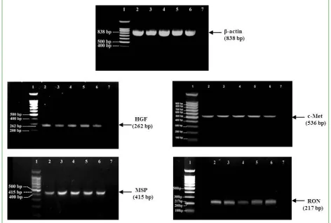
To determine whether human scalp hair follicles actually produce the hepatocyte growth family member genes, the gene expression for HGF, MSP, and their receptors c-Met and RON was investigated using RT-PCR. Total and poly (A) RNA (i.e mRNA) was successfully isolated from microdissected scalp hair follicles (Fig. 1). Prior to investigating the gene expression, the quality of cDNA was initially confirmed by PCR using primers specific for the positive control, housekeeping gene, -actin, a highly expressed cytoskeletal protein.
The detection of -actin as a housekeeping gene would denote that the isolated RNA from all experimental samples was of sufficient quality for reverse transcriptase PCR to be performed successfully. The expression of the -actin sequence which amplified by RT-PCR in all five individual samples were similar in size to that expected from the human sequence which is 838 bp (Fig. 2). The identity of the -actin PCR products was verified by sequence analysis.
Scalp hair follicles express the genes for HGF, MSP and their receptors
Int. J. Curr. Res. Biosci. Plant Biol. 2015, 2(1): 4-15 respectively (Fig. 2). The negative control, in which cDNA was omitted from the PCR reaction mix and replaced with nuclease free water, was clear of any bands. This demonstrated that all PCR products were originated from the amplification of the cDNA synthesis from the mRNA samples and demonstrated an absence of any contamination.
Sequence analysis was used to ascertain the identity of the PCR products. The sequenced PCR product of each gene was compared to the known expected human sequence. Thus, sequencing verified all genes against their relevant human sequences in GeneBank.
Fig. 1: Isolated human scalp hair follicle and its bulb. (A) Scalp skin, showing hair follicles and skin layers. (B) Isolated lower human scalp anagen hair follicle. (C) Isolated human hair bulb shows the
different parts of the hair follicle including the dermal sheath, hair matrix and dermal papilla. The hair
Int. J. Curr. Res. Biosci. Plant Biol. 2015, 2(1): 4-15
Fig. 2: Expression of HGF, MSP and their receptors in non-balding scalp hair follicles. Reverse
transcriptase PCR demonstrated expression of HGF, MSP and their receptors c-Met and RON in mRNA from five human hair follicle samples. Hair follicle cDNA samples were taken from non-balding scalp of five individuals. RT-PCR was performed using specific primers for target genes. The housekeeping gene, -actin, was used as a positive control. PCR products were applied to 1.5% agarose gel for electrophoresis with ethidium bromide staining. DNA was visualized with UV illumination. Lane 1 denotes 100bp DNA molecular size marker (range from 100-1,500), Lane 2-6 human hair follicle PCR products. Lane 7 contained the negative control in which nuclease free water was used as a template instead of cDNA. The cDNA amplification products were predicted to be in length as indicated.
Table 1. Localization of HGF, MSP, and their receptors c-Met and RON in hair bulb
components. Hair follicle components were
microdissected, and their expression of hepatocyte growth factor family member genes was investigated by RT-PCR. Analysis was performed on the dermal sheath, the hair matrix, and the dermal papilla microdissected from 150 scalp follicles from each of 3 individuals separately. Hair bulb components from all three individuals gave the same pattern of expression. , expressed; x, not expressed.
Gene Dermal
sheath
Hair matrix
Dermal papilla
HGF ××× MSP
c-Met ××× RON
Localization of HGF, MSP and their receptors in scalp hair bulb tissues
Int. J. Curr. Res. Biosci. Plant Biol. 2015, 2(1): 4-15 In contrast, the hair matrix samples expressed only MSP and its receptor RON (Table 1). In addition, cultured dermal papilla cells also expressed all four genes (Fig. 3).
The relative expression levels for HGF, MSP and their receptors in scalp follicles
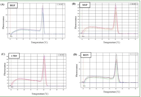
The relative expression levels of HGF, MSP, c-Met, and RON were investigated using relative quantitative real-time PCR. Since SYBR Green binds to any double stranded DNA, it is necessary to examine the specificity of the resulting PCR products of each gene. Melt-curve analysis allows the identification of any non-specific product which may
be amplified with these genes such as genomic DNA contamination and primer-dimers, as the presence of a non-specific product would show up as an additional peak in the melt-curve. The melt-curves for all examined genes contained only single peaks indicating that these reactions generated only one product for each gene in each of the five samples used, and no contaminating products were present (Fig. 4). It is apparent from the graphs that the melting temperature (the inflection point) occurred around 80 C in all investigated genes. Data from all five individual hair follicle samples were collected as cycle threshold (Ct) and the gene expression levels were calculated by normalizing the data against those of the endogenous control GAPDH in each sample. Fig. 4: Melt-curve analysis for HGF, MSP and their receptors, c-Met and RON. Melt-curves were generated by real-time PCR for HGF (A), MSP (B), c-Met (C) and RON (D) from five different
non-balding hair follicle samples. The melt-curves for all genes contain only one peak indicating that no contaminating
products are present in these reactions and the reactions in (A), (B), (C) and (D), all generated only one product.
All five non-balding hair follicle samples expressed HGF, MSP and the receptors c-Met and RON, with the relative expression levels of HGF (7.282 ± 0.657) being higher than that of its receptor, c-Met (4.442 ± 0.537) (p<0.0001) and with expression levels of MSP (11.081 ± 0.560) being higher than
Int. J. Curr. Res. Biosci. Plant Biol. 2015, 2(1): 4-15
Fig. 5: Relative expression levels of HGF, MSP and their receptors in non-balding hair follicles. Relative
quantitative real-time PCR was performed to analyze the relative expression levels of HGF, MSP, c-Met and RON in human non-balding scalp hair follicles. Expression levels were calculated by normalizing the values against those of the endogenous control (GAPDH). Data are the mean values ± SEM from five different individuals.
Discussion
The expression of HGF and its receptor, c-Met, was investigated in isolated non-balding scalp hair follicles, to examine the suggestion that HGF is a possible paracrine factor involved in hair growth. It has been revealed that HGF may act as a powerful modulator of hair growth and be involved in morphogenesis and cycling (Jindo et al., 1995; Lindner et al., 2000). In a variety of organs, HGF is known to exert its effect as a paracrine factor secreted by mesenchyme- derived cells acting on neighbouring epithelial or endothelial cells (Peus and Pittelkow, 1996; Young-Ran et al., 2001). The results of this study indicated that the dermal papilla and dermal sheath have the capacity to synthesise HGF and c-Met indicating their ability to produce c-Met which allow them to respond to HGF. It has been observed that the gene for HGF was strongly expressed in rat anagen, and slightly in telogen, follicles using cDNA extracted from skin sections (Yamazaki et al., 1999) and localised to the dermal papilla cells of mouse pelage anagen follicles (Lindner et al., 2000). Yu et al. (2004) reported c-Met expression in human scalp cultured dermal papilla cells and also in rat follicular matrix keratinocytes.
Since the dermal papilla is believed to be the main regulator of the hair follicle and can determine what sort of hair is produced (Jahoda et al., 2001; Randall, 2008), cultured dermal papilla cells are used extensively as a model system (Itami et al., 1995; Jahoda et al., 2001; Inamatsu et al., 2006; Hamada and Randall, 2006). Therefore, the expression of HGF and c-Met, was examined also in human cultured dermal papilla cells of non-balding scalp hair follicles. The detection of genes for both HGF and its receptor, c-Met, in the dermal papilla cells would enable this signalling system to work as an autocrine regulator. Dermal papilla cells have been shown to secrete autocrine regulatory factors which alter the growth of the dermal papilla cells in vitro (Thornton et al., 1998; Hamada and Randall, 2006). Since the size of the dermal papilla and number of cells it contains are directly related to the size of the hair produced by the follicles (Van Scott and Ekel, 1958; Elliott et. al., 1999), alteration in such factors could be important in alteration in hair follicle and hair size.
Int. J. Curr. Res. Biosci. Plant Biol. 2015, 2(1): 4-15 papilla and dermal sheath of human occipital scalp follicles (McElwee et al., 2004; Shorter, 2007) and also the bulb matrix (Shorter et al., 2008), this would suggests that this signalling pathway works as paracrine signalling system. Since the dermal papilla can express both MSP and the receptor RON, this could be an autocrine response, as suggested earlier in the discussion of HGF and its receptor. On the other hand it could be the more normal paracrine route with the signal being produced by either the dermal papilla or the dermal sheath or the matrix cells and received by the receptors in one of the other tissues. Therefore, MSP and its receptor RON seem good candidates for a paracrine role in alteration of hair growth.
References
Baer, P. C., Geiger, H., 2006. Different effects of growth factors on human renal early distal tubular cells in vitro. Kidney Blood Press. Res. 29, 225-230.
Banu, N., Price, D. J., London, R., Deng, B., Mark, M., Godowski, P. J., and Avraham, H., 1996. Modulation of megakaryocytopoiesis by human macrophage-stimulating protein, the ligand for the RON receptor. J. Immunol. 156, 2933-2940. Brunelleschi, S., Penengo, L., Lavagno, L., Santoro,
C., Colangelo, D., Viano, I., and Gaudino, G., 2001. Macrophage Stimulating Protein (MSP) evokes superoxide anion production by human macrophages of different origin. Br. J. Pharmacol. 134, 1285-1295.
Camp, E. R., Yang, A., Gray, M. J., Fan, F., Hamilton, S. R., Evans, D. B., Hooper, A. T., Pereira, D. S., Hicklin, D. J., and Ellis, L. M., 2007. Tyrosine kinase receptor RON in human pancreatic cancer. Cancer 109, 1030-1039. Carpenter, G., Liao, H. J., 2009. Trafficking of
receptor tyrosine kinases to the nucleus. Exp Cell Res. 315, 1556-66
Danilkovitch, A., Leonard, E. J., 2001. Anti-apoptotic action of macrophage stimulating protein (MSP). Apoptosis 6, 183-190.
Davies, G. C., Thornton, M. J., Jenner, T. J., Chen, Y. J., Hansen, J. B., Carr, R. D., Randall, V. A., 2005. Novel and established potassium channel openers stimulate hair growth in vitro: implications for their modes of action in hair follicles. J. Invest. Dermatol. 124, 686-694.
Elliott, K., Stephenson, T. J., Messenger, A. G., 1999. Differences in hair follicle dermal papilla volume are due to extracellular matrix volume and cell number: implications for the control of hair follicle size and androgen responses. J. Invest. Dermatol. 113, 873-877.
Gandino, L., Longati, P., Medico, E., Prat, M., Comoglio, P. M., 1994. Phosphorylation of serine 985 negatively regulates the hepatocyte growth factor receptor kinase. J. Biol. Chem. 269, 1815-1820.
Hamada, K., Randall, V. A., 2006. Inhibitory autocrine factors produced by the mesenchyme-derived hair follicle dermal papilla may be a key to male pattern baldness. Br. J. Dermatol. 154, 609-618.
Imaizumi, Y., Murota, H., Kanda, S., Hishikawa, Y., Koji, T., Taguchi, T., Tanaka, Y., Yamada, Y., Ikeda, S., Kohno, T., et al., 2003. Expression of the c-Met proto-oncogene and its possible involvement in liver invasion in adult T-cell leukemia. Clin. Cancer Res. 9, 181-187. Inamatsu, M., Tochio, T., Makabe, A., Endo, T.,
Oomizu, S., Kobayashi, E., Yoshizato, K., 2006. Embryonic dermal condensation and adult dermal papilla induce hair follicles in adult glabrous epidermis through different mechanisms. Dev. Growth Differ. 48, 73-86. Itami, S., Kurata, S., Sonoda, T., Takayasu, S.,
1995. Interaction between dermal papilla cells and follicular epithelial cells in vitro: effect of androgen. Br. J. Dermatol. 132, 527-532. Iwama, A., Yamaguchi, N., Suda, T., 1996.
STK/RON receptor tyrosine kinase mediates both apoptotic and growth signals via the multifunctional docking site conserved among the HGF receptor family. Embo J. 15, 5866-5875.
Jahoda, C. A., Oliver, R. F., Reynolds, A. J., Forrester, J. C., Gillespie, J. W., Cserhalmi-Friedman, P. B., Christiano, A. M., Horne, K. A., 2001. Trans-species hair growth induction by human hair follicle dermal papillae. Exp. Dermatol. 10, 229-237.
Jindo, T., Tsuboi, R., Imai, R., Takamori, K., Rubin, J. S., Ogawa, H., 1994. Hepatocyte growth factor/scatter factor stimulates hair growth of mouse vibrissae in organ culture. J. Investig. Dermatol. 103, 306-309.
Int. J. Curr. Res. Biosci. Plant Biol. 2015, 2(1): 4-15 growth factor/scatter factor on human hair follicle growth. J. Dermatol. Sci. 10, 229-232. Jindo, T., Tsuboi, R., Takamori, K., and Ogawa, H.
(1998). Local Injection of Hepatocyte Growth Factor//Scatter Factor (HGF//SF) Alters Cyclic Growth of Murine Hair Follicles. 110, 338-342. Kurihara, N., Iwama, A., Tatsumi, J., Ikeda, K., Suda, T., 1996. Macrophage-stimulating protein activates STK receptor tyrosine kinase on osteoclasts and facilitates bone resorption by osteoclast-like cells. Blood 87, 3704-3710. Kurihara, N., Tatsumi, J., Arai, F., Iwama, A., Suda,
T., 1998. Macrophage-stimulating protein (MSP) and its receptor, RON, stimulate human osteoclast activity but not proliferation: effect of MSP is distinct from that of hepatocyte growth factor. Exp. Hematol. 26, 1080-1085.
Lassus, P., Janer, J., Haglund, C., Karikoski, R., Andersson, L. C., Andersson, S., 2006. Consistent expression of HGF and c-met in the perinatal lung. Biol. Neonate 90, 28-33.
Lindner, G., Menrad, A., Gherardi, E., Merlino, G., Welker, P. I. A., Handjiski, B., Roloff, B., Paus, R., 2000. Involvement of hepatocyte growth factor/scatter factor and Met receptor signaling in hair follicle morphogenesis and cycling. FASEB J. 14, 319-332.
Livak, K. J., Schmittgen, T. D., 2001. Analysis of relative gene expression data using real-time quantitative PCR and Ct method. Methods 25, 402-408.
Matsumoto, K., Nakamura, T., 1992. Hepatocyte growth factor: molecular structure, roles in liver regeneration, and other biological functions. Crit. Rev. Oncog. 3, 27-54.
Matsuzaki, S., Canis, M., Pouly, J. L., Dechelotte, P., Okamura, K., Mage, G., 2005. The macrophage stimulating protein/RON system: a potential novel target for prevention and treatment of endometriosis. Mol. Hum. Reprod. 11, 345-349.
McElwee, K. J., Huth, A., Kissling, S., Hoffmann, R., 2004. Macrophage-stimulating protein promotes hair growth ex vivo and induces anagen from telogen stage hair follicles in vivo. J. Invest. Dermatol. 123, 34-40.
McKinnon, H., Gherardi, E., Reidy, M., Bowyer, D., 2006. Hepatocyte growth factor/scatter factor and MET are involved in arterial repair and atherogenesis. Am. J. Pathol. 168, 340-348. Medico, E., Mongiovi, A. M., Huff, J., Jelinek, M. A., Follenzi, A., Gaudino, G., Parsons, J. T.,
Comoglio, P. M., 1996. The tyrosine kinase receptors Ron and Sea control "scattering" and morphogenesis of liver progenitor cells in vitro. Mol. Biol. Cell 7, 495-504.
Merrick, A. E., 2000. The role of paracrine factors in androgen-regulated human hair growth. Ph.D. thesis, Department of Biomedical Sciences, University of Bradford.
Mizuno, S., Matsumoto, K., Li, M.-Y., Nakamura, T., 2005. HGF reduces advancing lung fibrosis in mice: a potential role for MMP-dependent myofibroblast apoptosis. FASEB J. 19, 580-582.
Montesano, R., Matsumoto, K., Nakamura, T., Orci, L., 1991. Identification of a fibroblast-derived epithelial morphogen as hepatocyte growth factor. Cell 67, 901-908.
Ohmichi, H., Koshimizu, U., Matsumoto, K., Nakamura, T., 1998. Hepatocyte growth factor (HGF) acts as a mesenchyme-derived morphogenic factor during fetal lung development. Development 125, 1315-1324. Peus, D., Pittelkow, M. R., 1996. Growth factors in
hair organ development and the hair growth cycle. Dermatol. Clin. 14, 559-572.
Randall, V. A., 2008. Androgens and hair growth. Dermatol. Ther. 21, 314-328.
Randall, V. A., Hibberts, N. A., Thornton, M. J., Merrick, A. E., Hamada, K., Kato, S., Jenner, T. J., de Oliveira, I., Messenger, A. G., 2001. Do androgens influence hair growth by altering the paracrine factors secreted by dermal papilla cells? Eur. J. Dermatol. 11, 315-320.
Roletto, F., Galvani, A. P., Cristiani, C., Valsasina, B., Landonio, A., Bertolero, F., 1996. Basic fibroblast growth factor stimulates hepatocyte growth factor/scatter factor secretion by human mesenchymal cells. J. Cell Phys. 166, 105-111. Rosen, E. M., Jaken, S., Carley, W., Luckett, P. M.,
Setter, E., Bhargava, M., Goldberg, I. D., 1991. Regulation of motility in bovine brain endothelial cells. J. Cell Physiol. 146, 325-335. Rubin, J. S., Chan, A. M., Bottaro, D. P., Burgess,
W. H., Taylor, W. G., Cech, A. C., Hirschfield, D. W., Wong, J., Miki, T., Finch, P. W., 1991. A broad-spectrum human lung fibroblast-derived mitogen is a variant of hepatocyte growth factor. Proc. Nat. Acad. Sci. United States of America 88, 415-419.
Int. J. Curr. Res. Biosci. Plant Biol. 2015, 2(1): 4-15 growth but not transformation [published erratum appears in Mol. Cell Biol. 1997 17(3), 1758]. Mol. Cell Biol. 16, 7072-7083.
Shimaoka, S., Tsuboi, R., Jindo, T., Imai, R., Takamori, K., Rubin, J. S., Ogawa, H., 1995. Hepatocyte growth factor/scatter factor expressed in follicular papilla cells stimulates human hair growth in vitro. J. Cell Phys. 165, 333-338.
Shorter, K., 2007. Investigation into the mechanisms of factors which alter hair growth. Ph.D. thesis, Department of Biomedical Sciences, University of Bradford
Shorter, K., Farjo, N. P., Picksley, S. M., Randall, V. A., 2008. Human hair follicles contain two forms of ATP-sensitive potassium channels, only one of which is sensitive to minoxidil. FASEB J. 22, 1725-1736.
Skeel, A., Yoshimura, T., Showalter, S. D., Tanaka, S., Appella, E., Leonard, E. J., 1991. Macrophage stimulating protein: purification, partial amino acid sequence, and cellular activity. J. Exp. Med. 173, 1227-1234.
Sonnenberg, E., Meyer, D., Weidner, K. M., Birchmeier, C., 1993. Scatter factor/hepatocyte growth factor and its receptor, the c-met tyrosine kinase, can mediate a signal exchange between mesenchyme and epithelia during mouse development. J. Cell Biol. 123, 223-235. Stern, C. D., Ireland, G. W., Herrick, S. E.,
Gherardi, E., Gray, J., Perryman, M., Stoker, M., 1990. Epithelial scatter factor and development of the chick embryonic axis. Development 110, 1271-1284.
Stoker, M., Gherardi, E., Perryman, M., Gray, J., 1987. Scatter factor is a fibroblast-derived modulator of epithelial cell mobility. Nature 327, 239-242.
Sulpice, E., Ding, S., Muscatelli-Groux, B., Berge, M., Han, Z. C., Plouet, J., Tobelem, G., Merkulova-Rainon, T., 2009. Cross-talk between the VEGF-A and HGF signalling pathways in endothelial cells. Biol. Cell 101, 525-539.
Thornton, M. J., Hamada, K., Messenger, A.G., Randall, V.A., 1998. Beard, but not scalp, dermal papilla cells secrete autocrine growth factors in response to testosterone in vitro. J. Invest. Dermatol. 111, 727-732.
Van Adelsberg, J., Sehgal, S., Kukes, A., Brady, C., Barasch, J., Yang, J., Huan, Y., 2001. Activation of hepatocyte growth factor (HGF) by endogenous HGF activator is required for metanephric kidney morphogenesis in vitro. J. Biol. Chem. 276, 15099-15106.
Van Scott, E. J., Ekel, T. M., 1958. Geometric relationships between the matrix of the hair bulb and its dermal papilla in normal and alopecic scalp. J. Invest. Dermatol. 31, 281-287.
Wang, M. H., Julian, F. M., Breathnach, R., Godowski, P. J., Takehara, T., Yoshikawa, W., Hagiya, M., Leonard, E. J., 1997. Macrophage stimulating protein (MSP) binds to its receptor via the MSP beta chain. J. Biol. Chem. 272, 16999-17004.
Wang, M.-H., Wang, D., Chen, Y.-Q., 2003. Oncogenic and invasive potentials of human macrophage-stimulating protein receptor, the RON receptor tyrosine kinase. Carcinogenesis 24, 1291-1300.
Weidner, K. M., Behrens, J., Vandekerckhove, J., Birchmeier, W., 1990. Scatter factor: molecular characteristics and effect on the invasiveness of epithelial cells. J. Cell Biol. 111, 2097-2108. Woolf, A. S., Kolatsi-Joannou, M., Hardman, P.,
Andermarcher, E., Moorby, C., Fine, L. G., Jat, P. S., Noble, M. D., Gherardi, E., 1995. Roles of hepatocyte growth factor/scatter factor and the met receptor in the early development of the metanephros. J. Cell Biol. 128, 171-184.
Yamazaki, M., Tsuboi, R., Lee, Y. R., Ishidoh, K., Mitsui, S., Ogawa, H., 1999. Hair cycle-dependent expression of hepatocyte growth factor (HGF) activator, other proteinases, and proteinase inhibitors correlates with the expression of HGF in rat hair follicles. J. Investig. Dermatol. Symp. Proc. 4, 312-315. Young-Ran, L., Masashi, Y., Shinichi, M., Ryoji,
T., Hideoki, O., 2001. Hepatocyte growth factor (HGF) activator expressed in hair follicles is involved in in vitro HGF-dependent hair follicle elongation. J. Dermatol. Sci. 25, 156-163. Yu, D., Cao, Q., He, Z., Sun, T. T., 2004.