Banerjee et al.Microbial Cell Factories 2010, 9:30 http://www.microbialcellfactories.com/content/9/1/30
Open Access
R E S E A R C H
Bio
Med
Central
© 2010 Banerjee et al; licensee BioMed Central Ltd. This is an Open Access article distributed under the terms of the Creative CommonsAttribution License (http://creativecommons.org/licenses/by/2.0), which permits unrestricted use, distribution, and reproduction in any medium, provided the original work is properly cited.Research
A novel prokaryotic vector for identification and
selection of recombinants: Direct use of the vector
for expression studies in
E. coli
Sampali Banerjee, Jitendra Kumar, Anjali Apte-Deshpande and Sriram Padmanabhan*
Abstract
Background: The selection of bacterial recombinants that harbour a desired insert, has been a key factor in molecular cloning and a series of screening procedures need to be performed for selection of clones carrying the genes of interest. The conventional cloning techniques are reported to have problems such as screening high number of colonies, generation of false positives, setting up of control ligation mix with vector alone etc.
Results: We describe the development of a novel dual cloning/expression vector, which enables to screen the recombinants directly and expression of the gene of interest. The vector contains Green fluorescence protein (GFP) as the reporter gene and is constructed in such a way that the E. coli cells upon transformation with this vector does not show any fluorescence, but readily fluoresce upon insertion of a foreign gene of interest. The same construct could be easily used for screening of the clones and expression studies by mere switching to specific hosts.
Conclusions: This is the first vector reported that takes the property of colour or fluorescence to be achieved only upon cloning while all the other vectors available commercially show loss of colour or loss of fluorescence upon cloning. As the fluorescence of GFP depends on the solubility of the protein, the intensity of the fluorescence would also indicate the extent of solubility of the expressed target protein.
Background
Gene cloning is a frequently used technique in molecular biology and there are several methods available for screening the recombinants like colony PCR screening, blue white screening, vector carrying toxic gene which gets inactivated upon insertion of any foreign gene, GFP fluorescence vectors wherein upon cloning, the GFP fluo-rescence disappears etc.
The method for screening of bacterial transformants that carry recombinant plasmid with the gene of interest, has become more rapid and simple by the use of vectors with visually detectable reporter genes. The blue white screen is one of the most common molecular techniques that allow detecting the successful ligation of gene of interest in vector [1]. The principle behind this technique is the genetic engineering of the lac operon in the bacte-rial host (E. coli) combined with subunit
complementa-tion (alpha complementacomplementa-tion) achieved with the cloning vector.
Another approach includes the use of toxic genes like
ccdB (used in pDEST vectors, Invitrogen), for positive
selection of right clones, where the cytotoxic gene ccdB
[2-4] gets inactivated if the target gene is inserted upstream of these toxic genes. Therefore the colonies that grow on selective media are only indicative of recombi-nants. The only disadvantage of this vector is that one need to clone the gene of interest into a separate expres-sion vector for expresexpres-sion studies and that would require a second round of screening of the recombinants using conventional methods. The recently published E. coli
toxic gene (TG) [5] also works in a similar fashion, although the mechanism of cytotoxic effect of TG has not yet been elucidated. For the above-mentioned methods, caution is required in selecting the expression host and the stop codon present in the target gene.
The green fluorescent protein (GFP) has emerged, in recent years, as a powerful reporter molecule for moni-toring gene expression, protein localization and
protein-* Correspondence: [email protected]
1 Lupin Limited, Biotechnology R & D, Gat #1156, Ghotawade Village, Mulshi
Taluka, Pune-411042, India
protein interaction. The use of GFP from the jellyfish
Aequorea victoria has been introduced as a tool for the study of gene expression and protein localization in vari-ous systems [6-9]. GFP has been expressed in bacteria, yeast, slime mold, plants, drosophila, zebra fish and in mammalian cells [10]. Inouye et al [11] have described a bacterial cloning vector with mutated Aequorea Victoria
GFP protein as an indicator for screening recombinant plasmids. The pGREENscript A plasmid when expressed in E. coli, produced colonies showing yellow colour in day light and strong green fluorescence under long-UV. Inserted foreign genes are selected on the basis of loss of the fluorescence caused by inactivation of the GFP pro-duction. While GFP solubility appears to be one of the limiting factors in whole cell fluorescence, Davis and
Vierstra [12] have reported about soluble derivatives of GFP for use in Arabidopsis thaliana.
All the above-described plasmids could also result in false positive clones, which is a major concern for researchers. In case of blue-white selection using beta galactosidase, sometimes the blue colour is lost due to chemical instability making it difficult to differentiate a recombinant versus a non-recombinant clone. Also, loss of GFP fluorescence due to medium composition is also known to lead to false positive results. To avoid these problems, we have designed a unique strategy to screen recombinants carrying the gene of interest in such a way that all the clones carrying the foreign gene would show fluorescence, with no exceptions.
Banerjee et al.Microbial Cell Factories 2010, 9:30 http://www.microbialcellfactories.com/content/9/1/30
Page 3 of 8
In this report we describe a novel cloning/expression vector for one-step screening and expression of foreign genes. The strategy uses the cloning of gene for GFP into any expression vector with a stop codon upstream of the ORF of GFP, other than the amber stop codon. Upon induction, the GFP would not express and hence would not fluoresce due to presence of the stop codon. In frame cloning of gene of interest upstream of GFP, would then excise the initial stop codon and the resultant fusion pro-tein would fluoresce. The gene of interest contains an amber stop codon and the recombinant screening is car-ried out in an amber suppressor E. coli strain. For expres-sion studies, the same clone can be used for checking expression in a non amber suppressor E. coli host. This is the first report of a vector where in situ screening of the transformants for the presence of recombinants is possi-ble without any false positive results.
Results
Modification of pBAD24M vector and cloning of GFP
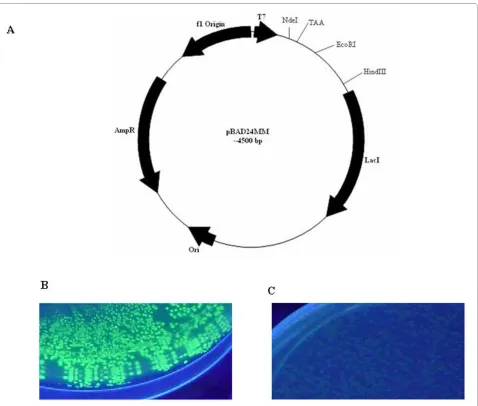
To construct the single step screening and expression vector, a stop codon (like TAA) other than the amber codon (TAG) was introduced between NdeI and EcoRI sites by site directed mutagenesis (SDM) to pBAD24M vector and the resultant construct was designated as pBAD24MM (Fig. 1A). The TAA stop codon of pBAD24MM vector does not allow expression of any for-eign gene when cloned downstream of the same stop codon.
As a reporter gene, the gene for GFP was cloned into vectors pBAD24M and pBAD24MM as EcoRI/HindIII fragment. Both the constructs were transformed into BL21 cells and plated on inducer containing media (13 mM arabinose). Figure 1B shows GFP fluorescence upon UV exposure in pBAD24M while the fluorescence was completely abolished in pBAD24MM (Fig. 1C) vector due to presence of the stop codon. This result indicates that the stop codon TAA interfered with the translation of the protein as expected.
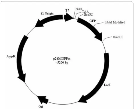
To clone any foreign gene at the NdeI site of the pBAD24MM-GFP vector, the internal NdeI site present in the gene for GFP ought to be removed without altering the amino acid sequence that eventually would also not affect the GFP fluorescence. The internal NdeI site of the GFP gene in the pBAD24M-GFP construct was hence mutated by SDM (data not shown) and the induction studies of such a construct showed no effect on GFP fluo-rescence. Such a modified GFP gene was later sub cloned into pBAD24MM vector and the resultant vector is desig-nated as p24MGFPm (Fig. 2).
Cloning of foreign genes in p24MGFPm vector and in situ screening of the recombinants
Our objective was to use the constructed vector p24MGFPm as a cloning vehicle for screening
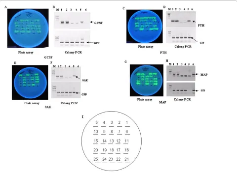
recombi-nants by a unique strategy of appearance of GFP fluores-cence only in recombinants carrying the target gene. All the target genes with amber stop codon (TAG) were used for the cloning and amber suppressor E. coli strain LE392 was used for expression of fusion proteins. Due to inser-tion of the target genes at NdeI/EcoRI site in frame with GFP, the stop codon TAA is removed and only the recom-binants with C-terminus GFP fusion would fluoresce in an amber suppressor strain upon induction and exposure to UV light. To substantiate our concept, we carried out cloning of genes like Staphylokinase (SAK), a soluble pro-tein from Staphylococcus aureus of ~15 kDa, human granulocyte colony stimulating factor (hGCSF) of 18 kDa, human parathyroid hormone (hPTH) of 4 kDa and E. coli
methionine amino peptidase (MAP) of ~26 kDa as NdeI/ EcoRI fragments in p24MGFPm vector so that the resul-tant clones would be C terminal GFP fusions. The recom-binants upon transfer to an amber suppressor E. coli
would glow after induction and could be selected by mere visualization under UV light. Hence, the ligation mix-tures of p24MGFPm and foreign gene(s) were trans-formed to an amber suppressor strain LE392 and plated on LB-amp media. Following day, the colonies were rep-lica plated on 13 mM arabinose containing media and after incubation at 30°C, plates were exposed under UV (long uv) showing some glowing and some non-glowing colonies (Fig. 3A, C, E and 3G). To confirm our hypothe-sis, both fluorescing and non-fluorescing colonies from all the four ligations were subjected to PCR amplification with GFP specific and gene specific primers individually. As shown in Figure 3, all glowing cells were PCR positive for GCSF (Fig. 3B, lanes 1 and 2), PTH (Fig. 3D, lanes 1 and 2), SAK (Fig. 3F, lanes 1 and 2) and MAP (Fig. 3H, lanes 1 and 2) genes and all non glowing colonies were
negative for all the above gene specific PCR (Fig. 3B, D, F, H, lanes 3 and 4). On the other hand all glowing and non-glowing colonies were positive for GFP PCR (Fig. 3B, D, F, H, lower panels) as expected.
Expression of GFP fusions in an amber suppressing strain LE392 and quantitation of GFP fluorescence
To show the expression of the fusion proteins of GFP, the plasmid DNA from all the glowing colonies of the four ligation mix namely GCSF,
p24MGFPm-PTH, p24MGFPm-SAK and p24MGFPm-MAP were introduced into competent cells of LE392 and induced with 13 mM arabinose for 4 hours at 37°C.
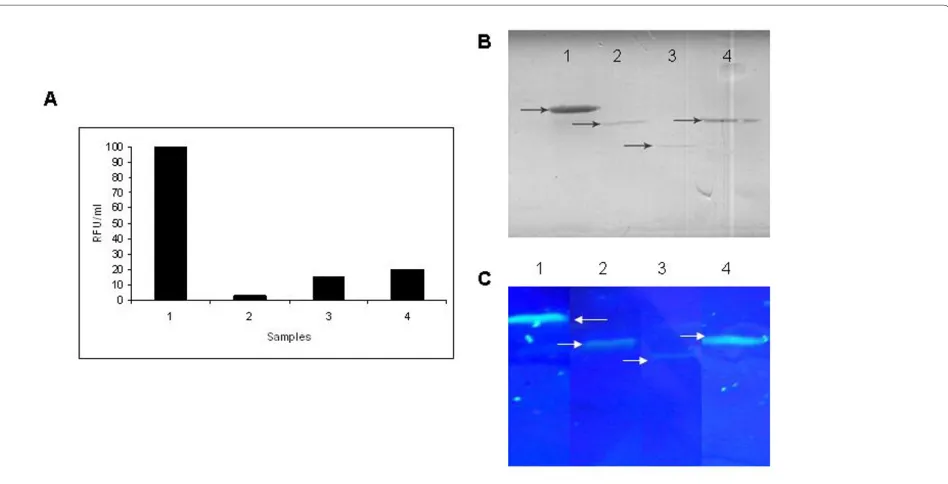
To gain insight into the levels of GFP expressed from all these fusions, GFP assay was carried out from all the sol-uble fractions of GFP fusion proteins from normalized cell density using the kit from Cell Bio labs, USA as per manufacturer's instructions along with denaturing SDS-PAGE followed by western blot. It is interesting to see that units of GFP fluorescence varied based on the
Banerjee et al.Microbial Cell Factories 2010, 9:30 http://www.microbialcellfactories.com/content/9/1/30
Page 5 of 8
bility of the target protein (Fig. 4A). The expression of all the GFP fusion proteins in the soluble fractions was con-firmed by western blot using anti-GFP antibody (Fig. 4B). The SDS-PAGE profile of the soluble fractions of the fusion proteins without sample boiling (Fig. 4C) showed that better solubility of the GFP fusions leads to higher fluorescence.
Expression of the genes of interest in a non amber suppressing E. coli strain BL21
The glowing clones of GCSF, SAK and MAP along with non-glowing clones from all the experimental sets were introduced in BL21 cells (a non amber suppressor E. coli
B strain) and expressions were induced with 13 mM L(+) arabinose. Expression of the heterologous proteins was analyzed on regular SDS-PAGE followed by Coomassie blue staining. As expected, the protein synthesis from glowing colonies was terminated at amber stop codon in all the constructs and proteins of expected molecular size for example, GCSF (18 kDa), SAK (15 kDa) and MAP (26 kDa) were seen on SDS-PAGE (Fig. 5, lanes 2). The non-glowing clones from all the sets did not express any recombinant protein since they were non-recombinants with TAA at the N terminus of the GFP gene (Fig. 5, lanes 1). The expression of PTH (~4 kDa) was not tested in this study since expression of untagged PTH in E. coli yields
very low amounts of PTH due to its RNA and the protein instability.
Discussion
In this study, we report the development of a dual-func-tion prokaryotic vector to be used for in situ screening of recombinants as well as for expression studies. In a con-ventional method, the transformants are screened either by carrying out the colony PCR or plasmid preparation to confirm the recombinants. Recently, use of many toxic
Figure 4 Effect of solubility of target protein on GFP fluorescence. A. Relative GFP fluorescence assay for GFP fusion proteins. B. Western blot of soluble fraction of GFP fusion proteins using anti-GFP antibody. Lane 1: MAP-GFP, Lane 2: GCSF-GFP, Lane 3: PTH-GFP, Lane 4: SAK-GFP. C. SDS-PAGE profile of the soluble fractions of the expressed fusion proteins followed by visualization under UV light. The induced cells expressing the fusion pro-teins were normalized to equal cell density and lysed by sonication. The soluble fractions were analyzed on SDS-PAGE followed by visualization upon UV exposure. Lane 1: MAP-GFP fusion (~55 kDa), lane 2: GCSF-GFP fusion protein (~41 kDa), lane 3: PTH-GFP fusion (~30 kDa), and lane 4: SAK-GFP fusion (~41 kDa). Arrows indicate the fusion proteins. Note the difference in fluorescence intensities of different fusion proteins directly reflecting the solubility of the target gene.
Figure 5 SDS-PAGE showing protein expression from one glow-ing and one non-glowglow-ing clones of p24MGFPm-MAP,
genes like ccdB, TG etc. have been reported for screening the recombinants. We have constructed a novel vector with GFP as a reporter gene for screening and expression of recombinants. For the first time we have successfully shown that upon insertion of any target gene upstream of GFP results in appearance of GFP fluorescence which provides direct evidence of presence of the target gene in the vector.
Earlier reports have suggested that GFP expression is affected by OmpT proteases as there are two putative OmpT protease sites in the coding region of gene for GFP [13] and with the observations that since OmpT expres-sion is low at 28-30°C, the GFP expresexpres-sion is pronounced at these temperatures [14,15]. Inclusion of inhibitors of OmpT like zinc chloride and copper chloride at 0.1 to 0.5 mM final concentration also are known to reduce OmpT expression in such cells [16]. Based on these literature reports,, we carried out all our GFP fluorescence plate assays at 30°C instead of regular 37°C though we did not observe any enhancement of fluorescence intensity upon addition of metal ions. To substantiate the use of amber stop codon in the target gene for recombinant screening, we have used E. coli LE 392 strain which is known to be a strong amber suppressor strain.
All other commercially avaliable vectors show loss of color or loss of fluorescence that may not be unfailing while the major advantage of the vector described in this report, takes the property of color or fluorescence obtained after cloning. This unique vector would also be applicable with any other reporter genes like beta galacto-sidase gene, luciferase gene, DsRed protein instead of the described gene for GFP in the same vector constructed similarly. It also provides researchers to skip to set up the control ligation mix (without insert) and the dephospho-rylation step (CIP or SAP step) since the religated vector would never glow and all the fluorescing colonies are indicative of only the recombinants and also indicative of correct reading frame of the inserted target gene. Since GFP fluorescence is brightest when it is expressed in sol-uble form [17], the intensity of the fluorescence after cloning the foreign gene would also indicate the extent of solubility of the fusion protein. We have cloned four genes namely hGCSF, hPTH, SAK and MAP as examples where GCSF is a known insoluble protein while SAK and MAP are known soluble proteins when expressed in E. coli. Since untagged PTH is known to be unstable in E.
coli [18], we did not study its expression in a non-amber
suppressing strain. Our plate assay data showing the least intensity with the GCSF-GFP fusion and maximum intensity with MAP-GFP along with similar data from GFP quantitation assay indicate a new possibility of use of this vector for predicting the solubility status of the for-eign gene. Moreover, upon switching to a non-suppressor strain like BL21, this construct would yield proteins with
no extra amino acid at the N-terminus. A prokaryotic vector indicating solubility of any foreign gene product based on cloning is the first report of its kind.
Conclusions
In conclusion, we report a novel dual purpose vector for
in situ recombinant screening and expression of genes in
bacterial system. The fluorescence intensity of the reporter gene with the fusion partner is indicative of the solubility index for the target protein.
Methods
Construction of vectors and clones
Modification of pBAD24M vector
The pBAD24M vector [18] was modified by introducing a STOP codon other than amber codon after NdeI site using site directed mutagenesis (SDM) kit following man-ufacturer's (Stratagene, USA) protocol. The primers used was as follows; forward: 5' T AGC ATG ACT GGT GGA CAG TAA ATG GGT CGC GGA TCC GAA TTC GA 3'
Reverse: 5' TC GAA TTC GGA TCC GCG ACC CAT
TTA CTG TCC ACC AGT CAT GCT A 3'.
The incorporated STOP is shown in bold and this mod-ified vector was designated as pBAD24MM.
Cloning of GFP in pBAD24M and pBAD24MM vectors
The GFP gene was PCR amplified from pGFPuv (Clonetech) by using the following primers; forward: 5' TCC CCC ATG GTA GAA TTC AGT AAA GGA GAA GAA CTT TTC ACT 3'; reverse: 5' CCG CCG GTC GAC AAG CTT TTA TTT GTA GAG CTC ATC CAT GCC 3' and cloned into pBAD24M and pBAD24MM as EcoRI/ HindIII fragment.
Modification of internal NdeI restriction site of the gene for GFP
Cloning of genes of interest is usually done at NdeI site of the vector so as to obtain an efficient translation initia-tion. As the gene for GFP contains an internal NdeI site (at 230 position), this site was removed by SDM using the following primers, forward: 5' GC TTT TCC CGT TAT CCG GAT CAC ATG AAA CGG CAT GAC 3'; reverse: 5' GTC ATG CCG TTT CAT GTG ATC CGG ATA ACG GGA AAA GC 3' without altering the amino acid (changes are shown by underline). This modification did not affect the GFP fluorescence as observed by the plate assay and the vector was designated as p24MGFPm. Cloning of foreign genes like GCSF, PTH, SAK and MAP into p24MGFPm vector
Banerjee et al.Microbial Cell Factories 2010, 9:30 http://www.microbialcellfactories.com/content/9/1/30
Page 7 of 8
min followed by 5 cycles of 94°C for 30 s, 50°C for 30 s and 72°C for 30 s; 25 cycles of 94°C for 30 s, 60°C for 30 s and 72°C for 30 s and final primer extension at 72°C for 5 min. The primers were: forward, 5' CCG CCGGAATTC-CATATGGCTATCTCAATCAAGACCCCAGAA 3' and reverse, 5'CCGCCGGAATTCAAGCTTCTATTCGTCGT GCGAGATTATCGC 3'. For cloning the hPTH gene, the primers were, forward, 5' CCG CCG GGA TCC CAT ATG TCT GTGCTC GAG ATT CGG TTA 3' and reverse 5' CCG CCG GAA TTC AAG CTT CTA AAA ATT GTG CAC ATC CTG 3' and the PCR was carried out in two steps with annealing temperature of 43°C for 5 cycles and anneal-ing temperature of 60°C for rest of the 25 cycles.
GFP plate assay
All the relevant constructs were introduced into compe-tent LE392 E. coli (hsdR514(rk-, m
k+), glnV(supE44), tryT
(supF58), lacY1 or Δ(lacIZY)6, galK2, galT22, metB1,
trpR55) cells and plated on LB agar containing ampicillin (100 μg/ml) and 13 mM arabinose as an inducer and incubated at 30°C for 16 hrs. Subsequently the plates were exposed to UV to observe the fluorescence.
GFP fluorescence on SDS-PAGE
GFP fluorescence for the fermentation samples was done as per the protocol described by Baird et al. [21] with lit-tle modification. Briefly, to prevent denaturation, protein samples were mixed with SDS-PAGE sample buffer and after incubating at 45°C for 10 minutes, centrifuged briefly and loaded onto SDS-PAGE. The gel was later visualised under UV light and photographs documented.
GFP quantitation assay
GFP quantitation fluorometric kit (Cell Bio labs, USA) that measures GFP fluorescence in a fluorometer was used for relative quantitation of GFP produced from all constructs under test. The relative quantity of GFP in sample was determined by comparing its fluorescence with different samples. The kit has a detection sensitivity limit of (0.1 ng of GFP/μl). A proprietary GFP quench solution is also included for determining auto fluores-cence of all the cell samples.
Expression studies
For all expression studies, suitable E. coli cells were taken and grown in Luria broth (LB) at 30°C or 37°C and induced at an OD600 0.8-1.0 with 13 mM arabinose. After
4 hrs of additional growth at 30°C or 37°C, all the samples were analyzed on SDS-PAGE.
Western blot
Protein samples were separated on 12% SDS-PAGE gels and electro blotted onto nitro cellulose membrane. The membrane was blocked with 3% BSA in TBST (10 mM Tris.Cl with 150 mM NaCl and 0.1% Tween 20) for 1 h at
37°C. The membrane was then incubated for 1 h at room temperature with rabbit anti-GFP antibody (diluted in TBST with 0.3% BSA). After three washes with TBST buffer, the membrane was incubated for 1 h with alkaline phosphatase conjugated anti rabbit IgG (diluted in TBST with 0.3% BSA). The membrane was washed three times and specific protein was visualized by adding BCIP/NBT solution (Bangalore Genei, India).
Competing interests
The authors declare that they have no competing interests.
Authors' contributions
SB generated the idea of using amber suppressor strain for in-situ screening of recombinants and participated in the design and coordination of the experi-ments along with drafting the manuscript. JK carried out cloning of most of the constructs described. AD constructed the vector pBAD24MM. SP con-ceived the concept, participated in the study design and critically evaluated the data generated. All the authors read and approved the final manuscript.
Acknowledgements
Authors are thankful to Dr Kamal Sharma, Managing Director, Lupin Limited, India for his constant encouragement and support. Authors are also thankful to Dr Veena Raiker for conducting the GFP assay. Authors also thank Mr Pankaj Salvi for helping with formatting of one of the figures used in this manuscript.
Author Details
Lupin Limited, Biotechnology R & D, Gat #1156, Ghotawade Village, Mulshi Taluka, Pune-411042, India
References
1. Gronenborn B, Messing J: Methylation of single stranded DNA in vitro introduces new restriction endonuclease cleavage sites. Nature 1978,
272:375-377.
2. Bernard P, Gabant P, Bahassi EM, Couturier M: Positive-selection vectors using the F plasmid ccdB killer gene. Gene 1994, 148:71-74.
3. Bernard P, Couturier M: The 41 carboxy-terminal residues of the miniF plasmid CcdA protein are sufficient to antagonize the killer activity of the CcdB protein. Mol Gen Genet 1991, 226:297-304.
4. Bernard P, Couturier M: Cell killing by the F plasmid CcdB protein involves poisoning of DNA-topoisomerase II complexes. J Mol Biol
1992, 226:735-45.
5. Mandi N, Kotwal P, Padmanabhan S: Construction of a novel zero background prokaryotic expression vector: potential advantages.
Biotechnol Lett 2009, 31:1905-1910.
6. Patterson GH, Knobel SM, Sharif WD, Kain SR, Piston DW: Use of green fluorescent protein and its mutants in quantitative fluorescence microscopy. Biophysical Journal 1997, 73:2782-2790.
7. Prasher DC, Eckenrode VK, Ward WW, Prendergast FG, Cormier MJ:
Primary structure of Aqueorea Victoria green fluorescent protein. Gene
1992, 111:229-233.
8. Chalfie M, Euskirche YTG, Ward WW, Prasher DC: Green fluorescent protein as a marker for gene expression. Science 1994, 263:802-805. 9. Inouye S, Tsuji FI: Aqueorea green fluorescent protein expression of the
gene and fluorescent characteristics of the recombinant protein. FEBS Letters 1994, 341:277-280.
10. Yang F, Larry GM, George NP: The molecular structure of green fluorescent protein. Nature Biotechnology 1996, 14:1246-1251. 11. Inouye S, Hidesato O, Yasuda K, Umesono K, Tsuji FI: A bacterial cloning
vector using a mutated Aequorea green fluorescent protein as an indicator. Gene 1997, 189:159-162.
12. Davis SJ, Vierstra RD: Soluble, highly fluorescent variants of green fluorescent protein (GFP) for use in higher plants. Plant Molecular Biology 1998, 36:581-8.
13. Shi H, Wen Su W: Display of green fluorescent protein on Escherichia coli
cell surface. Enzyme Microb Technol 2001, 28:25-34.
Received: 7 January 2010 Accepted: 11 May 2010 Published: 11 May 2010
This article is available from: http://www.microbialcellfactories.com/content/9/1/30 © 2010 Banerjee et al; licensee BioMed Central Ltd.
14. Stathopoulous C, Provence DL, Curtiss R III: Characterization of the Avian pathogenic E. coli heamagglutinin Tsh, a member of immunoglobulin A protease-type family for autotransporters. Infection and Immunology
1999, 67:772-781.
15. Ogawa H, Inouye S, Tsuji FI, Yasuda K, Umesono K: Localization, trafficking, and temperature-dependence of Aequorea green fluorescent protein in cultured vertebrate cells. Proceeding of the National Academy of Science, USA 1995, 92:11899-11903.
16. Beneyx F, Georgiou G: In vivo degradation of secreted fusion proteins by the E. coli outer membrane protease OmpT. Journal of Bacteriology
1990, 172:491-494.
17. Davis SJ, Vierstra RD: Soluble, highly fluorescent variants of green fluorescent protein (GFP) for use in higher plants. Plant Molecular Biology 1998, 36:581-8.
18. Rabbani SA, Yasuda T, Bennett HP, Sung WL, Zahab DM, Tam CS, Goltzman D, Hendy GN: Recombinant human parathyroid hormone synthesised in E. coli. Purification and characterization. J Biol Chem 1988,
263:1307-1313.
19. Banerjee S, Salunkhe SS, Apte-Deshpande AD, Mandi SN, Mandal G, Padmanabhan S: Over-expression of proteins using a modified pBAD24 vector in E. coli expression system. Biotechnol Lett 2009, 31:1031-1036. 20. Mandi N, Soorapaneni S, Rewanwar S, Kotwal P, Prasad B, Mandal G,
Padmanabhan S: High yielding recombinant Staphylokinase in bacterial expression system--cloning, expression, purification and activity studies. Protein Expr Purif 2009, 64:69-75.
21. Baird GS, Zacharias DA, Tsien RY: Biochemistry, mutagenesis, and oligomerization of DSRed, a red fluorescent protein from coral. PNAS USA 2000, 97:11984-11989.
doi: 10.1186/1475-2859-9-30