R E S E A R C H A R T I C L E
Open Access
Pulmonary function and exercise tolerance are
related to disease severity in pre-dialytic patients
with chronic kidney disease: a cross-sectional
study
Ruiter de Souza Faria
1*, Natália Fernandes
1,2, Júlio César Moraes Lovisi
1, Maycon de Moura Reboredo
1,2,
Murilo Sérgio de Moura Marta
3, Bruno do Valle Pinheiro
2,3and Marcus Gomes Bastos
1,2Abstract
Background:Chronic kidney disease (CKD) involves a progressive, irreversible loss of kidney function. While early-stage CKD patients may show changes in pulmonary function and lowered exercise tolerance, the role of the estimated glomerular filtration rate (eGFR) in these patterns remains unknown. The aim of this study was to investigated pulmonary function and exercise tolerance in pre-dialytic CKD patients.
Methods:A cross-sectional study was carried out with 38 adult volunteers divided into a control group (CG), consisting of 9 healthy adults, and 29 pre-dialytic CKD patients in stages 3 (G3), 4 (G4), and 5 (G5). All participants underwent spirometric and manovacuometric tests, a cardiopulmonary exercise test (CPET), a 6-minute walk test (6MWT), and laboratory tests.
Results:The significant differences was observed in maximal exercise tolerance, measured as peak oxygen
consumption percentage (VO2peak) (mL/kg/min) (CG = 28.9 ± 7.8, G3 = 23.3 ± 5.6, G4 = 21.4 ± 5.2, G5 = 20.2 ± 6.9; p = 0.03), and submaximal exercise tolerance, measured by 6MWT (m) (CG = 627.6 ± 37.8, G3 = 577.4 ± 66.1, G4 = 542.7 ± 57.3, G5 = 531.5 ± 84.2, p = 0.01). The eGFR was associated with pulmonary function-forced expiratory volume in the first second percentage (FEV1) (%) (r = 0.34, p = 0.02) and maximum inspiratory pressure (PImax) (r = 0.41, p = 0.02) - and exercise tolerance - VO2peak (mL/kg/min) (r = 0.43, p = 0.01) and 6MWT distance (m) (r = 0.55, p < 0.01).
Conclusion:Pre-dialytic CKD patients showed lower maximal and submaximal exercise tolerances than healthy individuals.
Keywords:Respiratory function tests, Exercise tolerance, Physical fitness, Kidney failure chronic
Background
Chronic kidney disease (CKD) is a worldwide public health problem. In Brazil, there are an estimated 2.9 mil-lion patients with an estimated glomerular filtration rate
(eGFR) lower than 45 mL/min/1.73 m2, which classifies
them as CKD stage 3B, 4 or 5 [1]. CKD often results in complications and comorbidities which compromise the
function of various organs and may lead to premature mortality [2].
Respiratory problems are common in patients undergo-ing dialysis treatment, and these arise from a variety of factors, such as interstitial and alveolar edema, pleural ef-fusion due to volume overload or increased capillary per-meability [3], pulmonary hypertension [4], haemosiderosis [5], and weakness of the pulmonary muscles [6]. Haemo-dialysis patients present increased interstitial fluid volume, weakened muscles, and decreased diffusion capacity [7]. Peritoneal dialysis causes increased intra-abdominal pres-sure, which results in changes in respiratory mechanics * Correspondence:[email protected]
1Interdisciplinary Nucleus of Study, Research and Treatment in Nephrology
(NIEPEN), Federal University of Juiz de Fora, José Lourenço Kelmer, 1300, São Pedro, 36036-330 Juiz de Fora, Minas Gerais, Brazil
Full list of author information is available at the end of the article
[8]. A study of 109 patients receiving renal replacement therapy over 36 months showed an association between inflammation and worsened respiratory function, as well as a higher relative risk of mortality among patients with the worst forced vital capacity [9]. In fact, most of the available literature evaluated the pulmonary function in dialysis patients. However, the relation between the de-crease in the eGFR and the respiratory function in pre-dialytic CKD patients remains unclear.
Various studies have shown that cardiovascular dis-eases, peripheral muscle dysfunction, anaemia and sed-entary lifestyle result in a reduction in exercise tolerance and quality of life and are associated with higher mortal-ity in CKD [10-13]. Renal transplant patients showed higher exercise tolerance than haemodialysis patients, which was attributed to improvement in kidney function associated with removal of uremic toxins [14]. Despite their potential relevance in CKD, exercise tolerance and pulmonary function have been little studied in pre-dialytic CKD patients [15] The aim of this study, there-fore, was to evaluate the respiratory function and the exercise tolerance of pre-dialytic CKD patients in stages 3, 4, and 5.
Methods
A cross-sectional study was performed from June through November 2011 as part of the Program of Secondary Pre-vention of Kidney Disease at the Interdisciplinary Nucleus of Studies, Research and Treatment in Nephrology of the Federal University of Juiz de Fora, Minas Gerais, Brazil.
The inclusion criteria were adult pre-dialytic CKD pa-tients in stages 3, 4 and 5 based on the CKD staging proposed by the Kidney Disease Outcomes Quality Ini-tiative [16]. The control group (CG) consisted of healthy individuals recruited from among the programme staff or their families. The study was approved by the Research Ethics Committee of the Federal University of Juiz de Fora and participants signed a consent form.
The exclusion criteria were as follows: patients older than 65 years; prior diagnoses of pulmonary diseases; current smokers, past smokers who quit less than 10 years before the study, or patients with a history of smoking more than 20 packs per year; cognitive or mus-culoskeletal conditions that would compromise test per-formance; unstable angina; an active infection in the previous 3 months; uncontrolled hypertension (systolic
blood pressure≥200 mmHg and/or diastolic blood
pres-sure≥120 mmHg); and the use of medication that could
affect respiratory musculature function (e.g. steroids or cyclosporine).
Participants first underwent medical (anamnesis and physical examination) and physical (musculoskeletal) assessment to identify any clinical conditions that could limit their participation in the study. Immediately
afterwards, blood samples were collected and tested for creatinine (mg/dL), potassium (mEq/L), haemoglobin (g/dL), calcium (mg/dL), phosphorus (mg/dL), albumin (g/dL), alkaline phosphatase (U/L), parathyroid hor-mone intact molecule (PTH-i) (pg/mL), total cholesterol (mg/dL), triglycerides (mg/dL), and venous gasometry. The eGRF was calculated from serum creatinine using Modification of Diet in Renal Disease equation [16].
The pulmonary function tests and functional capacity tests are exercise-dependent tests, and were therefore performed in different days so that the results would not be compromised because of fatigue in these tests. Evalu-ations were performed at 3 different visits. At first, the blood sample test and the 6-minute walk test (6MWT) were performed. In the second visit, the spirometry and the manovacuometry were performed with a interval of 1 hour between these tests. Cardiopulmonary exercise test (CPET) was performed only in the third visit. The visits were carried out between 15 to 30 days.
Assessment of respiratory function
Spirometry
Patients underwent spirometry tests with a KoKo® appar-atus (Koko Spirometer, Louisville, USA), following the recommendations of the Brazilian Society of Thoracic Medicine for tests of pulmonary function [17]. Before each test, the apparatus was calibrated with a 1 L syringe (Vitalograph Precision Syringe, Vitalograph, England), such that variability after 3 tests was < 3% (3 L). The forced ex-piratory volume in 1 second (FEV1), forced vital capacity
(FVC), and the ratio FEV1/FVC were measured. From at
least 2 sessions (with up to 8 attempts) the 2 highest values of FVC and FEV that differed by less than 0.15 L and peak expiratory flow (PEF) less than 10% or 0.5 L (whichever was greater) were selected. These values were standardised to percentages of expected values (%) based on data from the broader Brazilian population [18].
Manovacuometry
A Ger-ar® Classe B (São Paulo, Brazil) calibrated analog manovacuometer with an operational interval of ±
150 cmH2O was used to assess respiratory muscle
strength via exercises to measure maximum inspiratory pressure (PImax) and maximum expiratory pressure (PEmax). Subjects were tested in a seated position, wear-ing a nose clip. Three measurements were taken with in-tervals of 2 seconds during which values were recorded. To compare results between groups, measured values converted to the expected percentage (%) were used for the broader Brazilian population [19].
Assessment of exercise tolerance
Cardiopulmonary exercise test (CPET)
The CPET was carried out according to the recommen-dations of the American Thoracic Society [20] on an ergometric treadmill (Inbrasport®, Porto Alegre, Brazil) equipped with a computerised system for an exercise test (Ergo-PC Elite, Micromed Biotecnologia®, Brasília, Brazil). To analyse exhaled gases, patients wore a gas mask connected to a gas analyser (VO2000, Inbrasport®, Porto Alegre, Brazil). During the test, the electrocardio-gram and heart rate were monitored continuously via 3 cutaneous electrodes placed to record the CM5 lead. In addition, blood pressure was monitored using the aus-cultatory method every 2 minutes. The ramp protocol was used, with constantly increasing incline which varied depending on the tolerance of each individual, until physical exhaustion was evident, despite being encour-aged by the investigators, or until another criterion to interrupt the test was recorded. Peak oxygen consumption
(VO2peak) was defined as the highest O2 consumption
reached during the test. The values considered normal for VO2peak vary according to sex and age and are calculated using mathematical equations of the American College of Sports Medicine [21]. The anaerobic threshold (AT) was estimated using the V-slope method and ventilatory equiv-alents [22,23].
Six-minute walk test (6MWT)
The 6MWT was carried out according to the recom-mendations of the American Thoracic Society [24].
Individuals were instructed to walk as fast as possible during the 6 minutes on a flat 30-m track, and the dis-tance walked was recorded in meters. Patients were allowed to stop and rest during the test but were instructed to resume walking as soon as they felt able to do so. The two tests were completed on the same day, with an interval of 30 minutes between each, and the farthest distance each patient walked was used to calcu-late the percentage relative to the predicted distance [25]. At the end of the test, perceived levels of effort were obtained using the modified Borg scale [26].
Statistical analysis
The descriptive analysis and the normality test (Shapiro Wilk) were performed. The descriptive statistics was used to explore patterns in the demographic, clinical, and laboratory variables, and in the variables that assess exercise tolerance and respiratory function. The data were expressed as means and standard deviations or percentages, depending on the distribution. Initially the group was divided in patients and controls and Student’s T test and Chi Squared were utilized for comparison. Sub-sequently, patients were divided into groups correspond-ing to CKD stages 3, 4 and 5 and compared with the control group. Among group comparisons were carried out with normally distributed data using ANOVA. For non-normally distributed data chi-squared test was used. The correlations between variables were tested with
Pearson or Spearman’s correlation tests, based on the
Table 1 Descriptive analysis of clinical and laboratory data
Variables Control Stage 3 Stage 4 Stage 5 P
(CG = 9) (G3 = 10) (G$ = 10) (G5 = 9)
Age (years) 51.5 ± 7.5 56.8 ± 5.8 52.3 ± 8.5 56.3 ± 9.0 0.351
Sex (F/M) 4/5 5/5 6/4 5/4 0.914
BMI (kg/m2) 27.2 ±4.6 29.5 ± 5.7 26.3 ± 6.5 27.9 ± 5.6 0.655
CKD Etiology:
Hypertensive Nx - 5 3 2 <0.001*
Diabetic Nx - 1 2 2
CGN - 1 2 3
Others - 3 3 2
Comorbidities:
H - 10 9 9 <0.001*
DM - 2 4 2 <0.001*
Dyslipidemia - 6 6 8 <0.001*
eGFR (mL/min/1.73m2) 77.6 ± 9.8b,c,d 43.8 ± 8.7a,c,d 21.3 ± 4.2a,b,d 12.7 ± 3.7a,b,c <0.001*
Creatinine (mg/dL) 0.9 ± 0.1b,c,d 1.5 ± 0.2a,c,d 2.7 ± 0.6a,b,d 4.4 ± 1.3a,b,c <0.001*
Potassium (meq/L) 4.6 ± 0.4d 4.4 ± 0.5d 4.7 ± 0.9 5.4 ± 0.9 0.049
Calcium (mg/dL) 10.0 ± 0.6 9.9 ± 1.0 9.9 ± 0.7 10.2 ± 0.6 0.852
Phosphorus (mg/dL) 4.8 ± 2.2 3.6 ± 0.6 4.1 ± 0.8 4.9 ± 0.6 0.081
Alkaline phosphatase (U/L) 155.2 ± 41.1d 193.8 ± 38.3d 220.1 ± 51.1d 305.5 ± 144.7 0.014
PTHi (pg/mL) 71.2 ± 29.8d 77.0 ± 24.9d 114.2 ± 52.2d 337.2 ± 283.0 0.004
Hemoglobin (g/dL) 15.0 ± 1.8c,d 13.7 ± 1.4c 12.3 ± 1.3 12.3 ± 1.6 0.003
Albumin (g/dL) 3.9 ± 0.1 4.2 ± 0.3 3.6 ± 0.5 4.2 ± 0.5 0.102
Total cholesterol (mg/dL) 201.6 ± 41.5 207.8 ± 43.5 190.6 ± 40.7 215.4 ± 59.3 0.698
Triglycerides (mg/dL) 106.5 ± 45.9 147.6 ± 81.8 179.1 ± 120.0 252.1 ± 221.3 0.163
HCO3(mmol/L) 28.0 ± 0.8c,d 27.3 ± 2.5c,d 23.7 ± 3.8 22.0 ± 4.5 0.003
<0.001* = P-value for comparison between the control Group and the CKD groups (Stage 3,4 and 5);BMIBody Mass Index,NxNephropathy,CKDChronic kidney disease,CGNChronic glomerulonephritis,HHypertension,DMDiabetes Mellitus,eGFRestimated Glomerular Filtration Rate,PTHiParathyriod Hormone Intact Molecule,HCO3Bicarbonate.
Post hoe analysis: a = differs from CG at p<0.05; c = differs from G4 at p<0.05; d = differs from G5 at p<0.05.
Table 2 Assessment of pulmonary function (spirometry and manovacuometry)
Variables Control Stage 3 Stage 4 Stage 5 P
(CG = 9) (G3 = 10) (G4 = 10) (G5 = 9)
Spirometry
FVC (%) 118.0 ± 136.7 101.7 ± 20.2 98.3 ± 15.1 97.3 ± 21.1 0.115
FEV, (%) 111.0 ± 10.4 100.4 ± 18.4 95.0 ± 13.1 92.1 ± 19.1 0.126
FEV,/FVC (%) 94.9 ± 4.5 99.3 ± 7.8 97.5 = 5.3 95.0 ± 4.0 0.333
Manovacuometry
PImax (%) 93.3 ± 14.3 63.2 ± 27.1 62.0 = 12.5 69.9 ± 29.0 0.061
PEmax (%) 108.4 ± 16.5 94.1 ± 21.9 84.4 = 16.4 89.6 ± 25.6 0.192
distribution of the variables. The significance level of p < 0.05 and a confidence interval of 95% were used. Analysis was carried out using SPSS 13.0 software.
Results
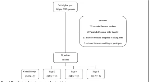
Of the 348 pre-dialytic CKD patients assessed for eligi-bility, 317 were excluded from the study for the reasons shown in Figure 1. Twenty-nine of the 31 patients who fulfilled the criteria for inclusion in the study agreed to participate: 10 in CKD stage 3 (G3), 10 in stage 4 (G4) and 9 in stage 5 (G5). The control group consisted of 9 healthy individuals.
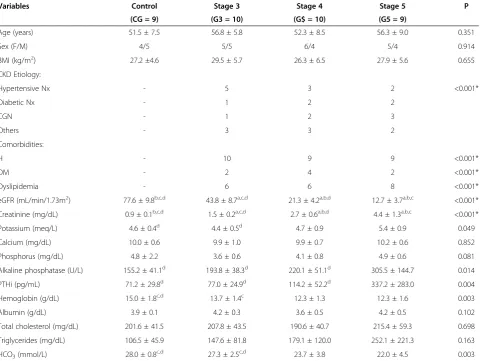
The mean time since diagnosis time was 4.47 ± 2.67 years (median 4 years). No statistical differences among groups in age, sex or body mass index were observed. The most prevalent cause of CKD was hypertensive ne-phropathy (34.4%), followed by chronic glomeruloneph-ritis (20.6%) and diabetic nephropathy (17.2%). The most
common comorbidities were hypertension (96.5%) and diabetes mellitus (24.1%). In the laboratory variables, there were significant differences in serum levels of po-tassium, alkaline phosphatase, PTH-i, haemoglobin, and bicarbonate (Table 1). Only 1 patient presented albumin levels of < 3.5 g/dL.
In this study, only 2 (6.8%) patients were anaemic, and these were incidents in the clinic and they had not yet reversed this condition, and used erythropoietin. About 37.9% of the patients used beta-blockers and all hyper-tensive patients were using ACE inhibitors and/or angio-tensin receptor blockers. Sodium bicarbonate, calcium chelating and vitamin D agents were used where neces-sary, in accordance with the guidelines of the Brazilian Society of Nephrology [27,28].
There were no significant differences in spirometric measures among groups. Among the manovacuometry variables, PImax (%) differed between groups, but the difference was not statistically significant (p = 0.06) (Table 2).
In the CPET, VO2peak was lower in the G4 and G5
groups than in the CG (p = 0.03), while values of relative power maximal (RPmax) (p < 0.01), oxygen
consump-tion at anaerobic threshold (VO2AT) (p < 0.01) and
maximum heart rate (HRmax) (p < 0.01) were lower in the 3 CKD groups than in the control. Likewise, distance walked in the 6MWT was lower in the G4 and G5 groups than in the CG (Table 3). Maximal exercise
toler-ance (VO2peak and RPmax) and submaximal exercise
tolerance (VO2AT and 6MWT), FEV1and FCV were
cor-related with eGFR (Table 4 and Figure 2). The criteria for discontinuation of CPET were the same as recommended
Table 3 Assessment of exercise tolerance (cardiopulmonary exercise test and six-minute walk test)
Variables Control Stage 3 Stage 4 Stage 5 P
(CG = 9) (G3 = 10) (G4 = 10) (G5 = 9)
Cardiopulmonary test
RPmax (W/kg) 4.9 ± 1.3 3.7 ± 0.9a 3.0 ± 1.2a 3.2 ± 1.0a 0.007
VO2peak (mL/kg/min) 28.9 ± 7.8 23.3 ± 5.6 21.4 ± 5.2a 20.2 ± 6.9a 0.033
VO2peak (%) 85.4 ± 18.2 75.8 ± 18.3 65.9 ± 16.0a 63.4 ± 16.0a 0.038
VO2AT (mL/kg/min) 20.5 ± 4.1 15.7 ± 3.5a 16.6 ± 3.8a 13.6 ± 3.8a 0.005
VEmax (l/min) 68.2 ± 20.3 60.8 ± 20 49.7 ± 20 56.9 ± 28.9 0.356
HRmax (bpm) 165 ± 14 146 ± 16a 139 ± 23a 129 ± 18a 0.003
HRmax (%) 97.9 ± 34.6 90.0 ± 11.1 82.8 ± 11.5a 79.3 ± 9.9a 0.003
SBPmax (mmHg) 197.5 ± 34.6 202.6 ± 25.8 204.2 ± 35.5 201.7 ± 34.3 0.976
DPBmax (mmHg) 89.7 ± 7.5 97.6 ± 9.2 101.8 ± 18.7 98.8 ± 9.6 0.203
6MWT
Distance (m) 627.6 ± 37.8 577.4 ± 66.1 542 ± 57.3a 531.5 ± 84.2a 0.012
% Predicted (%) 90.5 ± 7.6 93.9 ± 7.5 83.4 ± 9.5b 83.0 ± 8.9b 0.017
RPmaxMaximum Relative Power,VO2peakPeak Oxygen Consumption,VO2ATOxygen Consumption at Anaerobic Threshold,VEMinute Ventalation,HRmax Maximum Heart Rate,SBPmaxMaximum Systolic Blood Pressure,DBPmaxMaximum Diastolic Blood Pressure.
Post hoc analysis: a = differs from CG at p<0.05; b = differs from G3 at p<0.05.
Table 4 Correlations between the estimated glomerular filtration rate, spirometric variables, and exercise tolerance variables
Variables Estimated glomerular filtration rate (mL/min/1.73 m2)
r p
FVC (%) 0.348 0.041
FEV, (%) 0.349 0.020
RPmax (W/kg) 0.536 0.001
VO2peak (mL/kg/min) 0.430 0.008
VO2AT (mL/kg/min) 0.481 0.003
by the American Thoracic Society [20] however in all tests there were no complications; in addition, all tests were stopped when the patients reached physical exhaustion.
The correlation between pulmonary function and exer-cise tolerance was performed and a weak correlation
be-tween FVC (%) and VO2peak: r = 0.34 and p = 0.05; FVC
(%) and VO2peak (%): r = 0.37 and p = 0.03; and FEV1(%) and VO2peak (%): r = 0.37 and p = 0.03 were observed.
Discussion
In this study was observed that pre-dialytic CKD pa-tients showed lower maximal and submaximal exercise tolerances than healthy individuals. In addition, eGFR was correlated with reduced exercise tolerance and pul-monary function.
Most studies of respiratory function and exercise tol-erance in CKD patients have included patients under-going haemodialysis or peritoneal dialysis, treatments that can interfere with pulmonary function [7,8]. The few studies performed in pre-dialytic CKD patients have not excluded factors that may contribute to re-duced exercise tolerance and impaired pulmonary func-tion, such as smoking [11] or were carried out with children [29]. Because major confounders such as aging, smoking, previous pulmonary disease and medi-cations that can interfere with respiratory function were excluded, this study was able to assess only the impact of CKD and its associated changes in exercise tolerance and lung function.
So far, few studies have been performed on exercise tolerance in patients not yet on dialysis [15]. In CKD patients, particularly those undergoing dialysis, reduced exercise tolerance is associated with conditions such as anaemia, sedentary lifestyle, decreased muscular strength and resistance, and chronic inflammation [10,30-32].
Exercise tolerance can be assessed by relatively complex tests such as CPET or by simple low-cost tests such as 6MWT.
The incremental CPET quantifies VO2peak, which is
considered the gold standard for determining maximal exercise tolerance. In a recent review, Johansen and Painter [33] report a mean reduction of 50% - 80% of
VO2peak in pre-dialytic CKD patients. In this study,
VO2peak was also lower in pre-dialytic CKD patients
than in the CG and correlated with eGFR. Accordingly, in a study published by Clyne et al. [15], the authors showed in pre-dialytic CKD patients, that exercise tolerance reduces with progress of kidney disease and
VO2peak of these patients is approximately 60% lower
than healthy controls. The CPET also offers important information on submaximal exercise tolerance by deter-mining metabolic responses at the AT, which permit an assessment of the efficiency of aerobic metabolism. Dur-ing activities performed below the AT, energy sources are aerobic, and there is no sustained accumulation of lactate. On the other hand, anaerobic glycolysis occurs above this threshold, which results in the production and accumulation of lactate [34]. In the present study, reduced VO2AT, indicative of less efficient energy produc-tion by aerobic mechanisms, was observed in patients with lower eGFR. Clinically, the less efficient aerobic mechanism leads to greater muscular fatigue and lower exercise tolerance.
Submaximal exercise tolerance may also be quantified by the 6MWT. This test is easy to perform, requires no special equipment, is better tolerated by patients and is more representative of daily activities than the incre-mental CPET [24]. Some authors have reported lower distances in 6MWT in CKD patients when compared with healthy individuals [35-37]. In one such study, Cury
et al. [37] associated the reduction in functional capacity with inefficiencies in the uptake, transport, and use of O2 caused by dysfunction of cardiovascular, respiratory and muscular systems.
In the present study, also was recorded shorter dis-tances walked in the 6MWT by CKD patients and this result was more evident in those with lowest eGFR. Since the ventilatory response (minute ventilation) was not altered during the CPET in CKD patients, the de-creasing in exercise tolerance in these patients can be associated with disturbance in cardiovascular system or peripheral muscles. In addition, pulmonary function was assessed by spirometry and showed no differences be-tween groups, supporting the findings obtained in the CPET. Moreover, there was only a weak correlation be-tween pulmonary function and exercise tolerance.
The reduced exercise capacity in patients on haemodi-alysis may be due to changes in transport mechanisms and oxygen extraction. The transport of oxygen in these patients may be altered by reduced cardiac output, changes in maximum heart rate and decreased arterial oxygen by anemia, while the impairment of oxygen ex-traction may be due to uremic myopathy and disuse at-rophy [38-40]. In this study, the hemoglobin level was appropriate and not correlated with lung function and CPET related variables.
CKD patients may present conditions that result in ventilatory limitations, such as musculoskeletal weakness [37,41,42], interstitial edema and edema of small airways [13], pleural effusion [17] and osteoarticular changes in the thoracic vertebrae [43]. In the present study, there were no significant differences in spirometric test results between CKD patients and the CG, but a positive
correl-ation between eGFR, FVC, and FEV1 was observed,
which suggests some decline in pulmonary function associated with deteriorating kidney function. The rela-tively small number of patients that was studied and the small magnitude of the loss of pulmonary function may explain the lack of statistically significant differences be-tween groups in the spirometric data.
Decreased muscular strength may have multiple causes, including reduced carnitine level, hypovitaminosis D, hypotrophy of type-II muscle fibres, decreased energy use by muscle fibres, increased PTH-i [29], metabolic acidosis, chronic inflammation [44], decreases in oxidative metab-olism, decreases in serum levels of calcium and increases in protein catabolism [37]. In the present study, CKD pa-tients showed a tendency of lower values of PImax (%) than the CG (p = 0.06), in agreement with previous re-ports that skeletal muscles, including the respiratory mus-culature, presented lower strength and resistance over the course of CKD [9,22]. It is also possible that changes to other musculoskeletal groups, such as those in the lower limbs, also contributed to decreased exercise tolerance.
This study presented limitations. First, the fact that it is a cross-sectional study limits the interpretation of the impact of CKD on the observed results. Second, the ex-clusion of possible factors known to decrease pulmonary function limited the size of study sample. Third, these results cannot be interpreted as broadly representative, because the study was carried out in non-elderly patients at a single centre. Finally, it is important to emphasize that the patients who participated in this study were also participating in a CKD secondary prevention programme, received interdisciplinary treatment and were clinically stable and under close observation.
Conclusion
The present study showed that pre-dialytic CKD patients had a reduction in maximal and submaximal exercise tolerances. In this context, CPET can be used for exercise prescription before an inclusion in exercise programmes which should be started early in the course of the disease. Nevertheless, further studies are necessary to confirm these outcomes.
Abbreviations
(CKD):Chronic kidney disease; (eGFR): Estimated glomerular filtration rate; (CG): Control group; (G3): Stage 3; (G4): Stage 4; (G5): Stage 5;
(CPET): Cardiopulmonary exercise test; (6MWT): 6-minute walk test; (VO2peak) (%): Peak oxygen consumption percentage; (FEV1) (%): Forced expiratory volume in the first second percentage; (PImax): Maximum inspiratory pressure; (PTH-i): Parathyroid hormone intact molecule; (FVC): Forced vital capacity; (PEF): Peak expiratory flow; (PEmax): Maximum expiratory pressure; (VO2max): Peak oxygen consumption maximum; (AT): Anaerobic threshold; (RPmax): Relative power maximum; (VO2AT): Oxygen consumption at anaerobic threshold; (HRmax): Maximum heart rate.
Competing interests
The authors declare the following non-financial competing interests.
Authors’contributions
All the authors of this manuscript made substantial contributions to the conception and design, acquisition of data, and analysis and interpretation of data. All authors read and approved the final manuscript.
Author details
1Interdisciplinary Nucleus of Study, Research and Treatment in Nephrology
(NIEPEN), Federal University of Juiz de Fora, José Lourenço Kelmer, 1300, São Pedro, 36036-330 Juiz de Fora, Minas Gerais, Brazil.2Department of Medicine, Campus Universitário, Federal University of Juiz de Fora, Martelos, 36016-970 Juiz de Fora, Minas Gerais, Brazil.3Division of Pneumology, University Hospital of Federal University of Juiz de Fora, Rua Catulo Breviglieri, Santa Catarina, 36036110 Juiz de Fora, Minas Gerais, Brazil.
Received: 21 March 2012 Accepted: 8 July 2013 Published: 4 September 2013
References
1. Bastos MG, Kirsztajn GM:Chronic kidney disease: importance of early diagnosis, immediate referral and structured interdisciplinary approach to improve outcomes in patients not yet on dialysis.J Bras Nefrol2011,
33:93–108.
2. Keith DS, Nichols GA, Gullion CM, Brown JB, Smith DH:Longitudinal follow-up and outcomes among a population with chronic kidney disease in a large managed care organization.Arch Intern Med2004,164:659–663. 3. Bush A, Gabriel R:Pulmonary function in chronic renal failure: effects of
4. Yigla M, Nakhoul F, Sabag A, Tov N, Gorevich B, Abassi Z, Reisner SA:
Pulmonary hypertension in patients with end-stage renal disease.
Chest2003,123:1577–1582.
5. Kalender B, Erk M, Pekpak MA, Apaydin S, Ataman R, Serdengecti K, Sariyar M, Erek E:The effect of renal transplantation on pulmonary function.
Nephron2002,90:72–77.
6. Rocha CB, Araujo S:Evaluation of maximum respiratory pressures in chronic renal patients at the pre and post hemodialysis moment.J Bras Nefrol2010,32:105–111.
7. Hekmat R, Boskabady MH, Khajavi A, Nazary A:The effect of hoemodialysis on pulmonary function tests and respiratory symptoms in patients with chronic renal failure.Pak J Med Sci2007,23:862–866.
8. Siafakas NM, Argyrakopoulos T, Andreopoulos K, Tsoukalas G, Tzanakis N, Bouros D:Respiratory muscle strength during continuous ambulatory peritoneal dialysis (CAPD).Eur Respir J1995,8:109–113.
9. Nascimento MM, Qureshi AR, Stenvinkel P, Pecoits-Filho R, Heimburger O, Cederholm T, Lindholm B, Barany P:Malnutrition and inflammation are associated with impaired pulmonary function in patients with chronic kidney disease.Nephrol Dial Transplant2004,19:1823–1828.
10. Ulmer HE, Griener H, Schuler HW, Scharer K:Cardiovascular impairment and physical working capacity in children with chronic renal failure.
Acta Paediatr Scand1978,67:43–48.
11. Goldberg AP, Geltman EM, Gavin JR 3rd, Carney RM, Hagberg JM, Delmez JA, Naumovich A, Oldfield MH, Harter HR:Exercise training reduces coronary risk and effectively rehabilitates hemodialysis patients.
Nephron1986,42:311–316.
12. Mayer G, Thum J, Graf H:Anaemia and reduced exercise capacity in patients on chronic haemodialysis.Clin Sci (Lond)1989,76:265–268. 13. Carney RM, McKevitt PM, Goldberg AP, Hagberg J, Delmez JA, Harter HR:
Psychological effects of exercise training in hemodialysis patients.
Nephron1983,33:179–181.
14. Painter P, Messer-Rehak D, Hanson P, Zimmerman SW, Glass NR:Exercise capacity in hemodialysis, CAPD, and renal transplant patients.
Nephron1986,42:47–51.
15. Clyne N, Jogestrand T, Lins LE, Pehrsson SK:Progressive decline in renal function induces a gradual decrease in total hemoglobin and exercise capacity.Nephron1994,67:322–326.
16. K/DOQI:K/DOQI clinical practice guidelines for chronic kidney disease: evaluation, classification, and stratification.Am J Kidney Dis2002,39:S1–266. 17. Pereira CAC, Neder JA, SBPT:Diretrizes para Testes de Função Pulmonar.
J Pneumol2002,28:S1–S237.
18. Neder JA, Andreoni S, Castelo-Filho A, Nery LE:Reference values for lung function tests. I. Static volumes.Braz J Med Biol Res1999,32:703–717. 19. Neder JA, Andreoni S, Lerario MC, Nery LE:Reference values for lung
function tests. II. Maximal respiratory pressures and voluntary ventilation.Braz J Med Biol Res1999,32:719–727.
20. ATS:ATS/ACCP Statement on cardiopulmonary exercise testing.Am J Respir Crit Care Med2003,167:211–277.
21. Maeder M, Wolber T, Atefy R, Gadza M, Ammann P, Myers J, Rickli H:Impact of the exercise mode on exercise capacity: bicycle testing revisited.
Chest2005,128:2804–2811.
22. Beaver WL, Wasserman K, Whipp BJ:A new method for detecting anaerobic threshold by gas exchange.J Appl Physiol1986,60:2020–2027. 23. Reinhard U, Muller PH, Schmulling RM:Determination of anaerobic
threshold by the ventilation equivalent in normal individuals.
Respiration1979,38:36–42.
24. ATS:American Thoracic Society statement: guidelines for the six-minute walk test.Am J Respir Crit Care Med2002,166:111–117.
25. Enright PL, Sherrill DL:Reference equations for the six-minute walk in healthy adults.Am J Respir Crit Care Med1998,158:1384–1387. 26. Borg G:Psychophysical scaling with applications in physical work and the
perception of exertion.Scand J Work Environ Heal1990,16(Suppl 1):55–58. 27. Kirsztajn GM, Romão-Junior JE, Souza E, Soriano EA, Ribas DF, Andrada NC,
Bastos MG:Doença Renal Crônica (Pré-terapia Renal Substitutiva): Tratamento. http://www.projetodiretrizes.org.br/diretrizes11/doenca_renal_cronica_pre_ terapia_renal_substitutiva_tratamento.pdf.
28. Kirsztajn GM, Souza E, Romão Junior JE, Bastos MG, Meyer F, Andrada NC: Doença Renal Crônica (Pré-Terapia Renal Substitutiva): Diagnóstico.http:// www.projetodiretrizes.org.br/diretrizes11/doenca_renal_cronica_pre_ terapia_renal_substitutiva_diagnostico.pdf.
29. Coelho CC, Aquino ES, Lara KL, Peres TM, Barja PR, Lima EM:Consequences of chronic renal insufficiency on the exercise capacity, nutritional status, pulmonary function and respiratory musculature of children and adolescents.Rev Bras Fisioter2008,12:1–6.
30. Kettner-Melsheimer A, Weiss M, Huber W:Physical work capacity in chronic renal disease.Int J Artif Organs1987,10:23–30.
31. Clyne N, Jogestrand T, Lins LE, Pehrsson SK, Ekelund LG:Factors limiting physical working capacity in predialytic uraemic patients.Acta Med Scand 1987,222:183–190.
32. Johansen KL:Exercise in the end-stage renal disease population.J Am Soc Nephrol2007,18:1845–1854.
33. Johansen KL, Painter P:Exercise in individuals with CKD.Am J Kidney Dis 2012,59:126–134.
34. Neder JA, Nery LE:Teste de Exercício Cardiopulmonar.J Pneumol2002,
28(3):166–206. 2002,28:166–206.
35. Ling KW, Wong FS, Chan WK, Chan SY, Chan EP, Cheng YL, Yu WY:Effect of a home exercise program based on tai chi in patients with end-stage renal disease.Peritoneal dialysis international: journal of the International Society for Peritoneal Dialysis2003,23(Suppl 2):S99–S103.
36. Silva VG, Amaral C, Monteiro MB, Nascimento DM, Boschetti JR:Effects of inspiratory muscle training in hemodialysis patients.J Bras Nefrol2011,
33:62–68.
37. Cury JL, Brunetto AF, Aydos RD:Negative effects of chronic kidney failure on lung function and functional capacity.Rev Bras Fisioter2010,14:91–98. 38. Moore GE, Parsons DB, Stray-Gundersen J, Painter PL, Brinker KR, Mitchell JH:
Uremic myopathy limits aerobic capacity in hemodialysis patients.
Am J Kidney Dis1993,22:277–287.
39. Painter P, Hanson P, Messer-Rehak D, Zimmerman SW, Glass NR:Exercise tolerance changes following renal transplantation.Am J Kidney Dis1987,
10:452–456.
40. Kopple JD, Storer T, Casburi R:Impaired exercise capacity and exercise training in maintenance hemodialysis patients.J Ren Nutr2005,15:44–48. 41. Kostianev S, Kumchev E:External respiration in patients with chronic
renal failure in a predialysis state.Folia Med1994,36:23–26. 42. Faria RS, Silva VSA, Reboredo MM, Fernandes NMS, Bastos MG, Cabral LF:
Avaliação da Função Respiratória, Capacidade Física e Qualidade de Vida de Pacientes com Doença Renal Crônica Pré-Dialítica.J Bras Nefrol2008,
30:264–271.
43. Vieira WP, Gomes KWP, Frota NB, Andrade JECB, Vieira MRA, Moura FEA, Vieira FJF:Musculoskeletal manifestations in patients under hemodialysis.
Rev Bras Reumatol2005,45:357–364.
44. Workeneh BT, Mitch WE:Review of muscle wasting associated with chronic kidney disease.Am J Clin Nutr2010,91:1128S–1132S.
doi:10.1186/1471-2369-14-184
Cite this article as:Fariaet al.:Pulmonary function and exercise tolerance are related to disease severity in pre-dialytic patients with chronic kidney disease: a cross-sectional study.BMC Nephrology
201314:184.
Submit your next manuscript to BioMed Central and take full advantage of:
• Convenient online submission
• Thorough peer review
• No space constraints or color figure charges
• Immediate publication on acceptance
• Inclusion in PubMed, CAS, Scopus and Google Scholar
• Research which is freely available for redistribution