R E S E A R C H
Open Access
Ovarian teratoma in children: a plea for
collaborative clinical study
Justyna
Ł
uczak
*and Maciej Bag
ł
aj
Abstract
Background:Although teratomas are the most common histologic subtype of childhood ovarian germ cell tumors, their appropriate treatment in this age group still remains unclear. Paucity of research dedicated exclusively to both mature and immature teratomas of the ovary, contribute to decision making difficulties.
Therefore, we decided to review retrospectively our experience in treatment of pediatric ovarian teratomas in order to assess the epidemiology, presenting features, and diagnostic as well as surgical management of these lesions. Results:The study comprised 58 patients. Fifty percent of patients were between 9 and 15 years old. Mature teratoma was diagnosed in 55(94.83%) patients, while 3(5.17%) patients presented with immature teratoma. Twenty eight (50. 91%) girls with mature teratoma had laparotomy and 23 (41.82%) had laparoscopy performed as an initial operative approach. Ovarian tissue sparing technique (preservation of the ovarian tissue of the affected gonad) was applied in only 11.11% of patients operated in the first study period (years 1999–2003) and increased to 40.54% in the second half of our study (years 2004–2016). The extent of gonadal resection was not related with the size of the lesion. Bilateral lesions were noted in 8 patients with mature teratoma. All girls with immature teratoma were subjected to formal laparotomy. Two patients had stage III of the disease and one had stage IV. They underwent at least resection of the affected gonad. Adjuvant chemotherapy was given to all girls with immature teratoma after the surgery.
Conclusions:Under particular conditions ovarian-sparing surgery might be successfully applied in children with mature teratoma. Laparotomy is the treatment of choice in large masses, suspicious for malignancy and if surgical staging is required. High quality prospective multi-institutional studies are required in order to get an objective insight into biology and prognostic factors of teratomas in children.
Keywords:Ovarian teratoma, Ovarian neoplasms, Child
Background
Ovarian teratomas are the most common type of ovarian tumors in children. Nevertheless, some aspects of its path-ology, classification and management still remain unclear. Their embryology and genetic basis are not yet under-stood. Specific concerns regard their malignant potential and therefore the possible use of ovarian-sparing operative techniques and the suitability of chemotherapy in its treat-ment. According to the recent studies, ovarian-sparing procedures are recommended in case of mature teratoma, on specific conditions [1, 2]. However, variety of tissue architecture in teratomas creates a lot of uncertainty. Such as the significance of grade of immaturity or detection of Yolk sac tumor microfoci. In turn, difficulties in histologic
sampling might result in overlooking of some malignant cells, especially in the large tumors. Although surgery re-mains the re-mainstay of treatment, the indications for chemotherapy vary between the studies [2–7]. Paucity of research dedicated exclusively to both mature and imma-ture teratomas of the ovary, contribute to decision making difficulties. Therefore, we decided to review our experi-ence of pediatric ovarian teratomas at our institution in order to assess the epidemiology, presenting features, and diagnostic as well as surgical management of these lesions.
Material and methods
We analyzed retrospectively medical files of all consecutive patients aged 0–18 years, who underwent surgical proce-dures for ovarian teratoma between 1991 and 2016 at the University Department of Pediatric Surgery and Urology in Wroclaw, Poland. The demographic data, presenting * Correspondence:[email protected]
Pediatric Surgery and Urology Department, Wroclaw Medical University, 52 M. Sklodowskiej–Curie ST, 50-369 Wroclaw, Poland
symptoms and signs, results of laboratory and diagnostic studies (including ultrasound examination, additional im-aging studies and tumor markers), details of surgical proce-dures, and clinical outcomes (including preservation rate), were extracted in each case. Ovarian mass characteristics were evaluated by preoperative imaging (structure and size) or by description of the procedure (size). An ovarian lesion was described arbitrarily as large when its diameter was 10 cm or more in girls aged between 1 and 18 years and 5 cm or more in newborns and infants. Such classification was based on the previous experience of other authors, in order to obtain comparable results [8, 9]. A choice of an operative technique (either laparoscopic or open) depended solely on a surgeon’s preference. The extent of gonadal re-section was based on intraoperative findings and it ranged from total, when the whole gonad affected by lesion was re-moved, to partial resection, when at least the remnant of ovarian tissue was preserved. Preservation rates were com-pared taking into consideration the operative method, the histological type, the size of the mass, the presence of ovar-ian torsion and the study period (1991–2003 vs 2004– 2016). The analyzed data were subjected to the statistical evaluation with Chi square, Kruskal-Wallis and Barttlet test, Pearson’s correlation coefficient and logistic regression. The statistical software package EPIINFO Ver. 7.1.1.14 was used for all data analyses.
Results
Epidemiology and presentation
The study comprised 58 patients. The median of age was 12,0 years (SD = 4.2). Fifty percent of patients were
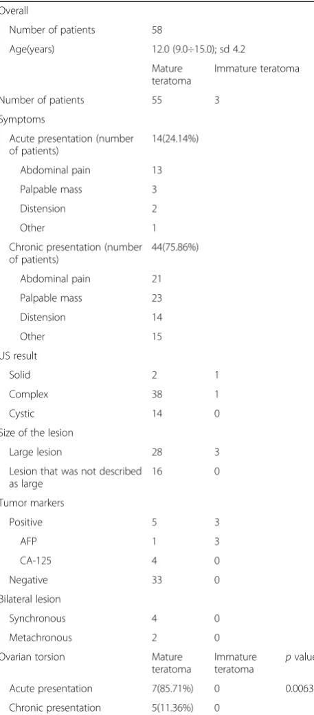
between 9 and 15 years old. Mature teratoma was diag-nosed in 55(94.83%) patients, while 3(5.17%) patients presented with immature teratoma (age: 8,12 and 15 years old). There were 44 girls (75.86%) with chronic presentation (no symptoms requiring immediate surgical intervention). Fourteen patients (24.14%) presented with acute symptoms (admitted to the hospital as emergency due to relevant pain or/and vomits, fever). Abdominal pain, palpable mass and distension were the most fre-quent clinical features noted in the whole study group. Palpable mass was the most frequent symptom in girls with chronic presentation (52.27%), while abdominal pain predominated in girls with acute presentation and was noted in 13 of them (92.86%); Fig.1. The most com-mon intraoperative finding additional to the tumor, in girls with acute presentation, was ovarian torsion (85.71% of patients with acute symptoms; p= 0.00634, χ2
= 7.45, Rr = 3.83, CI = 1.67÷8.82); Table 1. All three patients with immature teratoma presented with chronic symptoms.
Diagnostic studies
Mature teratomas
All of the patients underwent abdominal ultrasound scan (US). It showed a cystic structure in 14 girls (25.45%). A complex ovarian mass was noted in 38 (69.09%) and a solid mass in 2 girls (36.36%). Twenty-eight girls (50.91%) had a large tumor (with a diameter of more than 10 cm). Twenty-one patients (38.18%) had computed tomography (CT) or magnetic resonance(MRI) studies performed preoperatively. In 11
patients this additional examination led to the evaluation of tumor origin or its real structure changing the initial radiological description of the lesion (based on US scan). Statistical analysis revealed linear dependence between the size of the lesion and patient’s age (Pearson product-moment correlation coefficient:r= 0.29, N= 53, p= 0.039, Fig. 2). The results of tumor markers
evaluation (AFP –alpha-fetaprotein, β-hCG - beta sub-unit of human chorionic gonadotropin, CA125 - cancer antigen 125, LDH - lactate dehydrogenase) were avail-able for 38 girls (69.09%). They were elevated in 5 girls (9.09%). Only one marker was positive in four patients out of five: CA-125. We noted elevated level of AFP (37.45 ng/ml) in one patient aged 14 (Table1).
Immature teratomas
Abdominal US showed a complex ovarian lesion and a solid mass in one girl each. A heterogenous structure of one of the lesions was confirmed in CT. One patient had only computed tomography result available, it re-vealed a complex lesion. Calcifications were observed on imaging in two patients. Large tumor was diagnosed in all three girls. Tumor markers evaluation revealed ele-vated level of AFP in all girls (186,1680 and 6434 ng/ ml).
Treatment
Mature teratomas
Twenty-eight (50.91%) girls had laparotomy and 23 (41.82%) had laparoscopy performed as an initial opera-tive approach. Conversion to open procedure was noted in 4 girls (7.27%). The number of patients operated on with laparoscopy increased from 44.44 to 51.35% in the second part of our study (years 1991–2003 vs 2004– 2016). Ovarian tissue sparing technique (preservation of the ovarian tissue of the affected gonad) was applied in only 11.11% of patients operated in the first study period and increased to 40.54% in the second half of our study. The preservation rate was higher in the group of girls in whom laparoscopic technique was chosen. The extent of gonadal resection was not related with the size of the le-sion. Moreover, tissue sparing was possible in 32.26% of those with large lesions and 29.17% of girls with lesions not classified as large. The difference was not statistically significant; p= 0.806, χ2 = 0.605 (Table 2). Biopsy pro-cedure was performed in 21 cases and it was negative in all of them.
Immature teratomas
All girls with immature teratoma were subjected to for-mal laparotomy. Two patients had stage III of the dis-ease and one had stage IV (according to Children’s Oncology Group ovarian staging system) [10]. They underwent at least resection of the affected gonad. Bi-opsy procedure was performed all girls and it was nega-tive in all of them.
Histopathologic findings
Mature teratomas
In 18 cases there was ovarian dermoid cyst diagnosed. Table 1Comparison of the clinical data between mature and
immature teratomas
Overall
Number of patients 58
Age(years) 12.0 (9.0÷15.0); sd 4.2
Mature teratoma
Immature teratoma
Number of patients 55 3
Symptoms
Acute presentation (number of patients)
14(24.14%)
Abdominal pain 13
Palpable mass 3
Distension 2
Other 1
Chronic presentation (number of patients)
44(75.86%)
Abdominal pain 21
Palpable mass 23
Distension 14
Other 15
US result
Solid 2 1
Complex 38 1
Cystic 14 0
Size of the lesion
Large lesion 28 3
Lesion that was not described as large
16 0
Tumor markers
Positive 5 3
AFP 1 3
CA-125 4 0
Negative 33 0
Bilateral lesion
Synchronous 4 0
Metachronous 2 0
Ovarian torsion Mature
teratoma
Immature teratoma
pvalue
Acute presentation 7(85.71%) 0 0.00634
Immature teratomas
Grading (according to O’Connor and Norris) was avail-able only in one patient (G1) [11]. No Heifetz lesions were observed.
Final outcome
Mature teratomas
Bilateral, synchronous lesions were noted in 6 patients with mature teratoma. Metachronous disease in the contralateral ovary was observed in two patients with mature teratoma after the first operation within 10 months and 5 years. Respectively (Table 1). Follow-up was possible in 13 patients and lasted be-tween 1 and 12 years. All girls which have at least one ovary have normal menstruation. Some of them (30.77%) reported painful menstruations or are diag-nosed with simple cysts (without operative treatment).
Immature teratomas
Adjuvant chemotherapy (protocol TGM 95) was given to all girls with immature teratoma after the surgery. All three patients responded well to the treatment and were disease free after follow-up of 3,4 and 12 years respectively.
Discussion
Teratomas present diverse biological behavior and con-tinue to be the cause of many diagnostically and thera-peutically challenging issues. While it is commonly
accepted that mature teratomas of the ovary are benign neoplasms, the classification of the immature ones is still discussed. About 25% of all the pediatric GCTs present as tumors with more than one histologic type. In this situation, therapy and prognosis depend on the compo-nent with the highest malignancy [12–17]. In accordance with this approach pure immature teratomas were ex-cluded in the recent studies concerning pediatric malig-nant germ cell tumors. Conversely, patients with malignant germ cell tumors, even when these lesions contained mature teratoma or immature teratoma ele-ments, were not eligible for studies concerning terato-mas exclusively [2, 12, 17, 18]. Nevertheless, there are also studies were a lesion was considered as immature teratoma although the pathology report revealed a com-ponent of another malignant tumor [19]. Marks of the controversy regarding this topic are also reflected in the nomenclature of these lesions across the studies. The multiplicity of names describing immature teratomas (immature teratoma, malignant teratoma, teratoma with malignant elements, immature teratoma with malignant behavior) renders universally applicable classification of these lesions very difficult [5,12,17–21].
All the girls in our study had at least stage III of the disease. In a study from 2012, Schneider et al. ascertain that immature teratomas behave in a malignant fashion only if foci of malignant germ cell elements are present and if they are resected incompletely. According to their paper, tumors containing clusters of yolk sac tumor are
Fig. 2Patient’s age and size of the lesion
Table 2Ovarian tissue preservation rates in selected groups of patients
Study period
Overall Operation method Large lesion Torsion
Laparoscopy Laparotomy – + – +
1991–2016 29.31% 43.48% (27.27% large lesions) 20.00% (75.76% large lesions) 29.17% 32.26% 30.43% 25.00% p= 0.806
1991–2003 11.11% 14.28% 7.69% 12.50% 9.09% 7.14% 0.00%
likely responsible for the reports that immature teratoma may metastasize [2]. Nevertheless, in some recently pub-lished studies immature teratomas are still included in the evaluation of patients with malignant germ cell tu-mors and even at US National Cancer Institute website reads that “Immature teratomas can exhibit malignant behavior and metastasize” [7, 14, 22, 23]. Therefore, can we definitively classify them as benign?
Another important aspect in attempts to estimate the real nature of these lesions is the importance of the grade of immaturity. In a study published by Malignant Germ Cell Tumor International Collaborative it was re-vealed that grade was the most important risk factor for relapse in ovarian immature teratoma [6]. However, there is scarceness of papers reporting the use of a de-tailed grading system and its significance in children has been challenged by some former studies. Additionally, high immaturity itself is not associated with a poor prog-nosis if the tumor is completely resected [2]. Defining real nature of the lesion is indispensable in choosing optimum management. Obtaining the correct pathology report is crucial here. Clusters of malignant tumor can be easily overlooked and yolk sac tumor components might be very small. Microfoci of yolk sac tumor (Hei-fetz lesions) are not precisely defined among the studies, thereby classifying a lesion as malignant yolk sac tumor is particularly difficult [2, 5, 12]. Performing a central pathomorphological examination can be helpful in the case of these neoplasms. Perhaps new tumor markers, like the ones discovered by Feng et al. will help eliminate mistakes in pathomorphological diagnoses [24]. Grading was available only in one patient in our study and no Heifetz lesions were observed. This is a retrospective study and we cannot verify the results of the pathology report. Taking into consideration that all immature tera-toma cases were of stage III at least, it is questionable if no other malignant components were present.
Despite these unclear pathological aspects, commonly applied diagnostic work-up of ovarian teratomas include physical examination, ultrasound imaging and tumor markers evaluation. In most of the studies the peak inci-dence of these lesions is reported in early adolescence [2,12]. It was also confirmed by our study. Symptoms of ovarian teratoma do not differ from those observed in other ovarian masses. Some patients reveal acute symp-toms rising suspicion of ovarian torsion. The risk of tor-sion complicating a case of ovarian teratoma is approximately 3 to 16% in children [2, 25]. Those cases are rarely associated with malignancy. Tsai et al. revealed that young girls tend to have either torsion of a mature cystic teratoma or torsion without underlying condition, while older patients are more likely to present with tor-sion and a tumor [2, 26]. In our study ovarian torsion was the most common intraoperative finding additional
to the tumor in girls with acute presentation. The inci-dence of ovarian torsion was not higher in older girls (Kruskal-Wallis test,p= 0,765).
As most of the mature teratomas are slow-growing cystic lesions and there are some common characteristic findings, they are easily recognized on US. Nevertheless, their typical features might be less clear in case of pre-pubertal girls and when the lesion is large [13, 26, 27]. Our study poses limitations in that respect. The ultra-sound description was often very short and we were un-able to identify characteristic features for teratoma in each case. Diagnosis of immature type also poses prob-lems. The appearance of the tumor on ultrasound is non-specific, although predominance of solid compo-nent is a differentiating factor. Identification of fat, mural nodules and calcified components within the le-sion is typical for teratomas. However, there are reports suggesting that typical CT features of teratomas occur more often in mature than in immature lesions. Those findings suggest that CT or MRI imaging might be help-ful, rather for staging and assessing of tumor respectabil-ity [13,28,29].
Alphafetoprotein evaluation is an important prognos-tic factor in many malignant germ cell tumors treatment protocols and it is also used in follow-up of those pa-tients. Additionally, it is a characteristic marker of yolk sac tumor. Regarding its levels in ovarian teratomas, it was revealed that they are rarely elevated [2, 12]. In the recent Malignant Germ Cell Tumor International Col-laborative study regarding ovarian immature teratomas, patients with AFP levels higher than 1000 ng/mL were excluded because this level was considered more likely to indicate malignant elements [6]. However, this ap-proach was questioned by Terenziani et al. who used an AFP level cutoff “high for age” in their study [14]. An-other limitation is caused by the vide variation in AFP levels at birth and the variability in its half-time within the first year of life [2]. Multiple publications where cases of immature teratoma are analyzed without taking AFP levels into consideration makes it more difficult to form a credible assessment of this indicator [14,30,31]. Only one girl in the immature teratoma group had AFP levels < 1000 ng/mL in our study. Given the results of the recent research this is another factor calling into question the result of pathology report in our cases.
ultrasound after cystectomy even if there was no normal ovarian tissue visible preoperatively [13]. Although incom-plete resection is an important risk factor of recurrence, relapse is not inevitable in those cases. The use of laparo-scopic techniques in the treatment of ovarian teratomas has its supporters and opponents. If the suspicion of ma-lignancy is low and a surgeon is experienced in minimal invasive surgery, laparoscopic approach might be adequate [2, 6, 7, 12, 15, 21, 33]. Children’s Cancer Study Group demonstrated in their study that cyst fluid aspiration or spillage during surgery are not associated with relapse, thereby this factor, claimed by laparoscopy opponents as more often associated with minimal invasive methods, might not have such importance for the final outcome [12,30]. Our results are promising in that respect. We re-vealed that the use of laparoscopy increased in time and the preservation rate was higher in the laparoscopy group. Laparotomy is the treatment of choice in large masses, suspicious for malignancy, if surgical staging is required [13]. The preservation rate seemed no to be affected by the size of the lesion in our study.
An important aspect of the management of ovarian teratomas is their recurrence rate and the incidence of bilateral lesions. The recurrence rate after cystectomy vary between 3 to 13%. However, in the study were the recurrence rate was estimated to be of 10% the authors revealed that only 3% of the recurrent cases will require reoperation [13, 34]. There were studies suggesting that women with bilateral or multiple dermoid cysts may in-clude a subgroup of patients with a greater tendency to develop future ovarian germ cell neoplasms. Due to low risk of malignant transformation in case of mature cystic teratomas in children the treatment should be directed on the basis of age, fertility desire or presence of another pelvic pathology rather than the size or bilaterality [35– 37]. In the study by O’Neill et al. the biopsy of the contralateral macroscopically normal ovary revealed pathological lesions only in 1.1% of patients. Therefore, preoperative ultrasound in combination with careful in-spection of the contralateral ovary at the time of surgery offer a safe alternative to biopsy [13, 38]. Our results support this approach, none of the biopsies was positive in our study. Given the sensitivity of ultrasound in the detection of mature cystic teratomas, annual imaging seems appropriate as a postoperative surveillance. AFP monitoring is not recommended in case of completely resected ovarian teratomas without preoperatively ele-vated tumor markers [12,38].
The treatment of immature teratomas poses much more difficulties. Their possible malignant behavior re-quires appropriate risk classification. Among factors considered as those influencing the prognosis, except the previously mentioned, staging is the next unclear one. Since malignant tumors and benign ones cannot be
distinguished based only on intraoperative appearance, there are studies recommending staging in all tumors [2, 39]. However, Billimire et al. revealed that survival ap-pears to have been unaffected by deviations from surgi-cal guidelines when chemotherapy is administrated [39]. Different staging systems across the surgical centers ren-ders evaluation of their significance difficult [2, 6]. An-other controversial aspect is the use of chemAn-otherapy. Number of therapeutic options might be found in the literature. To name some of them: chemotherapy for re-currence, only for malignant rere-currence, in all cases of stage I grade 2 and higher or only in girls older than 15 [2,6,7,22,40]. In the German MAKEI study more than half of the children with tumor recurrence after watch and wait strategy had yolk sac tumor in addition to tera-toma. It might be one of the main counter-arguments to the results of the studies which did not find evidence that chemotherapy had any significant therapeutic ef-fects in pure teratomas and that it does not decrease re-lapses in pediatric population [40]. Recently created Malignant Germ Cell Tumor International Collaborative in its publications gathering data from four clinical trials recommends surgery alone for patients with stage I/II, grade 1/2 tumors and suggests conducting of a prospect-ive trial of observation after surgery for patients with grade 2/3, stage II-IV tumors [6]. This approach might be encouraged by the results of Park et al. as they re-vealed that most stage I malignant ovarian germ cell tumor recurrences can be successfully salvaged by sur-gery and BEP chemotherapy without compromising the overall survival [22]. Adjuvant chemotherapy according to protocol TGM 95 was given to all girls with immature teratoma in our study group. They all responded well to the therapy but it remains questionable if these were real pure immature teratoma lesions.
Schneider et al. revealed that patients with cytogeneti-cally abnormal immature teratomas often develop recur-rence [2]. An optimal consensus treatment strategy should be based on a detailed analysis of all possible fac-tors. In this regard, the above mentioned study shows that we might be still not aware of a number of them. The future molecular research may allow distinguishing low-risk from high-risk patients accurately [15].
Conclusions
Under particular conditions ovarian-sparing surgery might be successfully applied in children with mature teratoma. Laparotomy is the treatment of choice in large masses, suspicious for malignancy and if surgical staging is required. High quality prospective multi-institutional studies are required in order to get an objective insight into biology and prognostic factors of teratomas in children.
Availability of data and materials
The datasets used and analyzed during the current study are available from the corresponding author on reasonable request.
Authors’contributions
JL made substantial contributions to conception and design of this research study as well to acquisition, analysis and interpretation of data. She was responsible for writing and drafting the manuscript. MB he was involved in interpretation of the data, writing and drafting the manuscript as well as revising it critically for important intellectual content. Both authors read and approved the final manuscript.
Ethics approval and consent to participate
The study was approved by the Ethical Committee of the Medical University of Wroclaw - available on request.
Consent for publication
Not applicable.
Competing interests
The authors declare that they have no competing interests.
Publisher’s Note
Springer Nature remains neutral with regard to jurisdictional claims in published maps and institutional affiliations.
Received: 4 July 2018 Accepted: 21 August 2018
References
1. Özcan R, Kuruoğlu S, Dervişoğlu S, Eliçevik M, Emir H, Büyükünal C. Ovary-sparing surgery for teratomas in children. Pediatr Surg Int. 2013;29(3):233–7. 2. Schneider DT, Terenziani M, Cecchetto G, Olson TA. Gonadal and
extragonadal germ cell tumors, sex cord stromal and rare gonadal tumors. In: Schneider D, Brecht I, Olson T, Ferrari A, editors. Rare tumors in children and adolescents. Pediatric Oncology. Berlin: Springer; 2011. p. 327–402. 3. Ulbright TM. Germ cell tumors of the gonads: a selective review
emphasizing problems in differential diagnosis, newly appreciated, and controversial issues. Mod Pathol. 2005;18(Suppl 2):S61–79.
4. Heifetz SA, Cushing B, Giller R, Shuster JJ, Stolar CJ, Vinocur CD, et al. Immature teratomas in children: pathologic considerations: a report from the combined pediatric oncology group/Children’s cancer group. Am J Surg Pathol. 1998;22(9):1115–24.
5. Mann JR, Raafat F, Robinson K, Imeson J, Gornall P, Sokal M, et al. The United Kingdom Children's Cancer study Group's second germ cell tumor study: carboplatin, etoposide, and bleomycin are effective treatment for children with malignant extracranial germ cell tumors, with acceptable toxicity. J Clin Oncol. 2000;18(22):3809–18.
6. Pashankar F, Hale JP, Dang H, Krailo M, Brady WE, Rodriguez-Galindo C, et al. Is adjuvant chemotherapy indicated in ovarian immature teratomas? A combined data analysis from the malignant germ cell tumor international collaborative. Cancer. 2016;122(2):230–7.
7. Mangili G, Sigismondi C, Lorusso D, Cormio G, Candiani M, Scarfone G, et al. The role of staging and adjuvant chemotherapy in stage I malignant ovarian germ cell tumors (MOGTs): the MITO-9 study. Ann Oncol. 2017;28(2):333–8.
8. Rogers EM, Casadiego Cubides G, Lacy J, Gerstle JT, Kives S, Allen L. Preoperative risk stratification of adnexal masses: can we predict the optimal surgical management? J Pediatr Adolesc Gynecol. 2014;27(3):125–8. 9. Oltmann SC, Garcia N, Barber R, Huang R, Hicks B, Fischer A. Can we
preoperatively risk stratify ovarian masses for malignancy? J Pediatr Surg. 2010;45(1):130–4.
10. PDQ Pediatric Treatment Editorial Board. Childhood Extracranial Germ Cell Tumors Treatment (PDQ®): Health Professional Version. In:PDQ Cancer Information Summaries [Internet]. Bethesda: National Cancer Institute (US); 2002-.https://www.ncbi.nlm.nih.gov/books/NBK65877/. Accessed 18 Aug 2018. 11. O’Connor DM, Norris HJ. The influence of grade on the outcome of stage I
ovarian immature(malignant) teratomas and the reproducibility of grading. Int J Gynecol Pathol. 1994;13(4):283–9.
12. Mann JR, Gray ES, Thornton C, Raafat F, Robinson K, Collins GS, et al. Mature and immature extracranial teratomas in children: the UK Children's Cancer study group experience. J Clin Oncol. 2008;26(21):3590–7.
13. O’Neill KE, Cooper AR. The approach to ovarian dermoids in adolescents and young women. J Pediatr Adolesc Gynecol. 2011;24(3):176–80. 14. Terenziani M, Bisogno G, Boldrini R, Cecchetto G, Conte M, Boschetti L, et al.
Malignant ovarian germ cell tumors in pediatric patients: The AIEOP (Associazione Italiana Ematologia Oncologia Pediatrica) study. Pediatr Blood Cancer. 2017;64(11)
15. Goebel U, Calaminus G, Schneider DT, Schmidt P, Haas RJ. Management of Germ Cell Tumors in children: approaches to cure. Onkologie. 2002;25:14–2212. 16. Bouffet E, De Lumley LL, Habrand Paris-Igr JL, Lejars Tours O, Martelli Paris
H, Perel Bordeaux Y, et al. Strategie thérapeutique des tumeurs germinales malignes extra-cérébrales de l’enfant, TGM 95. Lille Cedex: In: Protocol text: Société Française d’Oncologie Pédiatrique (SFOP); 1994.http://www. oncauvergne.fr/index.php?option=com_content&view=section&layout= blog&id=36&Itemid=118. Accessed 18 Aug 2018.
17. Frazier AL, Hale JP, Rodriguez-Galindo C, Dang H, Olson T, Murray MJ. Revised risk classification for pediatric extracranial germ cell tumors based on 25 years of clinical trial data from the United Kingdom and United States. J Clin Oncol. 2015;33(2):195–201.
18. Billmire DF, Cullen JW, Rescorla FJ, Davis M, Schlatter MG, Olson TA. Surveillance after initial surgery for pediatric and adolescent girls with stage I ovarian germ cell tumors: report from the Children's oncology group. J Clin Oncol. 2014;32(5):465–70.
19. Yanai H, Matsuura H, Kawasaki M, Takada Y, Tabuchi Y, Yoshino T. Immature teratoma of the ovary with a minor rhabdomyosarcomatous component and fatal rhabdomyosarcomatous metastases: the first case in a child. Int J Gynecol Pathol. 2002;21(1):82–5.
20. Fakhr IM, el- Khalil SA, El Baradie TS, Shaalan MA, Shalaby LM, Nassif SL, et al. The role of surgical management in pediatric germ cell tumors (GCTs), NCI case series. J Egypt Natl Canc Inst. 2008;20(1):70–9.
21. L-Y YE, J-J WANG, D-R LIU, G-P DING, L-P CAO. Management of giant ovarian teratoma: a case series and review of the literature. Oncol Lett. 2012;4(4):672–6.
22. Park JY, Kim DY, Suh DS, Kim JH, Kim YM, Kim YT, et al. Outcomes of surgery alone and surveillance strategy in young women with stage I malignant ovarian germ cell tumors. Int J Gynecol Cancer. 2016;26(5):859–64. 23. National Cancer Institute.https://www.cancer.gov(2018). Accessed 18 Aug 2018. 24. Feng S, Huang S, Tong Y, Chen Z, Shen D, Wu D, et al. RNA-binding protein
LIN28 is a sensitive marker of pediatric yolk sac tumors. Pediatr Surg Int. 2016; 32(8):819–25.
25. Smith CJ, Bey T, Emil S, Wichelhaus C, Lotfipour S. Ovarian Teratoma with torsion masquerading as intussusception in 4-year-old child. West J Emerg Med. 2008;9(4):228–31.
26. Tsai TC, Wong LY, Wu HP. Ovarian torsion caused by teratoma masquerading as perforated appendicitis in a 5-year-old girl. Pediatr Neonatol. 2011;52(1):51–4.
27. Smorgick N, Maymon R. Assessment of adnexal masses using ultrasound: a practical review. Int J Womens Health. 2014;6:857–63.
28. Alotaibi MO, Navarro OM. Imaging of ovarian teratomas in children: a 9-year review. Can Assoc Radiol J. 2010;61(1):23–8.
29. Sisler CL, Siegel MJ. Ovarian teratomas: a comparison of the sonographic appearance in prepubertal and postpubertal girls. AJR Am J Roentgenol. 1990;154(1):139–41.
31. Chan JK, Gardner AB, Chan JE, Guan A, Alshak M, Kapp DS. The influence of age and other prognostic factors associated with survival of ovarian immature teratoma - a study of 1307 patients. Gynecol Oncol. 2016;142(3): 446–51.
32. Papic JC, Finnell SM, Slaven JE, Billmire DF, Rescorla FJ, Leys CM. Predictors of ovarian malignancy in children: overcoming clinical barriers of ovarian preservation. J Pediatr Surg. 2014;49(1):144–7. discussion 147-8
33. Briones-Landa CH, Ayala-Yáñez R, Leroy-López L, Anaya-Coeto H, Santarosa-Pérez MA, Reyes-Muñoz E. Comparison of laparoscopic vs. laparotomy treatment in ovarian teratomas. Ginecol Obstet Mex. 2010;78(10):527–32. 34. Rogers EM, Allen L, Kives S. The recurrence rate of ovarian dermoid cysts in
pediatric and adolescent girls. J Pediatr Adolesc Gynecol. 2014;27(4):222–6. 35. Taskinen S, Urtane A, Fagerholm R, Lohi J, Taskinen M. Metachronous benign
ovarian tumors are not uncommon in children. J Pediatr Surg. 2014;49(4):543–5. 36. Anteby EY, Ron M, Revel A, Shimonovitz S, Ariel I, Hurwitz A. Germ cell
tumors of the ovary arising after dermoid cyst resection: a long-term follow-up study. Obstet Gynecol. 1994;83(4):605–8.
37. Ayhan A, Bukulmez O, Genc C, Karamursel BS, Ayhan A. Mature cystic teratomas of the ovary: case series from one institution over 34 years. Eur J Obstet Gynecol Reprod Biol. 2000;88(2):153–7.
38. Templeman CL, Hertweck SP, Scheetz JP, Perlman SE, Fallat ME. The management of mature cystic teratomas in children and adolescents: a retrospective analysis. Hum Reprod. 2000;15(12):2669–72.
39. Billmire D, Vinocur C, Rescorla F, Cushing B, London W, Schlatter M, et al. Outcome and staging evaluation in malignant germ cell tumors of the ovary in children and adolescents: an intergroup study. J Pediatr Surg. 2004; 39(3):424–9. discussion 424-9