CHAUS W. N*
Jaipur National University, Jaipur. Rajasthan E-mail: [email protected]
Address for correspondence Chaus W N et al. / JGTPS/ 5(3)-(2014) 1839 - 1843
FORMULATION DEVELOPMENT OF CHITOSAN BASED SUB DERMAL IMPLANTS
INTRODUCTION
Rheumatic diseases are challenging task for orthopedic surgeons. Skeletal muscle disorders, affecting joints tendons, ligaments, bones, and muscle pains, can be broadly categorized either into Rheumatic diseases such as osteoarthritis, ankylosing spondylitis, or autoimmune diseases such as Rheumatoid arthritis, systemic lupus erythematosus.1
The common symptoms of rheumatic diseases are inflammation in one or more joints, stiffness around the joint that lasts for at least one hour in the early morning, constant or recurring pain and tenderness in a joint, difficulty while using or moving the joint normally, warmth and redness in a joint.1The response of the body
to the stress of tissue damage is known as inflammation. The inflammation is usually a defensive response of the body which involves a variety of chemical mediators such as histamin, prostaglandin, bradykinin, interleuckin 1(IL-1), tumor necrosis factor (TNF), nitric oxide, free oxygen radical.2, 3
NSAIDs are therefore the drugs of choice with occasional local treatment with steroids for the relief of pain and inflammation since NSAIDs modify the inflammation by reducing the levels of prostaglandins, bradykinins, and 5-HP4. The Vane hypothesis relates the
anti-inflammatory action of NSAIDs to their ability to inhibit the enzyme Cox-2 Selectively.5 Subcutaneous
implants, of drug pellets is known to be the first medical approach aiming to achieve prolonged and continuous administration of drugs. Subcutaneous implantation is currently one of the most utilized routes to investigate the potential of sustained delivery system. This is because ready accessibility of drugs to unusual absorption sites such as tumor, bone marrow, slow absorption of drugs at a fixed rate through subcutaneous tissue, low reactive nature of subcutaneous tissue to the foreign material, easy removal of the device at any time, if needed.4
Chitosan, obtained by deacetylation of chitin, is a natural, hydrophilic, nontoxic, biocompatible, and biodegradable polysaccharide suitable for applications in pharmaceutical technology. Chitosan is soluble at acidic pH, forming gels; hydrogels are also formed in the presence of negatively charged drugs or polyanions, and represent a sustained drug release form. The bio adhesive nature of Chitosan can be attributed to the same type of ionic interactions with mucosal membrane components. Mucoadhesive formulations have been developed for ocular, nasal, buccal, gastrointestinal and vaginal drug administration. Chitosan is able to promote Trans mucosal absorption of small polar drugs, including peptides, inducing a transient opening of the tight junctions of the cell membrane. Due to its polymeric nature, Chitosan has been widely investigated for a variety of micro particular pharmaceutical forms. Chitosan is also a candidate for potential applications in the delivery of radiopharmaceuticals, genes and peptides.6, 7
To investigate the wound healing efficacy of two chitosan films, chitt-aa and chitt-la, in comparison with a commercial preparation, omiderm (r), using punch biopsy This present research study was carried out to develop and evaluate the subcutaneous implants of Aceclofenac sodium by using soluble Chitosan. Aceclofenac sodium, the NSAID by using Chitosan as polymer and glycerin as a plasticizer, under aseptic conditions. The subcutaneous implants, weighing 10 mg were hardened by exposing them to hardening agents such as formaldehyde and the formulated implants were evaluated for thickness, wt variation, drug content uniformity, free formaldehyde, drug polymer interaction and sterility. In vitro drug release studies were conducted in phosphate buffer pH 7.4. The stability studies were carried out at ambient temperature for 3 months. For studying the tissue polymer compatibility at sub dermal region, in vivo studies were carried out in rabbits under the supervision of Staff, Department of Pathology, M.R. Medical College, Gulbarga, with the permission from the Ethical committee.
Keywords:Aceclofenac sodium, Chitosan, Subcutaneous, implants.
ABSTRACT
Chaus W. N.
*1,Shrivastava B.
1Rao K. P.
2Jadge D.R.
3Satpute K. L.
3Dongre O. S.
31
Jaipur National University,
Jaipur. Rajasthan, India.
2
H.K.E. Society’s College of
Pharmacy, Gulbarga, Karnataka
.
3
Dayanand College of Pharmacy,
Latur. Maharashtra. India
Journal of Global Trends in Pharmaceutical Sciences
Journal home page: www.jgtps.com
ISSN: 2230-7346
wounds in rate. The punch biopsy wounds were created in the abdominal region of male wistar rats. The films were evaluated in terms of transparency flexibility, adherence property, ease of removal from wounds without damaging underlying tissues and fluid accumulation. In addition, the wounds were examined for dryness, exudation, contraction, period of epithelialization and scar formation. Chitt-aa, chit-la and omiderm (r) films were comparable in terms of transparency, flexibility, adherence property, ease of film removal from wounds without damaging underlying tissues and fluid accumulation. Although there was no statistically significant difference in wound dryness and exudation between the film treated wounds and untreated wounds (control), a significant difference was obtained in complete wound closure (t100%), period of
epithelialization and scar formation. Both la and chit-aa were able to promote wound healing with minimal scar formation.8
MATERIALS AND METHODS
Aceclofenac sodium was obtained as a gift sample from Bio-Vaccine Hi-Tech Formulation, Hyderabad. Chitosan was purchased from S.D. Fine Chemicals Ltd; Mumbai. Glycerin and Formaldehyde were purchased from Ranbaxy Laboratories Ltd. Punjab. was purchased from Loba Chemicals Mumbai. All other chemicals used were of analytical grade.
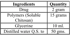
Table 1:Formulae of Implants Prepared
Ingredients Quantity Drug 2 gram Polymers (Soluble
Chitosan)
15 grams
Glycerine 10 ml. Distilled water Q.S. to 50 gms.
1. Preparation of Soluble Chitosan:
A) Preparation of N-trimethyl Chitosan chloride9: A mixture of 2g. of sieved Chitosan (93% deacetylated), 4.8g. of sodium iodide, 11 ml. of a 15% aqueous sodium hydroxide solution and 11.5 ml. of methyl iodide in 80ml. of 1-methyl-2-pyrrolidinone was stirred on a water bath of 60°C for 1 h. (Le Dung et. al., 1994). Special care was taken to keep the methyl iodide in the reaction mixture by using a Liebig condenser. The product was precipitated using ethanol and thereafter isolated by centrifugation. The N-trimethyl Chitosan iodide obtained after this first step was washed twice with ether on a glass filter to remove the ethanol. It was dissolved in 80 ml. of 1-methyl-2-pyrrolidinone and heated to 60°C, thus removing most of the absorbed ether. Subsequently, 4.8 g. of Nal, 11 ml. of 15% NaOH solution and 7 ml. of methyl iodide were added with rapid stirring and the mixture was heated on a water bath at 60°C for 30 min. An additional 2 ml. of methyl iodide and 0.6 g. of NaOH pellets were added and the stirring was continued for 1 h.
The product, prepared as described above, was dissolved in 40 ml. of a 10% NaCl aqueous solution, instead of HCl to exchange the iodide. The polymer was precipitated with ethanol, isolated by centrifugation and thoroughly washed with ethanol and ether. In vacuum drying yielding a white, water-soluble powder.
Viscosity:
Viscosity of water soluble Chitosan is 409.94 cps measured by using Brookfield Viscometer DV-III Ultra Programmable Rheometer.
B) Preparation of Aceclofenac sodium rod shaped implants 10:
Weighed quantity of Chitosan was dissolved in water and allowed to hydrate for 24 hours, Glycerin was added as a plasticizing agent and stirred for on hour. Aceclofenac sodium was dissolved separately in a small quantity of methanol and added to above polymeric solution the dough mass was extruded in to a 3mm diameter, rod shaped thread like implants are dried at room temp for 48 hours , After drying the implants were cut in to number of pieces of 6mm in length. The implants containing 10 mg of drug and have 155mg of total weight were prepared.
Hardening of rod shaped implants 10:
25 ml of Formaldehyde solution (37% v/v) was transferred in a petridish and kept in an empty glass dessicator. A wire mesh containing the implantable discs was placed on the top of the petridish and immediately the dessicator was closed. The discs were made to react with formaldehyde vapors for 12hours. Then they were removed and air dried for 72 hours so that the reaction between formaldehyde and Chitosan was completed. After wards the discs were kept in an open atmosphere in aseptic conditions for a week, to make sure that the residual formaldehyde gets evaporated.
EVALUATION OF SUB DERMAL IMPLANTS a) Measurement of implant thickness11: [Table-2]
The thickness of implants was measured with a screw gauge on a sample of three implants.
b) Weight of Implants11: [Table-2]
Weight variation was checked by weighing three implants individually.
c) Drug content uniformity test12: [Table-2]
Drug content of implants was estimated by removing a sample of three implants, of 6 mm in length and 3 mm thickness and analyzing the Drug content as follows. Each implant was cut in to small pieces and dissolved in a small quantity of methanol by heating at 600C on a water bath. After cooling the solution was
Table 2: Measurement of implant -Thickness, Weight and Drug content
Thickness Weight of Implants Drug content
SN Mean SD CV Mean SD CV Mean SD CV
1 3.01 0.0100 0.332 155.01 0.0152 0.0098 9.72 0.0916 0.9423 2 3.02 0.0115 0.380 154.07 0.545 0.3522 9.646 0.277 2.87 3 3.02 0.0264 0.874 155.01 0.0152 0.0098 9.576 0.066 0.689 * Each reading is a mean of three replicates.
* Each implant contains 10mg. of drug
d) Tests for sterility IP13:
The sterility test was conducted by Membrane Filtration Method on soyabean-casein digest medium and found to be implants are sterile.
Sterility test of prepared Implants
e) Test for free formaldehyde14:
To 1ml of 1 in 10 dilution preparation to be examined in a test-tube, 4 ml of water and 5ml of acetyl acetone solution were added. The tube was placed in a water bath at 400C for 40 minutes. The solution was not
more intensely colored than a reference solution prepared at the same time and in the same manner using 1ml of standard formaldehyde solution in place of the dilution of the preparation being examined. The comparison should be made by examining the tubes down their vertical axis.
f) Drug-polymers interaction study15:
The sub dermal implants of Aceclofenac sodium prepared with Chitosan, hardened with formaldehyde was tested for compatibility of the drug with the excipients used like, Chitosan, Glycerin and hardening agents by I.R. Study. The IR spectra of Aceclofenac sodium and its formulations were obtained by potassium bromide pellet method using Perkin Elmer FTIR series model 1615 spectrometer.
g) In-vitrodrug release studies16:
Implants were placed separately into a 10 ml vials containing 10 ml of phosphate buffer pH 7.4 the vials were sealed with rubber stoppers and kept in incubator shaker thermo stated at 370 + 0.50 C. The
dissolution fluid was changed for given time intervals and replaced with fresh 10 ml phosphate buffer pH 7.4. The
drug concentration in every dissolution fluid was analyzed spectrophotometrically at 295 nm after suitable dilution with phosphate buffer pH 7.4.
Table 3: Calibration Curve Data for Aceclofenac in Phosphate Buffer PH 7.4 (MAX= 275)
Sl. No. Concentration (mcg/ml) Absorbance*
1. 0 0.000
2. 4 0.072
3. 8 0.140
4. 12 0.210
5. 16 0.275
6. 20 0.343
* Average of three determinations.
Figure 1:Calibration Curve Data for Aceclofenac in Phosphate Buffer pH 7.4 (max= 275)
0 0.1 0.2 0.3 0.4
0 5 10 15 20
Concentration (mcg/ml.)
A
bs
or
ba
nc
e
Figure 2: Comparative cumulative percent drug released versus time (zero order kinetic model) plots of implants
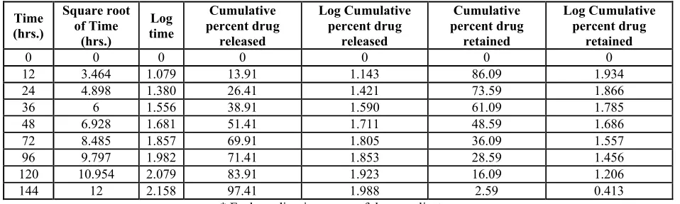
Table 4:In vitrorelease of Aceclofenac in phosphate buffer of pH 7.4 from implants prepared with soluble Chitosan and hardened for 12 hours using formaldehyde
Time (hrs.)
Square root of Time
(hrs.)
Log time
Cumulative percent drug
released
Log Cumulative percent drug
released
Cumulative percent drug
retained
Log Cumulative percent drug
retained
0 0 0 0 0 0 0
12 3.464 1.079 13.91 1.143 86.09 1.934 24 4.898 1.380 26.41 1.421 73.59 1.866
36 6 1.556 38.91 1.590 61.09 1.785
48 6.928 1.681 51.41 1.711 48.59 1.686 72 8.485 1.857 69.91 1.805 36.09 1.557 96 9.797 1.982 71.41 1.853 28.59 1.456 120 10.954 2.079 83.91 1.923 16.09 1.206
144 12 2.158 97.41 1.988 2.59 0.413
* Each reading is a mean of three replicates. * Each implant contains 10mg of drug
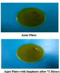
h) In- VivoStudies (Tissue-Polymer Interaction Studies)17, 18:
Two male white rabbits weighing around 2.5 kg were used for the study. The animals were housed individually in cages under environmentally controlled conditions (temperature 370C and 12 hr. lighting cycle). The animals were fed as usual with a standard rabbit diet that is commercially available and had access to water ad libitum. On the day of implantation the skin at the site of implantation (thigh) was cleaned by alcohol swab. Before implantation, the animals were applied with lignocaine, a local anesthetic gel. No.5, skin punch biopsy stainless steel forceps was used to take the tissue sample from the thigh region for histopathological studies.
RESULTS & DISCUSSION
Implants of Aceclofenac sodium were prepared employing Chitosan and hardened with formaldehyde for 12 hours. Prepared Implants gave the uniform results for drug content, thickness, weight variation and found to have uniform drug content 9.64 mg and 3.1 mm thickness with a mean weight of 155.01 mg. (Table 2) and in vitro drug release characteristics study. (Table 4).
At intervals during the incubation period, and at its conclusion, when the media was examined for macroscopic evidence of microbial growth, no evidence of microorganisms was found. So the Aceclofenac sodium implants pass the test for sterility.
The sample solution was not more intensely colored than the standard solution inferring that less than 20 mcg of free formaldehyde was present in 25 implants.
The same peaks as that of the pure drug Aceclofenac sodium were observed in implantable discs hardened with formaldehyde. The IR study showed there was no interaction between the Aceclofenac sodium and excipients used.
The drug release studies of Aceclofenac sodium in phosphate buffer pH 7.4 indicated 97.41% of drug release in 6 days (table 4). Further the implants
hardened with formaldehyde show first order rate kinetics. The mechanism of drug release was found to be diffusion. Chitosan implants were found to erode slowly giving out the drug Aceclofenac sodium in addition to diffusion mechanism. (Fig 2,) In-vivo studies in animals (Rabbits), revealed that at
sub dermal thigh region before and after one month of implantation , there was no inflammation at the site of implantation, foreign body grannuloma formation was not observed, necrosis / hemorrhage was not present. Thus Chitosan was found to be compatible with the tissues at sub dermal region.
CONCLUSION
Chitosan based sub dermal implants of Aceclofenac sodium having uniform characters can be prepared with minimum batch to batch variation. The sub dermal implants containing Chitosan and hardened with Formaldehyde for 12 hours is found to produce the most satisfactory drug release.
The Aceclofenac sodium implants can be used for the treatment of arthritis as they meet the criteria such as better patient compliance, improved therapeutic outcome and minimum incidence of adverse effects.
ACKNOWLEDGEMENT:
The authors are thankful to Ethical Committee, M. R. Medical College, Gulbarga for in-vivo studies in animals. Authors are also thankful to Cipra Labs Hyderabad for FTIR Spectra.
REFERENCES
1. Health Topics, National Institute of Arthritis, Bethesda- MD, 2000, p. 01-12.
2. Paul A. Insel, Analgesic – antipyretic and anti-inflammatory agents and drugs employed in the treatment of gout, Goodman and Gilman’s The Pharmacological Basis of Therapeutics, 9th
edition, McGraw-Hill Publications,1996,618 3. Robert E. Willette, Anti-inflammatory analgesics,
10th edition, Lippincott Raven Publishers, NY,
1998, 711
4. Jain, N.K. Advance’s in controlled and novel drug delivery, first edition, CBS publishers and Distributors,4596/1-A,11 Dariya Gunj, New Delhi; 2001, page 204-231.
5. Ronal E. Barne, Nonsteroidal anti-inflammatory drugs, Principles of Medicinal Chemistry by William Foye, 4THedition, B. I. Waverly Pvt. Ltd.,
1995,537
6. Muthuswamy K, Ravi TK, Govindharajan G, Gopalakrishnan S. “The use of Soluble Chitosan as drug carrier”, Ind. J. Pharm. Edn., July-Sept. 2004; 38(3): 138-140.
7. Murthy, R.S.R., Implantable Therapeutic Systems, Advances in Controlled and Novel Drug Delivery Systems, N. K. Jain, 1st edition, CBS Publishers
and Distributors, 2001, 204
8. Tan veer Ahmed Khan et. al., kok kniang peh, 2002, “Mechanical bioadhesive strength and biological evaluation of Soluble Chitosan film for wound dressing” School of Pharmaceutical Sciences, University of Science, Malaysia.
9. Sieval AB, Thanau M, Kotze AF, Verhod JC, Brussee J, Junginger HE. “Preparation and NMR characterization of highly substituted N-trimethyl Soluble Chitosan chloride”, Elsevier Carbohydrate Polymers; 36 (1998): 157-165.
10. Mohammed Majid Iqbal Sanjeev Gupta Shanti Sagar and Mohammed Ibrahim, “Design and evaluation of subcutaneous implantable drug
delivery” Annals of Phytomedicine 1(2): 30-38, 2012
11. Sandeep j jaybhaye, ravindra kamble, appala raju, anil bhandari, s a sreenivas.
“Design of chitosan based progesterone subdermal implants for synchronization of estrus cycle in animals” International Journal of Pharmacy and Pharmaceutical Sciences , Vol 4, Issue 2, 2012 12. Sanjeev.L.Gupta, K. Purushotham Rao, KPR
Choudary, S.Pratima. “Preformulation Studies of Biodegradable Drug Implants of Meloxicam for Orthopedic Patient Care” Journal of Pharmaceutical Science and Technology Vol. 3 (1), 2011,494-498
13. Rao. K. Purushothama. Jaybhaye S.Jb, Ravindra Kamblec, Anil Bhandarib and Pratima Sd “Designing of Diclofenac Sodium Biodegradable Drug Implant for Speedy Fracture Healing.” J. Chem. Pharm. Res., 2010, 3(1):330-337
14. Test for Sterility, A—111, The Indian Pharmacopoeia, 3rdedition, 1985
15. Test for free formaldehyde, A—60, The Indian Pharmacopoeia, 3rdedition, 1985
16. Shivkumar, H.N., Sarasija, Suresh, Venkatram, S., Ind J Pharm Sci, 2002:64(3), 412
17. Negrin, C. M., Delgado, A., Llabres, M., Methadone implants for methadone maintenance treatment in vitro and in vivo animal studies, J Control Rel, 2004,95(30 , 413
18. Howard, c. Aurd, Textbook of Pathology, 6th
edition, Nicholas G. Papovach,120
All © 2010 are reserved by Journal of Global Trends in Pharmaceutical Sciences
How to cite this article:
Chaus W. N.*,Shrivastava. B.Rao. K. P, Jadge D.R.Satpute K. L. Dongre. O. S: Formulation development of Chitosan