ISSN:2278 7496
AJPER Jan – Mar. 2020, Vol 9, Issue 1(11-19)
RP-HPLC METHOD DEVELOPMENT AND VALIDATION FOR THE ESTIMATION OF
MICAFUNGIN IN BULK AND INJECTABLES FORMULATION
Vivek Jain1, Priyanka Sharma*1, Navjot Singh1, Prabhat Kumar Jain2
1NRI Institute of Pharmacy, Bhopal, Madhya Pradesh, India
2Scan Research Laboratories, Bhopal, Madhya Pradesh, India
*Corresponding Author’s E mail: [email protected]
Received 22Oct. 2019; Revised 01Nov. 2019; Accepted 07Nov. 2019, Available online 15 Jan. 2020
ABSTRACT
Micafungin is an antifungal drug. It belongs to the antifungal class of compounds known as echinocandins and exerts its effect by inhibiting the synthesis of 1, 3-beta-D-glucan, an integral component of the fungal cell wall. A reversed-phase high performance liquid chromatography (RP-HPLC) method was developed and validated for the estimation of micafungin in bulk and injectable formulation. The separation was achieved on Thermo C18 analytical column (250 mm × 4.6 mm i.d., 5.0
μm) using 20mM KH2PO4: acetonitrile (pH 3.0 with orthophosphoric acid) in the ratio 20:80 v/v as
mobile phase and at a flow rate of 1.0 ml/min. Detection was carried out using a UV detector at 264 nm. The total chromatographic analysis time per sample was about 7.0min with micafungin eluting at retention time of about 4.569±0.3min. The method was validated for accuracy, precision, specificity, linearity and sensitivity. Validation studies demonstrated that this HPLC method is simple, specific, rapid, reliable and reproducible. The standard curve was linear over the concentration range of 5-25μg/ml with r2 close to one (0.999). The limit of detection (LOD) and limit of quantitation (LOQ) obtained for micafungin were 0.32μg/ml and 1.05μg/ml respectively. The developed and validated method was successfully applied for the quantitative analysis of mycamine 50mg injection. The high recovery and low relative standard deviation confirm the suitability of the proposed method for the determination of micafungin in injectable formulation.
Keywords: Analytical method development, Reversed phase HPLC method, ICH guidelines, Tablet dosage forms, Accuracy and precision
INTRODUCTION
Fungal infections are caused by microscopic organisms that can invade the epithelial tissue. The fungal
kingdom includes yeasts, molds, rusts and mushrooms. Fungi, like animals, areheterotrophic, i.e. they
obtain nutrients from the environment and not from the endogenous sources. Some of these fungi are
pathogenic and can produce mild to severe fungal infections. An antifungal agent is a drug that selectively
eliminates fungal pathogens from a host with minimal toxicity to the host 1. They can be categorized in
to several categories according to different pharmacophores and different mechanisms. Polyene
antifungal drugs like amphotericin B, nystatin interacts with sterols in the cell membrane to form
channels through which small molecules leak from the inside of the fungal cell to the outside. Azoles
like fluconazole, itraconazole, ketoconazole, clotrimazole, voriconazole, posaconazole etc. inhibit
cytochrome P450-dependent enzymes (particularly C14-demethylase) involved in the biosynthesis of
ergosterol, which is required for fungal cell membrane structure and function.
Allyl amines like naftifine, terbinafine inhibit ergosterol biosynthesis at the level of squalene epoxidase.
Antimetabolites like 5-Fluorocytosine act as an inhibitor of both DNA and RNA synthesis via the
intracytoplasmic conversion of 5-fluorocytosine to 5-fluorouracil.Echinocandins like anidulafungin,
caspofungin and micafungin are used for systemic fungal infections in immunocompromised patients
and they inhibit the synthesis of glucan in the cell wall via the enzyme 1, 3-β glucan synthase2, 3.
Analytical method Development and validation for newly introduced pharmaceuticals is of importance,
as drug or drug combination may not be official in pharmacopoeia and so analytical method for
quantification is not available. To check and ensure the quality standards of drug molecules and their
formulation various analytical methods are employed. Most of the drugs in single or multi component
dosage forms can be analyzed by HPLC method because of the associated advantages like speed, greater
sensitivity, improved resolution, specificity, accuracy, precision, reusable columns and ease of
automation in this method 4, 5, 6.Micafungin (trade name Mycamine)
[5-[(1S,2S)-2-
[(3S,6S,9S,11R,15S,18S,20R,21R,24S,25S,26S)-3-[(1R)-3-amino-1-hydroxy-3-oxopropyl]-
11,20,21,25-tetrahydroxy-15-[(1R)-1-hydroxyethyl]-26-methyl-2,5,8,14,17,23-hexaoxo-18-[[4-[5-(4-pentoxyphenyl)-1,2-oxazol-3-yl]benzoyl]amino]-1,4,7,13,16,22 –hexazatricyclo [22.3.0.09,13] hepta-
cosan-6-yl]-1,2-dihydroxyethyl]-2-hydroxyphenyl] hydrogen sulfate (Fig. 1). Micafungin is a cyclic
semisynthetic derivative of the echinocandin-like lipopeptideFR-901379 isolated from the culture broth
of Coleophoma empetri, a plant pathogen associated with postharvest fruit rot in cranberries 7,8. It has an
empirical formula of C56H70N9NaO23S, a molecular weight of 1292.26 g/mol 9. Micafungin selectively
inhibit 1,3-β-D-glucan, which is required for fungal cell wall synthesis. It has been approved for the
treatment of esophageal candidiasis and for prophylaxis of candida and aspergillus infections in patients
undergoing hematopoietic stem cell transplantation10, 11. The drug was first launched in Japan in
December 2002 and then also approved by US food and drug administration in March 200512, 13. Several
methods have been employed for the estimation of micafungin alone and combination with other drugs
by UV and RP-HPLC methods in bulk drug and plasma samples 14-17. Sheng et al reported stability
indicating HPLC method for determination of micafungin and related substances 18 and Joshi et al
reported RP-UPLC method for determination of micafungin and related substances19.But there is no
simple and easy method for the analysis of micafungin. Hence, it is necessary to develop a rapid, accurate
and validated RP-HPLC method for the determination of micafungin in bulk and injectables formulation.
reversed phase HPLC method, using PDA detection, for the estimation of micafungin in bulk and
injectables formulation. The developed method validated according to ICH guidelines 20.
O S O O OH OH OH HO N NH H N N HN N H HN O O N O O O O O O O O O O O HO HO HO HO HO O OH NH2 H H
Figure 1 Chemical structure of micafungin
Materials and Methods
Instrumentation
Liquid chromatographic system from Waters model no 784 comprising of manual injector, water 515
binary pump for constant flow and constant pressure delivery and UV-Visible detector connected to
software Data Ace for controlling the instrumentation as well as processing the generated data. Weighing
was done on a Digital Micro Balance (CX-265) manufactured by Citizen Scale (I) Pvt. Ltd.
Reagents and chemicals
Analytically pure sample of micafungin was a generous gift from Glaxo SmithKline Pharmaceuticals
Ltd,Mumbaialong with their analytical reports.Potassium di hydrogen phosphates (AR grade), disodium
hydrogen phosphate (AR grade), OPA and acetonitrile (HPLC Grade) was purchased from E. Merck Ltd.
Worli, Mumbai, India.All other chemical used were of analytical grade. Triple distilled water was used
for whole experiment was generated in house.Mycamine 50 mg injectionGlaxo SmithKline
Pharmaceuticals Ltd,Mumbai, India was purchased from local market.
Diluents
A mixture of 20mM KH2PO4: acetonitrile(pH 3.0 with orthophosphoric acid) in the ratio 20:80 v/v was
used in RP-HPLC as diluents.
Selection of mobile phase
Initially to estimate micafungin simultaneously, number of mobile phases in different ratios was tried.
Taking into consideration the system suitability parameter like RT, tailing factor, number of theoretical
acetonitrile(pH 3.0 with orthophosphoric acid) in the ratio 20:80 v/v run as isocratic system. The mobile
phase was filtered through 0.45 m filter paper and then degassed by Sonication. Flow rate employed for
analysis was 1 ml/min.
Chromatographic conditions
The isocratic mobile phase consisted of 20mM KH2PO4: acetonitrile(pH 3.0 with orthophosphoric acid)
in the ratio 20:80 v/v, flowing through the column at a constant flow rate of 1.0 ml/min. The mobile
phase was filtered through nylon 0.22 µm membrane filters and was degassed before use (30
min).AThermo (C-18) column (5 µm, 250mm x 4.60mm) was used as the stationary phase. By
considering the chromatographic parameter, sensitivity and selectivity of method for drugs, 264.0 nm
was selected as the detection wavelength for UV-Visible detector.
Standard preparation
Standard stock solution
Accurately weighed 10 mg of micafungin was transferred into 10 ml volumetric flask, dissolved in 5ml
of diluentsand volume was made up to 10ml with diluents to get concentration of solution 1000 g/ml
(Stock-A).
Working standard solution
From stock solutions of micafungin, 1 ml was taken and diluted up to 10 ml. from this solution 0.5, 1.0,
1.5, 2.0, 2.5 ml solutions were transferred to 10ml volumetric flasks and make up the volume up to 10
ml with diluents, gives standard drug solution of 5, 10, 15, 20, 25 µg/ ml concentration.
Sample preparation
For analysis of the injectables formulation, weight equivalent to weight 10 mg of micafungin was
transferred to 10 ml volumetric flask and dissolved in diluents. The solution was shaking vigorously for
20 min and filtered through Whattman filter paper no.41, then volume was made up to mark with diluents.
From the above solution 1 ml of solution was taken and diluted to 10 ml with diluents to get a solution
containing 100µg/ml. From the above solution 1 ml of solution was taken and diluted to 10 ml with
diluents to get a solution containing 10 µg/ml of micafungin. The amounts of micafunginin injection
were calculated by extrapolating the value of area from the calibration curve. Analysis procedure was
repeated six times with formulation.
Results and discussion
Chromatography
The mobile phase was chosen after several trials with methanol, isopropyl alcohol, acetonitrile, water
and buffer solutions in various proportions and at different pH values. A mobile phase consisting of
achieve maximum separation and sensitivity. Flow rates between 0.5 and 1.5 min were studied. A flow
rate of 1 ml/min gave an optimal signal-to-noise ratio with a reasonable separation time. Using a
reversed-phase C18 column, the retention times for micafunginwas observed to be 4.569±0.3min. Total
time of analysis was less than 7 min. The maximum absorption of micafunginwas detected at 264nm and
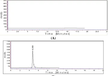
this wavelength was chosen for the analysis Fig. 2
(A)
(B)
Figure 2 Chromatograms of (A) Blank mobile phase (B) micafungin (10μg/ml) as reference substances
System suitability
System suitability parameters such as number of theoretical plates, HETP and peak tailing are
determined. The results obtained are shown in Table 1. The number of theoretical plates for micafungin
was 2127.
Table 1 Results of system suitability parameters
Parameters Micafungin
AUC* 971.4593
No. of Theoretical
Plates 2127
Tailing Factor* 0.945
Retention time*
4.569 Calibration
range(μg/ml) 5-25
Linearity
The calibration curve was linear over the concentration range of 5-25μg/ml for micafungin. The linearity
was represented by a linear regression equation as follows:
Y(micafungin) = 65.52conc-0.821 (r2 = 0.999)
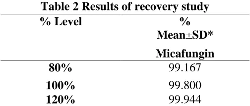
Accuracy
Recovery studies were performed to calculate the accuracy of developed method to preanalysed sample
solution, a definite concentration of standard drug (80%, 100%, and 120%) was added and then its
recovery was analyzed. The value of percentage RSD was found less than 2 (0.647, 0.659 and 0.438)
show good recovery at all three level 80, 100 and 120% respectively. Each level was made in triplicate
Table 2.
Table 2 Results of recovery study
% Level %
Mean±SD*
Micafungin
80% 99.167
100% 99.800
120% 99.944
* Value of three replicate and three concentrations.
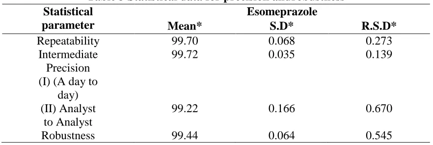
Precision
Repeatability
Five dilutions in three replicates were analyzed in the same day for repeatability and results were found
within acceptable limits (RSD < 2) as shown in Table 3.
Intermediate precision
Five dilutions in three replicates were analyzed on two different days and by two analysts for
day-to-day and analyst-to-analyst variations and results were found within acceptable limits (RSD < 2) as
shown in Table 3.
Robustness
As per ICH norms, small, but deliberate variations in concentration of the mobile phase were made to
check the method’s capacity to remain unaffected. The ratio of mobile phase was change from, 20mM
KH2PO4: acetonitrile(20:80 % v/v), to (25: 75% V/V) and method is found robust as RSD is again found
Table 3 Statistical data for precision androbustness Statistical
parameter
Esomeprazole
Mean* S.D* R.S.D*
Repeatability 99.70 0.068 0.273
Intermediate Precision (I) (A day to
day)
99.72 0.035 0.139
(II) Analyst to Analyst
99.22 0.166 0.670
Robustness 99.44 0.064 0.545
*Mean of 15 determinations (three replicates at five concentration level)
Detection Limit and Quantitation Limit
The LOD and LOQ of developed method were calculated based on the standard deviation of response
and slope of the linearity curve Table 4.
Table 4 LOD and LOQ
Name LOD
(g/ml)
LOQ (g/ml)
Micafungin 0.32 1.05
Analysis of marketed formulation
The assay value of drugs was close to 100, SD and % RSD are less than 2 indicate the no interference of
excipients in the estimation of drug Table 5.
Table 5Assay of tablet formulation S.
No.
Parameter Micafungin
1. Mean 99.12
2. S. D. 0.215
3. % RSD 0.265
Mean of nine determinations Conclusion
The proposed HPLC method was validated as per the International Conference on Harmonisation (ICH)
Q2B Guidelines, and was found to be applicable for routine quantitative analysis of micafungin by HPLC
in pharmaceutical dosage form. The results of linearity, precision, accuracy and specificity, were proved
to be within the limits. The method provides selective quantification of micafungin with no interference
from other formulation excipients. The proposed method was highly reproducible, reliable, rapid, robust
and specific. Therefore, a high percentage of recovery and the run time of less than seven minutes allow
Acknowledgments
The authors would like to thank the Mr. Prabhat Kumar Jain, Geeta Parkhe and All supporting staff of
Scan Research Laboratories, Bhopal (M.P.) who helped in the experiments during research work.
References
1. Borne RF. In Foye’s Principles of Medicinal Chemistry, Ed.; Willims DA, Lemke TL. 5th edition,
Lippincott Williums and Wilkins pub., Philadelphia. 2002; 751-793.
2. Enoch DA, Ludlam HA and Brown NM. Invasive fungal infections: a review of epidemiology and
management options. J Med Microbiol. 2006; 55: 809-818.
3. Georgopapadakou NH and Walsh TJ. Antifungal agents: chemotherapeutic targets and immunologic
strategies. Antimicrob Agents Chemother. 1996; 40: 270-91.
4. Vander Wal S and Snyder LR. Photometric detection at 185 nm for high-performance liquid
chromatography with either isocratic or gradient elution: Assay of mixtures of polyethylene glycol
oligomers. J Chromatogr. 1983; 225: 463-474.
5. Poole CF and Schutte SA. Contemporary Practice of Chromatography, Elsevier, Amsterdam, 1984:
pp. 375
6. Krull IS. In Chromatography and Separation Chemistry: Advances and Developments, 2nd. ed., ACS
Symposium Series 297, ACS, Washington, DC, 1986: pp. 137.
7. Tawara S, Ikeda F, Maki K, Morishita Y, Otomo K, Teratani N, Goto T, Tomishima M, Ohki H and
Yamada A.In vitro activities of a new lipopeptide antifungal agent, FK463, against a variety of
clinically important fungi. Antimicrob. Agents Chemother. 2000;44: 57–62.
8. Olatinwo RO, Schilder, A and Kravchenko AN. Incidence and causes of postharvest fruit rot in stored
Michigan cranberries. Plant Dis. 2004:88: 1277–1282.
9. Vehreschild JJ and Cornely OA. Micafungin sodium, the second of the echinocandin class of
antifungals: Theory and practice. Future Microbiol. 2006;1: 161–170.
10.Scott LJ. Micafungin: a review of its use in the prophylaxis and treatment of invasive Candida
infection. Drugs 2012;72(16):2141-65
11.Hatano K, Morishita Y, Nakai T, Ikeda F. Antifungal mechanism of FK463 against Candida albicans
and Aspergillus fumigatus. J. Antibiot. 2002; 55:219-222
12.Carver PL. Micafungin. Ann. Pharmacother. 2004; 38:1707-21
13.Zaas AK, Steinbach WJ. Micafungin: The US perspective. Expert Rev. Anti-infect. Ther. 2005;
14.Kumar SM, Shetty SK and SatyanarayanND.Development and validation of UV spectrophotometric
methods for the estimation of micafungin in bulk and pharmaceutical formulations.International
Journal of Pharmaceutical, Chemical and Biological Sciences.2018; 8(2): 204-209.
15.Nakagawa S, Kuwabara N, Kobayashi H, Shimoeda S, Ohta S and Yamato S. Simple
column-switching HPLC method for determining levels of the antifungal agent micafungin in human plasma
and application to patient samples. Biomed. Chromatogr. 2013; 27:551–555
16.Martens-Lobenhoffer J, Rupprecht V and Bode-Boger SM. Determination of micafungin and
anidulafungin in human plasma: UV- or mass spectrometric quantification. J. Chromatogr. B. 2011;
879:2051–2056
17.Uranishi H, Nakamura M, Nakamura H, Ikeda Y, Otsuka M, Kato Z and Tsuchiya T. Direct-injection
HPLC method of measuring micafungin in human plasma using a novel hydrophobic/hydrophilic
hybrid ODS column. J. Chromatogr. B. 2011; 879:1029–1032
18.Shengsheng Z, Xiang M, Xin S, Yongwei L and Zuyue S. Development and Validation of a
Stability-Indicating High Performance Liquid Chromatographic (HPLC) method for the determination of
related Substances of Micafungin Sodium in drug substances. Int J Mol Sci. 2013; 14(11):21202–
21214
19.Joshi S, Majmudar F and Vyas N. Development and Validation of Analytical Method for
Determination of Micafungin and Its Related Substances in Bulk by RP-UPLC. International Journal
of Pharmaceutical Sciences and Research. 2016; 7(3): 1211-1218.
20.Code Q2 (R1) -Text on Validation of Analytical Procedures: Text and Methodology Current Step 4