SOUND PROPAGATION THROUGH BONE TISSUE
H Aygün, C Barlow, and L Yule. Solent Acoustics, School of Media Arts and Technology, Southampton Solent University, Southampton, SO14 0YN, UK.
S. Y. Liu School of Naval Architecture & Ocean Engineering, Huazhong University of
Science & Technology, P. R. China.
1
INTRODUCTION
Osteoporosis has been recognized as an established and well-defined disease that affects more
than 75 million people in the Europe, United States and Japan1. Osteoporosis causes more than 8.9
million fractures annually worldwide, of which more than 4.5 million occur in the Europe and United States. The lifetime risk for a wrist, hip or vertebral fracture has been estimated to be in the order of 30% to 40% in developed countries – in other words, very close to that for coronary heart disease. Osteoporosis is not only a major cause of fractures, it also ranks high among diseases that cause people to become bedridden with serious complications. These complications may be life threatening in ageing people because it is estimated that the burden of osteoporosis will increase four-fold by the year 20502.
The prevention of osteoporosis and its associated fractures is a requirement to maintain the independence, well-being and quality of life of the elderly population in World. As the skeleton develops during childhood, more bone is added than is being taken away. During early adulthood, the amounts removed and added are the same. If however, more bone is removed than is being added, akin to skeletal bio-corrosion, we have a condition called osteoporosis. Osteoporosis literally means ‘porous bone’ and describes a period of largely asymptomatic bone loss leading to skeletal fragility and increased risk of fracture3. One in three women and one in five men over the age of 50 will break a bone attributed to osteoporosis. A quarter of hip fracture subjects die within 12 months, whilst a quarter of those remaining never regain independent status.
During acoustic excitation (e.g. from a loudspeaker) of an air-filled porous solid most of the acoustic energy travels in the pores and the acoustical properties are described well by assuming that the solid frame is rigid. On the other hand intimate mechanical contact between a transducer and a porous elastic medium excites waves predominantly in the solid frame. The inherent anisotropy of cancellous bone means that the acoustical properties vary with transmission direction. Tortuosities deduced from audio-frequency measurements in air-filled bone replicas, assuming rigid-porous behavior, have shown a strong anisotropy4. To predict fast wave transmission it is also necessary to allow for elastic anisotropy also. A method of including the effects of anisotropy in Biot model5, 6, 7 introduces an angle dependent tortuosity.
To date, no satisfactory evidence exists either supporting or refuting the usefulness of quantitative ultrasound worldwide, so that further research clearly is warranted. Nonetheless, there is enough evidence in other populations to suggest that QUS may be an acceptable, low cost and readily-accessible alternative to DXA measurements of BMD in the managements of osteoporosis in Hispanics8. BMD tests are not widely available, or are used predominantly for research, in part because of the high capital costs of DXA. BMD test alone are not optimal for the detection of individuals at high risk of fracture. The fracture risk is very high when osteoporosis is present, but by no means negligible when BMD is normal. Current recommendations for the assessment of patients for BMD and fracture risk have several difficulties, and none is suitable for international use.
and perforation introduced into materials, the amplitude, arrival time, and shape of the detected structural borne sound wave are different from the one initially observed in original materials. In another word it means that, when normal and osteoporotic bones are subjected to vibration, the resulting detected responses have different shapes and amplitudes. Differences between normal and osteoporotic bones are the sign of osteoporosis being diagnosed by structural vibration technique.
2
EXPERIMENTAL PROCEDURE
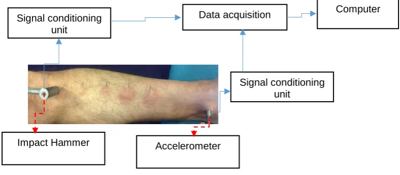
Experiments have been carried out on a male tibia as shown in Figure 1. An impact hammer, PCB086C03, is used to generate structural borne acoustic waves. The response is detected by using an accelerometer, MME-KS901.100. The Impact hammer and accelerometer are connected to signal conditioning units, DJB-VB/01, which fed a data acquisition system, (NI-USB-4431), which is connected to a computer as seen in Figure 1. Electronic interference is removed by 10000 acquisition averages per second. The measurement used is as follow input force has been applied to bone surface at five different locations along the bone and response has been detected under the knee cap on the surface of tibia for each measurements.
Figure 1: Measurement set-up.
3
RESULTS
Measurements have been performed on a healthy human male tibia and on an unhealthy female tibia in order to investigate wave propagation through the bone and to determine the natural frequency of the bone. Firstly, force has been applied to five different positons, including ankle too, on the surface of the bone by using an impact hammer.
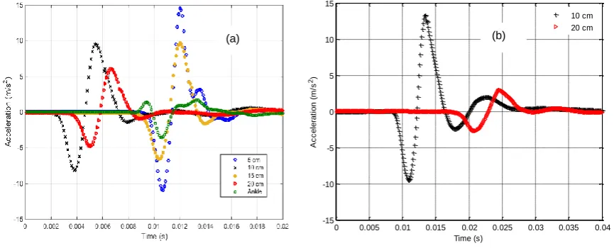
Vibrating the bone has generated structural borne acoustic waves travelling through the bone and along the surface of the bone. The responses have been detected at the top of human tibia just under the knee cap. The input forces and their associated responses are shown in Figure 2 and Figure 3. Detected responses can’t be compared because initial forces applied to bone by an impact hammer are not repeatable. Unwanted noise has been filtered by using the Wavelet toolbox in MATLAB. Fast Fourier Transform (FFT) has been used to convert filtered signals into the frequency domain.
Signal conditioning unit
Signal conditioning unit
Data acquisition Computer
Figure 2: a-) Input force applied to five different locations on male tibia by an impact hammer. b-) Input force applied to two different locations on famale tibia by an impact hammer.
Figure 3: a-) Responses detected by an accelerometer under knee cap for male tibia, b-) Responses detected by an accelerometer under knee cap for female tibia.
Frequency response signals have been calculated by using a transfer function method given as follows;
𝑇(𝑓) =
𝐴(𝑓)𝐹(𝑓) (1)
where 𝐴(𝑓) is the response of the system in the frequency domain, and 𝐹(𝑓) is the force applied to system in the frequency domain.
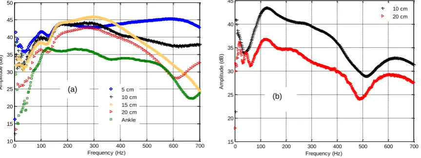
Transmission function curves of male and female tibia are given in frequency domain in Figure 4. When the distance between the accelerometer and the impact hammer increases, the amount of structural borne acoustic energy transmitted through the bone mostly reduces throughout the frequency range. This attenuation of acoustic energy might be due to the distance between input and outputs, soft tissue of the bone, or changes in bone diameter along the tibia surface.
0 0.005 0.01 0.015 0.02 0.025 0.03 0.035 0
0.05 0.1 0.15 0.2 0.25
F
o
rc
e
(
V
o
lt
a
g
e
)
Time (s)
10 cm 20 cm
0 0.005 0.01 0.015 0.02 0.025 0.03 0.035 0.04 -15
-10 -5 0 5 10 15
A
c
c
e
le
ra
ti
o
n
(
m
/s
2)
Time (s)
10 cm 20 cm
(a)
(b)
Hz and 127.1 Hz respectively. The amplitude of the natural frequency of the male bone is between 36 dB and 46 dB.
Increasing the distance between accelerometer and impact hammer causes the natural frequency of male bone to shift from higher frequency to lower frequencies.
The natural frequency of female tibia obtained under the knee cap at distance of 10 cm and 20 cm from accelerometer are given as 126.7 Hz and 121 Hz respectively as shown in Figure 4b. The amplitude of natural frequency of female bone is between 36 dB and 44 dB. More acoustic energy was attenuated throughout frequency range when the distance between accelerometer and impact hammer is increased.
Figure 4: Transfer functions versus frequency, a-) for male tibia, and b-) for female tibia.
4
CONCLUSION
An experimental investigation has been carried out to determine the possibility of using a structural borne acoustic wave technique to detect the variation of sound propagation in the bone structure. Both tibias tested have different dimensions and size.
Varying the distance between the accelerometer and impact hammer mostly changes the natural frequency of the male tibia and the amplitude of response while it slightly changes the natural frequency of the female tibia and reduced the amplitude of response a lot throughout the frequency range. The results show that tibia has an anisotropic structure which has an important effect on measured natural frequencies.
Further work is needed to carry out measurements on more healthy and unhealthy male and female tibias.
5
ACKNOWLEDGEMENT
This work has been supported by Southampton Solent University, Research & Enterprise / ID No: 1027.
6
REFERENCES
1. The world health report 2004: changing history. Geneva, World Health Organization, 2004.
0 100 200 300 400 500 600 700 10 15 20 25 30 35 40 45 50 A m p lit u d e ( d B ) Frequency (Hz) 5 cm 10 cm 15 cm 20 cm Ankle
2. Cooper C., Campion G., and Melton L. J., 3rd (1992) Hip fractures in the elderly: a world-wide projection. Osteoporosis International 2, pp.285-289.
3. Consensus Development Conference: prophylaxis and treatment of osteoporosis. 1991.
American Journal of Medicine. 90, pp. 107-110.
4. Attenborough K., Qin Q., Fagan M. J., Shin H-C., and Langton C. M., 2005. Measurements of tortuosity in stereolithographical bone replicas using audio-frequency pulses. Journal of
the Acoustical Society of America 118, pp. 2779-2782.
5. Aygün H., Attenborough K., Lauriks W., and Langton M.C., 2010, Ultrasonic wave propagation in Stereolithographical bone replicas. Journal of the Acoustical Society of
America 127 (6), pp. 3781-3789.
6. Aygün H., Attenborough K., Lauriks W., Rubini P.A., and Langton M.C., 2011, “Wave propagation in Stereolithographical (STL) bone replicas at oblique incidence.” Applied
Acoustics 72 (7), pp. 458-463.
7. Aygün H., and Barlow C., 2015, Ultrasonic wave propagation through porous ceramics at different angles of propagation. Applied Acoustics 88, pp. 6-11.
8. Hans D and Kreig M A 2009. Quantitative ultrasound for the detection and management of