Turkish Journal of Fisheries and Aquatic Sciences 17: 135-143 (2017)
www.trjfas.org ISSN 1303-2712 DOI: 10.4194/1303-2712-v17_1_16
RESEARCH PAPER
© Published by Central Fisheries Research Institute (CFRI) Trabzon, Turkey in cooperation with Japan International Cooperation Agency (JICA), Japan
Cd2+ and Pb2+ Induced Structural, Functional and Compositional Changes
in The Liver and Muscle Tissue of Crucian Carp (Carassius auratus
gibelio): an FT-IR Study
Introduction
Heavy metals are serious environmental pollutants because of their persistency, non biodegradability and higher accumulative tendency in the living tissue (Begum et al. 2005, Gupta and Karthikeyan 2016). Among heavy metals, cadmium (Cd) and lead (Pb) are the prominent toxic metals (Lim et al. 2008, Senthil Kumar et al. 2008) with no known nutritive values (Poole et al. 2005, Xu et al. 2008) and are abundant in nature. The common sources of Cd and Pb pollution are contaminated soils, sediments and waters due to natural and anthropogenic activities which cause their entry into the food chain and generating various adverse effects in animals and humans (Chai et al. 2014, Chakraborty et al. 2012, Khan et al. 2014). Though freshwater ecosystems have certain physico-chemical and biological mechanisms to counteract or eliminate the adverse effects of pollutants; however, toxicants may induce changes in normal growth, reproduction and behavior or may be fatal to freshwater organisms (Rand et al. 2003).
Cadmium has the ability to disturb various cellular functions and can damage the structures of
different cellular compartments (Nemmiche et al. 2007), because of the higher affinity of Cd2+ ions to
biological structures consisting of sulfhydryl (-SH), carboxyl and phosphate groups. This may cause the inhibition of numerous enzymes and disturbance of important metabolic processes including lipid metabolism (Krishnakumar et al. 2012, Murugavel and Pari 2007). On the other hand, Pb toxicity may occur through the ionic Pb2+ replacement with certain
divalent ions such as Zn, Fe and by calcium mimicry (Tellis et al. 2014) causing neurological disorders, genotoxicity, muscular spasms, haematological alterations, paralysis and mortality in the exposed freshwater fish (Martinez et al. 2004, Grosell et al. 2006, Monteiro et al. 2011). However, the mechanism of Cd and Pb induced molecular alterations in the tissues and cells are still not clear.
Fish play an important role in balanced and nutritious diet containing a vital source of proteins and long chain polyunsaturated fatty acids with high quantity of fat soluble vitamins. However it can also be a source of trace metal exposure due to excessive amount of elements they can contain, in which some are highly toxic to human (Carvalho et al. 2005). The nutritive value of fish greatly depends on their
Sher Ali Khan
1, Xiaoyu Liu
1,2,*, Hong Li
1, Jingna Li
1, Peiyuan Zhou
1, Zia ur Rehman
4, Rehman
Ullah Khan
51 College of Food Science & Technology, Huazhong Agricultural University Wuhan, 430070, P. R. China. 2Key Laboratory of Environmental Correlative Dietology, Ministry of Education Wuhan, 430070, P. R. China. 3 Agricultural Research Institute Tarnab Peshawar, Pakistan.
4 Key Laboratory of Animal Genetics, Breeding and Reproduction, Huazhong Agricultural University Wuhan, 430070, P. R.
China.
5 Food Technology Centre, Pakistan Council of Scientific and Industrial Research Labs. Peshawar, Pakistan.
* Corresponding Author: Tel.: 008613517257126; E-mail: [email protected]
Received 16 June 2016 Accepted 10 August 2016
Abstract
Contamination of aquatic ecosystems with toxic metals such as Cd2+ and Pb2+ is a serious issue in the industrialized
world, which can affect freshwater fish even at low concentrations. The aim of the present study was to investigate the effect of Cd2+ and Pb2+ alone or in combination on the biochemical constituents of liver and muscle in Crucian carp using Fourier
Transform Infrared Spectroscopy (FT-IR). Results from the spectral analysis revealed significant decline in protein and increase in lipids in the two tissues with marked effect caused by the combined exposure of Cd2+ and Pb2+. In case of liver,
alteration in the intensity and band area at amide I resulted in a differential response of structural protein for the exposed groups. Furthermore, a decrease in the α-helix and alterations in the nucleic acids content was also observed in both liver and muscle of the exposed fish. Moreover, biochemical alterations in the vital tissue of freshwater fish due to toxic contaminants can be used as a marker of environmental pollution with the help of FT-IR spectroscopy.
136 S. A. Khan et al. / Turk. J. Fish. Aquat. Sci. 17: 135-143 (2017)
biochemical constituent which is affected by polluted water because of their direct contact with the toxicant in contaminated waters (Burger et al. 2002). Metals usually accumulate in the high fatty tissues of muscle, or in some specific organs, based on the lipophilic nature of the toxic chemical and how they are metabolized by the organism. Once enter to an organism, metals tend to remain in various tissues and may continuously accumulate with subsequent exposures. The bioaccumulation of metals mostly depends on the species, feeding habits and life style of the exposed fish. Although fish muscle is not an active part in accumulating heavy metals, there is evidence that certain metals in the fish muscles exceeded the acceptable range in some polluted regions. Therefore, studies on metal toxicity of fish are of vital importance in terms of food safety perspectives (Palaniappan and Renju 2009, Uysal et al. 2008).
Fourier Transform Infrared (FT-IR)
spectroscopy is a non-perturbing and sensitive analytical technique with practical advantages. Application of FT-IR to biological sample is started at the mid of this century. Recently, this technique has become an independent and advance modality in terms of high sensitivity in detecting changes in the functional groups belonging to the specific components of tissue such as proteins, lipids, carbohydrates and nucleic acids (Karthikeyan and Easwaran 2013). Because of high sensitivity, this technique is capable of providing a strong insight on the structural and functional changes induced by various factors (Lu et al. 2011, Palaniappan and Renju 2009, Staniszewska et al. 2014). The liver and muscle tissue of fish under toxic metal exposure draw much of our attention due to detoxification and accumulation of metals (Khan et al. 2014). Crucian carp is an important food fish and a good experimental model, indicating the effects of organic and inorganic pollutants in different studies (Zhang et al. 2007, Shao et al. 2010, Khan et al. 2014). However, there is limited information about the individual and combine effect of water born Cd and Pb on the biochemical alteration of liver and muscle of Crucian carp. In the present study an attempt was made to elucidate the structural, functional and compositional changes induced by environmentally relevant Cd and Pb using FT-IR spectroscopy.
Materials and Methods
Chemical and Reagents
Cadmium chloride and Lead nitrate of purity > 99%, Nitric acid, Acetic acid (conc. glyacial), Sodium thiaosulphate, EDTA (disodium salt of EDTA), Potassium iodide crystal, Megnisum sulphate, Erichrome black T, Ammonium chloride and Ammonium hydroxide were purchased from Sinopharm Chemical Reagents Co., Ltd (Beijing,
China). The deionized water used for preparation of reagents and elemental stock solutions were passed through Millipore purification apparatus (Millipore, MA, USA) to a resistivity higher than 18.2 MΩ·cm. ICP-Multi-element certified reference materials
(CRM) were obtained from PerkinElmer
No.N9300281, 1 Shelton, Connecticut, USA. All the chemicals were analytical grade and used without any further purification.
Fish Acclimation and Experimental Condition
Crucian carp with mean body weight 92±4.2 g and mean length 12±2.6 cm were obtained from a freshwater fish breeding base in Wuhan, China and immediately transported to the laboratory in plastic container. On arrival, fish were released to 200 L plastic tank having dechlorinated tap water with continuous supply of oxygen. Tap water was dechlorinated by exposure to light followed by one day aeration with stone aerators before release of fish. Water quality was regularly monitored prior and later during experimentation according to the standard methods of APHA (1992). The optimum condition (total hardness 156.32±4.43 mgL-1 as CaCO
3, temp.
22.41±2.11 oC, pH 7.6±0.31 , dissolved oxygen
8.26±0.68 mgL-1) for water quality was maintained
till the end of the experiment. Fish were acclamated to the laboratory condition for a peroid of 1 week in a laboratory tanks (50 cm × 30 cm × 30 cm) under natural photoperiod. During acclimation fish were fed with artificial feed once a day until a day before termination of acclimation period. Half of the aquarium water was renewd everyday to clean the residual feed and ammonia produced by fish. All the experiments were carried out according to the guidelines of Chinese Law for Animal Health Protection and Instructions for Granting Permits for Animal Experimentation for Scientific Purposes [Ethics approval No. SCXK (YU) 2005-0001].
Exposure to Cd2+ and Pb2+
All the acclimated fish were randomly divided into four different groups: control group, Pb group, Cd group and Cd+Pb group without making any distinction between sexes. Control group was kept under similar experimental condition but without any addition of test chemical while Pb group was exposed to 30 µgL-1 Pb as Pb(NO
3)2. The Cd group was
exposed to 100 µgL-1 Cd in the form of CdCl 2,
exposure to these concentrations might significantly inhibit the activity of antioxidant enzymes and induce a pro-oxidant condition in the various tissues of freshwater fish. Moreover, these concentrations are somewhat related to the contamination levels of rivers and lakes in China (An et al. 2010, Bing et al. 2013, Li et al. 2013, Wang et al. 2012, Yang et al. 2009, Zhou et al. 2007). At completion of the exposure period, fish from all the groups were sacrificed and tissues like liver and white muscles were isolated and stored at -80 °C until analysis.
Sample Preparation
The liver and muscle tissues were lyophilized for 12h to remove its water content completely. The samples were then ground with the help of an agate mortar and pestle to bring it in powdered form. Finely powdered tissues were mixed with pre-dried potassium bromide in a ratio of 1:100 respectively and subjected to a high pressure (3000 Psi) for 5 min in an evacuated die to produce a transparent sample pellet of 1 mm thickness and 13 mm diameter for use in FTIR spectrophotometer.
FT-IR Analysis
FT-IR spectra were recorded on NEXUS 470 spectrophotometer installed at Central Lab. of Food Science and Technology College, Huazhong Agricultural University. The pellets were scanned at room temperature in the spectral range of 4000~500 cm-1 at a resolution of 4 cm-1, with air as the
background. Special care was taken during pellet preparation by taking equal amount of sample and applying same pressure to maintain the same thickness of pellets. Thus the spectra possibly related to the intensities of the absorption bands and to the concentration of the corresponding functional groups (Cakmak et al. 2006, Dogan et al. 2007). All the spectra obtained were analyzed by ORIGIN 9.0 software (Origin Lab CO., Northampton, MA, USA).
Statistical Analysis
Statistical analysis was performed by SPSS 16 software, Chicago USA. All the experiments were replicated 3 times. One way analysis of variance followed by Duncan Multiple Range Test (DMRT) was performed to differentiate the corresponding band area values of control and experimental animals in each group. A probability level (P-value) of less than 0.05 was regarded as statistically significant.
Results
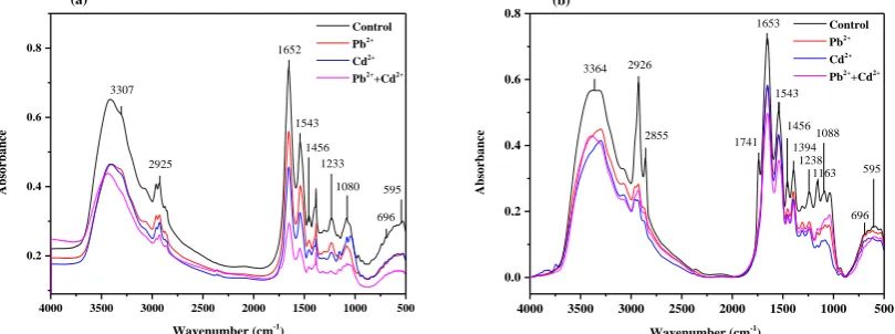
The present study was conducted to explore the structural, functional and compositional changes in the liver and muscle tissues of Crucian carp exposed to environmentally relevant Cd2+ and Pb2+ for 21days
using FT-IR spectroscopy. The representative FT-IR spectra of control, Pb2+, Cd2+ and Cd2++Pb2+ exposed
fish liver and muscle in the region of 4000 to 500 cm -1are given in Figure 1 a,b. Shifts in peak positions,
changes in intensities, and band areas of the infrared spectrum were exploited to get important structural and functional information about the studied tissues (Hayashi et al. 2007). The observed peak positions of the spectra for the studied organs and their assignments according to the previous literature are presented in Table 1 (Palaniappan and Renju 2009, Senthil Kumar and Rajkumar 2014, Sivakumar et al. 2014).
As the spectra of the two tissues with multiple bands originate from the functional groups of various biomolecules including proteins, lipids, polysaccharides and nucleic acids, the detailed spectral features were investigated in two distinct regions for liver (3700 to 3000 cm-1 and 1800 to 800
cm-1) and three distinct regions for muscle (3600–
3100 cm-1, 3050–2800 cm-1 and 1800–800 cm-1) as
shown in Figure 2 a,b and Figure 3 a,b,c respectively. Structural variations in the studied tissues were monitored with help of changes in the frequency of the respective bands, while the compositional changes
4000 3500 3000 2500 2000 1500 1000 500
0.2 0.4 0.6 0.8
1233
696 1456
1080 595
1543 1652
2925
Absor
ban
ce
Wavenumber (cm-1)
Control
Pb2+
Cd2+
Pb2+
+Cd2+
3307
(a)
4000 3500 3000 2500 2000 1500 1000 500
0.0 0.2 0.4 0.6 0.8
696 595 1088
1163 1238 1456
1394 1543
1741 1653
2855 2926
Absor
ban
ce
Wavenumber (cm-1)
Control
Pb2+
Cd2+
Pb2++Cd2+
3364
(b)
138 S. A. Khan et al. / Turk. J. Fish. Aquat. Sci. 17: 135-143 (2017)
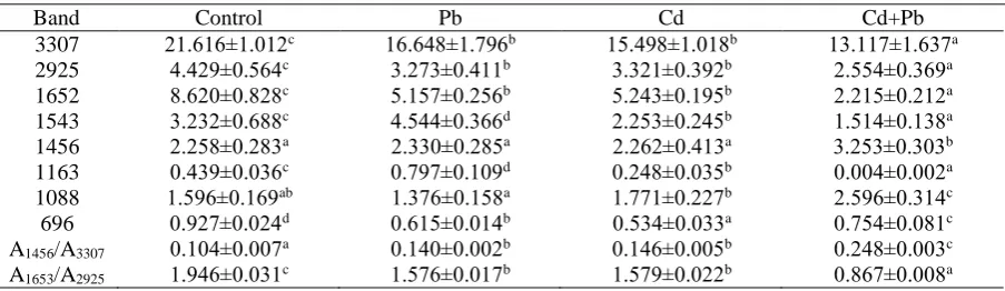
in the corresponding molecules were deduced from the accurate area under the characteristic band (Garip and Severcan 2010, Toyran et al. 2006). As shown in Fig 2a, the detail spectra of control and exposed liver tissue in the region of 3700 cm-1 to 2400 cm-1 consist
of two broad bands. The first band at ~3307 cm-1 is
mainly assigned to amide A: N− H stretching of proteins with little contribution from inter molecular O− H group. The second band at ~2925 cm-1 is
assigned to asymmetric stretching of CH2 which
mainly corresponds to lipids with little contribution from protein, carbohydrates and nucleic acids. On the other hand, Fig 2b depicts the spectral details of liver tissue in the region of 1800 cm-1 to 800 cm-1,
representing several bands at ~1652, ~1543, ~1456, ~1233, ~1080, ~696 and ~595 cm-1 corresponding to
amide I: C=O stretch of α-helix protein, amide II: N−H bending and C−N stretching of proteins, CH3
bending of lipids with little contribution from proteins, C–O asymmetric stretching of glycogen and
nucleic acids, symmetric PO2− stretching of
phospholipids and phosphodiester in nucleic acids, ring breathing mode in DNA basis and O-H deformation respectively. It can be seen from Fig 2a,b and Table 2, the absorption frequencies and band areas of the selected bands were decreased in the exposed groups with the exception of increase in band areas at ~1543 cm-1, ~1456 cm-1, ~1088 cm-1 in Pb2+,
Cd2++Pb2+ and Cd2+, Cd2++Pb2+ exposed groups
respectively.
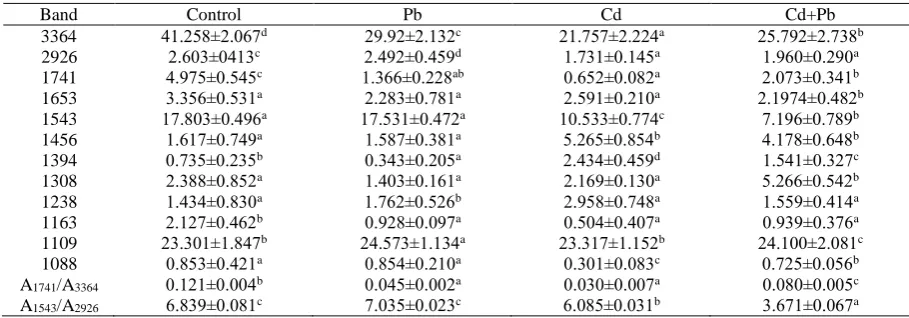
Fig 3a,b,c shows the detail spectral features of control, Cd2+, Pb2+ and Pb2++Cd2+ exposed fish
muscles. The absorption band at 3364 cm-1 in the
spectral region of 3600 cm-1 to 3100 cm-1 mainly
corresponds to amide A and amide B: N–H stretching of proteins (Fig 3a). The bands assigned to ~2926 cm -1 in the region of 3050 cm-1 to 2800 cm-1 belong to
asymmetric stretch of CH2 investigating main<ly
lipids with minor contributions from protein, carbohydrates and nucleic acids (Fig 3b). The spectral
Table 1. General vibrational peak assignment of the FT-IR spectra and band position observed for the liver and muscle tissue of Crucian carp after 21 days exposure to Pb2+, Cd2+ and Pb2++Cd2+.
Band Assignment Liver Muscle
3364 Amide A and Amide B: mainly N–H stretching of proteins +
3307 Mainly N− H stretching of proteins with the little contribution from O− H
stretching of polysaccharides and intermolecular H bonding: amide A +
2926 CH2asym. stretch: mainly lipids, with the little contribution from proteins,
carbohydrates, nucleic acids + +
1741 Ester C=O stretch: triglycerides, cholesterol esters +
1653 Amide I (C= O stretching of α-helix protein) + +
1543 Amide II (N−H bending and C−N stretching of proteins) + +
1456 CH3bending mainly lipids, with the little contribution from proteins + +
1394 COO− symmetric stretch: fatty acids and amino acids +
1308 CH3 CH2stretching of collagen +
1238 PO2− asym. Stretch: mainly phospholipids and phosphodiester in nucleic acids + +
1163 C–O asym. stretching of glycogen and nucleic acids + +
1109 CO–O–C asymmetric stretching: glycogen and nucleic acids +
1088 PO2− sym. Stretch: mainly phospholipids and phosphodiester in nucleic acids + +
696 Ring breathing mode in the DNA bases +
595 O-H deformation +
3 7 0 0 3 6 0 0 3 5 0 0 3 4 0 0 3 3 0 0 3 2 0 0 3 1 0 0 3 0 0 0
0 .2 0 .4 0 .6 0 .8
1 8 0 0 1 6 0 0 1 4 0 0 1 2 0 0 1 0 0 0 8 0 0
0 .2 0 .4 0 .6 0 .8
A
b
s
o
r
b
a
n
c
e
( a )
( b )
A
b
s
o
r
b
a
n
c
e
W a v e n u m b e r ( c m- 1)
C o n t r o l P b2 +
C d2 +
P b2 +
+ C d2 +
Figure 1. The average FT-IR spectra of Crucian carp’s liver, representing the control, Pb2+, Cd2+ and
region from 1800 cm-1 to 800 cm-1 showing various
bands at ~1741, ~1653, ~1543, ~1456, ~1394, ~1238, ~1163 and ~1088 cm-1 corresponding to C=O stretch
of triglycerides and cholesterol esters, amide I: C=O stretching of protein α-helix, amide II: N−H bending and C−N stretching of proteins, CH3 bending of lipids
with minor contribution from proteins, COO− symmetric stretching of fatty acids and amino acids, CH3 and CH2 stretching of collagen, PO2−
asymmetric stretching of mainly phospholipids and phosphodiester in nucleic acids, C–O asymmetric stretching of glycogen and nucleic acids, and PO2−
symmetric stretch of phospholipids and phosphodiester in nucleic acids respectively. Significant variations in the band areas and absorption frequencies of the selected bands among control and exposed groups can be seen from Table 3 and Fig 3. Comparing to control group, a decrease in the absorption frequency and bands areas of the exposed groups were observed at wavenumber 3364 cm-1 and
1741 cm-1 to 1543 cm-1 in the region of 3600 cm-1 to
3100 cm-1 (Fig 3a) and 1800 cm-1 to 800 cm-1
respectively. While the band areas significantly
increased at ~2926 cm-1 for Pb2+, at ~1456 cm-1 to
1394 cm-1 for Cd2+ and Cd2++Pb2+, at ~1308 cm-1 for
Cd2++Pb2+, at ~1238 cm-1 for all the treated groups
and at ~1109 cm-1 for Pb2+ and Cd2++Pb2+ exposed
group.
Discussion
Freshwater contamination with toxic metal is a serious issue in the developing world due to high anthropogenic pressure and industrial expansion (Sun et al. 2015). An extensive literature is available on the bioaccumulation of toxic metals in freshwater fish including Cd2+ and Pb2+ (Hosseini Alhashemi et al.
2012, Low et al. 2015). However, fewer studies have explained their effects on the structural and compositional changes in various tissues of the exposed organisms (Krishnakumar et al. 2012, Palaniappan and Renju 2009). In the present study, an attempt was made to investigate the individual and combined effect of Cd2+ and Pb2+ on the structural and
compositional changes in the liver and muscle of Crucian carp at concentrations closely related to
3600 3500 3400 3300 3200 3100
0.0 0.2 0.4 0.6 0.8
3050 3000 2950 2900 2850 2800
0.0 0.2 0.4 0.6 0.8
1800 1600 1400 1200 1000 800
0.0 0.2 0.4 0.6 0.8
A
b
sor
b
an
ce
Wavenumber (cm-1
)
Control Pb2+ Cd2+ Pb2++Cd2+
Figure 2. The average FT-IR spectra of Crucian carp’s muscle, representing the control, Pb2+, Cd2+ and Pb2++Cd2+
exposed groups for 21days in the region of 3600-3100 cm-1 (a), 3050-2800 cm-1 (b) and 1800-800 cm-1 (c).
Table 2: Changes in the selected FT-IR band area values and band area ratios of the selected bands of control, Pb2+, Cd2+ and Cd2++Pb2+ exposed Crucian carp’s liver tissue.
Band Control Pb Cd Cd+Pb
3307 21.616±1.012c 16.648±1.796b 15.498±1.018b 13.117±1.637a
2925 4.429±0.564c 3.273±0.411b 3.321±0.392b 2.554±0.369a
1652 8.620±0.828c 5.157±0.256b 5.243±0.195b 2.215±0.212a
1543 3.232±0.688c 4.544±0.366d 2.253±0.245b 1.514±0.138a
1456 2.258±0.283a 2.330±0.285a 2.262±0.413a 3.253±0.303b
1163 0.439±0.036c 0.797±0.109d 0.248±0.035b 0.004±0.002a
1088 1.596±0.169ab 1.376±0.158a 1.771±0.227b 2.596±0.314c
696 0.927±0.024d 0.615±0.014b 0.534±0.033a 0.754±0.081c
3307
/A
1456
A 0.104±0.007a 0.140±0.002b 0.146±0.005b 0.248±0.003c
2925
/A
1653
A 1.946±0.031c 1.576±0.017b 1.579±0.022b 0.867±0.008a
Values are means ± SD for five fish in each group
140 S. A. Khan et al. / Turk. J. Fish. Aquat. Sci. 17: 135-143 (2017)
contaminated environment by FT-IR spectroscopy (Bing et al. 2013, Khan et al. 2014, Wang et al. 2013, Zhou et al. 2007).
The FT-IR spectra of the two tissues for control and exposed groups of Crucian carp revealed significant differences in terms of band intensities and band areas (Fig 1). The band at ~3364 cm-1in the
spectra of muscle tissue of Crucian carp, which mainly correspond to amide A and amide B: N− H stretching of protein revealed significant reduction in band areas of intoxicated groups (Fig3a). The absorption band at ~3307 cm-1 in the liver tissue
originates from (amide A) N− H stretching and O− H stretching modes in water, since water was removed during sample preparation, thus the band can only be considered due to protein and polysaccharide in the sample. As seen from Fig 2a, a significant reduction in the band areas of the three exposed groups suggests a proportional decline or deterioration in the content of protein and polysaccharides. One of the possible mechanisms responsible for protein modification and misfolding is oxidative stress, because toxic metals such as Cd2+ and Pb2+ are the potential producer of
oxidative stress (Ashry et al. 2010, Pathak and Khandelwal 2006).
The band at ~2925 cm-1 arising from the olefinic
region of CH2 asymmetric stretching mainly monitors
lipids (Bogomolny et al. 2008, Bozkurt et al. 2010). Reduction in the corresponding band intensities (Table 2 & 3) in the exposed groups revealed a decrease in the proportion of unsaturation in acyl chain of lipid, which indicates increase in lipid peroxidation (Garip and Severcan 2010) with highest lipid peroxidation being caused by the combined exposure of Cd2++Pb2+. This can also be deduced
from the selected band area ratios at A1456/3307 and
A1741/3364 in table 2 and 3 respectively. Increase in the
content of lipid was thought to be important for regulation of membrane functions in a cell (Ibarguren et al. 2014). However, in our study, disturbed metabolism of lipids might be the possible reason for increase in lipid peroxidation in Cd2+ and Cd2++Pb2+
intoxicated liver and muscle tissue.
The band at ~1652 cm-1 and ~1543 cm-1 (Fig 2b
& Fig 3c) corresponds to amide I and amid II, investigating the structural proteins, respectively. Intoxication of Cd2+ and Pb2+ shows decrease in band
areas with maximum decline in the band area due to combined exposure of the two metals. Contrary to the Cd2+ and Cd2++Pb2+ exposed groups, exposure to Pb2+
caused elevation of band area (4.544±0.366) at ~1543 cm-1 in the liver tissue (Table 2) which further
revealed that intoxication of Cd2+ and Cd2+ +Pb2+
might decrease the α-helical structure of protein in the studied tissues. In another study, intoxication of Zn also decreased the α-helical structure of protein in the muscle of Labeo rohita (Palaniappan and Renju 2009). The band area at ~1394 cm-1 (Table 3)
attributed to symmetric stretch of carboxylate, increased from 0.735±0.235 to 2.434±0.459 and 1.541±0.327 in Cd2+ and Cd2++Pb2+ exposed group
respectively but decreased in the Pb2+ exposed group
(0.343±0.205). This might confirm the partial oxidation of protein and lipid at ~1652 cm-1 and
~2925 cm-1 position respectively, which resulted in
high content of fatty acid and amino acid at ~1394 cm-1 (Palaniappan and Pramod 2011). A significant
increase in the band area was noticed at ~1308 cm-1
for Cd2++Pb2+ exposed group (Table 3), which mainly
explain changes in collagen, a common fibrous protein, with many important functions and an indicator of several pathological conditions (Sivakumar et al. 2014). Similarly, changes in the intensity of phospodiester band at ~1088 cm-1 showed
an increase in the content of nucleic acids of liver but decrease in muscle. Previously a decrease in the nucleic acid content was also observed in the arsenic intoxicated brain tissue of Labeo rohita (Palaniappan and Vijayasundaram 2008). Significant changes in the band areas at 3307, 1543, 1163, 696 of liver tissue and 3364, 2926, 1741, 1653, 1543, 1456, 1394, 1163 of muscles tissue further suggested that Cd had more drastic effect on the tissue architecture as compared to Pb. In a previous study, Tatrai et al. (2001) observed
Table 3. Changes in the selected FT-IR band area values and band area ratios of the selected bands of control, Pb2+, Cd2+ and Cd2++Pb2+ exposed Crucian carp’s muscle tissue
Band Control Pb Cd Cd+Pb
3364 41.258±2.067d 29.92±2.132c 21.757±2.224a 25.792±2.738b
2926 2.603±0413c 2.492±0.459d 1.731±0.145a 1.960±0.290a
1741 4.975±0.545c 1.366±0.228ab 0.652±0.082a 2.073±0.341b
1653 3.356±0.531a 2.283±0.781a 2.591±0.210a 2.1974±0.482b
1543 17.803±0.496a 17.531±0.472a 10.533±0.774c 7.196±0.789b
1456 1.617±0.749a 1.587±0.381a 5.265±0.854b 4.178±0.648b
1394 0.735±0.235b 0.343±0.205a 2.434±0.459d 1.541±0.327c
1308 2.388±0.852a 1.403±0.161a 2.169±0.130a 5.266±0.542b
1238 1.434±0.830a 1.762±0.526b 2.958±0.748a 1.559±0.414a
1163 2.127±0.462b 0.928±0.097a 0.504±0.407a 0.939±0.376a
1109 23.301±1.847b 24.573±1.134a 23.317±1.152b 24.100±2.081c
1088 0.853±0.421a 0.854±0.210a 0.301±0.083c 0.725±0.056b
3364
/A
1741
A 0.121±0.004b 0.045±0.002a 0.030±0.007a 0.080±0.005c
2926
/A
1543
that Cd caused more severe oxidative stress, membrane damage and inhibition of protein synthesis than Pb during exposure of type II pneumocytes to Cd and Pb.
The pronounced mechanism of Cd2+ and Pb2+
toxicity is the production of reactive oxygen species which further aggravate the normal homeostasis of the cells thereby affecting the biochemical integrity (Dewanjee et al. 2013, Souid et al. 2013). However, cells have marked defense mechanism to overcome the production of oxidatively modified protein with an increased proteolysis (Chondrogianni et al. 2014). This might be the possible reason for altered protein structure and concentration in the present study coinciding with earlier observations about Cd intoxicated liver tissue of rate by FT-IR analysis (Krishnakumar et al. 2012). Lipid peroxidation is another offshoot of oxidative damage caused by Cd and Pb due to indirect generation of free radicals (Sun et al. 2011). These free radicals attack cell membranes and cause disintegration of their vital cellular component such as protein and lipids resulting in lipid peroxides production (Krishnakumar et al. 2012, Brucka-Jastrzębska 2010). However, each toxic metal has unique toxicity mechanism, for example Pb2+ has the ability to compete for diverse
polyvalent cations (Ca, Zn, Mg) in their binding sites (Garza et al. 2006). This evolutionary characteristic of Pb2+ may affect several physiological functions with
the onset of structural and compositional change in the cells and tissues, whereas Cd has the ability to reduce the activity of glutathione reductase, which is a pre requisite for membrane integrity (Tatrai et al. 2001). Thus, it is imperative to further investigate the toxicity mechanism of these metals and their interactions in details.
The findings of the present stuady suggest that exposure to Cd2+ and Pb2+ alone or in combination at
relatively low concentrations can significantly change the biochemical constituents of liver and muscle in Crucian carp. In general, a decrease in the protein and increase in lipid contents were observed from the spectra of the two tissues with marked changes caused by the combined exposure of the two metals. Alterations in the intensity and band areas at amide I responded differently to the exposed groups, revealing a differential response of the structural protein to Cd2+
and Pb2+ exposure in the liver. Also, exposure to Cd2+
and Pb2+ caused conformational changes in protein
structure with depleted α-helix and alterations in the nucleic acids content. Moreover, FT-IR spectroscopy was found an effective and rapid technique in monitoring the lucid effect of environmental contaminants on biochemical integrity.
Acknowledgments
This work was supported by the National Natural Science Foundation of China (Grant No. 31171694, 31471655).
References
An, Q., Wu, Y., Wang, J. & Li, Z. 2010. Assessment of dissolved heavy metal in the Yangtze River estuary
and its adjacent sea, China. Environmental
Monitoring and Assessment, 164:1-4, 173-87. doi: 10.1007/s10661-009-0883-z
Ashry, K. M., Sayed, Y. S., Khamiss, R. M. & El-Ashmawy, I. M. 2010. Oxidative stress and immunotoxic effects of lead and their amelioration with myrrh (Commiphora molmol) emulsion. Food
and Chemical Toxicology, 48:1, 236-41.
doi:10.1016/j.fct.2009.10.006
Begum, A., Amin, M. N., Kaneco, S. & Ohta, K. 2005. Selected elemental composition of the muscle tissue of three species of fish, Tilapia nilotica, Cirrhina mrigala and Clarius batrachus, from the fresh water Dhanmondi Lake in Bangladesh. Food Chemistry, 93:3, 439-43. doi:10.1016/j.foodchem.2004.10.021 Bing, H., Wu, Y., Liu, E. & Yang, X. 2013. Assessment of
heavy metal enrichment and its human impact in lacustrine sediments from four lakes in the mid-low reaches of the Yangtze River, China. Journal of
Environmental Sciences, 25:7, 1300-09.
doi:10.1016/S1001-0742(12)60195-8
Bogomolny, E., Argov, S., Mordechai, S. & Huleihel, M. 2008. Monitoring of viral cancer progression using FTIR microscopy: a comparative study of intact cells and tissues. Biochimica et Biophysica Acta, 1780:9, 1038-46. doi:10.1016/j.bbagen.2008.05.008
Bozkurt, O., Severcan, M. & Severcan, F. 2010. Diabetes induces compositional, structural and functional alterations on rat skeletal soleus muscle revealed by FT-IR spectroscopy: a comparative study with EDL
muscle. Analyst, 135, 3110–9. doi:
10.1039/c0an00542h
Brucka-Jastrzębska, E. 2010. The Effect of Aquatic Cadmium and Lead Pollution on Lipid Peroxidation and Superoxide Dismutase Activity in Freshwater Fish. Polish Journal of Environmental Studies, 19 6, 1139-50.
Burger, J., Gaines, K. F., Boring, C. S., Stephens, W. L., Snodgrass, J., Dixon, C., McMahon, M., Shukla, S., Shukla, T. & Gochfeld, M. 2002. Metal levels in fish from the Savannah River: potential hazards to fish and other receptors. Environmental Research, 89:1, 85-97. doi:10.1006/enrs.2002.4330
Cakmak, G., Togan, I. & Severcan, F. 2006. 17Beta-estradiol induced compositional, structural and functional changes in rainbow trout liver, revealed by FT-IR spectroscopy: a comparative study with nonylphenol. Aquatic Toxicology, 77:1, 53-63. doi:10.1016/j.aquatox.2005.10.015
Carvalho, M. L., Santiago, S. & Nunes, M. L. 2005. Assessment of the essential element and heavy metal content of edible fish muscle. Analytical and Bioanalytical Chemistry, 382 426–32.
Chai, M., Shi, F., Li, R. & Shen, X. 2014. Heavy metal contamination and ecological risk in Spartina alterniflora marsh in intertidal sediments of Bohai Bay, China. Marine Pollution Bulliton, 84:1-2, 115-24. doi:10.1016/j.marpolbul.2014.05.028
Chakraborty, P., Babu, P. V. R. & Sarma, V. V. 2012. A study of lead and cadmium speciation in some estuarine and coastal sediments.' Chemical Geology, 294-295, 217-25.
142 S. A. Khan et al. / Turk. J. Fish. Aquat. Sci. 17: 135-143 (2017)
K., Catalgol, B., Friguet, B., Grune, T. & Gonos, E. S. 2014. Protein damage, repair and proteolysis.'
Molicular Aspects of Medicine, 35, 1-71.
doi:10.1016/j.mam.2012.09.001
Dewanjee, S., Sahu, R., Karmakar, S. & Gangopadhyay, M. 2013. Toxic effects of lead exposure in Wistar rats: involvement of oxidative stress and the beneficial role of edible jute (Corchorus olitorius) leaves. Food and
Chemical Toxicology, 55, 78-91.
doi:10.1016/j.fct.2012.12.040
Dogan, A., Siyakus, G. & Severcan, F. 2007. FTIR spectroscopic characterization of irradiated hazelnut (Corylus avellana L.). Food Chemistry, 100:3, 1106-14. doi:10.1016/j.foodchem.2005.11.017
Garip, S. & Severcan, F. 2010. Determination of simvastatin-induced changes in bone composition and structure by Fourier transform infrared spectroscopy in rat animal model. Journal of Pharmaceutical and
Biomedical Analysis, 52:4, 580-8.
doi:10.1016/j.jpba.2010.01.044
Garza, A., Vega, R. & Soto, E. 2006. Cellular mechanisms of lead neurotoxicity. Medical Science Monitor, 12, RA57-65.
Grosell, M., Gerdes, R. M. & Brix, K. V. 2006. Chronic toxicity of lead to three freshwater invertebrates – Brachionus calyciflorus, Chironomus tentans, and Lymnaea stagnalis. Environmental Toxicology and Chemistry, 25, 97–104.
Gupta, A. D. & Karthikeyan, S. 2016. Individual and combined toxic effect of nickel and chromium on biochemical constituents in E. coli using FTIR spectroscopy and Principle component analysis. Ecotoxicology and Environmental Safety, 130, 289-94. doi:10.1016/j.ecoenv.2016.04.025
Hayashi, K., Hara, H., Asvarujanon, P., Aoyama, Y. & Luangpituksa, P. 2007. Ingestion of insoluble dietary fibre increased zinc and iron absorption and restored growth rate and zinc absorption suppressed by dietary phytate in rats. British Journal of Nutrition, 86:04, 443.
Hosseini Alhashemi, A., Sekhavatjou, M. S., Hassanzadeh Kiabi, B. & Karbassi, A. R. 2012. Bioaccumulation of trace elements in water, sediment, and six fish species from a freshwater wetland, Iran. Microchemical Journal, 104, 1-6. doi:10.1016/j.microc.2012.03.002 Ibarguren, M., Lopez, D. J. & Escriba, P. V. 2014. The
effect of natural and synthetic fatty acids on membrane structure, microdomain organization, cellular functions and human health. Biochimica et
Biophysica Acta, 1838:6, 1518-28.
doi:10.1016/j.bbamem.2013.12.021
Karthikeyan, S. & Easwaran, R. 2013. Analysis of a curve fitting model in the amide region applied to the muscle tissues of an edible fish: Labeo rohita fingerlings. Journal of Biological Physics and
Chemistry, 13 125–30. doi:
10.4024/13KA13A.jbpc.13.04
Khan, S. A., Liu, X., Li, H., Fan, W., Shah, B. R., Li, J., Zhang, L., Chen, S. & Khan, S. B. 2015a. Organ specific antioxidant defenses and FT-IR spectroscopy of muscles in Crucian carp (Carassius auratus gibelio) exposed to environmental Pb2+. Turkish Journal of
Biology. doi:10.3906/biy-1410-3
Khan, S. A., Liu, X. & Shah, B. R. 2014. Impact Of Acute Toxicity Of Lead Acetate On The Level Of Essential Trace Metals And Histopathological Changes In Crucian Carp (Carassius Auratus Gibelio). The
Journal of Animal & Plant Sciences, 24:5, 1405-14. Khan, S. A., Zhou, P., Liu, X., Li, H., Li, J., Rehman, Z. U.
& Ahmad, I. 2015b. Response of vitamins A, E, hematological and serum biochemical markers in Crucian carp (Carassius auratus gibelio) exposed to environmental Pb(2+) and Cd(2+). Acta Biochimica
Polonica, 62:3, 581-7. doi: 10.18388/abp.2015_1036 Krishnakumar, N., Prabu, S. M. & Sulfikkarali, N. K. 2012.
Quercetin protects against cadmium-induced
biochemical and structural changes in rat liver revealed by FT-IR spectroscopy. Biomedicine &
Preventive Nutrition, 2:3, 179-85.
doi:10.1016/j.bionut.2012.03.010
Li, F., Huang, J., Zeng, G., Yuan, X., Li, X., Liang, J., Wang, X., Tang, X. & Bai, B. 2013. Spatial risk assessment and sources identification of heavy metals in surface sediments from the Dongting Lake, Middle China. Journal of Geochemical Exploration, 132, 75-83. doi:10.1016/j.gexplo.2013.05.007
Lim, H.-S., Lee, J.-S., Chon, H.-T. & Sager, M. 2008.
Heavy metal contamination and health risk
assessment in the vicinity of the abandoned Songcheon Au–Ag mine in Korea. Journal of
Geochemical Exploration, 96:2-3, 223-30.
doi:10.1016/j.gexplo.2007.04.008
Low, K. H., Zain, S. M., Abas, M. R., Md Salleh, K. & Teo, Y. Y. 2015. Distribution and health risk assessment of trace metals in freshwater tilapia from three different aquaculture sites in Jelebu Region (Malaysia). Food
Chemistry, 177, 390-6.
doi:10.1016/j.foodchem.2015.01.059
Lu, X., Webb, M. A. H., Talbott, M. J., Van Eenennaam, J. P., Doroshov, S. I. & Rasco, B. A. 2011. A study of biochemical parameters associated with ovarian atresia and quality of caviar in farmed white sturgeon (Acipenser transmontanus) by Fourier Transform Infrared (FT-IR) Spectroscopy. Aquaculture, 315:3-4, 298-305. doi:10.1016/j.aquaculture.2011.01.048 Martinez, C. B. R., Nagae, M. Y., Zaia, C. T. B. V. & Zaia,
D. A. M. 2004. Acute morphological and physiological effects of lead in the neotropical fish Prochilodus lineatus. Brazilian Journal of Biology, 64, 797–807.
Monteiro, V., Cavalcante, D. G., Vilela, M. B., Sofia, S. H. & Martinez, C. B. 2011. In vivo and in vitro exposures for the evaluation of the genotoxic effects of lead on the Neotropical freshwater fish Prochilodus lineatus. Aquatic Toxicology, 104:3-4, 291-8. doi:10.1016/j.aquatox.2011.05.002
Murugavel, P. & Pari, L. 2007. Diallyl tetrasulfide protects cadmium-induced alterations in lipids and plasma lipoproteins in rats. Nutrition Research, 27:6, 356-61. doi:10.1016/j.nutres.2007.04.012
Nemmiche, S., Chabane-Sari, D. & Guiraud, P. 2007. Role of alpha-tocopherol in cadmium-induced oxidative stress in Wistar rat's blood, liver and brain. Chemico
Biological Interaction, 170:3, 221-30.
doi:10.1016/j.cbi.2007.08.004
Palaniappan, P. R. & Pramod, K. S. 2011. The effect of titanium dioxide on the biochemical constituents of the brain of Zebrafish (Danio rerio): an FT-IR study.
Spectrochimica Acta: Part A Molicular and
Biomolicular Spectroscopy, 79:1, 206-12.
doi:10.1016/j.saa.2011.02.038
Technology, 52:1, 37-41.
doi:10.1016/j.infrared.2008.11.001
Palaniappan, P. R. & Vijayasundaram, V. 2008. Fourier transform infrared study of protein secondary structural changes in the muscle of Labeo rohita due
to arsenic intoxication. Food and Chemical
Toxicology, 46:11, 3534-9.
doi:10.1016/j.fct.2008.09.001
Pathak, N. & Khandelwal, S. 2006. Oxidative stress and apoptotic changes in murine splenocytes exposed to
cadmium. Toxicology, 220:1, 26-36.
doi:10.1016/j.tox.2005.11.027
Poole, J. H., Tyack, P. L., Stoeger-Horwath, A. S. & Watwood, S. 2005. Animal behaviour: elephants are capable of vocal learning. Nature, 434:7032, 455-6. doi:10.1038/434455a
Qu, R., Wang, X., Wang, Z., Wei, Z. & Wang, L. 2014. Metal accumulation and antioxidant defenses in the freshwater fish Carassius auratus in response to single and combined exposure to cadmium and hydroxylated multi-walled carbon nanotubes. Journal of Hazardius
Material, 275, 89-98.
doi:10.1016/j.jhazmat.2014.04.051
Rand, G. M., Wells, P. G. & McCarty, L. S. 2003. Introduction to aquatic toxicology. New York: Taylor and Francis. doi:10.1016/B978-0-12-411574-3.00001-3
Senthil Kumar, K., Sajwan, K. S., Richardson, J. P. & Kannan, K. 2008. Contamination profiles of heavy metals, organochlorine pesticides, polycyclic aromatic hydrocarbons and alkylphenols in sediment and oyster collected from marsh/estuarine Savannah GA, USA. Marine Pollution Bulliton, 56:1, 136-49. DOI: 10.1016/j.marpolbul.2007.08.011
Senthil Kumar, R. & Rajkumar, P. 2014. Characterization of minerals in air dust particles in the state of Tamilnadu, India through FTIR, XRD and SEM analyses. Infrared Physics & Technology, 67, 30-41. doi:10.1016/j.infrared.2014.06.002
Shao, X.-p., Liu, W.-b., Xu, W.-n., Lu, K.-l., Xia, W. & Jiang, Y.-y. 2010. Effects of dietary copper sources and levels on performance, copper status, plasma
antioxidant activities and relative copper
bioavailability in Carassius auratus gibelio.
Aquaculture, 308:1-2, 60-65.
doi:10.1016/j.aquaculture.2010.07.021
Sivakumar, S., Khatiwada, C. P. & Sivasubramanian, J. 2014. Comparative study of desferrioxamine and deferiprone protects against aluminum induced compositional, structural and functional changes in liver tissue of mice (mus musculus) investigated by FT-IR spectroscopy. Biomedicine & Preventive
Nutrition, 4:2, 231-38.
doi:10.1016/j.bionut.2013.10.007
Souid, G., Souayed, N., Yaktiti, F. & Maaroufi, K. 2013. Effect of acute cadmium exposure on metal accumulation and oxidative stress biomarkers of Sparus aurata. Ecotoxicology and Environmental Safety, 89, 1-7. doi:10.1016/j.ecoenv.2012.12.015 Staniszewska, E., Malek, K. & Baranska, M. 2014. Rapid
approach to analyze biochemical variation in rat organs by ATR FTIR spectroscopy. Spectrochimica
Acta Part A: Molicular and Biomoliclular
Spectroscopy, 118, 981-6.
doi:10.1016/j.saa.2013.09.131
Sun, S. Q., Wang, G. X., He, M. & Cao, T. 2011. Effects of Pb and Ni stress on oxidative stress parameters in three moss species. Ecotoxicology and Environmental
Safety, 74:6, 1630-5.
doi:10.1016/j.ecoenv.2011.04.002
Sun, Z., Mou, X., Tong, C., Wang, C., Xie, Z., Song, H., Sun, W. & Lv, Y. 2015. Spatial variations and bioaccumulation of heavy metals in intertidal zone of the Yellow River estuary, China. Catena, 126, 43-52. doi:10.1016/j.catena.2014.10.037
Tatrai, E., Kovacikova, Z., Hudak, A., Adamis, Z. & Ungvary, G. 2001. Comparative in vitro toxicity of cadmium and lead on redox cycling in type II pneumocytes. Journal of Applied Toxicology, 21:6, 479-83.
Tellis, M. S., Lauer, M. M., Nadella, S., Bianchini, A. & Wood, C. M. 2014. Sublethal mechanisms of Pb and
Zn toxicity to the purple sea urchin
(Strongylocentrotus purpuratus) during early
development. Aquatic Toxicology, 146, 220-9. doi:10.1016/j.aquatox.2013.11.004
Toyran, N., Lasch, P., Naumann, D., Turan, B. & Severcan, F. 2006. Early alterations in myocardia and vessels of the diabetic rat heart: an FTIR microspectroscopic study. Biochemistry Journal, 397:3, 427-36. DOI: 10.1042/BJ20060171
Uysal, K., Emre, Y. & Köse, E. 2008. The determination of heavy metal accumulation ratios in muscle, skin and gills of some migratory fish species by inductively coupled plasma-optical emission spectrometry (ICP-OES) in Beymelek Lagoon (Antalya/Turkey).
Microchemical Journal, 90:1, 67-70.
doi:10.1016/j.microc.2008.03.005
Wang, F., Wang, W. X. & Huang, X. P. 2012. Spatial distribution of gut juice extractable Cu, Pb and Zn in sediments from the Pearl River Estuary, Southern China. Marine Environmental Research, 77, 112-9. doi:10.1016/j.marenvres.2012.03.002
Wang, S. L., Xu, X. R., Sun, Y. X., Liu, J. L. & Li, H. B. 2013. Heavy metal pollution in coastal areas of South China: a review. Marine Pollution Bulliten, 76:1-2, 7-15. doi:10.1016/j.marpolbul.2013.08.025
Xu, Y., Feng, L., Jeffrey, P. D., Shi, Y. & Morel, F. M. 2008. Structure and metal exchange in the cadmium carbonic anhydrase of marine diatoms. Nature, 452:7183, 56-61. doi:10.1038/nature06636
Yang, Z., Wang, Y., Shen, Z., Niu, J. & Tang, Z. 2009. Distribution and speciation of heavy metals in sediments from the mainstream, tributaries, and lakes of the Yangtze River catchment of Wuhan, China. Journal of Hazardius Material, 166:2-3, 1186-94. doi:10.1016/j.jhazmat.2008.12.034
Zhang, X., Xie, P., Li, D. & Shi, Z. 2007. Hematological and plasma biochemical responses of crucian carp (Carassius auratus) to intraperitoneal injection of extracted microcystins with the possible mechanisms
of anemia. Toxicon, 49:8, 1150-7.
doi:10.1016/j.toxicon.2007.02.009