Iran J Public Health, Vol. 45, No.6, Jun 2016, pp.806-813
Original Article
Prevalence of Virulence Factors and Vancomycin-resistant Genes
among
Enterococcus faecalis
and
E. faecium
Isolated from
Clin-ical Specimens
Mona NASAJ
1, Seyed Masoud MOUSAVI
1, Seyed Mostafa HOSSEINI
1, *Mohammad
Reza ARABESTANI
1, 21. Dept. of Microbiology, Faculty of Medicine, Hamadan University of Medical Sciences, Hamadan, Iran 2. Brucellosis Research Center, Hamadan University of Medical Sciences, Hamadan, Iran
*Corresponding Author: Email: [email protected]
(Received 10 Sep 2015; accepted 15 Dec 2015)
Introduction
Vancomycin-resistant Enterococci (VRE) strains were reported first in United Kingdom and France in 1986, and after that in the United States (1). At present, these organisms are significantly isolated in hospitals around the world, especially in patients with hemato-oncological diseases and hospitalized in intensive care units (2). Currently, nine types of vancomycin-resistance have been described in Enterococci, eight of these types cor-respond to acquired resistance, i.e., vanA, vanB, vanD, vanE, vanG, vanL,vanM and vanN; one type -VanC- have been identified in speciesof
Enterococ-cus gallinarum and E. casseliflavus–E. flavescens intrin-sically resistant to low levels of vancomycin but remain susceptible to teicoplanin and have been found in human intestinal tract (3, 4).
Among vancomycin-resistance genotypes in Enter-ococci, vanA and vanB possess greatest clinical signi-ficance (5). The vanA genotype is the most com-monly genotype in VRE worldwide, and is associ-ated with the transfer of a considerable amount of vancomycin resistance from Enterococci, particularly, vancomycin-resistant E. faecium (VREF) to Staphy-lococcus aureus (1, 6). The vanA-type genes play an
Abstract
Background: The aim of this study was to determine the occurrence of virulence determinants and vancomycin- re-sistant genes among Enterococcus faecalis and E. faeciumobtained from various clinical sources.
Methods: The study was performed on the 280 enterococcal isolated from clinical specimens in Hamadan hospitals, western Iran in 2012-14. Antibiotic susceptibility testing was performed using disk diffusion and Minimal Inhibitory Concentration (MIC) methods. The presence of vancomycin-resistant genes and virulence genes was investigated us-ing PCR.
Results: Totally 280 enterococcal isolates were identified as follows: E. faecalis (62.5%),E. faecium (24%) and Enterococ-cus spp (13.5%). The results of antibiotic susceptibility testing showed that resistance rates to vancomycin and teicop-lanin in E. faecalis and E. faecium isolates were 5% and 73%, respectively. Of Sixty vancomycin-resistant Enterococci
strains, fifty-one isolates were identified as E. faecium (VREfm) and nine as E. faecalis (VREfs). Prevalence of esp, hyl,
and asa1 genes were determined as 82%, 71.6%, and 100%, respectively in E. faecium strains; and 78%, 56/6%, and 97%, respectively in E. faecalis strains.
Conclusion: The increased frequency of VREF, as seen with rapid rise in the number of vanA isolates should be con-sidered in infection control practices.
important role in inducing resistance against both teicoplanin and vancomycin, while the level of re-sistance to vancomycin in strains sheltering vanB -type genes are not constant but somewhat variable (MICs, 4 to 1,024 mg/l). Moreover, remain sus-ceptible to teicoplanin (MICs, ≤ 2 mg/l) in vitro (4). The low-level resistance against such antibio-tics as vancomycin vancomycin (vancomycin [MIC], 64–128 mg/ml) and susceptibility or in-termediate resistance to teicoplanin (teicoplanin MIC, 8–16 mg/ml) is one of the main features of the vanD phenotype (7).
Although these organisms lacking strong virulent factors, but enterococcal infection is intricated by the intrinsic resistant / tolerant to many important an-timicrobial agents including cephalosporin, linco-mycin, cotrimoxazole, and low levels of penicillin and aminoglycosides, and as well as by the ability to acquire resistance to penicillins, chlorampheni-col, tetracyclines, aminoglycosides (high-level) and vancomycin either by mutation, or by acquisition of plasmids or transposons (8, 9). Thus, the inci-dence of antimicrobial resistant Enterococci, espe-cially VRE is a persisting clinical problem in pub-lic health care facilities in all geographical areas (9). Cytolysin (Cyl), gelatinase (GelE) and aggregation substance (Agg) are virulence factors identified in
E. faecalis that can influence the relationship be-tween parasite and host, on other hand, esp and hyl
have been found in both E. faecalis and E. faecium
(10, 11). “These virulence factors may causes in-creasing persistence of enterococci in the nosocomial environment, and consequently inter and intra-hospitals dissemination” (10).
Regarding the virulence determinants in clinical isolates of Enterococci that may result in promoting emergence of infections and persistence of this organism in nosocomial settings and consequently leads to increased resistance (5), this study was un-dertaken to determine the antimicrobial suscepti-bility patterns and virulence factors including esp
(enterococcal surface protein), asa1 (aggregation sub-stance), gelE (gelatinase), hyl (hyaluronidase) in clinical isolates of E. faecalis and E. faecium.
Materials and Methods
Identification of enterococcal isolates
One hundred and seventy-five E. faecalis and sixty-seven E. faecium isolates were collected from dif-ferent clinical specimens submitted in three teach-ing hospitals located in Hamedan/ western Iran, from Dec 2012 to May 2014. The origins of iso-lates were as follows: urine 200 (82.6%), tracheal 17 (7%), blood 8 (3.3%), wound 6 (2.5%), abscess and lower respiratory tract 6 (2.5%), and body flu-ids 5 (2.1%). Enterococcus genus were identified us-ing routine microbiological methods (9) then, PCR targeting D-alanine- D-alanine ligases specif-ic for E. faecalis (ddl E. faecalis) and E. faecium (ddl E. faecium) was used to confirm phenotypic speciation (12).
DNA extraction
Enterococci chromosomal DNA was extracted by boiling method. Briefly, 3-5 clones of overnight bac-terial culture was suspended in 500 µl of sterile dis-tilled water, boiled for 10-15 min, and then cen-trifuging at 14000 g for 5 min to pellet cell debris (13).
Detection of E. faecalis and E. faecium spe-cies by PCR
PCR reactions of ddl E.faecalis and ddl E.faecium
Antibiotic susceptibility testing
Antibiotic susceptibility testing of 175 E. faecalis
strains and 67 E. faecium strains was performed us-ing Kirby-Bauer disk diffusion method on Muller-Hinton agar in according to the Clinical and La-boratory Standards Institute (CLSI) guidelines (14). Antimicrobial agents used in this study were in-cluded: vancomycin (30 µg), teicoplanin (30 µg), tetracycline (30 µg), erythromycin (15 µg), norflox-acin (10 µg), ciprofloxnorflox-acin (5 µg), nitrofurantoin (300 µg), quinopristin-dalfopristin [synercid (15 µg)] (Mast co., UK), chloramphenicol (30 µg),
li-nezolid (30 µg), gentamicin (10 µg), and ampicillin (10 µg)(HiMedia Mumbai Co., India).
Determination of minimum inhibitory concentra-tion (MIC) of the glycopeptide antibiotics i.e. van-comycin and teicoplanin (Sigma-Aldrich, Poole, Co., UK) for E. faecalis and E. faecium isolates was done using microdilution broth method and Ca-tion Adjustment Muller Hinton Broth (CAMHB) medium according to the CLSI guidelines (14). The E. faecalis ATCC 29212 (Vancomycin sensi-tive), E. faecalis ATCC 51299 (vanB positive), E. faecalis E206 (vanA positive) were used as quality control strains for performing antimicrobial tests.
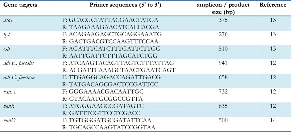
Table 1:Primers used in this study
Gene targets Primer sequences (5' to 3') amplicon / product
size (bp) Reference
asa1 F: GCACGCTATTACGAACTATGA
R: TAAGAAAGAACATCACCACGA 375 13
hyl F: ACAGAAGAGCTGCAGGAAATG
R: GACTGACGTCCAAGTTTCCAA 276 13
esp F: AGATTTCATCTTTGATTCTTGG
R: AATTGATTCTTTAGCATCTGG 510 13
ddl E. faecalis F: ATCAAGTACAGTTAGTCTTTATTAG
R: ACGATTCAAAGCTAACTGAATCAGT 941 12
ddl E. faecium F: TTGAGGCAGACCAGATTGACG
R: TATGACAGCGACTCCGATTCC 658 12
vanA F: GGGAAAACGACAATTGC
R: GTACAATGCGGCCGTTA 732 12
vanB F: ATGGGAAGCCGATAGTC
R: GATTTCGTTCCTCGACC 635 12
vanD F: TGTGGGATGCGATATTCAA
R: TGCAGCCAAGTATCCGGTAA 500 14
Detection of van determinants
Isolates with a vancomycin and teicoplanin Mini-mum Inhibitory Concentration (MIC) of 2 µg/ml were analyzed by PCR for the presence of the genes encoding the vancomycin-resistance deter-minants vanA, vanB, vanD using specific primers (Table 1). The PCR reaction was performed in a volume of 20 µl and contained: 2 µl template
DNA, 1 µl of each primer, 10 µl of Master Mix, 6 µl of sterile distilled water on a Eppendorf and Biorad thermocycler (ASTEC Co., Japan) with an initial denaturation at 94 °C for 3 min, 30 cycles of amplification (denaturation at 94 °C for 1 min,
annealing at 54 °C for 1 min, and extension at 72 °C for 1 min), and a final extension at 72 °C for 7 min (12).
Detection of virulence genes esp, hyl, and asa1
by PCR
chromosome DNA), 1µl of each primer for genes
esp and asa1, 12.5 µl of Master Mix, and 5.5 µl of sterile distilled water; the second 20 µl PCR mix-ture contained 2 µl of template DNA, 1 µl of each primer for hyl, 10 µl of Master Mix, and 6 µl of ste-rile distilled water. The PCR conditions included a pre-denaturation step at 95 ºC for 10 min, followed by 30 cycles of 1 min at 94 ºC, 1 min at 56 ºC and 1 min at 72 ºC. A final extension step was performed at 72 ºC for 10 min (13). The E. faecalis ATCC 29212 (asa1 positive), E. faecium C68 (hyl and esp positive) were used as quality control strains.
Statistical analysis
Data were analyzed statistically using Chi-Square test and difference was considered significant at
P<0.05 by SPSS software version 19 (Chicago, IL, USA).
Results
Enterococci isolates
Of 280 enterococcal isolates, 190 (67.8%) isolates were identified as E. faecalis, 75 (26.8%) as E. fae-cium and15 (5.4%) as Enterococcus spp., using bio-chemical methods. Overall, 175 (62.5%) E. faecalis
strains (5.62%) and 67 E. faecium strains (24%) were confirmed by the PCR method. A total of 38 strains (13.5%) have remained to the Enterococcus
genus. The most of isolates 95 (39.3%) were col-lected from internal ward, followed by Outpatient ward with 72 (29.8%), Nephrology 26 (10.7%), ICU 22 (9.1%), Emergency 21 (8.7%), Burn 3 (1.2%), and Surgeryandcardiaccare 3 (1.2%).
Antimicrobial susceptibility testing
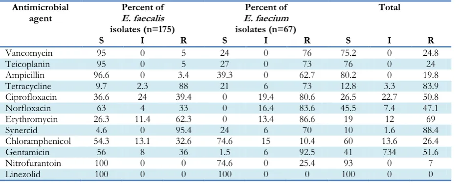
Resistance to the majority antibiotics except for chloramphenicol, tetracycline, and quinopristin-dalfopristin was higher in E. faecium isolates than
E. faecalis isolates. However, they showed good rate of sensitivity to linezolid (100%), nitrofuran-toin and chloramphenicol (74.6%). All isolates of
E. faecalis were susceptible to nitrofurantoin. None of the Enterococcus isolates was resistant to linezolid. The susceptibility patterns of E. faecium and E. faecalis to antibiotics are presented in Table 2. Of 175 isolates of E. faecalis, resistance of 9 and sensitivity of 166 isolates to vancomycin and tei-coplanin were confirmed by microdilution broth method.
Table 2:Antibiotic resistance behavior of Enterococci isolates with disk diffusion
Antimicrobial
agent Percent ofE. faecalis isolates (n=175)
Percent of E.faecium isolates (n=67)
Total
S I R S I R S I R
Vancomycin 95 0 5 24 0 76 75.2 0 24.8 Teicoplanin 95 0 5 27 0 73 76 0 24 Ampicillin 96.6 0 3.4 39.3 0 62.7 80.2 0 19.8 Tetracycline 9.7 2.3 88 21 6 73 12.8 3.3 83.9 Ciprofloxacin 36.6 24 39.4 0 19.4 80.6 26.5 22.7 50.8 Norfloxacin 63 4 33 0 16.4 83.6 45.5 7.4 47.1 Erythromycin 26.3 11.4 62.3 0 13.4 86.6 19 12 69 Synercid 4.6 0 95.4 24 6 70 10 1.6 88.4 Chloramphenicol 54.3 13.1 32.6 74.6 15 10.4 60 13.6 26.4 Gentamicin 56 8 36 1.5 6 92.5 41 734 51.6 Nitrofurantoin 100 0 0 74.6 0 25.4 93 0 7 Linezolid 100 0 0 100 0 0 100 0 0
Of 67 isolates of E. faecium, 51 strains were resis-tant to vancomycin by disk diffusion method, but resistance of 49 strains to vancomycin was con-firmed by Microdilution Broth. Two strains of E.
Analysis of vanA-vanB-vanD types vancomy-cin resistance genes
All VREfs and 49 of VREfm strains (96.7%) had high-level resistance to vancomycin and teicopla-nin carried the vanA gene. Two VREfm isolates (3.3%) had moderate-Level resistant to vancomy-cin with MIC=8 µg /ml and were insensitive to teicoplanin, carried the vanB gene, vanD gene was identified in none of VRE strain.
Prevalence of virulence genes in E. faecalis and E. faecium strains
Among the E. faecalis strains, the asa1 gene was the most prevalent factor, followed by the esp and hyl
genes; additionally, in E. faecium strains, the asa1 gene was the highest prevalence and hyl gene has the lowest frequency, followed by the esp gene.
Table 3:Results of MIC for glycopeptides antibiotics of vancomycin and teicoplanin in E. faecium and E. faecalis
strains
Antimicrobial
agent E. faecium E. faecalis
Teicoplanin Vancomycin Teicoplanin Vancomycin
Sensitivity Status S R S I R S R S R
MIC (µg/ml) 8≤ 32≥ 4≤ 8-16 32≥ 8≤ 32≥ 4≤ 32≥
Number 18 49 16 2 49 166 6 166 9
Percent (%) 26.9 73.1 24 3 73 95 5 95 5
Results of statistical analysis
Using SPSS software, there were significant corre-lations between the disk diffusion agar and broth microdilution methods for vancomycin and tei-coplanin antibiotics (P≤0.001); and between PCR and MICVanco and MICTeico results (P≤0.001) in
strains of E. faecalis and E. faecium.
Discussion
In the present study,of 280 enterococcal isolates, 175 isolates were identified as E. faecalis, 67 as E. fae-cium and38 as Enterococcus spp. The prevalence of
E. faecalis strains were reported 76% and 55.5% respectively (15, 9). In Iran, the prevalence of E. faecalis and E. faecium strains were reported 77.5% and 22.5%, 70.4% and 18.5%, 85.3% and 10.8%, 28% and 71%, respectively (8, 16-18). VRE strains are resistant to different classes of antibiotics si-multaneously. Rising high-level resistance to peni-cillin, ampicillin and aminoglycosides has been demonstrated in recent years, especially in vanco-mycin-resistant E. faecium strains. Multidrug-resistant strains of Enterococci, especially E. faecalis
and E. faecium are serious problems in treatment patients with enterococcal infections due to
im-proper use of antibiotics (19). In the current study, 85% of strains of E. faecalis and all E. faecium
strains had multidrug resistance, but strains of vancomycin-resistant compared to susceptible strains were resistant to greater number of antibi-otic classes.
In the current study, the prevalence of vancomy-cin resistance was found 24%, which was consis-tent with some previous (9, 20). The prevalence of vancomycin resistance in Enterococcus strains were reported 23.3% and 29%, respectively. Similar studies in Iran reported the prevalence of vanco-mycin resistance in Enterococcus strains as 24.10%, 16.9%, 14.6%, 25%, 22%, respectively (16, 18, 21-23). According to typing results of genotypes for resistance to vancomycin, vanA-type was the most common genotype seen among VRE strains ob-tained in Hamadan and vanB is the second. Ma-jority of the VRE isolates (96.8%) have the vanA
gene, 3.3% of VRE isolates with vanC, and vanB
genotype was identified in any VRE strains (24). All VREfm strains (100%) carrying the vanA gene; but vanB-C-D-E-G genotypes were not reported (25). The frequency of genes vanA and vanB
respective-ly (16). Increase in the prevalence of VRE, espe-cially E. faecium, in different countries has been attributed mainly to the incidence and diffusion of
vanA and vanB positive VRE, which exhibited some virulence factors such as Esp (esp), cytolysin (cyl), and hyaluronidase (hyl). The extracellular sur-face protein (esp), encoded by the chromosomal
esp gene, found on pathogenicity island in multi-drug-resistant pathogenic lineages of both E. faeca-lis and E. faecium strains. Esp is a cell wall-associated protein which contribute to the col-onization and persistence of E. faecalis strains in ascending infections of the urinary tract. In addi-tion, Esp may participate in biofilm formaaddi-tion, and may also be involved in antimicrobial resis-tance (26, 27). Blood and wound infections caused by Enterococci strains are mediated by Esp protein (28). In a study, the gene esp was detected only in
E. faecalis strains (29). However, incidence of the gene esp in clinical E. faecium are increasing com-pared to clinical E. faecalis isolates (30).
In the present study, prevalence of the genes esp
and hyl were significantly higher among ampicillin-resistant VREfm isolates (53.7%, 37.3%) compared to ampicillin-susceptible VREfm isolates (19.4%, 22.4%). This finding is in accordance with the re-ported results of other related studies (32-34). In our study, the frequency of the gene asa1, (which encodes aggregation substance) among E. faecalis
was as high as 97 percent and among E. faecium
strains was as high as 100 percent. This gene has a high incidence in E. faecalis, as well. Results of stu-dies on clinical E. faecium isolates are contradictory. Previous studies detected this virulence factor among 5%, 65% of VREfm and 2.7%, 60% of VREfs strains (26, 35). S Jahangiri et al. (11) did not found gene asa1 in either 49 of VREfm strains or 17 of VSEfm strains.
Hyaluronidase, coded by the chromosomal gene
hyl, is a degradative enzyme associated with tissue damage that influence on the hyaluronic acid (hya-luronate, HA) (33). We found the hyl gene among 49.3% of VREfm isolates and 22.4% of VSEfm iso-lates, which is in accordance to Rice et al. results (36), who detected the hyl gene among 71% of the United Kingdom VREfm isolates; but it was in con-trast to another study (11), who detected gene hyl
among 80% of VSEfm isolates and in 28.5% of VREfm isolates. Most of esp-positive isolates were resistant to more than 3 antibiotics (11, 37). Laud B et al. (38) demonstrated that the strong correla-tion between the carriage of gene esp and antimi-crobial resistance could be due to the higher con-jugation frequencies in strains carrying the esp
gene than strains lacking this gene. E. faecium
strains carrying the gene esp were resistant to more than 90% of the antibiotics tested and 64% of E. faecium strains were resistant to vancomycin (5). Considering these results, the gene esp facilitates E. faecium isolates ability to acquire antibiotic re-sistance genes. The expression level of gene esp
depending on growth conditions constantly vary between strains of E. faecium and is associated with initial connection and biofilm formation (39).
Conclusion
Due to increasing resistance rate of Enterococci to most common antibiotics, applying the preventive and control measures are required.
Ethical considerations
Ethical issues (Including plagiarism, informed consent, misconduct, data fabrication and/or fal-sification, double publication and/or submission, redundancy, etc.) have been completely observed by the authors.
Acknowledgement
The authors declare that there is no conflict of in-terests.
References
1. Xu H, Tian R, Chen D, Xiao F, Nie ZY, Hu YJ, et al. (2011). Nosocomial spread of hospital-adapted CC17 vancomycin-resistant Enterococcus faecium in a tertiary-care hospital of Beijing, China. Chin Med J, 124 (4):498-503.
en-terococci in hemato-oncological patients. Biomed Pap Med Fac Univ Palacky Olomouc Czech Repub, 150 (1):117-20.
3. Batistão DWdF, Gontijo-Filho PP, Conceição N, Oliveira AG, Ribas RM (2012). Risk factors for vancomycin-resistant enterococci colonization in critically ill patients. Mem Inst Oswaldo Cruz, 107(1):57-63.
4. Hegstad K, Giske CG, Haldorsen B, Matuschek E, Schønning K, Leegaard TM, et al (2014). Per-formance of the EUCAST disk diffusion me-thod, the CLSI agar screen meme-thod, and the Vi-tek 2 automated antimicrobial susceptibility test-ing system for detection of clinical isolates of en-terococci with low-and medium-level VanB-type vancomycin resistance: a multicenter study. J Clin Microbiol, 52 (5):1582-9.
5. Sharifi Y, Hasani A, Ghotaslou R, Varshochi M, Hasani A, Aghazadeh M, et al (2012). Survey of virulence determinants among vancomycin re-sistant Enterococcus faecalis and Enterococcus faecium isolated from clinical specimens of hos-pitalized patients of North west of Iran. Open Mi-crobiol J, 6:34-39.
6. Ji GY, Son BR, Kim JW (2014). Development of a Novel Immunochromatographic Assay for Rap-id Detection of VanA Ligase-Producing Vanco-mycin-Resistant Enterococci. J Microbiol Biotechnol, 24:427-30.
7. Song JY, Cheong HJ, Seo YB, Kim IS, Heo JY, Noh JY, et al (2013). Clinical and microbiological characteristics of vancomycin-resistant en-terococci with the VanD phenotype and vanA genotype. Jpn J Infect Dis, 66 (1):1-5.
8. Feizabadi MM, Sayadi S, Shokrzadeh L, Parvin M,YadegaryniA D (2008). Increase in prevalence of vancomycin resistant isolates of Enterococcous faecium at Labbafinejad hospital. Clin Infect Dis, 3 (2):123-26.
9. Mira M-U, Deana M, Zora J, Vera G, Biljana M, Bil-jan R (2014). Prevalence of different enterococcal species isolated from blood and their susceptibili-ty to antimicrobial drugs in Vojvodina, Serbia, 2011-2013. Afr J Microbiol Res, 8 (8):819-24. 10. Kariyama R, Mitsuhata R, Chow JW, Clewell
DB, Kumon H (2000). Simple and reliable mul-tiplex PCR assay for surveillance isolates of van-comycin-resistant enterococci. J Clin Microbiol, 38 (8):3092-5.
11. Jahangiri S, Talebi M, Eslami G, Pourshafie MR (2010). Prevalence of virulence factors and
an-tibiotic resistance in vancomycin-resistant En-terococcus faecium isolated from sewage and clinical samples in Iran. Indian J Med Microbiol, 28 (4):337-41.
12. Dutka-Malen S, Evers S, Courvalin P. Detection of glycopeptide resistance genotypes and identifi-cation to the species level of clinically relevant en-terococci by PCR. J Clin Microbiol, 1995;33(1):24-7.
13. Creti R, Imperi M, Bertuccini L, Fabretti F, Orefici G, Di Rosa R, et al (2004). Survey for virulence determinants among Enterococcus faecalis isolated from different sources. J Med Microbiol, 53 (p1):13-20.
14. Clinical and Laboratory Standards Institute. Performance Standards for Antimicrobial Susceptibility Testing; Twenty-Third Informational Supplement. CLSI document M100-S23. Wayne, PA: Clin Labora Stand Institu. 2013.
15. Mulla S, Patel KG, Panwala T, Rewadiwala S (2012). Prevalence of enterococci with higher resistance level in a tertiary care hospital: a matter of con-cern. Nat J Med Rse, 2 (1):25-27.
16. Ghalandarzadeh DZ, Javadpour S, Kargar M (2013). Frequency of vana & vanb genes in vancomycin-resistant enterococci isolated from clinical speci-mens at shahid mohammadi hospitals bandar abbass. J Microbiol Wrd, 6 (114):23-33.
17. Mohammadi F, Tabaraie B, Sadeghifard N, Ghafoo-rian S, Maleki A, Davoodian E, et al (2011). Evaluation of drug resistance frequency among
Entrococci faecium and E. faecalis strains and detec-tion of VanA/B genes in vancomycin resistance isolated by PCR method in ilam and kermanshah hospitals. J Ilam Univ Med Sci, 19:1-8.
18. Rafiei Tabatabaei S, Karimi A, Navidinia M, Fallah F, Tavakkoly Fard A, Rahbar M (2012). A study on prevalence of vancomycin-resistant enterococci carriers admitted in a children hospital in Iran.
Ann Biol Res,3 (12):5441-45.
19. Daikos GL, Bamias G, Kattamis C, Zervos MJ, Chow JW, Christakis G, et al. (2003). Struc-tures, locations, and transfer frequencies of ge-netic elements conferring high-level gentamicin resistance in Enterococcus faecalis isolates in Greece.
Antimicrob Agents Chemother, 47 (12):3950-3. 20. Khair H, VanTassell P, Henderson J, Warren
21. Hoseinizadeh A, Abtahi H, ShojaPour M, Akbari M, Nazari R , Sofian M (2012). Prevalence and antimicrobial susceptibility pattern of vancomy-cin resistant enterococci isolated from clinical sample of educational hospitals in Arak. Arak Med Univ J, 15 (6):11-6.
22. Nateghian A, Robinson J, Arjmandi K, Vosough P, Karimi A, Behzad A, et al. (2011). Epidemi-ology of vancomycin-resistant enterococci in children with acute lymphoblastic leukemia at two referral centers in Tehran, Iran: a descriptive study. Int J Infect Dis, 15 (5):e332-5.
23. Shaghaghian S, Pourabbas B, Alborzi A, Askarian M, Mardaneh J (2012). Vancomycin-Resistant
Entrococci colonization in chronic hemodialysis pa-tients and its risk factors in southern Iran (2005-2006). Iran Red Crescent Med J, 14 (10):686-691. 24. Praharaj I, Sujatha S, Parija SC (2013). Phenotypic &
genotypic characterization of vancomycin re-sistant Enterococcus isolates from clinical speci-mens. Indian J Med Res, 138(4): 549–556.
25. Resende M, Caierão J, Prates JG, Narvaez GA, Dias CA, d'Azevedo PA (2014). Emergence of vanA vancomycin-resistant Enterococcus faecium in a hos-pital in Porto Alegre, South Brazil. J Infect Dev Ctries, 8 (2):160-7.
26. Comerlato CB, Resende MCCd, Caierao J, Caierão J, d'Azevedo PA (2013). Presence of virulence fac-tors in Enterococcus faecalis and Enterococcus faecium
susceptible and resistant to vancomycin. Mem Inst Oswaldo Cruz, 108 (5):590-5.
27. Sharifi Y, Hasani A, Ghotaslou R, Aghazadeh M, Milani M, Bazmany A (2013). Virulence and an-timicrobial resistance in Enterococci isolated from urinary tract infections. Adv Pharm Bull, 3 (1):197-201.
28. Kafil HS, Mobarez AM, Moghadam MF (2012). Multidrug resistant and most virulent Enterococcus faecium (strain 2653), isolated from hospitalized patient wound in Iran. Schol J Med, 2 (3):36-9. 29. Shankar V, Baghdayan AS, Huycke MM, Lindahl
G, Gilmore MS (1999). Infection-derived Enter-ococcus faecalis strains are enriched in esp, a gene en-coding a novel surface protein. Infect Immun, 67 (1):193-200.
30. Eaton TJ, Gasson MJ (2001). Molecular Screening of Enterococcus Virulence Determinants and Po-tential for Genetic Exchange between Food and
Medical Isolates. Appl Environ Microbiol, 67 (4):1628-35.
31. Medeiros AW, Pereira RI, Oliveira DVd, Martins PD, d'Azevedo PA, Van der Sand S, et al. (2014). Molecular detection of virulence factors among food and clinical Enterococcus faecalis strains in South Brazil. Braz J Microbiol, 45 (1):327-32. 32. Leavis HL, Willems RJ, Top J, Spalburg E, Mascini
EM, Fluit AC, et al (2003). Epidemic and non-epidemic multidrug-resistant Enterococcus faecium.
Emerg Infect Dis, 9 (9):1108-1115.
33. Terkuran M, Erginkaya Z, ÜNAL E, Güran M, Kızılyıldırım S, Ugur G, et al. (2014). The rela-tionship between virulence factors and vanco-mycin resistance among Enterococci collected from food and human samples in Southern Tur-key. Ankara Univ Vet Fak Derg, 61:133-40. 34. Worth L, Slavin M, Vankerckhoven V, Goossens
H, Grabsch EA, Thursky KA (2008). Virulence determinants in vancomycin-resistant Enterococcus faecium vanB: clonal distribution, prevalence and significance of esp and hyl in Australian patients with haematological disorders. J Hosp Infect, 68 (2):137-44.
35. Kowalska-Krochmal B, Dworniczek E, Dolna I, Bania J, Wałecka E, Seniuk A, et al (2011). Re-sistance patterns and occurrence of virulence de-terminants among GRE strains in southwestern Poland. Adv Med Sci, 56 (2):304-10.
36. Rice LB, Carias L, Rudin S, Vael C, Goossens H, Konstabel C et al (2003). A potential virulence gene, hylEfm, predominates in Enterococcus faecium
of clinical origin. J Infect Dis, 187 (3):508-12. 37. Duprè I, Zanetti S, Schito AM, Fadda G, Sechi LA
(2003). Incidence of virulence determinants in clinical Enterococcus faecium and Enterococcus faecalis
isolates collected in Sardinia (Italy). J Med Microbiol, 52 (Pt 6):491-8.
38. Lund B, Edlund C (2003). Bloodstream isolates of Enterococcus faecium enriched with the enter-ococcal surface protein gene, esp, show increased adhesion to eukaryotic cells. J Clin Microbiol, 41 (11):5183-5.