Introduction
Dyslipidaemia refers to the combination of elevated levels of total and LDL-cholesterol (LDL-C) (including triglycerides), combined with decreased levels of HDL-cholesterol (HDL-C), and is considered to be a disorder of lipoprotein metabolism. There is an undisputed association between elevated levels of total and LDL-C, as a major modifiable risk factor, and coronary heart disease. LDL-C transports around two-thirds of all the circulating cholesterol in the human body. Elevated levels of LDL, in particular, are considered to constitute a primary risk factor for the occurrence of atherosclerosis and associated ischaemic coronary events.1,2,3,4
Atherosclerosis as a risk factor
Atherosclerosis is an inflammatory disease associated with lipid and metabolic abnormalities, which cause alterations in the arteries and is considered to be the major cause of cardiovascular disease.5 Atherosclerotic plaques are initiated by the so-called
fatty streak or initial lesion. These initial lesions arise from localised increases in the lipid content of lipoproteins and, in particular, of the fraction of lipoproteins pertaining to low-density lipoprotein, or LDL. These lipoproteins bind to constituents of the extracellular matrix in the intima of arteries, increasing the lipid-rich particles within the arterial wall. Lipoprotein particles in the extracellular space of the intima may undergo oxidative modifications, forming oxidised lipoproteins that support a pathogenic role
in atherogenesis. Oxidative stress plays an important role in cholesterol metabolism. Oxidised low-density lipoprotein is toxic to the vascular network, whereas high-density lipoprotein (HDL) acts as an antioxidant. Oxidative stress is believed to be a major cause of plaque rupture and resultant thrombosis; both are late events in the progression of atherosclerosis.6
Reduced levels of HDL-C and excess adiposity are important risk factors for CVD, because the so-called reverse cholesterol transport that is mediated by this high-density lipoprotein provides for an independent pathway for lipid removal, away from atheroma formation.
Adipocyte dysfunction
Pathophysiological and metabolic consequences of excess adiposity appear as central phenomena in the pathway to CVD. Excessive levels of circulating glucose and triglycerides cause energy imbalances, which lead to adipocyte hypertrophy and hyperplasia. The subsequent result is an inflammatory process within adipose tissue.7 Excesses of circulating nutrients can then
not be absorbed and the capacity of the adipocyte to store triglycerides and glucose is overwhelmed, causing adipocyte dysfunction. This dysfunction is characterised by infiltration of inflammatory cells and elevated pro-inflammatory cytokines that activate additional inflammatory pathways.7
Statins: why do they cause muscle pains?
Nicolene van der Sandt, BPharm, MPharm Candidate at Sefako Makgatho Health Sciences University
Jaco Schoeman, BPharm, MSc (Pharmacology), Community Service Pharmacist at Brits District Hospital
Natalie Schellack, BCur, BPharm, PhD (Pharmacy), Associate Professor, Department of Pharmacy, Sefako Makgatho Health Sciences University
Correspondence to: Natalie Schellack, e-mail: [email protected]
Keywords:statins, atherosclerosis, hypercholesterolaemia, statin-associated muscle symptoms, myopathy
Abstract
Atherosclerosis is a systemic diffuse disease, with a complex role of lipoprotein abnormalities associated with an increased risk for cardiovascular disease (CVD) events. These abnormalities include increased levels of low-density lipoprotein cholesterol (LDL-C), elevated triglycerides and low levels of high-density lipoprotein cholesterol (HDL-C). By agreement, statin therapy is the agent of choice for the reduction of LDL-C. Despite being the most commonly prescribed lipid-lowering agent with an exceptional safety profile and good tolerability, 10–25% of statin users experience muscle toxicity. This is known as statin-associated muscle symptoms (SAMS) which range from myalgia to rare life-threatening cases of rhabdomyolysis, in the presence of normal or elevated creatine kinase (CK). Several mechanisms have been proposed to describe the pathophysiology. However, not one completely captures the leading cause of SAMS. Despite its muscle toxicity and elusive pathophysiology, statins remain the drugs of choice in hypercholesterolaemia. Accurate diagnosis and specific individual management of SAMS can drastically improve quality of life and decrease CVD event risk. This article provides an overview of SAMS, the management and treatment thereof.
Transport and metabolism of lipoproteins
The most important lipids in the body are phospholipids, cholesterol and the triglycerides, with the latter two also constituting the major plasma lipids. These lipids are transported in the bloodstream as lipoprotein complexes. The liver is the primary organ responsible for the metabolism of lipoproteins. The latter complexes fall primarily into one of three categories, namely HDL, LDL and VLDL (refer to Table I). The category of intermediate-density lipoprotein (IDL) is typically grouped with LDL in the clinical practice setting (IDL is also referred to as LDL1, with the more characteristic connotation with LDL actually referring to LDL2).1Table I. Important terminology pertaining to the density of
commonly-occurring lipoprotein complexes1,2
Acronym Definition/Description
LDL Low-density lipoprotein. LDL is sub-divided into LDL1 (or intermediate-density lipoprotein) and LDL2 (the typical LDL that constitutes the major component of low-density lipoprotein)
VLDL Very-low-density lipoprotein
HDL High-density lipoprotein (sub-fractions of HDL also exist, namely HDL2 and HDL3)
Statins in the use of dyslipidaemias
Statins have become the most commonly used lipid-lowering class since their introduction in 19878,9 with a worldwide use of
roughly 100 million prescriptions per year.8,10 This revolutionised
the treatment of hypercholesterolaemia, a major CVD risk.10,11
In South Africa, statins are still commonly prescribed after four decades and remain the lipid-lowering agents of choice. Here, the vast majority of statin prescriptions are attributed to simvastatin (64.6%), followed by atorvastatin (22.2%), rosuvastatin (10.9%), pravastatin (1.6%), fluvastatin (0.6%) and lovastatin (0.2%).12,13
The main pharmacological effects of statins are to lower LDL-C levels used for both primary and secondary prevention of CVD.13
Statins lower LDL-C by competitively inhibiting 3-hydroxy-3-methylglutaryl coenzyme A (HMG-CoA) reductase reducing the production of endogenous cholesterol, in addition to other products of the mevalonate pathway.10 Decreased intracellular
cholesterol levels trigger upregulation of LDL-specific receptor expression consequently accelerating removal of LDL-C from the blood, further reducing hyperlipidaemia.9,13 Furthermore, statins
not only modestly reduce triglycerides, but also mildly increase HDL-C.13
Statins are generally well tolerated14 with an acceptable risk–
benefit ratio9 and an excellent safety record.14 Nonetheless, with
their worldwide common use, two main adverse effects have come to light, respectively, hepatotoxicity with a transient increase in liver enzymes, and muscle toxicity.15,16 With 10%–25% of patients
complaining of statin-associated muscle symptoms (SAMS) (ranging from pain or aching to stiffness, tenderness, cramping and even weakness) the latter has received noteworthy attention.17
Studies have, however, indicated that SAMS complaints are more
commonly found in observational cohort studies (11%–29%) compared to randomised clinical trials (1%–5%).18 Furthermore,
no difference has been found in the measurements of muscle strength or exercise performance in statin treated patients compared to those who received placebo.18 This data possibly
reflects the specific exclusion of patients with musculoskeletal complaints prior to randomisation.19 Thus clinical trial results
are unlikely to accurately depict the true prevalence of muscle symptoms.19 Regardless, up to 75% of patients discontinue the
use of statins within two years of initiation.10,18,20 Moreover, in 65%
of former statin users, the main reason for statin non-adherence or discontinuation was the onset of side-effects, of which SAMS was the predominant complaint.18
SAMS not only affects a patient’s quality of life, but, importantly, adherence to potentially life-saving treatment.10 Fortunately, rare
occurrence of serious muscle toxicity with currently marketed statins exists17 and the majority of SAMS resolves within weeks to
months of discontinuation.8,18
Statin-associated muscle symptoms (SAMS)
Due to the variety of terms used and no consensus on a single definition, statin-induced myopathy has been rarely recorded in clinical trials.9 Myopathy is an all-encompassing term used to referto all muscle complaints, from common clinical benign myalgia to fatal rhabdomyolysis.10,18 In addition, it also refers to a creatine
kinase level ten times greater than the upper limit of normal (ULN) with or without associated muscle symptoms.11 This not
only creates difficulties in the effort to draw reliable conclusions, but also complicates the establishment of accurate diagnostic criteria.9 Factors further complicating the latter are the absence
of a validated muscle symptom questionnaire,10 the fact that
symptoms are measured subjectively and that there is no ‘gold standard’ diagnostic test.
The European Society of Cardiology has since proposed the term statin-associated muscle symptoms (SAMS). This is a comprehensive term, comprising all muscle-related complaints, which is further subdivided by the presence or absence of elevated creatine kinase (CK).18 The clinical presentation of SAMS
is highly varied, as reflected by the diversity of definitions used in literature.21 Nonetheless, the most common SAMS are myalgia,
fatigue, muscle weakness, muscle tenderness and cramps.8
The onset of new symptoms may arise with an increased statin dose, with the introduction of an interacting drug and often occurs more promptly in patients re-exposed to the same statin. Moreover, in physically active individuals, an increase in the incidence of SAMS has been identified.18 Discomfort and
weakness usually transpire within the first four to six weeks of statin initiation, however it may also present after several years of treatment.18
Commonly experienced pain and weakness in typical SAMS are symmetrical and proximal, predominantly affecting large muscle groups (thighs, buttocks, calves, and back muscles).8 Additionally,
SAMS with CK elevations ten times greater than the upper limit of normal (ULN) is nearly one per 10 000 per year.11 Although the
incidence of rhabdomyolysis is rare with an estimated incidence of one in 100 000 per year, this life-threatening condition is associated with very high CK levels, myoglobinaemia and/or myoglobinuria and increased risk of renal failure.8
Pathophysiology of statin-induced muscle
injury
The precise mechanism of statin-induced muscle injury remains elusive. However, it does appear to be related to statin blood concentration, which is influenced by the drug’s pharmacokinetics and its potential for drug interactions.22 Lipophilic statins, such
as simvastatin, enter the cell membrane through diffusion. Compared to hydrophilic statins, higher distribution of the lipophilic statin is seen in non-hepatic cells, possibly explaining the affect it has on muscles.22 Although muscle toxicity may occur
in patients initiated on the starting dose of a statin, symptoms are more likely to occur in higher doses.9 Other factors that cause
elevated statin concentrations include (Box 1) advanced age, frailty, small body frame, deteriorating renal function, infection, hypothyroidism. Drug-related factors include polypharmacy exacerbating drug–drug interactions and statins used in combination with cytochrome P450 or P-glycoprotein 1 inhibiting agents. Lastly, by possibly influencing statin plasma concentration and consequently altering statin–drug interaction, the role of pharmacogenomics may be considered.18The theory that SAMS
are related to reduced ubiquinone muscle levels has not yet been proved.18
Several mechanisms have been proposed to enhance understanding of the possible pathophysiology behind SAMS with the focus on three main theories18:
• Statin-induced instability due to reduced levels of cholesterol in skeletal myocyte membranes
• Depletion of either coenzyme Q10 (coQ10) or isoprenoids (farnesyl pyrophosphate and geranyl pyrophosphate)
• Mitochondrial dysfunction
However, progress is slow as SAMS have been difficult to induce in preclinical models, clinical studies demonstrate inconsistency in myopathic changes, and direct correlation to symptoms or creatine kinase elevations.10
Several less recognised hypotheses exist and it is possible that SAMS may have a multifactorial mechanism. Although further investigations are needed to define the pathogenesis of SAMS,10
the three main hypotheses will be discussed.
1. Decreased cholesterol synthesis
Cholesterol is a basic component of the cell membrane, contributing to its stability.15Reductions in cholesterol therefore
change membrane fluidity, leading to alterations in the function of sodium, potassium and chloride channels and subsequently damaging myocytes causing myopathy.15The vulnerability of
skeletal muscle can be explained by the high sensitivity of this
tissue to the statin-induced inhibition of cholesterol synthesis.22
Statins have also been associated with increased expression of atrogin-1, a gene linked to muscle atrophy, as patients with statin-associated myopathy exhibited increased mRNA levels of atrogin-1 in biopsies of quadriceps muscles.15In contrast to the
presented evidence it has been found in experimental models that non-statin-like lowering agents (importantly fibrates) induce myopathy through distinct non-overlapping pathways.10
Conversely, a decrease in cholesterol synthesis through the inhibition of squalene synthase does not result in myopathy.10
Furthermore, inherited disorders of the distal cholesterol pathway lower cholesterol levels but are not associated with myotoxicity. However, when analysing all the possible mechanisms of SAMS, reduced cholesterol synthesis with membrane destabilisation is the most unlikely causative hypotheses.
2. Depletion of coenzyme (Co) Q10
Coenzyme Q10 is a basic electron transporter of the respiratory chain of the mitochondria, preventing oxidative stress and regenerating active antioxidants, vitamin C and E in the mitochondria.10As statins inhibit the production of mevalonate, a
precursor of CoQ10, one theory explains that the adverse effects on muscle involve the interruption in the synthesis of CoQ10. Several studies have reported lower levels of CoQ10 in the circulation of skeletal muscle cells with higher levels of lactate/pyruvate ratios in statin-treated patients with or without symptoms. This indicates abnormal mitochondrial function and provides the foundation for the CoQ10 hypothesis.10Although studies have
reported that decreased CoQ10 may result from lower LDL-C, the primary transporter of CoQ10,10 others argue that this may be
unmasking previously undiagnosed mitochondrial pathology.23
Nevertheless, changes in plasma and intramuscular CoQ10 levels are inconsistent, correlating imperfectly with pathological
Box 1. Factors resulting in elevated blood statin concentration A. Predisposing risk factors
Type Example
Anthropometric Age > 75 caution is advised Female
Low BMI Asian Genetic factors
Diet Copious consumption of grapefruit/cranberry juice
Related history Elevated CK levels > 10 x ULN Inflammatory/inherited metabolic, neuromuscular, muscle dysfunction
B. Secondary pathology
Surgery Surgery with high metabolic demands Concurrent
conditions
Hypothyroidism
Renal/hepatic/biliary impairment Severe trauma
Acute infections/HIV/DM Vitamin D deficiency High levels of
physical activity
Exercise
changes10 and supplementation with CoQ10 has failed to produce
equivocal results.
3. Mitochondrial dysfunction
Calcium homeostasis is crucial for normal muscle cell function.15
Statins are proposed to induce mitochondrial depolarisation and calcium release. This results in cytoplasmic waves with subsequent calcium release by the sarcoplasmic reticulum as a possible mechanism of caspase activation and induction of apoptosis.15 Based on these observations, it is likely that statins
decrease mitochondrial function, attenuate energy production, and alter muscle protein degradation,8 all of which may contribute
to the onset of muscle symptoms.8 Evidence of increased lipid
peroxidation and oxidative stress after statin treatment also exists.15 Muscle biopsy studies in a limited number of patients
with SAMS and normal CK levels suggested a role for abnormal mitochondrial function.8 Conversely, other studies in patients
with statin-induced SAMS with CK elevations were unable to demonstrate structural abnormalities in muscle cells.8
Lastly, although rare, it has been suggested that statins may trigger idiopathic inflammatory myositis or immune-mediated necrotising myopathy.8 Thus, dependent upon statin exposure,
male gender, diabetes, and genetic background, statins may increase the risk for development of anti-HMG-CoA-reductase antibodies.8 Overall, notwithstanding promising preclinical
data, it is still not clear what the underlying pathophysiological mechanism(s) in patients with SAMS are.
Diagnosis of SAMS
Several pointers may assist in the diagnosis of a drug-induced condition; drug exposure must precede the onset of injury, secondary pathology should be ruled out, rapid injury reoccurs on repeat exposure and conversely, improves with discontinuation of the causative agent.23 Nevertheless, the diagnosis of SAMS is
problematic as symptoms are subjective and no validated muscle symptom questionnaire or ‘gold standard’ diagnostic test exists.18
Although statin-induced CK elevations are rare and routine monitoring of CK levels is not recommended, the European Atherosclerosis Society (EAS) recommends that individual assessments for the likelihood of SAMS consider the nature of the muscle symptoms, creatine kinase (CK) elevation, and their temporal association with statin initiation, discontinuation, and re-challenge. However, even if an asymptomatic elevation of CK is detected, the clinical significance is unclear. Individual patient drug–placebo clinical trials have been suggested as an approach to confirm SAMS diagnosis, however this approach is not feasible in the routine outpatient setting. Instead, if elevated CK levels and/ or symptoms decrease after cessation of either statin use or the interacting drug, or reappear within a month of drug re-challenge, SAMS is most likely the reason.18
Management of SAMS
An important consideration is that most SAMS guidelines’ recommendations are based on expert opinion as limited
comprehensive studies on the treatment of SAMS have been published.18 In accordance with these guidelines SAMS
predisposing risk factors must be evaluated and secondary pathology excluded (see Box 1), the indication for statin use reviewed and the concomitant use of agents with muscle-related side-effects evaluated (Box 2).18 Once secondary causes and
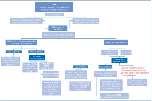
predisposing factors have been excluded, the need for continued statin therapy should be assessed.10,18 (See Figure 1.)
Box 2. Drugs with muscle-related adverse affects
Class Example
Anti-inflammatory drugs Glucocorticoids Antipsychotics Risperidone, haloperidol Immunosuppressant Methotrexate
Antiviral agents Protease inhibitors Lipid-lowering agents Gemifibrozil
Substance abuse Alcohol, opioids, amphetamines
As adapted18
Management of SAMS based on serum
creatine kinase (SCK)
Generally, SAMS complaints are associated with normal, mild to moderate CK elevations (< 4 × ULN). In low-risk CVD patients with SAMS the need for, as well as risk with statin continuation versus therapeutic lifestyle changes benefit must be assessed. Conversely in high-risk CVD patients with SAMS, the SAMS burden versus continued statin therapy benefit must be considered. The reason may be established with cessation of statin treatment during a washout period, followed by one or more re-challenges.18
In low CVD risk patients with SAMS and CK > 4 × ULN, statin use should be stopped and the necessity assessed. If continued statin therapy is required, low dosages of alternative statins (seeTreatment of SAMS) may be used with CK monitoring. With high-risk CVD and concomitant SAMS in the presence of CK of > 4 × ULN (but < 10 × ULN), continued statin therapy is required with concomitant CK monitoring.18
If CK levels exceed 10 x ULN, statin use must be stopped (at least momentarily) and reuse of the specific statin regimen should not occur. Once there is a reduction in CK levels, low dose statin therapy may be reinitiated with CK monitoring. If, however, CK levels remain elevated, causes of underlying myopathy should be evaluated in addition to referral to a neuromuscular specialist.18
Treatment of SAMS
If the management of SAMS indicates the need for continued lipid-lowering treatment, a number of options are available.18
1. Statin-based therapy
Effort should be made to keep high-risk patients on statin therapy, as these agents are the only lipid-lowering agents with comprehensive evidence proving clinical end-point reduction.18
Switching to an alternative statin
There are two available treatment options if SAMS or CK abnormalities resolve after discontinuation of the causative statin. Either re-initiation with lower dosing of the primary statin, or the use of an alternative statin. The aim is to achieve the target LDL-C level with the maximum tolerable dose with minimal SAMS.18
The use of fluvastatin is associated with less myopathy compared to the use of lovastatin, simvastatin, or atorvastatin. Furthermore, cases of fatal rhabdomyolysis with the use of fluvastatin have not yet been reported.10
As high statin doses and drug–drug interactions are risk factors for SAMS, the use of rosuvastatin, metabolised by CYP2C9, in patients receiving multiple medication, has a theoretical benefit. Additionally, lower dosages are safe and effective in SAMS
patients and reduction in LDL-C cholesterol comparable to that of atorvastatin has been shown.10
Non-daily dosing of statins
If the above-mentioned options are not tolerated to reduce LDL-C levels, the treatment strategy of choice is alternate day or twice-weekly dosing18 with long-acting statins administered in low
dosages or at a reduced frequency.23 Potential suitable agents,
with respectively 20 and 15 hour half-lives, both rosuvastatin and atorvastatin may decrease LDL cholesterol levels whilst possibly reducing adverse effects. Limited data exists on atorvastatin alternate day dosing and non-daily dosing of rosuvastatin appears tolerable and may decrease LDL-C levels in SAMS. However, the reduction in CVD risk still has to be evaluated.10
2. Non-statin-based lipid-lowering therapy
In the presence of complete statin intolerance, persistent LDL-C levels above the targeted goal and high CVD risk patients, despite maximally tolerated statin dosage to reduce LDL cholesterol,18
the addition of an alternative lipid-lowering agent should be considered.23
The drug of choice to achieve targeted LDL-C levels is ezetimibe, followed by the use of bile acid sequestrates (BAS) or fibrates in combination with ezetimibe.18
AIM
Achieve targeted LDLC levels with minimum tolerable statin dose
SAMS complaint Evaluate predisposing risk factors
and exclude secondary causes continuation or re-challenge Assess if SAMS favour statin Management
of SAMS
Measure CK and creatinine levels SAMS with normal or moderately
increased CK (< 4x ULN) Low CVD Risk
Assess therapeutic lifestyle changes bene t to continued
statin use
High CVD Risk 2-4 weeks washout period Resolved SAMS
Continue with current statin
therapy
Persistent SAMS
Change statin
Resolved SAMS
Continue therapy
Persistent SAMS
Change statin Consider slternate
day dosing Use alternative LL
agents
SAMS and elevated CK
CK >4x ULN, but <10x ULN
6 week statin washout period Low CVD Risk
Assess therapeutic lifestyle changes bene t
to continued statin use If use is important use
low dose alternative statin with CK
monitoring
High CVD Risk Continue statin therapy
+ CK monitoring,
Persistent SAMS Use
low dose alternative statin or non-daily dosing
with e ective statin
Improved SAMS
Use initial statin at starting dose
Consider rhabdomyolysis in patients with severe muscular pain and signs of myoglobinemia or myoglobinuria
If rhabdomyolysis, statin should not be
reintroduced
>10x ULN
LL = Lipid Lowering
Ezetimibe
Ezetimibe decreases LDL cholesterol levels by targeting the NPC1L1 transporter and inhibiting intestinal cholesterol absorption. Compared to higher dosages of statin monotherapy, similar results in LDL cholesterol reductions have been achieved with the addition of ezetimibe to existing statin therapy. This combination is both safe and well tolerated,23 which may benefit
patients with SAMS who do not achieve their targeted LDL-C levels. Although ezetimibe has been shown to reduce CVD events,18
comprehensive data on the risk reduction of CVD morbidity and mortality is lacking.10,14
Bile acid sequestrate
Despite reducing cholesterol absorption and CVD events,23 the
extent of BAS LDL-C reduction is dose dependent18 together with
high rates of gastrointestinal intolerance.23 However, compared to
previous formulations, colesevelam (not available in South Africa) may lead to enhanced patient compliance as it is easier to take and better tolerated with an improved adverse effect profile.18,23
Opportunely, greater decreases in LDL-C have been seen in the combination of ezetimibe and BAS.18,23
Fibrates
Fenofibrate can lower LDL-C in patients with high baseline levels in the absence of hypertriglyceridaemia. In addition, it is easy to take with a worthy safety profile in diabetic patients with CVD risks. Regrettably, increased serum creatinine levels have been shown during treatment and no added CVD benefits have being demonstrated. Compared to gemfibrozil, combination use of fenofibrate with a statin showed no increased risk for rhabdomyolysis.18,23
Niacin
Niacin does reduce LDL-C levels and may be used in combination with BAS and ezetimibe in statin-intolerant patients that require a large reduction in LDL-C levels. The use of this agent is, however, limited by its significant adverse effect profile23 and although
niacin monotherapy reduces CVD morbidity and mortality, no significant CVD benefit has been demonstrated.18
Lastly, several complementary and novel treatment strategies have been proposed with differing levels of success. These strategies are presented in Table II.
Novel LDL therapies in the management
of SAMS
Two novel therapies provide potential alternative treatment in the management of persistent SAMS; proprotein convertase subtilisin/kexin type 9 (PCSK9) inhibitors and cholesteryl ester transfer protein (CETP) inhibitors.
The pivotal role of the LDL-receptor and PCSK9
The protein constituents of lipoprotein complexes are referred to as apolipoproteins, which are grouped into four different classes, namely A, B, C and E. The most significant apolipoprotein
associated with LDL-cholesterol, is apolipoprotein B-100 (ApoB). The latter provides for an attachment to the binding site for low-density lipoprotein on its LDL-receptor.1,2
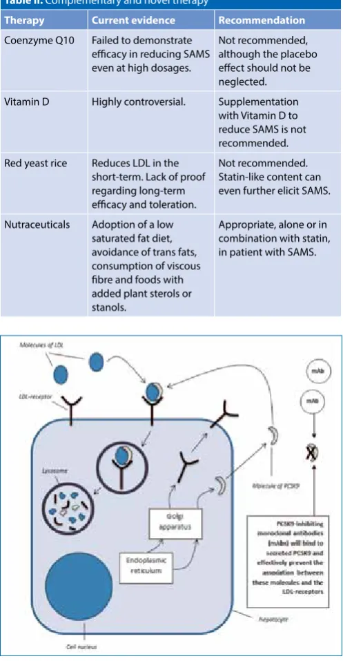
When LDL binds to its receptor on the cell surface of selected tissue cells, especially hepatocytes, the resultant internalisation of LDL provides for its lysosomal degradation. This is subsequently followed by the so-called recycling of the LDL-receptor (LDL-r) back to the cell’s plasma membrane. Once repositioned, it is able to bind yet another molecule of LDL. This is an ongoing process. However, in an attempt to maintain normal, stable levels of LDL-cholesterol in the body, the uncontrolled recycling of the LDL-r may be counteracted by the effects of PCSK9 (i.e. by proprotein Table II. Complementary and novel therapy18
Therapy Current evidence Recommendation
Coenzyme Q10 Failed to demonstrate efficacy in reducing SAMS even at high dosages.
Not recommended, although the placebo effect should not be neglected. Vitamin D Highly controversial. Supplementation
with Vitamin D to reduce SAMS is not recommended. Red yeast rice Reduces LDL in the
short-term. Lack of proof regarding long-term efficacy and toleration.
Not recommended. Statin-like content can even further elicit SAMS.
Nutraceuticals Adoption of a low saturated fat diet, avoidance of trans fats, consumption of viscous fibre and foods with added plant sterols or stanols.
Appropriate, alone or in combination with statin, in patient with SAMS.
LDL: low-density lipoprotein, mAb: monoclonal antibody, PCSK9: proprotein convertase subtilisin/ kexin type 9
Figure 2: Simplified diagram of a prototypical hepatocyte, illustrating
convertase subtilisin/kexin type 9), which promotes the breakdown of these receptors. (Refer to Figure 2.) PCSK9, a serine protease that is expressed in particularly significant quantities within the liver (the main source of circulating PCSK9), intestines and kidneys, may therefore be regarded as a modulator of LDL-cholesterol levels in the plasma. It effectively acts to reduce the number of LDL-receptors on the cell surface of hepatocytes.3,4
In turn, PCSK9 undergoes endogenous inactivation by two different proprotein conertases, referred to as furin and PC5/6A, within hepatocytes.17
PSCK9 inhibitors
The most advanced subcutaneously administered PSCK9 inhibitors (evo-locumab, alirocumab, and bococizumab [not available in South Africa yet]) have shown high reduction in LDL-C in a variety of patient groups (including those with statin intolerance), and additionally a low rate of muscle symptoms. These agents are well tolerated with no significant liver function abnormalities or CK elevations.18
Cholesteryl ester transfer protein inhibitors (CETP)
Both anacetrapib and evacetrapib equally lower LDL cholesterol and have shown significant increases in HDL-C by mediating the heteroexchange of triglycerides and cholesteryl esters between lipoproteins. The molecular base for LDL-C reduction is still uncertain, however improved fractional removal rates for LDL apolipoprotein B from plasma are involved. Fortunately, no muscular toxicities have been identified.18
Conclusion
Dyslipidaemia is a disorder associated with liver lipoprotein metabolism, which includes combined elevated levels of total and LDL-C and decreased HDL-C levels. In addition to adipocyte dysfunction, atherosclerosis is also a risk factor for CVD events. Nearly four decades after statins were introduced and tens of millions of scripts have been filled, the mechanism behind SAMS remains elusive. Despite no consensus on a true unifying mechanism, it would appear that SAMS is multifactorial. Due to varied use of terms and no consigning definition, SAMS is more common than previously believed although current evidence suggests the need for a diagnostic manual to distinguish muscle pain from SAMS. In recent years great improvements have been made in the management of SAMS. Management is aimed at
risk vs benefit and in the discontinuation and reintroduction of statins, although novel drug development could greatly aid in providing additional treatment options. Fortunately, statins can be interchanged, discontinued and reintroduced in order to improve quality of life as SAMS are rarely life-threatening.
References
1. Brenner GM, Stevens CW. Pharmacology. 4th ed. Philadelphia: Elsevier Saunders, 2013.
2. Talbert RL. Dyslipidemia, edited by JT DiPiro, RL Talbert, GC Yee, et al, 8th ed. New York:
McGraw-Hill Medical, 2011.
3. Zhang X, Zhu L, Chen J, et al. Safety and efficacy of anti-PCSK9 antibodies: a meta-analysis of 25 randomized, controlled trials. BMC Medicine. 2015. 13:123. DOI:10.1186/s12916– 015–0358–8.
4. Lambert G, Sjouke B, Choque B, et al. The PCSK9 decade. Journal of Lipid Research.2012; 53:2515–2524.
5. Barquera S, Pedroza-Tobías A, Medina C, et al. Global overview of the epidemiology of ath-erosclerotic cardiovascular disease. Arch Med Res 2015;46(5):328–338.
6. Libby P. The pathogenesis, prevention, and treatment of atherosclerosis. In: Kasper D, Fauci A, Hauser S, et al. eds. Harrison’s Principles of Internal Medicine, 19e.New York, NY: McGraw-Hill. 2015. http://accesspharmacy.mhmedical.com/content.aspx?bookid=1130&S ectionid=79743366. Accessed January 21, 2016.
7. Balagopal PB, de Ferranti SD, Cook S, et al. Nontraditional risk factors and biomark-ers for cardiovascular disease: mechanistic, research, and clinical considerations for youth: a scientific statement from the American Heart Association. Circulation 2011 Jun 14;123(23):2749–2769.
8. Sathasivam S. Statin induced myotoxicity. Eur J Intern Med 2012;23(4):317–324. 9. Di Stasi SL, MacLeod TD, Winters JD, et al. Effects of statins on skeletal muscle: a perspective
for physical therapists. Phys Ther 2010 Oct;90(10):1530–1542.
10. Joy TR, Hegele RA. Narrative review: statin-related myopathy. Ann Intern Med 2009;150(12):858–868.
11. Thompson PD, Clarkson PM, Rosenson RS. An assessment of statin safety by muscle ex-perts. Am J Cardiol 2006;97(8):S69–S76.
12. Raal FJ, Blom DJ, Naidoo S, et al. Prevalence of dyslipidaemia in statin-treated patients in South Africa: results of the DYSlipidaemia International Study (DYSIS). Cardiovasc J Afr 2013 Sep;24(8):330–338.
13. Patel S. A review of available cholesterol-lowering medicines in South Africa. SA Pharma-ceutical Journal 2013;80(9):20–25.
14. Stock J. Statin-associated muscle symptoms EAS Consensus Panel paper focuses on this neglected patient group. Atherosclerosis 2015;1(242):346–350.
15. Apostolopoulou M, Corsini A, Roden M. The role of mitochondria in statin‐induced myopa-thy. Eur J Clin Invest 2015;45(7):745–754.
16. Rajput M. Lipid-modifying therapy. Continuing Medical Education 2009;27(3). 17. McKenney JM, Davidson MH, Jacobson TA, et al. Final conclusions and
recommenda-tions of the national lipid association statin safety assessment task force. Am J Cardiol 2006;97(8):S89–S94.
18. Stroes ES, Thompson PD, Corsini A, et al. Statin-associated muscle symptoms: impact on statin therapy-European Atherosclerosis Society Consensus Panel Statement on Assess-ment, Aetiology and Management. Eur Heart J 2015 May 1;36(17):1012–1022.
19. Gulum AH, Hume AL. Statins: An update on clinical issues and selected adverse effects. The Journal for Nurse Practitioners 2015;11(3):287–294.
20. Newman CB, Tobert JA. Statin intolerance: reconciling clinical trials and clinical experience. JAMA 2015;313(10):1011–1012.
21. Rosenson RS, Baker SK, Jacobson TA, et al. An assessment by the statin muscle safety task force: 2014 update. Journal of clinical lipidology 2014;8(3):S58–S71.
22. Norata GD, Tibolla G, Catapano AL. Statins and skeletal muscles toxicity: from clinical trials to everyday practice. Pharmacological Research 2014;88:107–113.